Embed Size (px)
Citation preview
National Center for Emerging and Zoonotic Infectious Diseases
Distinguishing Primary from Secondary Bloodstream Infections (BSIs) for the NHSN Patient Safety Component
Katherine Allen-Bridson, RN, BSN, MScPH, CICLead- Protocol and Validation TeamSurveillance BranchDivision of Healthcare Quality PromotionCenters for Disease Control & Prevention
April 26, 2018
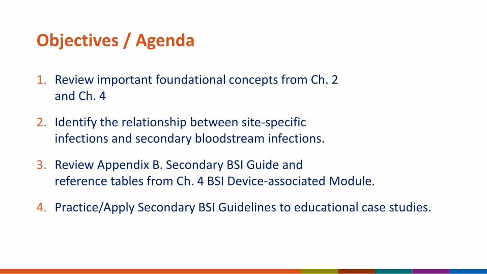
Objectives / Agenda
1. Review important foundational concepts from Ch. 2 and Ch. 4
2. Identify the relationship between site-specific infections and secondary bloodstream infections.
3. Review Appendix B. Secondary BSI Guide and reference tables from Ch. 4 BSI Device-associated Module.
4. Practice/Apply Secondary BSI Guidelines to educational case studies.
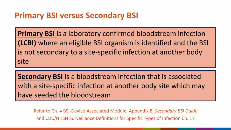
Primary BSI versus Secondary BSI
Refer to Ch. 4 BSI-Device-Associated Module, Appendix B. Secondary BSI Guide and CDC/NHSN Surveillance Definitions for Specific Types of Infection Ch. 17
Primary BSI is a laboratory confirmed bloodstream infection (LCBI) where an eligible BSI organism is identified and the BSI is not secondary to a site-specific infection at another body site
Secondary BSI is a bloodstream infection that is associated with a site-specific infection at another body site which may have seeded the bloodstream
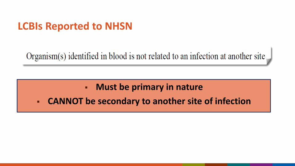
LCBIs Reported to NHSN
Must be primary in nature CANNOT be secondary to another site of infection
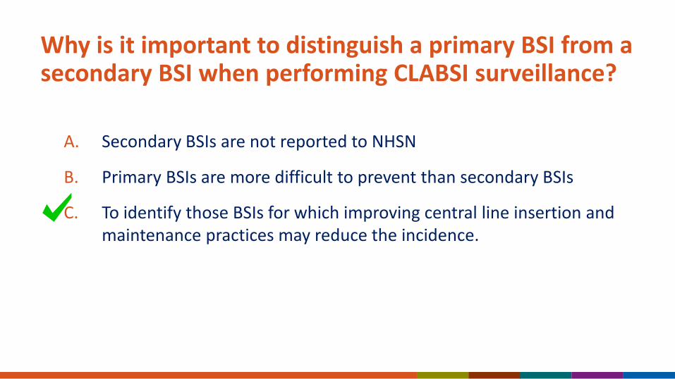
Why is it important to distinguish a primary BSI from a secondary BSI when performing CLABSI surveillance?
A. Secondary BSIs are not reported to NHSN
B. Primary BSIs are more difficult to prevent than secondary BSIs
C. To identify those BSIs for which improving central line insertion and maintenance practices may reduce the incidence.
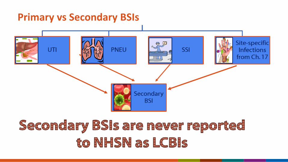
Primary vs Secondary BSIs
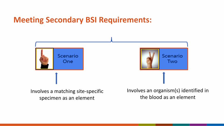
Meeting Secondary BSI Requirements:
Involves a matching site-specific specimen as an element
Involves an organism(s) identified in the blood as an element
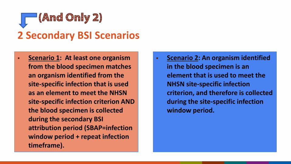
2 Secondary BSI Scenarios
Scenario 1: At least one organism from the blood specimen matches an organism identified from the site-specific infection that is used as an element to meet the NHSN site-specific infection criterion AND the blood specimen is collected during the secondary BSI attribution period (SBAP=infection window period + repeat infection timeframe).
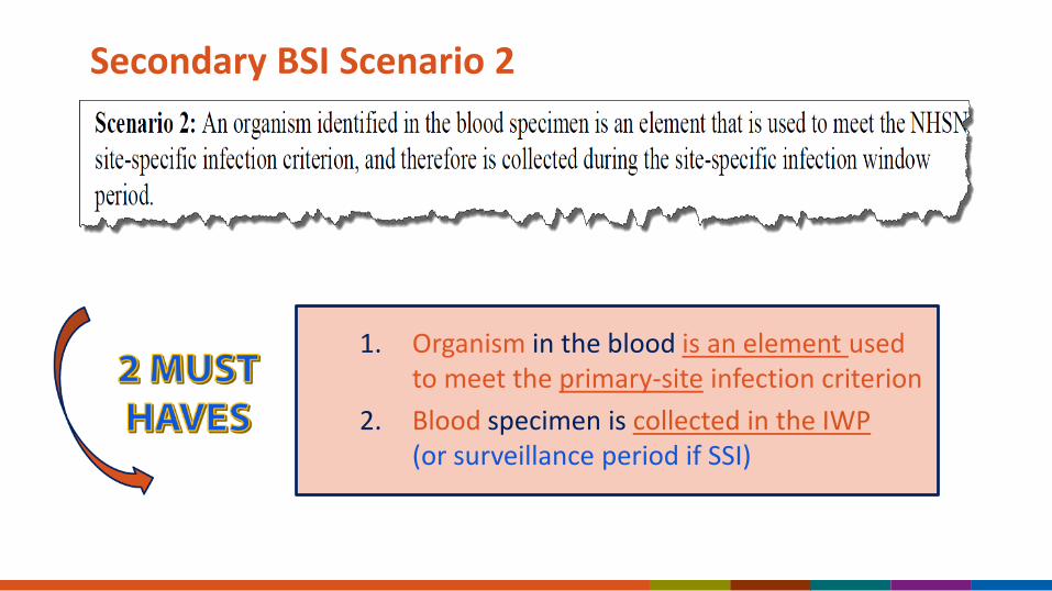
Scenario 2: An organism identified in the blood specimen is an element that is used to meet the NHSN site-specific infection criterion, and therefore is collected during the site-specific infection window period.
Secondary Bloodstream Infections Scenario 1
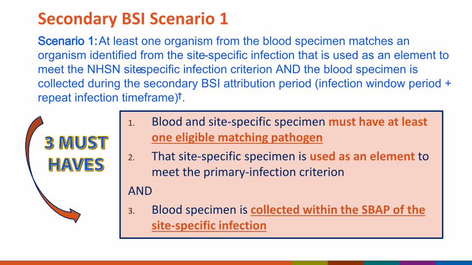
Secondary BSI Scenario 1
1. Blood and site-specific specimen must have at least one eligible matching pathogen
2. That site-specific specimen is used as an element to meet the primary-infection criterion
AND3. Blood specimen is collected within the SBAP of the
site-specific infection
Scenario 1: At least one organism from the blood specimen matches an organism identified from the site-specific infection that is used as an element to meet the NHSN site-specific infection criterion AND the blood specimen is collected during the secondary BSI attribution period (infection window period + repeat infection timeframe)†.
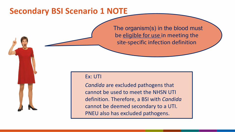
Secondary BSI Scenario 1 NOTE
Ex: UTICandida are excluded pathogens that cannot be used to meet the NHSN UTI definition. Therefore, a BSI with Candida cannot be deemed secondary to a UTI. PNEU also has excluded pathogens.
The organism(s) in the blood must be eligible for use in meeting the site-specific infection definition
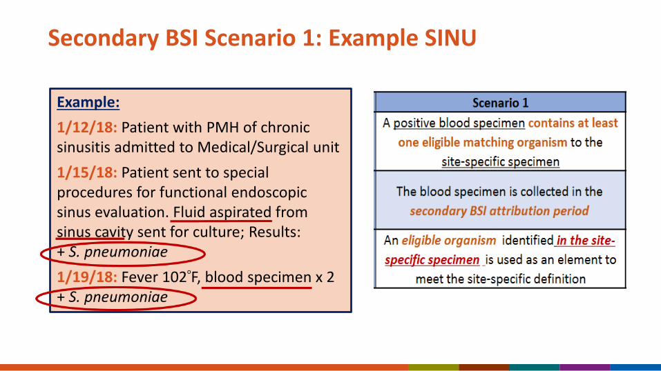
Secondary BSI Scenario 1: Example SINU
Example: 1/12/18: Patient with PMH of chronic sinusitis admitted to Medical/Surgical unit1/15/18: Patient sent to special procedures for functional endoscopic sinus evaluation. Fluid aspirated from sinus cavity sent for culture; Results: + S. pneumoniae1/19/18: Fever 102oF, blood specimen x 2 + S. pneumoniae

Secondary BSI Scenario 1
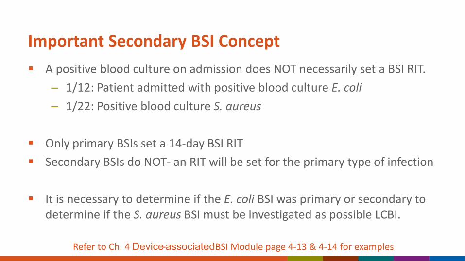
Important Secondary BSI Concept A positive blood culture on admission does NOT necessarily set a BSI RIT.
– 1/12: Patient admitted with positive blood culture E. coli– 1/22: Positive blood culture S. aureus
Only primary BSIs set a 14-day BSI RIT Secondary BSIs do NOT- an RIT will be set for the primary type of infection
It is necessary to determine if the E. coli BSI was primary or secondary to determine if the S. aureus BSI must be investigated as possible LCBI.
Refer to Ch. 4 Device-associatedBSI Module page 4-13 & 4-14 for examples
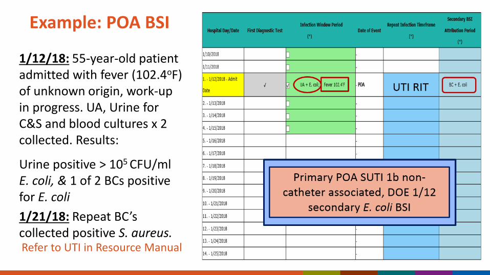
Example: POA BSI
1/12/18: 55-year-old patient admitted with fever (102.4oF) of unknown origin, work-up in progress. UA, Urine for C&S and blood cultures x 2 collected. Results:
Urine positive > 105 CFU/ml E. coli, & 1 of 2 BCs positive for E. coli1/21/18: Repeat BC’s collected positive S. aureus.Refer to UTI in Resource Manual


Secondary Blood Stream Infections Scenario 2
Secondary BSI Scenario 2
1. Organism in the blood is an element used to meet the primary-site infection criterion
2. Blood specimen is collected in the IWP (or surveillance period if SSI)
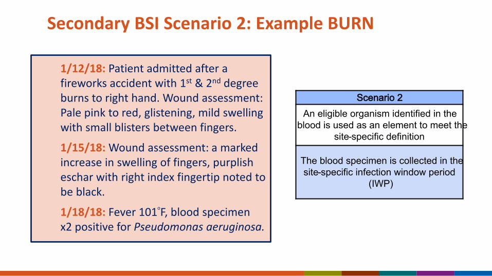
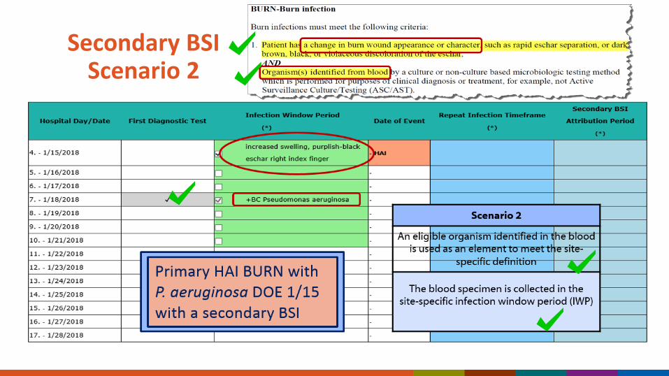
Secondary BSI Scenario 2: Example BURN
1/12/18: Patient admitted after a fireworks accident with 1st & 2nd degree burns to right hand. Wound assessment: Pale pink to red, glistening, mild swelling with small blisters between fingers.
1/15/18: Wound assessment: a marked increase in swelling of fingers, purplish eschar with right index fingertip noted to be black.
1/18/18: Fever 101oF, blood specimen x2 positive for Pseudomonas aeruginosa.
Scenario 2An eligible organism identified in the
blood is used as an element to meet the site-specific definition
The blood specimen is collected in the site-specific infection window period
(IWP)
Secondary BSI Scenario 2
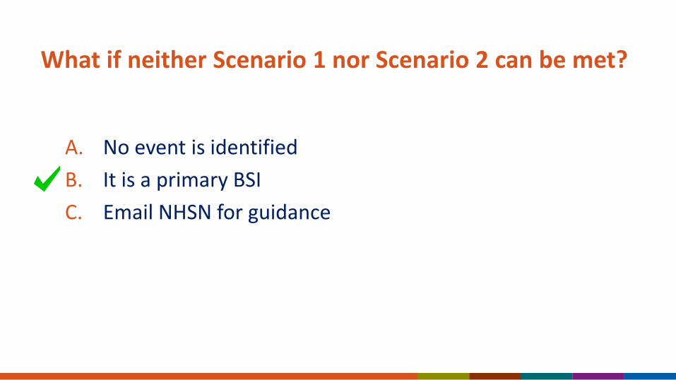
What if neither Scenario 1 nor Scenario 2 can be met?
A. No event is identifiedB. It is a primary BSIC. Email NHSN for guidance
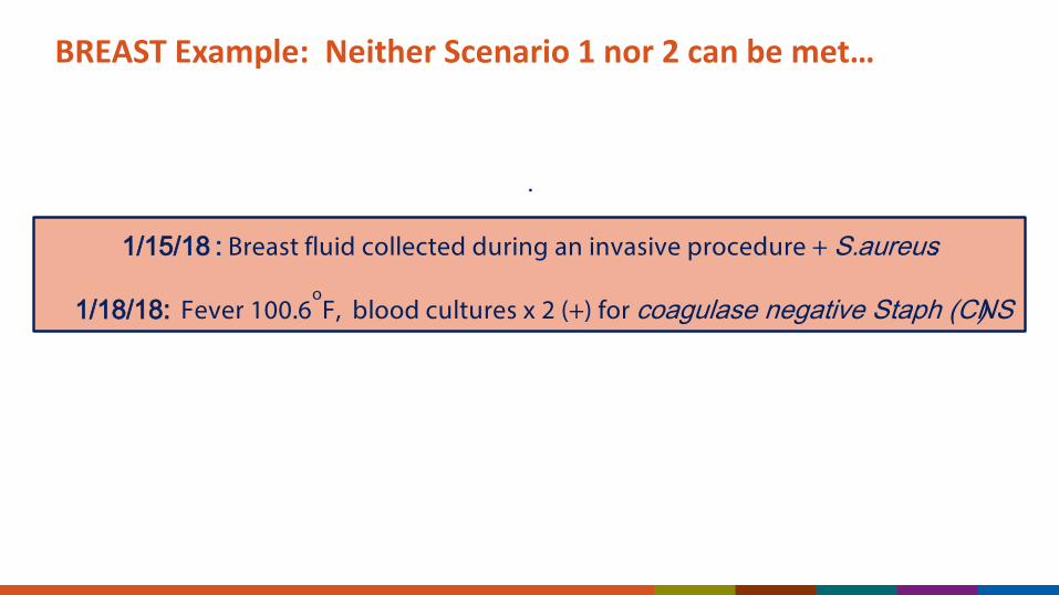
.
1/15/18 : Breast fluid collected during an invasive procedure + S. aureus.
1/18/18: Fever 100.6oF, blood cultures x 2 (+) for coagulase negative Staph (CNS)
BREAST Example: Neither Scenario 1 nor 2 can be met…
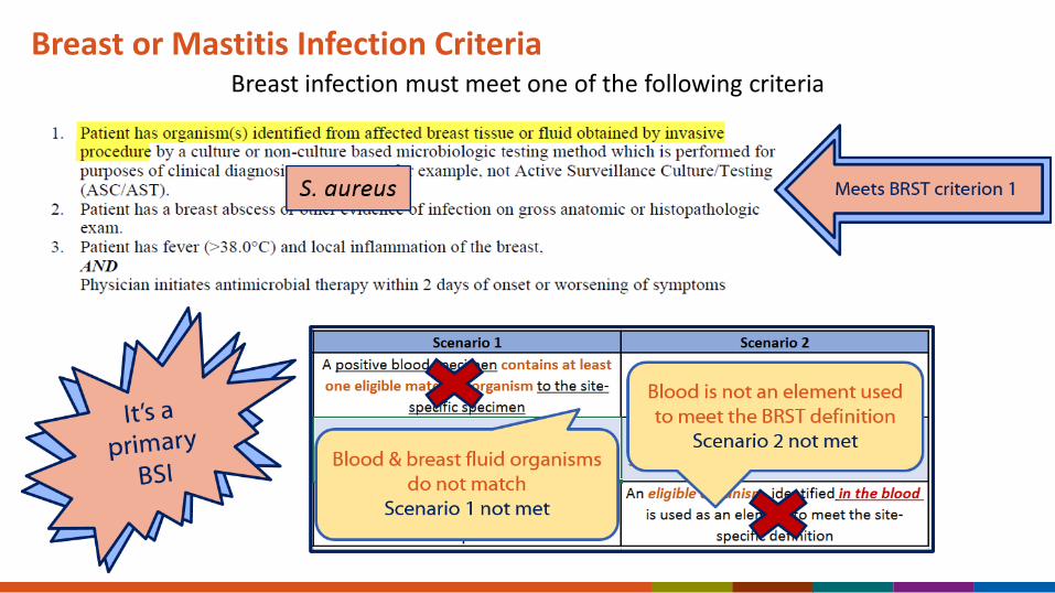
Breast or Mastitis Infection CriteriaBreast infection must meet one of the following criteria
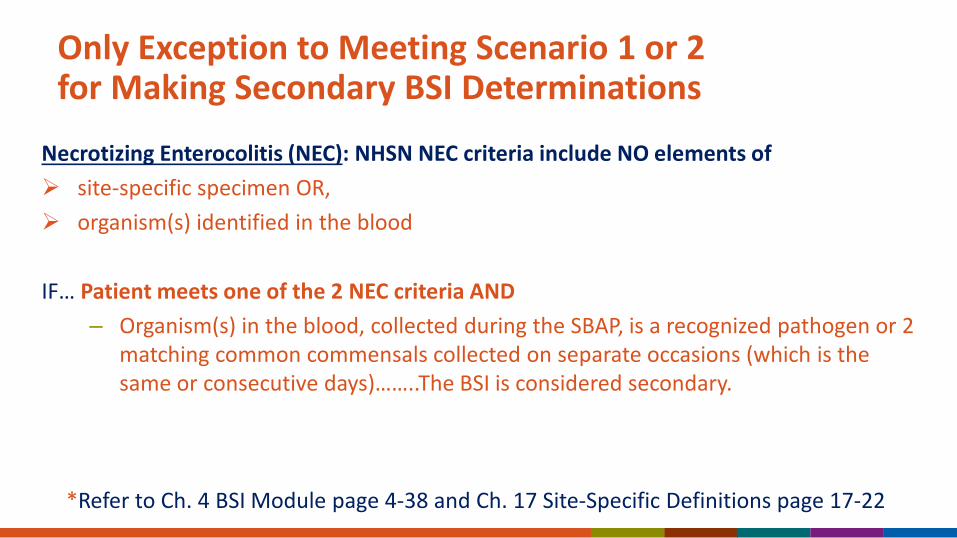
Only Exception to Meeting Scenario 1 or 2 for Making Secondary BSI Determinations
Necrotizing Enterocolitis (NEC): NHSN NEC criteria include NO elements of site-specific specimen OR, organism(s) identified in the blood
IF… Patient meets one of the 2 NEC criteria AND– Organism(s) in the blood, collected during the SBAP, is a recognized pathogen or 2
matching common commensals collected on separate occasions (which is the same or consecutive days)……..The BSI is considered secondary.
*Refer to Ch. 4 BSI Module page 4-38 and Ch. 17 Site-Specific Definitions page 17-22
Assigning Pathogens in Secondary BSIs
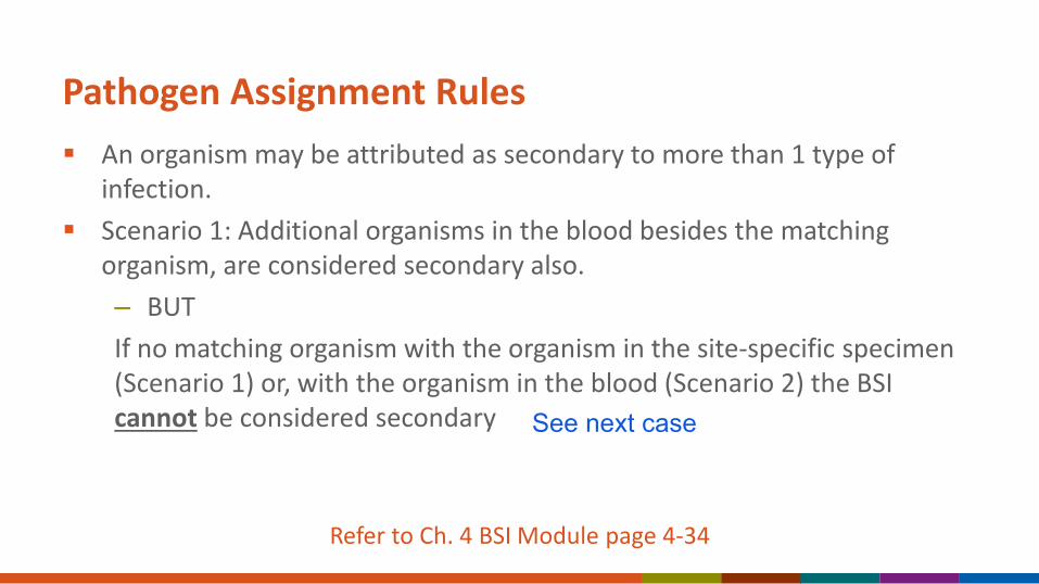
Pathogen Assignment Rules An organism may be attributed as secondary to more than 1 type of
infection. Scenario 1: Additional organisms in the blood besides the matching
organism, are considered secondary also. – BUTIf no matching organism with the organism in the site-specific specimen (Scenario 1) or, with the organism in the blood (Scenario 2) the BSI cannot be considered secondary
Refer to Ch. 4 BSI Module page 4-34
See next case
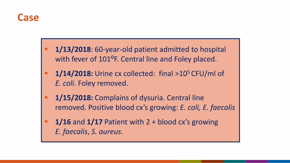
Case
1/13/2018: 60-year-old patient admitted to hospital with fever of 101⁰F. Central line and Foley placed.
1/14/2018: Urine cx collected: final >105 CFU/ml of E. coli. Foley removed.
1/15/2018: Complains of dysuria. Central line removed. Positive blood cx’s growing: E. coli, E. faecalis
1/16 and 1/17 Patient with 2 + blood cx’s growing E. faecalis, S. aureus.
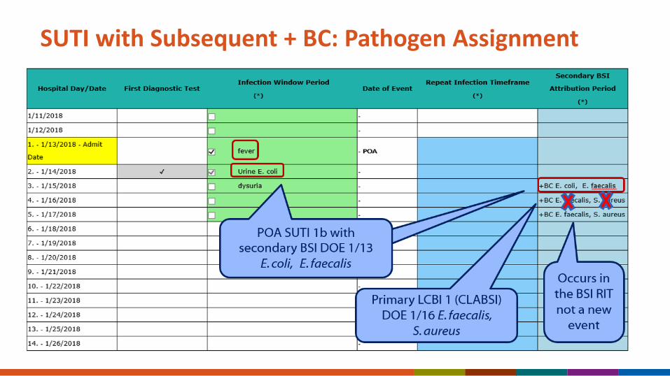
SUTI with Subsequent + BC: Pathogen Assignment

Secondary BSITools, Education & Reference
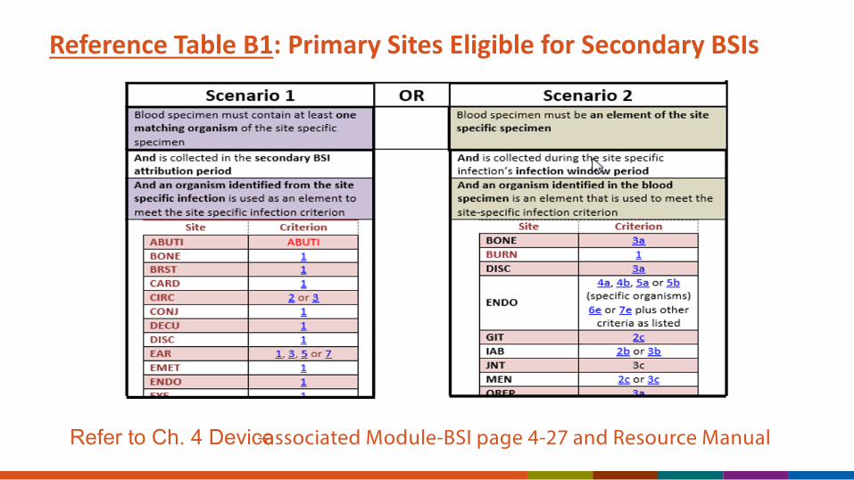
Reference Table B1: Primary Sites Eligible for Secondary BSIs
Refer to Ch. 4 Device-associated Module-BSI page 4-27 and Resource Manual
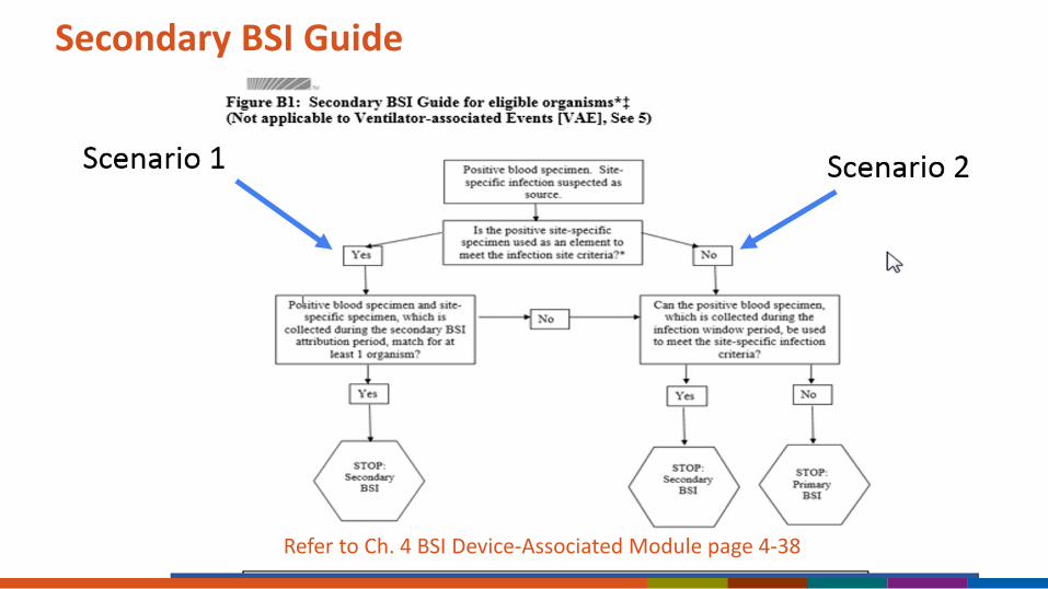
Secondary BSI Guide
Refer to Ch. 4 BSI Device-Associated Module page 4-38
Training Resources:
https://www.cdc.gov/nhsn/pdfs/pscmanual/4psc_clabscurrent.pdf
https:// www.cdc.gov/nhsn/training/patient-safety-component/index.html
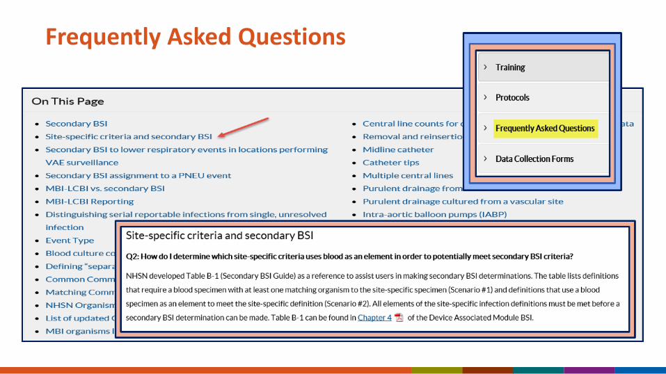
Frequently Asked Questions

Case Studies
Case Study 1 February 1: 35-year-old woman admitted with complaints of right
scapular pain and fever. Superficial laceration to back right scapula from falling into outdoor grill 5 days earlier. Wound is scabbed over in places but purulent drainage is noted from center. Admit for IV antibiotics.
February 3: No improvement noted with broad spectrum antibiotic coverage. Wound is swelling, red and warm to touch. Drainage from wound is sent for culture, positive for Streptococcus pyogenes.
February 8: Patient fever continues, and WBCs continue to increase, now at 20,000 WBC/μL. Blood cultures are collected and final results are positive for methicillin-resistant S. pyogenes.
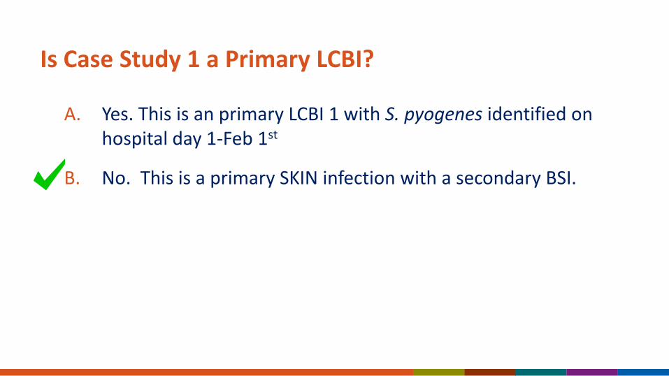
Is Case Study 1 a Primary LCBI?
A. Yes. This is an primary LCBI 1 with S. pyogenes identified on hospital day 1-Feb 1st
B. No. This is a primary SKIN infection with a secondary BSI.
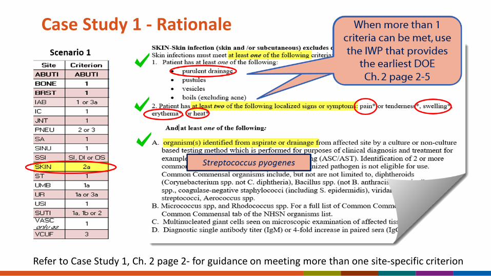
Case Study 1 - Rationale
Refer to Case Study 1, Ch. 2 page 2- for guidance on meeting more than one site-specific criterion

Case Study 1 Rationale:
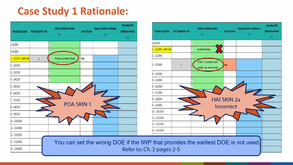
Case Study 1 Rationale:
You can set the wrong DOE if the IWP that provides the earliest DOE is not used. Refer to: Ch. 2 pages 2-5
Meeting More than One Criteria of Same Type of Infection?
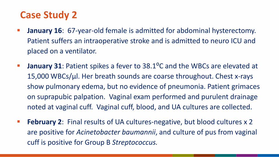
Case Study 2 January 16: 67-year-old female is admitted for abdominal hysterectomy.
Patient suffers an intraoperative stroke and is admitted to neuro ICU and placed on a ventilator.
January 31: Patient spikes a fever to 38.1⁰C and the WBCs are elevated at 15,000 WBCs/μl. Her breath sounds are coarse throughout. Chest x-rays show pulmonary edema, but no evidence of pneumonia. Patient grimaces on suprapubic palpation. Vaginal exam performed and purulent drainage noted at vaginal cuff. Vaginal cuff, blood, and UA cultures are collected.
February 2: Final results of UA cultures-negative, but blood cultures x 2 are positive for Acinetobacter baumannii, and culture of pus from vaginal cuff is positive for Group B Streptococcus.
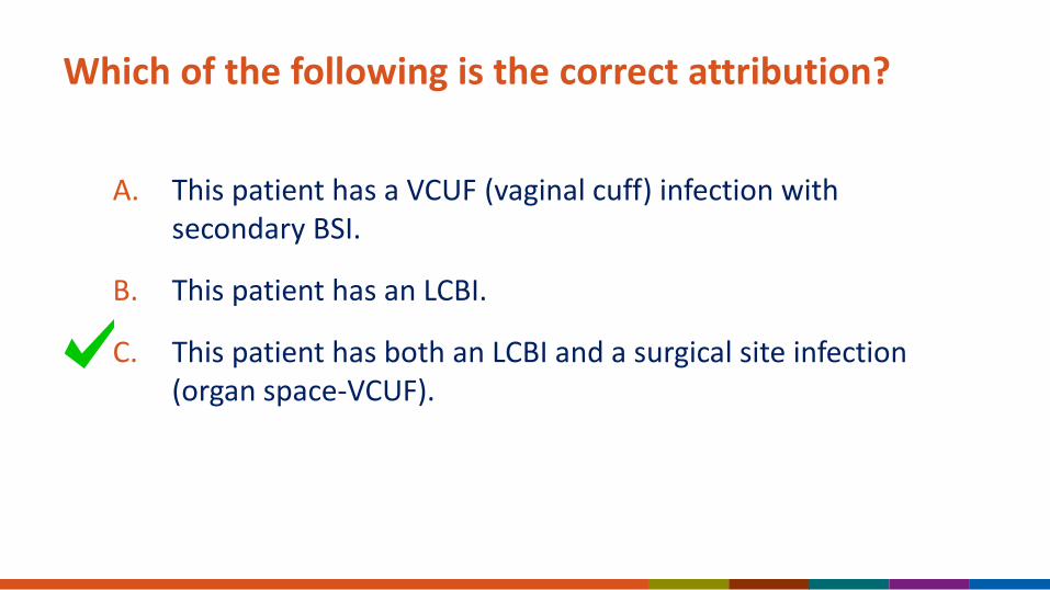
Which of the following is the correct attribution?
A. This patient has a VCUF (vaginal cuff) infection with secondary BSI.
B. This patient has an LCBI.
C. This patient has both an LCBI and a surgical site infection (organ space-VCUF).
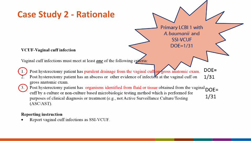
Case Study 2 - Rationale
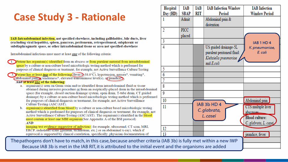
Case Study 3
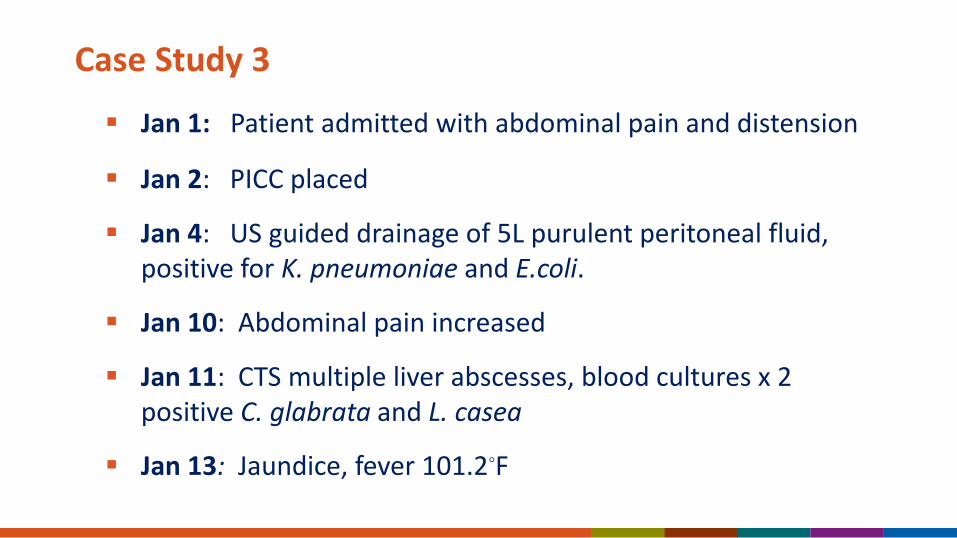
Jan 1: Patient admitted with abdominal pain and distension
Jan 2: PICC placed
Jan 4: US guided drainage of 5L purulent peritoneal fluid, positive for K. pneumoniae and E.coli.
Jan 10: Abdominal pain increased
Jan 11: CTS multiple liver abscesses, blood cultures x 2 positive C. glabrata and L. casea
Jan 13: Jaundice, fever 101.2◦F
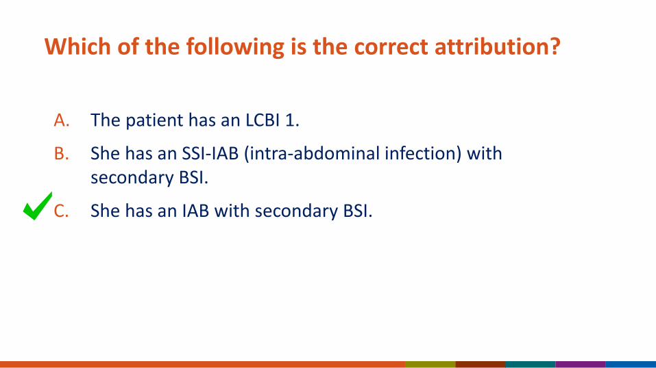
Which of the following is the correct attribution?
A. The patient has an LCBI 1.
B. She has an SSI-IAB (intra-abdominal infection) with secondary BSI.
C. She has an IAB with secondary BSI.
Case Study 3 - Rationale
The pathogens don’t have to match, in this case, because another criteria (IAB 3b) is fully met within a new IWP Because IAB 3b is met in the IAB RIT, it is attributed to the initial event and the organisms are added
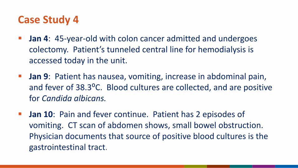
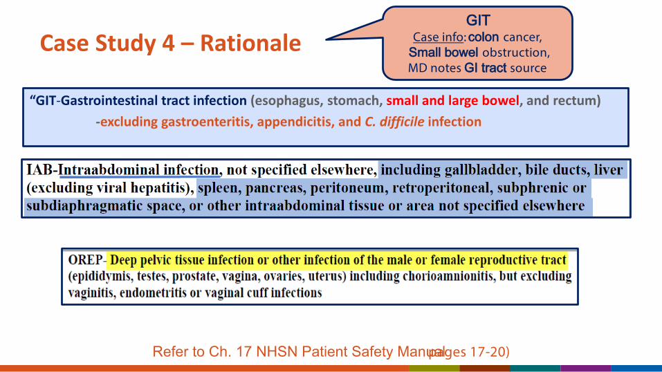
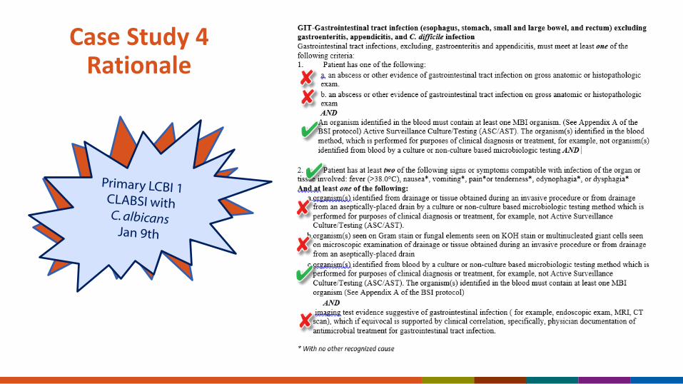
Case Study 4 Jan 4: 45-year-old with colon cancer admitted and undergoes
colectomy. Patient’s tunneled central line for hemodialysis is accessed today in the unit.
Jan 9: Patient has nausea, vomiting, increase in abdominal pain, and fever of 38.3⁰C. Blood cultures are collected, and are positive for Candida albicans.
Jan 10: Pain and fever continue. Patient has 2 episodes of vomiting. CT scan of abdomen shows, small bowel obstruction. Physician documents that source of positive blood cultures is the gastrointestinal tract.
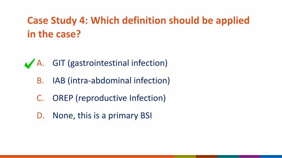
Case Study 4: Which definition should be applied in the case?
A. GIT (gastrointestinal infection)
B. IAB (intra-abdominal infection)
C. OREP (reproductive Infection)
D. None, this is a primary BSI
Case Study 4 – Rationale
“GIT-Gastrointestinal tract infection (esophagus, stomach, small and large bowel, and rectum) -excluding gastroenteritis, appendicitis, and C. difficile infection
Refer to Ch. 17 NHSN Patient Safety Manual pages 17-20)
GITCase info: colon cancer,
Small bowel obstruction, MD notes GI tract source
Case Study 4Rationale
Case Study 5 2/14: A 41-year-old female has been in your unit for 2
weeks. She has a central line through which she has been receiving hemodialysis since admission.
2/17: Her central line insertion site is red, and has purulent drainage, which is cultured and positive for Pseudomonas aeruginosa.
2/19: She develops fever of 39°C and shaking chills. Two sets of blood cultures are sent.
2/21: Final blood culture results are positive for Pseudomonas aeruginosa.
Case Study 5 Is this an LCBI?
A. Yes, the patient has an LCBI 1 with P. aeruginosa
B. No, this patient has a primary SKIN infection with P. aeruginosa and a secondary BSI with the same organism.
C. No. The patient has a VASC infection with P. aeruginosa and a secondary BSI with the same organism.
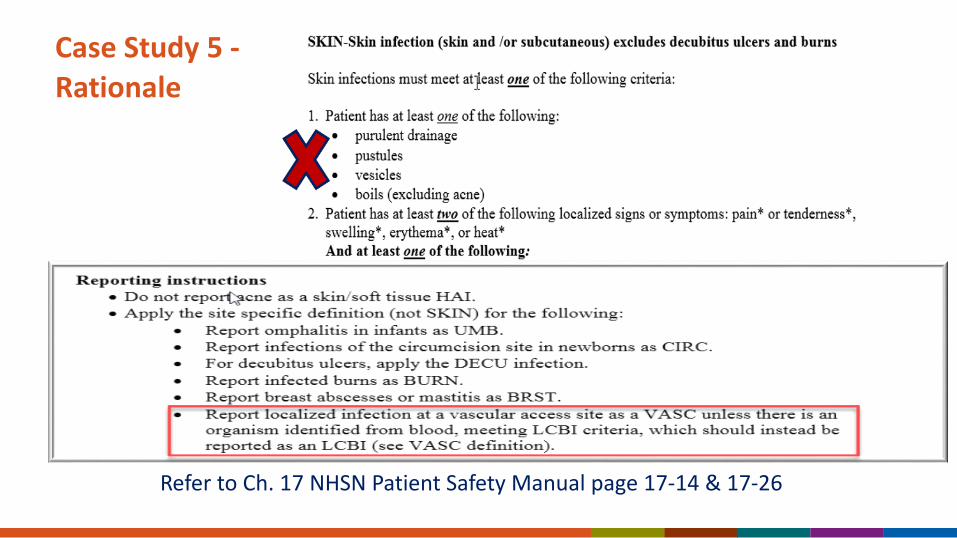
Case Study 5 -Rationale
Refer to Ch. 17 NHSN Patient Safety Manual page 17-14 & 17-26
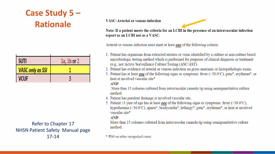
Case Study 5 –Rationale
Refer to Chapter 17 NHSN Patient Safety Manual page
17-14
Case Study 5 Rationale
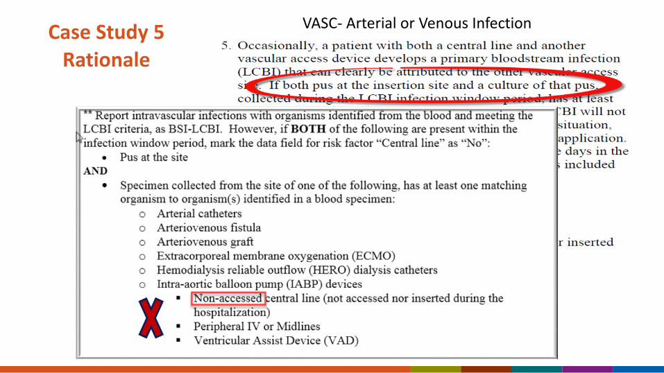
VASC- Arterial or Venous Infection
Case Study 6
February 1: Patient admitted to PICU 4 months status post allogeneic stem cell transplant for acute myeloid leukemia. Port in place and was accessed on admission. Her current weight is 25 kg.
February 8: Patient becomes disoriented and hypotensive. 2 sets of blood culture are collected. Both are positive for Enterococcus faecium.
February 9: Patient has nausea, emesis, diarrhea, and abdominal pain.
February 10: She is diagnosed with Grade III graft-versus-host disease by endoscopy.
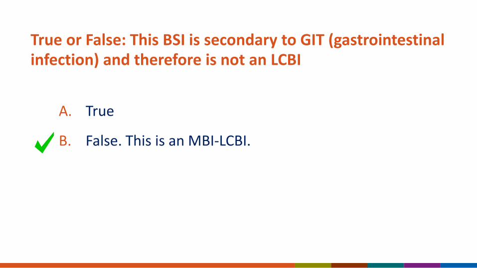
True or False: This BSI is secondary to GIT (gastrointestinal infection) and therefore is not an LCBI
A. True
B. False. This is an MBI-LCBI.
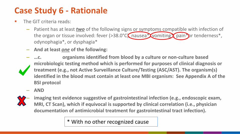
Case Study 6 - Rationale The GIT criteria reads:
– Patient has at least two of the following signs or symptoms compatible with infection of the organ or tissue involved: fever (>38.0°C), nausea*, vomiting*, pain*or tenderness*, odynophagia*, or dysphagia*
– And at least one of the following:– …c. organisms identified from blood by a culture or non-culture based
microbiologic testing method which is performed for purposes of clinical diagnosis or treatment (e.g., not Active Surveillance Culture/Testing (ASC/AST). The organism(s) identified in the blood must contain at least one MBI organism: See Appendix A of the BSI protocol
– AND– imaging test evidence suggestive of gastrointestinal infection (e.g., endoscopic exam,
MRI, CT Scan), which if equivocal is supported by clinical correlation (i.e., physician documentation of antimicrobial treatment for gastrointestinal tract infection).
* With no other recognized cause
Case Study 6 - Rationale
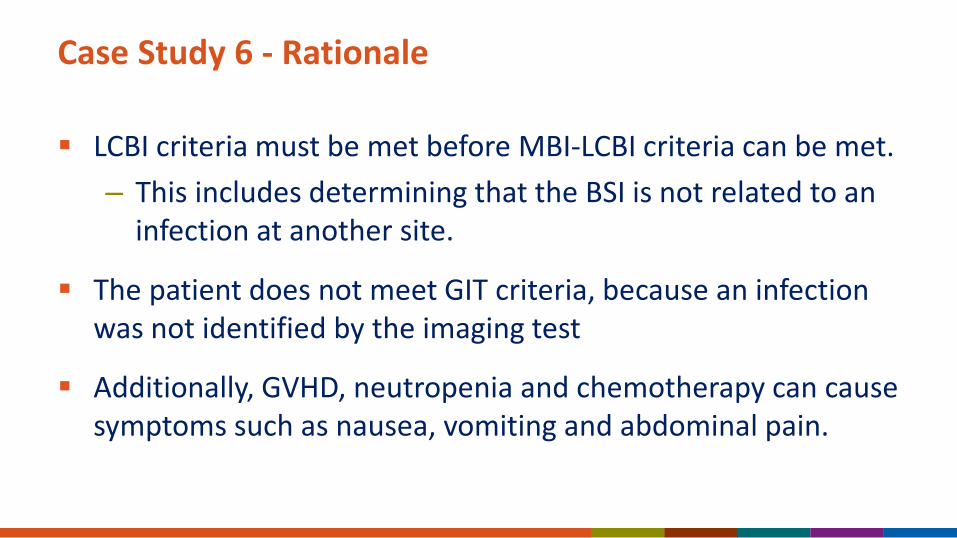
LCBI criteria must be met before MBI-LCBI criteria can be met.– This includes determining that the BSI is not related to an
infection at another site.
The patient does not meet GIT criteria, because an infection was not identified by the imaging test
Additionally, GVHD, neutropenia and chemotherapy can cause symptoms such as nausea, vomiting and abdominal pain.
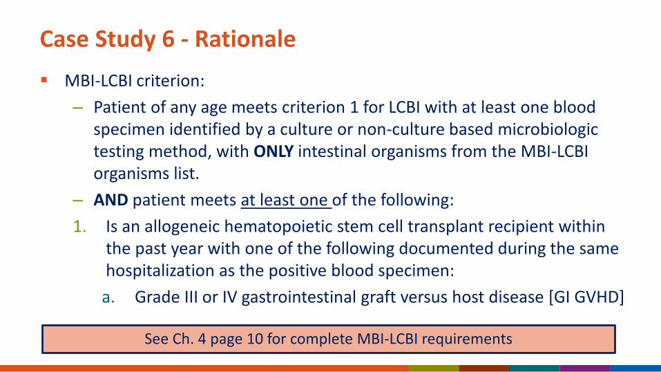
Case Study 6 - Rationale MBI-LCBI criterion:
– Patient of any age meets criterion 1 for LCBI with at least one blood specimen identified by a culture or non-culture based microbiologic testing method, with ONLY intestinal organisms from the MBI-LCBI organisms list.
– AND patient meets at least one of the following:1. Is an allogeneic hematopoietic stem cell transplant recipient within
the past year with one of the following documented during the same hospitalization as the positive blood specimen:a. Grade III or IV gastrointestinal graft versus host disease [GI GVHD]
See Ch. 4 page 10 for complete MBI-LCBI requirements
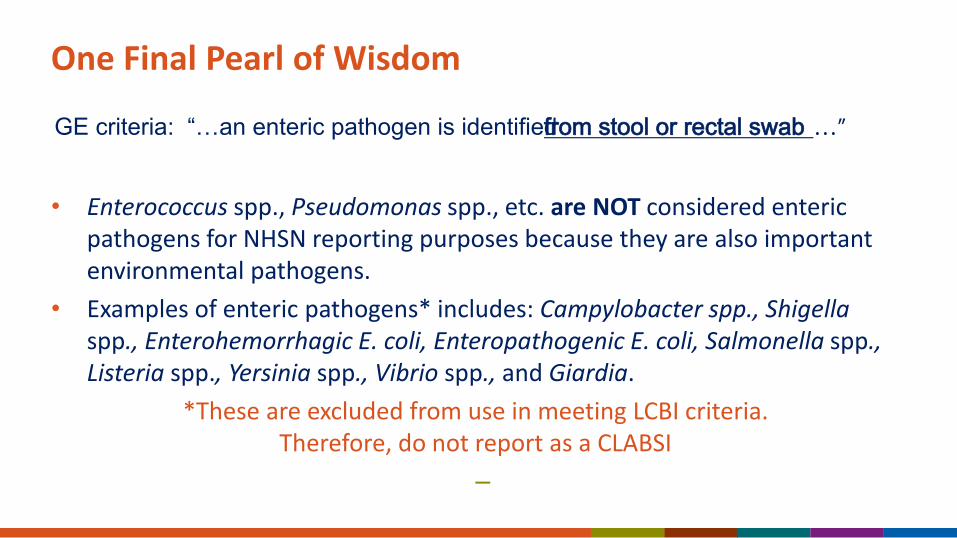
One Final Pearl of Wisdom
• Enterococcus spp., Pseudomonas spp., etc. are NOT considered enteric pathogens for NHSN reporting purposes because they are also important environmental pathogens.
• Examples of enteric pathogens* includes: Campylobacter spp., Shigella spp., Enterohemorrhagic E. coli, Enteropathogenic E. coli, Salmonella spp., Listeria spp., Yersinia spp., Vibrio spp., and Giardia.
*These are excluded from use in meeting LCBI criteria. Therefore, do not report as a CLABSI
–
GE criteria: “…an enteric pathogen is identified from stool or rectal swab …”
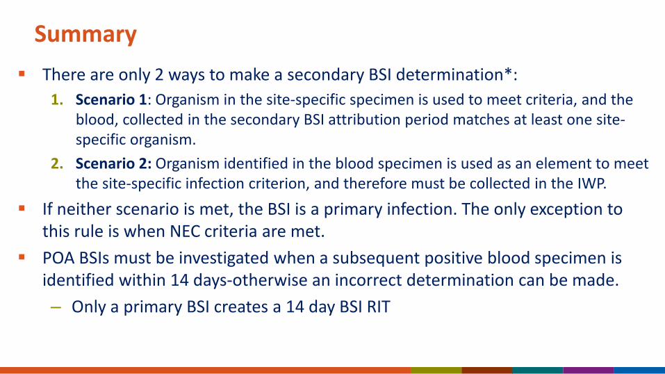
Summary There are only 2 ways to make a secondary BSI determination*:
1. Scenario 1: Organism in the site-specific specimen is used to meet criteria, and the blood, collected in the secondary BSI attribution period matches at least one site-specific organism.
2. Scenario 2: Organism identified in the blood specimen is used as an element to meet the site-specific infection criterion, and therefore must be collected in the IWP.
If neither scenario is met, the BSI is a primary infection. The only exception to this rule is when NEC criteria are met.
POA BSIs must be investigated when a subsequent positive blood specimen is identified within 14 days-otherwise an incorrect determination can be made.– Only a primary BSI creates a 14 day BSI RIT
Summary continued…
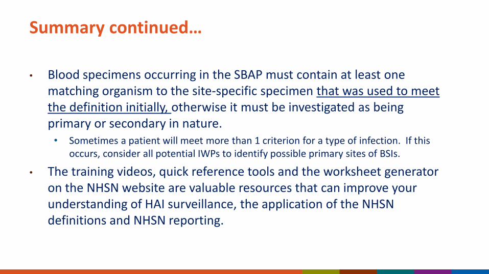
• Blood specimens occurring in the SBAP must contain at least one matching organism to the site-specific specimen that was used to meet the definition initially, otherwise it must be investigated as being primary or secondary in nature.
• Sometimes a patient will meet more than 1 criterion for a type of infection. If this occurs, consider all potential IWPs to identify possible primary sites of BSIs.
• The training videos, quick reference tools and the worksheet generator on the NHSN website are valuable resources that can improve your understanding of HAI surveillance, the application of the NHSN definitions and NHSN reporting.

Case #1
Table Top Group CasesSecondary BSIs
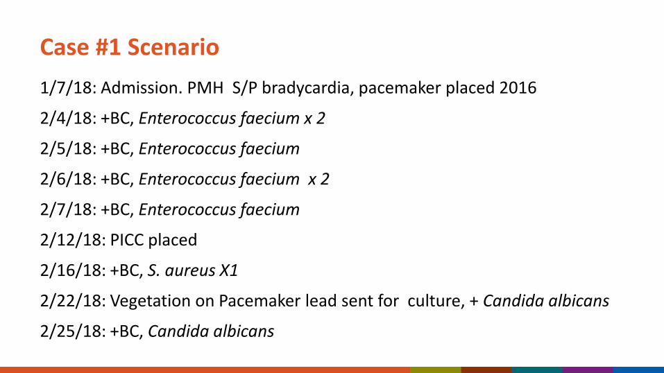
Case #1 Scenario1/7/18: Admission. PMH S/P bradycardia, pacemaker placed 2016
2/4/18: +BC, Enterococcus faecium x 2
2/5/18: +BC, Enterococcus faecium
2/6/18: +BC, Enterococcus faecium x 2
2/7/18: +BC, Enterococcus faecium
2/12/18: PICC placed
2/16/18: +BC, S. aureus X1
2/22/18: Vegetation on Pacemaker lead sent for culture, + Candida albicans
2/25/18: +BC, Candida albicans
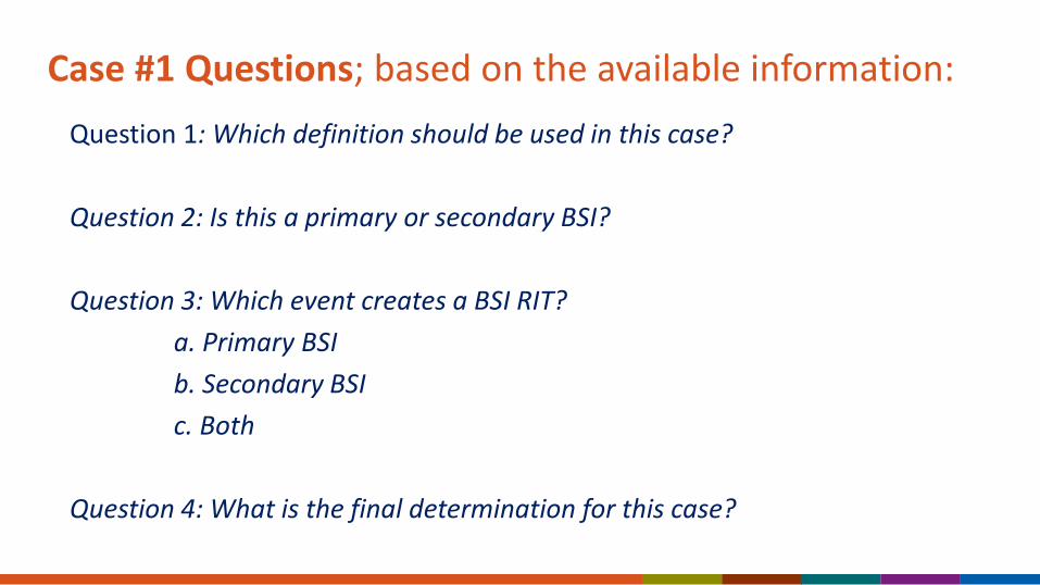
Case #1 Questions; based on the available information:Question 1: Which definition should be used in this case?
Question 2: Is this a primary or secondary BSI?
Question 3: Which event creates a BSI RIT?a. Primary BSIb. Secondary BSIc. Both
Question 4: What is the final determination for this case?

Case #1 Answers & Rationale
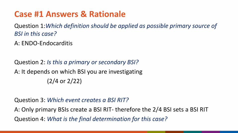
Case #1 Answers & RationaleQuestion 1:Which definition should be applied as possible primary source of BSI in this case?A: ENDO-Endocarditis
Question 2: Is this a primary or secondary BSI? A: It depends on which BSI you are investigating
(2/4 or 2/22)
Question 3: Which event creates a BSI RIT?A: Only primary BSIs create a BSI RIT- therefore the 2/4 BSI sets a BSI RITQuestion 4: What is the final determination for this case?
Case #1 Answers & Rationale
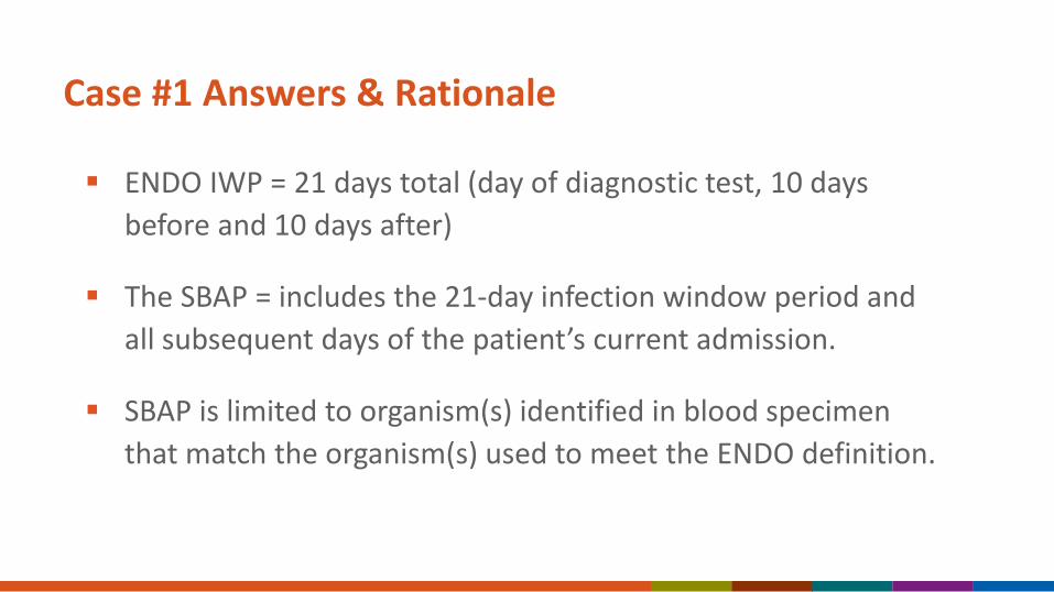
ENDO IWP = 21 days total (day of diagnostic test, 10 days before and 10 days after)
The SBAP = includes the 21-day infection window period and all subsequent days of the patient’s current admission.
SBAP is limited to organism(s) identified in blood specimen that match the organism(s) used to meet the ENDO definition.
Case #1 Answers & Rationale
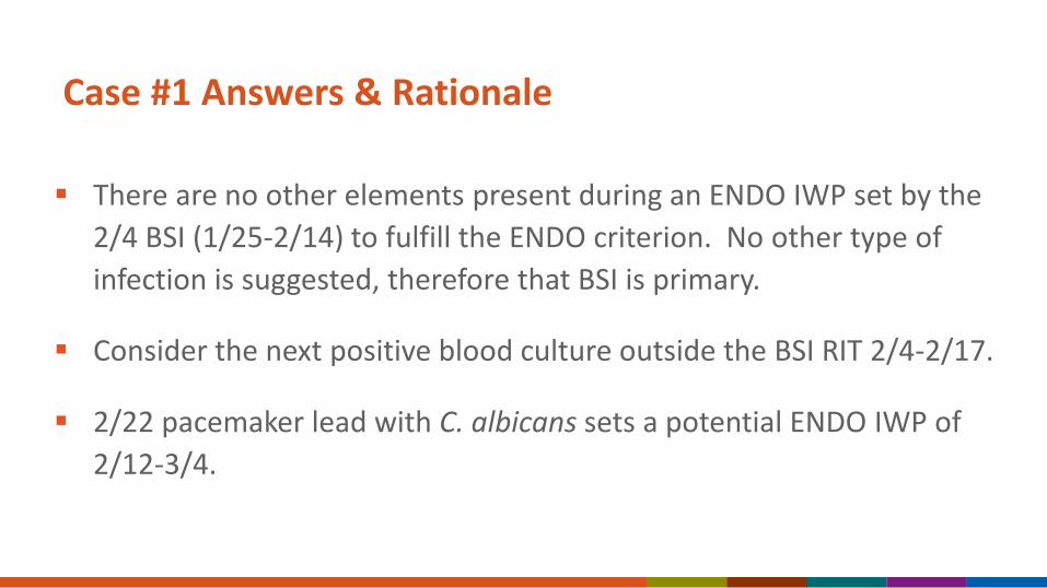
There are no other elements present during an ENDO IWP set by the 2/4 BSI (1/25-2/14) to fulfill the ENDO criterion. No other type of infection is suggested, therefore that BSI is primary.
Consider the next positive blood culture outside the BSI RIT 2/4-2/17.
2/22 pacemaker lead with C. albicans sets a potential ENDO IWP of 2/12-3/4.
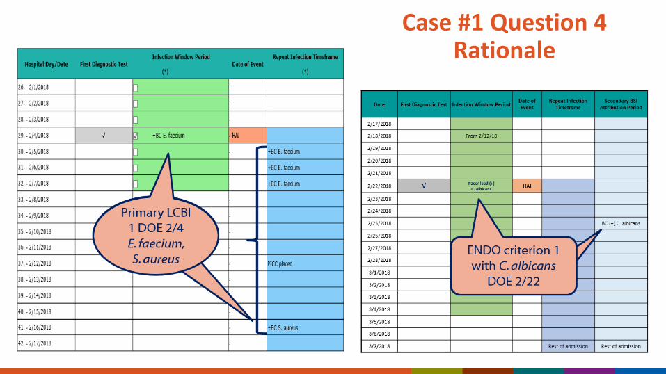
Case #1 Question 4 Rationale

Case #2

Case #2 Scenario
2/2/18: Admit with fever and chills of unknown origin, knee has been sore last few days but no obvious specific symptoms.
2/8/18: Knee fluid drained, sent for culture which grew S. aureus
2/14/18: Increased pain Left knee, unable to bend it due to swelling
2/17/18: +blood specimen S. aureus
2/22/18: +blood specimen S. aureus, S. pneumoniae
2/24/18: +blood specimen Strep pneumoniae, Neisseria gonorrhea
Case #2
Question 1: Which definition should be used in this case?
Question 2: What is the final determination for this case?

Case #2 Answers & Rationale
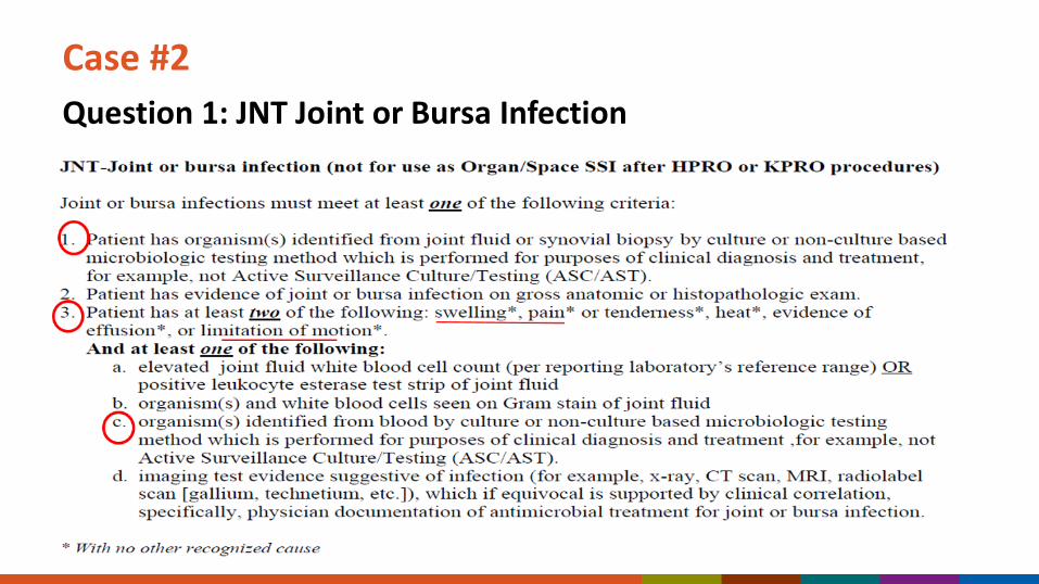
Question 1: JNT Joint or Bursa InfectionCase #2
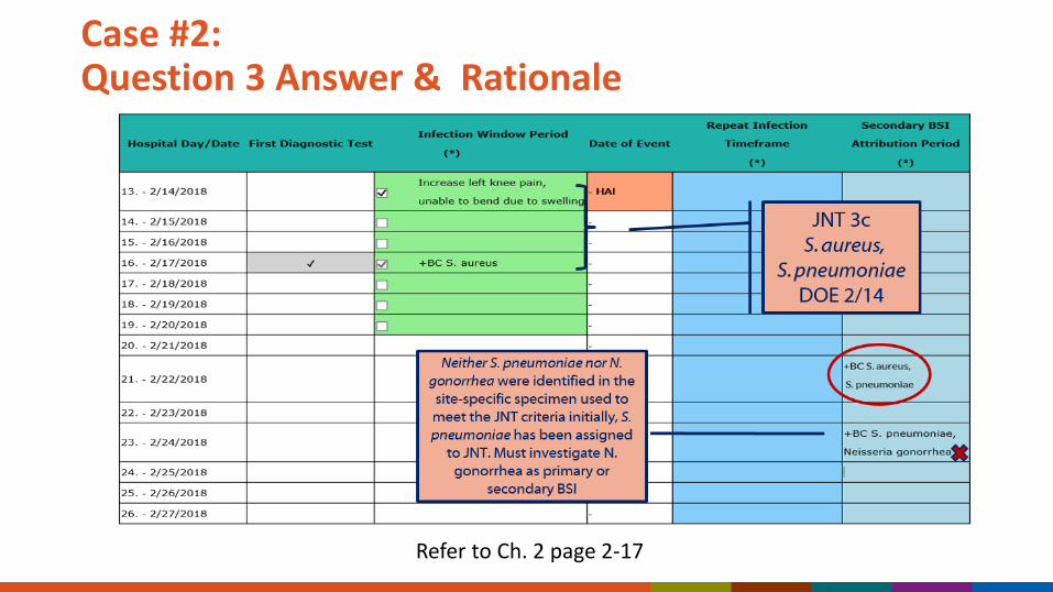
Case #2: Question 3 Answer & Rationale
Refer to Ch. 2 page 2-17