Embed Size (px)
Citation preview

Chapter 16
Distraction Osteogenesis
Hossein Behnia, Azita Tehranchi and Golnaz Morad
Additional information is available at the end of the chapter
http://dx.doi.org/10.5772/54647
1. Introduction
Distraction osteogenesis (DO) is defined as the formation of new bone between the vascularsurfaces of osteotomized bone segments, separated gradually by distraction forces. [1] Theincipient concept of distraction osteogenesis, as first described for correction of limb lengthdiscrepancies by Codivilla [2] in 1905, represented an osteotomized femur subjected torepeated forces of traction and counter-traction. This technique achieved a length increase of3 to 8 cm; an amount that surpassed that attainable by other methods common at that time.Codivilla asserted that confronting the resistance of the muscles surrounding a bone isinevitable if the discrepancies are to be corrected. Abbott and Saunders later used the techniquefor elongation of tibia. [3] Distraction osteogenesis proved advantageous over other conven‐tional methods for management of bone defects, particularly bone grafting, in that it providedsimultaneous expansion of the functional soft tissue matrix, referred to as distraction histogen‐esis. [1] The method however, remained undeveloped until it resurged in 1950s by Ilizarov,leading to several successful endeavors increasing the length of the extremities. In 1992,McCarthy [4] expanded the application of distraction osteogenesis to the craniofacial skeletonby attempting to ameliorate mandibular length deficiency in patients with hemifacial micro‐somia and Nager’s syndrome. Accomplishing an average increase of 20 mm in the mandibularlength in these preliminary cases, craniofacial DO rendered promising insights for treatmentof craniofacial skeletal abnormalities. Hitherto, a plethora of treatment protocols and modal‐ities have evolved in order to improve the outcomes of craniofacial DO.
2. The biology of distraction osteogenesis
Distraction osteogenesis initiates by surgically simulating bone fractures via osteotomy of thedeficient bone. Normal fracture healing occurs through a cascade of molecular and cellular
© 2013 Behnia et al.; licensee InTech. This is an open access article distributed under the terms of the CreativeCommons Attribution License (http://creativecommons.org/licenses/by/3.0), which permits unrestricted use,distribution, and reproduction in any medium, provided the original work is properly cited.

events triggered in response to injury. Formation of hematoma followed by chondrogenesisand angiogenesis eventually leads to the formation of hard callus by means of intramembra‐nous and endochondral ossification. The resultant woven bone subsequently remodels into amore mature lamellar bone to restore the strength and function of the organ. [5], [6] Duringdistraction osteogenesis however, the application of mechanical forces to the bone segmentsalters the repair process of the osteotomized bone segments, characteristic of fracture healing,to a regenerative process. [5] Evaluation of mechanotransduction mechanisms has demon‐strated that tensile forces increase the expression of bone morphogenic proteins by osteoblastsand stimulate intramembranous bone formation. [7] This regenerative effect of gradualtraction on tissue growth was originally designated by Ilizarov as the Law of Tension-Stress.[8] The process of distraction osteogenesis incorporates 3 major phases. The Latency phase isthe period Which starts immediately subsequent to the creation of osteotomy and lasts till thecommencement of distraction. This delay allows for tissue organization and formation of calluswhich bridges the gap between the two osteotomized bone surfaces. [5], [9], [10] Aronson etal conducted an animal study to test the outcome of different latency durations on boneregenerations. [11] Contrasting to other concurrent studies, it was observed that bone forma‐tion was most reliable when no latency period was considered prior to distraction; hence thesuggestion that latency phase may not be essential. A recent study appraising the benefits ofthe latency period on the outcomes of dentoalveolar DO proved that although a latency perioddid not enhance the amount and density of the newly formed bone, it slightly increased bonematuration. However, it was assumed that despite the minor effect of the latency period onthe regenerated bone, this phase may be crucial for soft tissue regeneration. [12] Regardless ofthe existing controversy on the importance of the latency phase, a review on the correspondingliterature demonstrated that a 5-7 day latency period is the most recommendable protocol forthe various indications of craniofacial DO. [1]The second phase known as the Distraction periodis characterized by the application of distraction forces. Histologically, this phase begins withconfiguration of a Fibrous Inter-Zone (FIZ) at which dense fibers of collagen demonstrate alongitudinal arrangement parallel to the direction of the distraction forces. In between thecollagen bundles osteoblastic activity creates a zone of Micro-Column Formation (MCF). Thetwo ends of the FIZ are characterized by areas of primary mineralization, thus dubbed asPrimary Mineralization Front (PMF). [5], [10], [13] The distraction phase continues with activesynthesis of fibrous tissue in central areas and active mineralization at the ends, till the acquiredamount of elongation is gained. [14] As the major features of the distraction phase, Ilizarovunderscored the significance of the rate and rhythm of distraction forces on the quality andquantity of the newly regenerated bone. The rate and the rhythm, respectively defined as thespeed and the frequency of the applied distraction forces were assessed in an animal study.The results, reported in 1989, suggested distraction of 1 mm per day applied as 0.25 mm per6 hours as the ideal distraction rate and rhythm for limb elongation. This was while slowerdistraction rates led to premature consolidation of the bone and faster distraction wasaccompanied by hindrance of osteogenesis. Moreover, he claimed that more satisfying resultswere obtained when distraction forces were applied with higher frequencies. [15] The currentstandard protocols of craniofacial DO seem to apply distraction forces at a rate of 1 mm perday 2-3 times. Nevertheless, these features may be altered in different patients. [1] Subsequent
A Textbook of Advanced Oral and Maxillofacial Surgery450

to the cessation of distraction, the third phase, designated as the Consolidation phase begins.This stage of distraction osteogenesis is distinguished by the growth of mineralization in acentripetal pattern. Moreover, transverse bridging connects the micro-columns of bone in theIFZ, leaving a honeycomb appearance. By the end of this phase, bone is being remodeled intoa more mature lamellar bone, strong enough to function. [10] The duration of this period canbe determined based on the amount of distraction. One month consolidation has beensuggested for each centimeter of distraction. [16]
3. Mandibular distraction osteogenesis
The primary attempts for mandibular distraction osteogenesis date back to the 1970s whenanimal studies were designed to restore surgically shortened canine mandibles via gradualdistraction. [17], [18] McCarthy et al were the first to apply gradual distraction for lengtheningthe human mandible. [4] The preliminary report of their study presented four children whounderwent unilateral and bilateral mandibular expansion for management of unilateralmicrosomia and Nager’s syndrome, respectively; 18 to 24 mm of mandibular distraction wasachieved. Common indications for mandibular distraction are summarized in Table 1.
Syndromic
Goldenhar Syndrome
Pierre Robin Sequence
Treacher Collins Syndrome
Nager’s Syndrome
Non-Syndromic mandibular hypoplasia (Unilateral/ Bilateral)
Retrognathia
Narrow mandible
Hemifacial Microsomia
Obstructive Sleep Apnea
Temporomandibular joint
Lack of TMJ translation
TMJ Ankylosis
Segmental bone defects
Alveolar Deficiencies
Table 1. Common indications for mandibular distraction osteogenesis
3.1. Mandibular lengthening
Mandibular hypoplasia, a condition associated with length deficiency in the mandibularramus or/ and body, is a manifestation of impairment of mandibular growth caused bysyndromic or non-syndromic congenital conditions or as a result of trauma. Depending on theseverity of the deficiency, mild to severe esthetic and functional problems arise which obligate
Distraction Osteogenesishttp://dx.doi.org/10.5772/54647
451

intervention. Mandibular hypoplasia has been traditionally managed by bone grafting,orthognathic surgery, and orthodontic therapy. These treatment approaches may contributeto considerable morbidity and unsatisfactory results in many situations, not to mentioninstances in which treatment appears unfeasible. The advent of craniofacial distractionosteogenesis has provided an alternative treatment modality to eliminate the shortcomings ofconventional protocols for management of this craniofacial discrepancy. [4] In correction ofmandibular hypoplasia in angle’s class II patients where great amounts of advancement (> 10mm) are required, DO has shown promising results with negligible relapse. This is probablydue to the simultaneous expansion of the surrounding soft tissue. [19] In comparison, thebilateral sagittal split osteotomy, the most common treatment choice, not only provides lessstable results with advancements larger than 6 mm, but is also likely to be accompanied byserious adverse events, namely neurosensory disturbance of inferior alveolar nerve anddisorders of the temporomandibular joint. [20], [21] On the other hand, bone grafting is anothercommon choice of treatment for severe hypoplastic mandibles. [22], [23] Yet, the procedureseems not to be very desirable, since it may be associated with donor site morbidity andresorption of the graft. Another important indication for mandibular distraction osteogenesisis a lack of condylar translation. In growing patients with mandibular hypoplasia in whomcondylar translations occurs normally during mandibular movements, functional orthodontictreatment may be of greater merit for restoration of the deficiency. Nevertheless, DO is atechnique-sensitive procedure and demands patient compliance. Therefore, until randomizedcontrolled trials have proved it beneficial over other treatment options for mandibularhypoplasia, it remains an alternative rather than a replacement for the existing treatmentmodalities. [20], [21]A variety of distraction devices have been designed and introduced forclinical implications; each associated with pros and cons. Extraoral distractors which are fixedin place by transcutaneous pins, are generally easier to manipulate and allow for multidirec‐tional distraction. However, the psychosocial problems consequent to the presence of thedevice as well as facial scarring led to the emergence of intraoral distractors. [24], [25] McCarthyintroduced the first intraoral distraction device in 1995. [26] Intraoral distractors are of threetypes: tooth-borne, bone-borne and hybrid distractors. [25] Finite element analysis of intraoraldistraction devices demonstrated the hybrid type to be the most stable under masticatoryloads, while tooth-borne distractors were the most reliable in transferring the expansion to thebone. [27] The morbidity associated with bone-borne devices appear to be higher than tooth-borne distractors. [28] While tooth-borne devices seem beneficial as they facilitate subsequenttooth movement, concerns such as greater mandibular expansion at the alveolar sectioncomparing to the basal bone may arise with this type of distractor. [25] Shetye et al demon‐strated that application of intraoral distractors may be associated with higher incidence ofminor adverse events, with no effect on treatment outcome. This is while the occurrence ofmajor incidents is more likely when extraoral distraction devices are used. [29]Mandibularhypoplasia is generally divided into two groups of unilateral and bilateral hypoplasia. A meta-analysis of mandibular distraction osteogenesis demonstrated the most common indicationfor unilateral DO to be hemifacial/craniofacial microsomia. [30] Unilateral craniofacialmicrosomia is a genetic disorder that affects the derivatives of the first and the second brachialarch and is initially characterized by abnormal growth of the mandibular ramus. The asym‐
A Textbook of Advanced Oral and Maxillofacial Surgery452

metric growth of the mandible may gradually affect the growth of the surrounding structures,a fact that encourages surgeons to begin treating patients at early ages. The resultant facialasymmetry has been corrected via unilateral DO particularly in growing children. [31], [32]The authors analyzed the posteroanterior cephalometric changes subsequent to unilateraldistraction osteogenesis in 10 patients. [33] Improvements in the piriform angle, intergonialangle, and the occlusal cant revealed the influence of the treatment on the maxillary andmidfacial growth. It is highly suggested that the treatment be continued with functionalorthodontic therapy in growing children. Functional appliances can act to obtain symmetryduring growth. We have performed the combination of distraction osteogenesis and functionalorthodontic therapy in a group of our patients. [34] It is advisable that the patient be followeduntil the end of growth and if necessary the functional orthodontic therapy be continued.
3.1.1. Lengthening for asymmetry
Case 1
An 8-year-old male patient with a history of right condylar trauma at birth presented withfacial asymmetry, cant of the occlusal plane, deviation of the midline, and a deep-bite maloc‐clusion (Figure 1-A, B, C). A horizontal osteotomy was made at the body of the right ramusbelow the mandibular foramen and a custom-made unidirectional extraoral distractor wasfixed in place (Figure 1-D, E). Following a 7-day latency period, the distractor was activatedat a rate of 1mm/ day. Distraction was stopped after the ramus was elongated by 22 mm (Figure1-F). Subsequent to removal of the distractor a hybrid functional appliance was used to managethe posterior right open-bite created as a result of mandibular lengthening (Figure 1-G, H).Functional therapy continued for 3 years when fixed orthodontic therapy was initiated in orderto restore the position of impacted left canine (Figure 1-I, J).
3.1.2. Unilateral mandibular hypoplasia
Case 2
A 6-year-old male patient with a history of trauma at age 2, presented with facial asymmetryand midline deviation due to unilateral mandibular hypoplasia (Figure 2-A, B). A horizontalosteotomy was done in the right ramus and a unidirectional intraoral distractor (KLS Martin,Tuttlingen, Germany) was fixed in place (Figure 2-C). Distraction was initiated with an obliquevector (Figure 2-D). Consequently, along with a posterior open bite, the teeth were deviatedto the opposite side to a considerable extent (Figure 2-E). This was corrected via cross elastictraction (Figure 2-F). The patient was followed during growth and no deviation or facialasymmetry occurred; hence no need for further orthodontic treatment.
3.1.3. Hemifacial microsomia
Case 3
A 9-year-old male patient with hemifacial microsomia type 2 A was planned to receivedistraction osteogenesis for treatment of facial asymmetry (Figure 3-A, B). Mandibular ramus
Distraction Osteogenesishttp://dx.doi.org/10.5772/54647
453

was elongated using an extraoral distractor. Following a 2-month consolidation period, thedistractor was removed. Orthodontic functional therapy was started to correct the posterioropen bite (Figure 3-C, D). Orthodontic therapy was continued for 5 years (Figure 3-E, F).
Figure 1. A. Pre-distraction facial appearance. Facial asymmetry and cant of occlusal plane is apparent. B. Pre-distraction intraoral view. C.
Panoramic view. D. Horizontal osteotomy was made at the body of the right ramus below the mandibular foramen. E. A custom-made
unidirectional extraoral distractor was fixed in place. F. Ramus was elongated by 22 mm. G. The posterior open-bite was created at the right side as
a result of mandibular lengthening. H. A hybrid functional appliance was used to manage the posterior right open-bite. I. Facial appearance 3 years
post-distraction. J. Panoramic view 3 years post-distraction.
3.1.2. Unilateral mandibular hypoplasia
Case 2
A 6-year-old male patient with a history of trauma at age 2, presented with facial asymmetry and midline deviation due to
unilateral mandibular hypoplasia (Figure 2-A, B). A horizontal osteotomy was done in the right ramus and a unidirectional
intraoral distractor (KLS Martin, Tuttlingen, Germany) was fixed in place (Figure 2-C). Distraction was initiated with an oblique
vector (Figure 2-D). Consequently, along with a posterior open bite, the teeth were deviated to the opposite side to a considerable
extent (Figure 2-E). This was corrected via cross elastic traction (Figure 2-F). The patient was followed during growth and no
deviation or facial asymmetry occurred; hence no need for further orthodontic treatment.
(a) (b) (c)
(e) (d) (f)
(g) (h)
(i) (j)
Figure 1. (a) Pre-distraction facial appearance. Facial asymmetry and cant of occlusal plane is apparent. (b) Pre-distrac‐tion intraoral view. (c) Panoramic view. (d) Horizontal osteotomy was made at the body of the right ramus below themandibular foramen. (e) A custom-made unidirectional extraoral distractor was fixed in place. (f) Ramus was elongat‐ed by 22 mm. (g) The posterior open-bite was created at the right side as a result of mandibular lengthening. (h) Ahybrid functional appliance was used to manage the posterior right open-bite. (i) Facial appearance 3 years post-dis‐traction. (j) Panoramic view 3 years post-distraction.
A Textbook of Advanced Oral and Maxillofacial Surgery454

3.1.4. Hemifacial microsomia
Case 4
A 17 year-old female patient with hemifacial microsomia presented with facial asymmetry andmidline deviation to the right. The right maxillary canine was impacted (Figure 4-A-E). Pre-distraction orthodontic therapy included maxillary expansion and repositioning the impactedcanine into the arch (Figure 4-F, G). Subsequently, unilateral osteotomy in the ramus wasperformed and an extraoral distraction device was fixed in place. With a rate of 1mm per day,distraction was continued until adequate elongation was obtained (Figure 4-H-K ). Fixedorthodontic treatment was ongoing during the consolidation period (Figure 4-L). Finalmaxillary and mandibular arch coordination was achieved through bimaxillary orthognathicsurgery (Figure 4-M-S).
Figure 2. A. Pre-distraction facial appearance demonstrates facial asymmetry due to childhood trauma. B. Intraoral view shows midline deviation.
C. A horizontal osteotomy was made at the body of the right ramus and an intraoral distractor was fixed in place. D. Distraction was initiated with
an oblique vector. E. Post-distraction intraoral view. Teeth were deviated to the opposite side. F. Deviation was corrected via cross elastic traction.
3.1.3. Hemifacial microsomia
Case 3
A 9-year-old male patient with hemifacial microsomia type 2 A was planned to receive distraction osteogenesis for treatment of
facial asymmetry (Figure 3-A, B). Mandibular ramus was elongated using an extraoral distractor. Following a 2-month
consolidation period, the distractor was removed. Orthodontic functional therapy was started to correct the posterior open bite
(Figure 3-C, D). Orthodontic therapy was continued for 5 years (Figure 3-E, F).
(a) (b) (c)
(e) (d)
(f)
Figure 2. (a) Pre-distraction facial appearance demonstrates facial asymmetry due to childhood trauma. (b) Intraoralview shows midline deviation. (c) A horizontal osteotomy was made at the body of the right ramus and an intraoraldistractor was fixed in place. (d) Distraction was initiated with an oblique vector. (e) Post-distraction intraoral view.Teeth were deviated to the opposite side. (f) Deviation was corrected via cross elastic traction.
Distraction Osteogenesishttp://dx.doi.org/10.5772/54647
455

3.1.5. Facial asymmetry
Case 5
A 13-year-old female patient presented with mandibular deformity due to left condylarankylosis (Figure 5-A). The patient had received a costochondral graft at age 6 and the functionof the joint was restored (Figure 5-B). The remaining facial asymmetry was planned to beresolved via distraction osteogenesis. Using an extraoral custom-made distraction device, theleft ramus was elongated by 18 mm (Figure 5-C). The resultant posterior open bite wascorrected via 3 years of hybrid functional therapy followed by fixed orthodontic treatment(Figure 5-D, E, F).
3.1.6. Mandibular asymmetry due to condylar ankylosis
Case 6
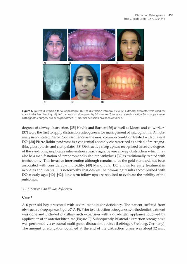
A 16-year-old female patient presented with mandibular asymmetry due to left condylarankylosis (Figure 6-A, B). At age 8, the patient had undergone a condylectomy procedure fortreatment of the condylar ankylosis. She was then a candidate for distraction osteogenesis.Elongation of the left ramus (20 mm) was achieved by an extraoral distraction device (LeibingerMultiguide, Freiburg, Germany) (Figure 6-C, D). Eight months following removal of thedistractor, the patient was orthodontically prepared for an orthognathic surgery. The surgeryincluded Le Fort I and bilateral sagittal split osteotomies as well as genioplasty. A normal classI occlusion was obtained (Figure 6-E, F).
(a) (b) (c)
(e) (d) (f)
Figure 3. (a) Pre-distraction facial asymmetry due to hemifacial microsomia. (b) Pre-distraction intraoral view. (c) Post-distraction intraoral view. Unilateral posterior open was created. (d) Orthodontic functional therapy was started tocorrect the posterior open bite. (e) Five years post-distraction intraoral view. (f) Facial appearance 5 years post-distrac‐tion.
A Textbook of Advanced Oral and Maxillofacial Surgery456

(a) (b) (c)
(d) (e) (f)
(g) (h) (i)
(j) (k)
(l) (m) (n)
(o) (p) (q)
Figure 4. (a) Pre-distraction facial asymmetry. (b) Pre-distraction intraoral view. (c) Pre-distraction panoramic view. (d)Pre-distraction lateral cephalometric view. (e) Pre-distraction posteroanterior (PA) cephalometric view. (f) Orthodon‐
Distraction Osteogenesishttp://dx.doi.org/10.5772/54647
457

tics included maxillary expansion. (g) The impacted canine was brought into the arch. (h) Post-distraction facial ap‐pearance. (i) Post-distraction intraoral view. (j) Post-distraction panoramic view. (k) Post-distraction lateralcephalometric view. (l) Post-distraction PA cephalometric view. (m) Post-orthognathic surgery facial appearance. (n)Post-orthognathic surgery intraoral view. (o) Post-orthognathic surgery panoramic radiograph. (p) Lateral cephalo‐metric radiograph. (q) Post-orthognathic surgery PA cephalometric radiograph.
Figure 5. A. Pre-distraction facial asymmetry. B. Pre-distraction panoramic view. Bone screws remained from a previous costochondral bone graft
can be observed. C. Immediate post-distraction panoramic view. Mandibular ramus elongated by 18 mm. D. Posterior open bite was corrected via
functional therapy and fixed orthodontic therapy. E. Six years post-distraction facial appearance. F. Normal occlusion was obtained.
3.1.6. Mandibular asymmetry due to condylar ankylosis
Case 6
A 16-year-old female patient presented with mandibular asymmetry due to left condylar ankylosis (Figure 6-A, B). At age 8, the
patient had undergone a condylectomy procedure for treatment of the condylar ankylosis. She was then a candidate for distraction
osteogenesis. Elongation of the left ramus (20 mm) was achieved by an extraoral distraction device (Leibinger Multiguide,
Freiburg, Germany) (Figure 6-C, D). Eight months following removal of the distractor, the patient was orthodontically prepared for
an orthognathic surgery. The surgery included Le Fort I and bilateral sagittal split osteotomies as well as genioplasty. A normal
class I occlusion was obtained (Figure 6-E, F).
(a) (b)
(c)
(e)
(d)
(f)
Figure 5. (a) Pre-distraction facial asymmetry. (b) Pre-distraction panoramic view. Bone screws remained from a previ‐ous costochondral bone graft can be observed. (c) Immediate post-distraction panoramic view. Mandibular ramuselongated by 18 mm. (d) Posterior open bite was corrected via functional therapy and fixed orthodontic therapy. (e)Six years post-distraction facial appearance. (f) Normal occlusion was obtained.
3.2. Bilateral hypoplasia
Similar to unilateral mandibular hypoplasia, several etiologies are documented for bilateralhypoplasia including syndromic conditions, condylar fracture due to trauma, and class IImalocclusion. Along with undesirable facial appearance and disorders in the masticatorysystem, micrognathia which itself may be symmetric or asymmetric, can cause mild to lethal
A Textbook of Advanced Oral and Maxillofacial Surgery458
degrees of airway obstruction. [35] Havlik and Bartlett [36] as well as Moore and co-workers[37] were the first to apply distraction osteogenesis for management of micrognathia. A meta-analysis indicated Pierre Robin sequence as the most common condition treated with bilateralDO. [30] Pierre Robin syndrome is a congenital anomaly characterized as a triad of microgna‐thia, glossoptosis, and cleft palate. [38] Obstructive sleep apnea; recognized in severe degreesof the syndrome, implicates intervention at early ages. Severe airway obstruction which mayalso be a manifestation of temporomandibular joint ankylosis [39] is traditionally treated withtracheotomy. This invasive intervention although remains to be the gold standard, has beenassociated with considerable morbidity. [40] Mandibular DO allows for early treatment inneonates and infants. It is noteworthy that despite the promising results accomplished withDO at early ages [40]- [42], long-term follow-ups are required to evaluate the stability of theoutcomes.
3.2.1. Severe mandibular deficiency
Case 7
A 6-year-old boy presented with severe mandibular deficiency. The patient suffered fromobstructive sleep apnea (Figure 7-A-F). Prior to distraction osteogenesis, orthodontic treatmentwas done and included maxillary arch expansion with a quad-helix appliance followed byapplication of an anterior bite plate (Figure G). Subsequently, bilateral distraction osteogenesiswas performed via extraoral multi-guide distraction devices (Leibinger, Freiburg, Germany).The amount of elongation obtained at the end of the distraction phase was about 32 mm;
Figure 6. A. Pre-distraction facial appearance. B. Pre-distraction intraoral view. C. Extraoral distractor was used for mandibular lengthening. D.
Left ramus was elongated by 20 mm. E. Two years post-distraction facial appearance. Orthognathic surgery has been performed. F. Normal
occlusion has been obtained.
3.2. Bilateral hypoplasia
Similar to unilateral mandibular hypoplasia, several etiologies are documented for bilateral hypoplasia including syndromic
conditions, condylar fracture due to trauma, and class II malocclusion. Along with undesirable facial appearance and disorders in
the masticatory system, micrognathia which itself may be symmetric or asymmetric, can cause mild to lethal degrees of airway
obstruction.[35] Havlik and Bartlett[36] as well as Moore and co-workers [37] were the first to apply distraction osteogenesis for
management of micrognathia. A meta-analysis indicated Pierre Robin sequence as the most common condition treated with
bilateral DO.[30] Pierre Robin syndrome is a congenital anomaly characterized as a triad of micrognathia, glossoptosis, and cleft
palate.[38] Obstructive sleep apnea; recognized in severe degrees of the syndrome, implicates intervention at early ages. Severe
airway obstruction which may also be a manifestation of temporomandibular joint ankylosis[39] is traditionally treated with
tracheotomy. This invasive intervention although it remains to be the gold standard, has been associated with considerable
morbidity.[40] Mandibular DO allows for early treatment in neonates and infants. It is noteworthy that despite the promising results
accomplished with DO at early ages [40]-[42], long-term follow-ups are required to evaluate the stability of the outcomes.
3.2.1. Severe mandibular deficiency
Case 7
A 6-year-old boy presented with severe mandibular deficiency. The patient suffered from obstructive sleep apnea (Figure 7-A-F).
Prior to distraction osteogenesis, orthodontic treatment was done and included maxillary arch expansion with a quad-helix
appliance followed by application of an anterior bite plate (Figure G). Subsequently, bilateral distraction osteogenesis was
performed via extraoral multi-guide distraction devices (Leibinger, Freiburg, Germany). The amount of elongation obtained at the
end of the distraction phase was about 32 mm; though not equal on both sides (Figure H-J). Obstructive sleep apnea was
completely resolved in this patient. Treatment was continued with functional orthodontic therapy; however, the patient was only
followed for 2 years (Figure K-P).
(a) (b) (c)
(e) (d) (f)
Figure 6. (a) Pre-distraction facial appearance. (b) Pre-distraction intraoral view. (c) Extraoral distractor was used formandibular lengthening. (d) Left ramus was elongated by 20 mm. (e) Two years post-distraction facial appearance.Orthognathic surgery has been performed. (f) Normal occlusion has been obtained.
Distraction Osteogenesishttp://dx.doi.org/10.5772/54647
459
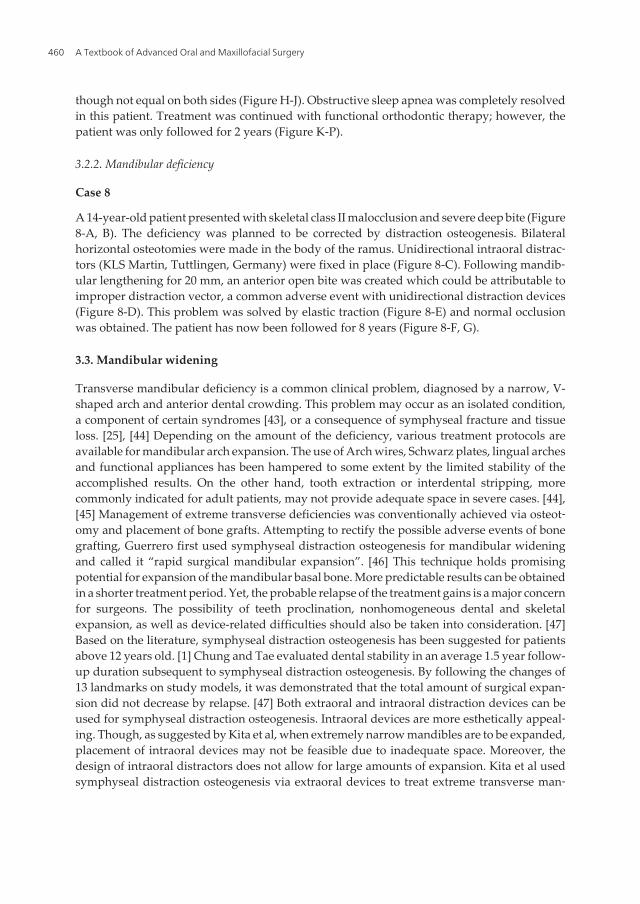
though not equal on both sides (Figure H-J). Obstructive sleep apnea was completely resolvedin this patient. Treatment was continued with functional orthodontic therapy; however, thepatient was only followed for 2 years (Figure K-P).
3.2.2. Mandibular deficiency
Case 8
A 14-year-old patient presented with skeletal class II malocclusion and severe deep bite (Figure8-A, B). The deficiency was planned to be corrected by distraction osteogenesis. Bilateralhorizontal osteotomies were made in the body of the ramus. Unidirectional intraoral distrac‐tors (KLS Martin, Tuttlingen, Germany) were fixed in place (Figure 8-C). Following mandib‐ular lengthening for 20 mm, an anterior open bite was created which could be attributable toimproper distraction vector, a common adverse event with unidirectional distraction devices(Figure 8-D). This problem was solved by elastic traction (Figure 8-E) and normal occlusionwas obtained. The patient has now been followed for 8 years (Figure 8-F, G).
3.3. Mandibular widening
Transverse mandibular deficiency is a common clinical problem, diagnosed by a narrow, V-shaped arch and anterior dental crowding. This problem may occur as an isolated condition,a component of certain syndromes [43], or a consequence of symphyseal fracture and tissueloss. [25], [44] Depending on the amount of the deficiency, various treatment protocols areavailable for mandibular arch expansion. The use of Arch wires, Schwarz plates, lingual archesand functional appliances has been hampered to some extent by the limited stability of theaccomplished results. On the other hand, tooth extraction or interdental stripping, morecommonly indicated for adult patients, may not provide adequate space in severe cases. [44],[45] Management of extreme transverse deficiencies was conventionally achieved via osteot‐omy and placement of bone grafts. Attempting to rectify the possible adverse events of bonegrafting, Guerrero first used symphyseal distraction osteogenesis for mandibular wideningand called it “rapid surgical mandibular expansion”. [46] This technique holds promisingpotential for expansion of the mandibular basal bone. More predictable results can be obtainedin a shorter treatment period. Yet, the probable relapse of the treatment gains is a major concernfor surgeons. The possibility of teeth proclination, nonhomogeneous dental and skeletalexpansion, as well as device-related difficulties should also be taken into consideration. [47]Based on the literature, symphyseal distraction osteogenesis has been suggested for patientsabove 12 years old. [1] Chung and Tae evaluated dental stability in an average 1.5 year follow-up duration subsequent to symphyseal distraction osteogenesis. By following the changes of13 landmarks on study models, it was demonstrated that the total amount of surgical expan‐sion did not decrease by relapse. [47] Both extraoral and intraoral distraction devices can beused for symphyseal distraction osteogenesis. Intraoral devices are more esthetically appeal‐ing. Though, as suggested by Kita et al, when extremely narrow mandibles are to be expanded,placement of intraoral devices may not be feasible due to inadequate space. Moreover, thedesign of intraoral distractors does not allow for large amounts of expansion. Kita et al usedsymphyseal distraction osteogenesis via extraoral devices to treat extreme transverse man‐
A Textbook of Advanced Oral and Maxillofacial Surgery460

Figure 7. -A. Pre-distraction facial appearance. B. Profile view. C. Intraoral view. D. Pre-distraction panoramic view. E. Pre-distraction lateral
cephalometric view. F. Pre-distraction posteroanterior cephalometric view. G. Pre-distraction orthodontic treatment included maxillary expansion
via a quad-helix appliance. H. Post-distraction facial appearance. I. Profile view. J. Lateral cephalometric view. Two years post-distraction
appearance. L. Profile. M. Intraoral view. N. Two years post-distraction panoramic view. O. Post-distraction lateral cephalogram. P. Two years post-
distraction PA cephalogram.
3.2.2. Mandibular deficiency
Case 8
(a) (b) (c)
(e) (d) (f)
(g) (h) (i) (j)
(k)
(o) (p)
(l) (m)
(n)
Figure 7. (a) Pre-distraction facial appearance. (b) Profile view. (c) Intraoral view. (d) Pre-distraction panoramic view.(e) Pre-distraction lateral cephalometric view. (f) Pre-distraction posteroanterior cephalometric view. (g) Pre-distrac‐tion orthodontic treatment included maxillary expansion via a quad-helix appliance. (h) Post-distraction facial appear‐ance. (i) Profile view. (j) Lateral cephalometric view. (k) Two years post-distraction appearance. (l) Profile. (m) Intraoralview. (n) Two years post-distraction panoramic view. (o) Post-distraction lateral cephalogram. (p) Two years post-dis‐traction PA cephalogram.
Distraction Osteogenesishttp://dx.doi.org/10.5772/54647
461

dibular deficiencies in patients with hypoglossia-hypodactyly syndrome. [43]A plethora ofinvestigations and modifications have attributed to enhanced efficiency of mandibulardistraction osteogenesis. Yet, the technique is not exempt of adverse events. Diverse rates ofincidence have been reported for different mandibular distraction osteogenesis procedures.Shetye et al [29] classified the potential adverse events associated with mandibular DO intothree groups: minor incidents indicated those with no influence on the outcome. Moderate and
(a) (b) (c)
(e) (d)
(f) (g)
Figure 8. (a) Pre-distraction facial appearance. (b) Pre-distraction intraoral view. (c) Unidirectional intraoral distractorswere fixed in place. (d) Following mandibular lengthening for 20 mm, an anterior open bite was created. (e) Anterioropen bite was corrected by elastic traction. (f) Eight years post-distraction facial appearance. (g) Intraoral view.
A Textbook of Advanced Oral and Maxillofacial Surgery462

major incidents were both defined as events that result in undesirable outcome and can orcannot be resolved via invasive procedures, respectively. Their 16 year follow-up of 141patients who underwent mandibular DO for unilateral or bilateral mandibular lengtheningdemonstrated that minor and moderate incidents were reported in 26.99% and 20.35%,respectively; while in only 5.31% of patients did major events occur. The majority of majorincidents included TMJ ankylosis and derangements as well as fibrous union. Nevertheless,taken all the above-mentioned complications into considerations, investigators seem to beunanimous in the safety of distraction osteogenesis.
3.4. Maxillary distraction osteogenesis
The Principles of distraction osteogenesis have been applied for correction of transverse andsagittal discrepancies of the maxilla and the midface associated with orofacial clefts and severalsyndromes. Midfacial distraction osteogenesis was first evaluated in animal studies performedon sheep [48] and dogs [49]. A preliminary human report of maxillary and midfacial advance‐ment through the application of a distraction device was published by Cohen et al in 1997. [50]Two children with cleft lip and palate, midfacial hypoplasia, and class III malocclusionunderwent treatment with distraction osteogenesis. Up to 11 mm advancement of the midfacialcomplex was achieved in both patients. Two years later, Mommaerts introduced a techniquefor maxillary expansion using a transpalatal distractor. [51] In comparison to rapid palatalexpansion, the treatment protocol most frequently used for maxillary expansion, palataldistraction osteogenesis was asserted to eliminate particular adverse events such as alveolarbending, tooth tipping, buccal cortex fenestration, and relapse. Common indications formaxillary and midface distraction osteogenesis are summarized in Table 2.
Orofacial Clefts
Craniosynostosis
Crouzon’s Syndrome
Apert’s Syndrome
Pfeiffer Syndrome
Midface deficiencies of other causes
Alveolar deficiencies
Table 2. Common indications for maxillary and midface distraction osteogenesis.
3.5. Maxillary and midfacial advancement
The majority of cleft lip and palate patients suffer from degrees of maxillary hypoplasia, eitheras a primary manifestation of the cleft or secondary to attempts for cleft repair. This oftencomplex discrepancy is conventionally corrected through a series of surgeries includingdifferent osteotomies. The inception of distraction osteogenesis for advancement of maxillary-midface in cleft patients brought new insight into the treatment of these patients. A meta-analysis of conventional osteotomies and distraction osteogenesis, along with manysimilarities between the two techniques, suggested distraction osteogenesis to be advanta‐
Distraction Osteogenesishttp://dx.doi.org/10.5772/54647
463

geous for it eliminates the need for bone grafts. [52] Moreover, it was demonstrated that mostprotocols postponed treatment with conventional osteotomies until growth was completed.In contrast, distraction osteogenesis was more frequently performed in growing patients;although, overcorrection was recommended to preclude relapse. Different types of extraoraland intraoral distractors have been established for maxillary distraction osteogenesis. Extrao‐ral distractors have the capacity for multidirectional maxillary advancement and the vectorscan be changed during the process. [53] Yet, many patients have difficulty accepting extraoraldevices primarily due to the unappealing appearance and discomfort. [54] Moreover, theexternal position of these devices makes them prone to loosening and fracture following anaccidental trauma. [53] Rigid external distraction (RED) device is fixed to the cranium. Thisallows for protection of maxillary teeth comparing to other types of extraoral devices whichare anchored to the maxilla. [54] The stability of maxillary advancement with RED wasevaluated in a 3-year prospective study. To avoid the possible interference of growth in theoutcomes, the study was performed on adult patients. The relapse was reported to be 22% after3 years. [55] Internal distractors cause less psychosocial problems for the patient and are lesslikely to be loosened or displaced during the distraction period or following traumatic forces.Moreover, being more easily tolerated by patients, an internal device can be maintained duringthe consolidation phase for as long as deemed necessary for prevention of relapse. [53], [54],[56] Nevertheless, installation of intraoral distractors may not be always feasible due toinadequate space. In addition, intraoral distractors provide unidirectional bone movement;hence demanding precise positioning. [53] Complications such as fracture and collapse of thecleft alveolar bone has been reported with intraoral devices used for Le Fort I distraction. [56]Picard et al described a rigid internal device (RID) with the ability to provide unrestrictedlengthening for total or segmental advancement of the maxilla. In 19 syndromic, cleft, andtraumatic patients treated with this distractor, an average advancement of 9.6 mm wasachieved. [54] A retrospective study comparing extraoral and intraoral distractors for midfaceadvancement in syndromic patients demonstrated no significant difference regarding thecomplication rate and amount of lengthening between the two types. Accordingly, bothdistractors were asserted to be safe and it was suggested that choosing a device be individu‐alized based on each patients needs and toleration. [53]Mild to moderate cases of maxillaryhypoplasia which were traditionally corrected via Le Fort I osteotomy, have been successfullytreated with anterior maxillary distraction osteogenesis. [57] Le Fort I osteotomy shows anegative impact on velopharyngeal competence and speech while this problem is rarely seenwith anterior maxillary distraction osteogenesis. [57] Nonetheless, more severe cases maynecessitate Le Fort I distraction.
3.5.1. Cleft lip and palate and class III malocclusion
Case 9
A 17-year-old female patient with cleft lip and palate presented with a class III malocclusionand anterior and posterior cross bite (Figure 9 A-C). Following pre-distraction orthodontictreatment, maxillary advancement was performed through Le Fort I osteotomy and REDdevice (KLS Martin, Figure 9-D). Maxilla was advanced by 18 mm (Figure 9-E, F). Orthodontic
A Textbook of Advanced Oral and Maxillofacial Surgery464

treatment continued for a year. Meanwhile, the patient received a removable partial prosthesisto replace the anterior missing teeth. Subsequently, the patient underwent rhinoplasty andprimary lip repair (Figure 9-G-J). The procedures for lip repair are still ongoing.
patients treated with this distractor, an average advancement of 9.6 mm was achieved. [54] A retrospective study comparing
extraoral and intraoral distractors for midface advancement in syndromic patients demonstrated no significant difference
regarding the complication rate and amount of lengthening between the two types. Accordingly, both distractors were asserted to
be safe and it was suggested that choosing a device be individualized based on each patients needs and toleration.[53]Mild to
moderate cases of maxillary hypoplasia which were traditionally corrected via Le Fort I osteotomy, have been successfully treated
with anterior maxillary distraction osteogenesis.[57] Le Fort I osteotomy shows a negative impact on velopharyngeal competence
and speech while this problem is rarely seen with anterior maxillary distraction osteogenesis.[57] Nonetheless, more severe cases
may necessitate Le Fort I distraction.
3.5.1. Cleft lip and palate and class III malocclusion
Case 9
A 17-year-old female patient with cleft lip and palate presented with a class III malocclusion and anterior and posterior cross bite
(Figure 9 A-C). Following pre-distraction orthodontic treatment, maxillary advancement was performed through Le Fort I
osteotomy and RED device (KLS Martin, Figure 9-D). Maxilla was advanced by 18 mm (Figure 9-E, F). Orthodontic treatment
continued for a year. Meanwhile, the patient received a removable partial prosthesis to replace the anterior missing teeth.
Subsequently, the patient underwent rhinoplasty and primary lip repair (Figure 9-G-J). The procedures for lip repair are still
ongoing.
Figure 9. A. Pre-distraction appearance. B. Profile view. C. Lateral cephalometric view. D. Maxillary distraction osteogenesis was performed using
an RED device. E. Post-distraction facial appearance. F. Post-distraction lateral cephalometric view. G. Two years post-distraction facial appearance.
H. Profile. I. Intraoral view. J. Lateral cephalometric view.
Distraction osteogenesis has also proved valuable for treatment of patients affected with craniosynostosis. This condition caused as
a result of premature fusion of cranial sutures is a clinical feature of particular syndromes such as Crouzon, Apert, and Pfeiffer. In
the severe expression of these syndromes, it may be crucial to initiate treatment as early as 1 year of age.[58] Le Fort III and
monobloc osteotomies are frequently used for management of craniosynostosis.[59] During the recent years, distraction osteogenesis
has become popular for correction of syndromic maxillary hypoplasia. The amount of advancement of midface that can be
(a) (b) (c)
(e) (d) (f)
(g) (h) (i) (j)
Figure 9. (a) Pre-distraction appearance. (b) Profile view. (c) Lateral cephalometric view. (d) Maxillary distraction os‐teogenesis was performed using an RED device. (e) Post-distraction facial appearance. (f) Post-distraction lateral ceph‐alometric view. (g) Two years post-distraction facial appearance. (h) Profile. (i) Intraoral view. (j) Lateral cephalometricview.
Distraction osteogenesis has also proved valuable for treatment of patients affected withcraniosynostosis. This condition caused as a result of premature fusion of cranial sutures is aclinical feature of particular syndromes such as Crouzon, Apert, and Pfeiffer. In the severeexpression of these syndromes, it may be crucial to initiate treatment as early as 1 year of age.[58] Le Fort III and monobloc osteotomies are frequently used for management of craniosy‐nostosis. [59] During the recent years, distraction osteogenesis has become popular forcorrection of syndromic maxillary hypoplasia. The amount of advancement of midface thatcan be achieved by distraction osteogenesis is generally greater than the amount obtained byconventional osteotomies such as Le Fort III and monobloc osteotomy. [60] Long-term follow
Distraction Osteogenesishttp://dx.doi.org/10.5772/54647
465

ups of syndromic patients who underwent maxillary-midface advancement with distractionosteogenesis have proved the stability of the results. [59], [61], [62]
3.6. Maxillary expansion
Maxillary transverse deficiency is a condition associated with anterior and posterior dentalcrowding, unilateral or bilateral cross-bite, as well as TMJ and respiratory problems. Expansionof maxillary bone during growth is usually feasible through orthodontic treatments. However,with skeletal maturation, a combination of surgical and orthodontic techniques may beinevitable in order to accomplish adequate expansion. When the condition is accompaniedwith cleft lip and palate, it poses even greater challenges for treatment. Treatment of maxillaryconstriction can be performed by means of Le Fort I osteotomy and expansion. This protocolallows for multidirectional expansion of the maxillary complex; however, the resistance of thepalatal fibromucosa diminishes the stability of the results. Another treatment option estab‐lished for this deformity is surgically assisted rapid maxillary expansion (SARME). Thistechnique eliminates soft tissue resistance via distraction histogenesis and can be based uponeither tooth-borne or bone-borne devices. Tooth-borne distractors transfer forces to the teeth,leading to tooth-related adverse events such as root resorption, tooth tipping, and corticalfenestration. In contrast, bone-borne devices; first introduced by Mommaerts as a transpalataldistractor (TPD), are exempt of these undesirable effects for they directly apply forces to thebone. [63], [64] Application of bone-borne distractors becomes of paramount importanceparticularly when insufficient tooth support exists due to tooth missing and impaction. [65]Yet, they require a secondary surgery for removal. [63], [64] It is noteworthy that despite thedisadvantages commonly considered for tooth-borne device, no considerable difference indental tipping and stability has been found between tooth-borne and bone-borne maxillarydistractors. [64], [66]
3.6.1. Skeletal class III malocclusion and narrow maxilla
Case 10
A 25-year-old female patient presented with dental and skeletal class III malocclusion and anarrow maxilla. Both maxillary and mandibular midlines had a shift to the right side. A 2-mmreverse over jet and anterior and posterior cross bites were present (Figure 10-A-D). Restrictionin mandibular movement was found on examination. Treatment plan included SARPE viaSmile distractor (Titamed). Transverse distraction was started at a rate of 1mm/ day andcontinued until 10 mm expansion was achieved (Figure 10-E, F). Post-distraction fixedorthodontic treatment closed the resultant gap between the two central incisors and reposi‐tioned the right lateral incisor into the dental arch (Figure 10-G-J).
3.6.2. Maxillary transverse deficiency
Case 11
A 20-year-old male patient presented with a class III malocclusion, maxillary transversedeficiency, severe anterior crowding, anterior open-bite, and bilateral cross-bite (Figure 11-A-
A Textbook of Advanced Oral and Maxillofacial Surgery466

E). The treatment plan included SARME with a bone-borne distractor followed by orthognathicsurgery in order to respectively correct the transverse deficiency and the open bite. Anosteotomy was made in the palatal midline, between the roots of the two central incisors andthe distraction device (Smile distractor, Titamed) was placed (Figure 11-F-H). Following a 7-day latency period, the distractor was activated at a rate of 1mm/ day. When expansion of 10mm was achieved, activation was stopped and the device was maintained for a 2-monthconsolidation period (Figure 11-I). The device was kept for another 4 months until the space
(a) (b) (c)
(e) (d) (f)
(g) (h) (i)
(j)
Figure 10. (a) Pre-distraction facial appearance. (b) Profile. (c) Intraoral view. (d) Pre-distraction intraoral view, theright lateral incisor is in a palatal position. (e) Post-distraction intraoral view. Maxilla expanded by 10mm. (f) Post-dis‐traction radiograph. (g) Post-orthodontic treatment facial appearance. (h) Profile. (i) Intraoral view. (j) Post-orthodon‐tic intraoral view. The right lateral incisor is repositioned into the arch.
Distraction Osteogenesishttp://dx.doi.org/10.5772/54647
467

created between the central incisors was closed by orthodontic forces. Subsequently, thepatient was orthodontically prepared for orthognathic surgery, Le Fort I osteotomy (Figure11-K). Arch coordination was obtained. The patient is still under orthodontic treatment (Figure11-L-P). It is worth mentioning that in this patient, alignment of maxillary teeth could havebeen achieved by extraction of premolars and fixed orthodontic therapy. However, thistreatment protocol would impede maxillary and mandibular arch coordination. On the otherhand, it is important that the amount of maxillary expansion is proportional to the mandibulararch.
3.7. Alveolar distraction osteogenesis
Alveolar distraction osteogenesis is pre-implant/ pre-prosthetic procedure which tends torestore the alveolar deficiencies and prepare the alveolar ridge for further rehabilitativetreatments. Alveolar distraction osteogenesis was first evaluated in an animal model by Blocket al. [67] Chin and Toth extended its application to human. [68] Alveolar ridge augmentationis frequently conducted via the use of different types of bone grafts. However, distractionosteogenesis not only decreases the complications and the duration of treatment, but alsoallows for reconstruction of large defects by simultaneously expanding the surrounding softtissue. [69] Studies have suggested the amount of newly formed bone resorption prior toimplant placement to be greater with onlay bone grafting in comparison to distractionosteogenesis. Peri-implant bone loss was comparable between the two techniques. [70], [71]On the other hand, the amount of augmentation gained with distraction osteogenesis wasreported to be significantly greater than that obtained with inlay bone grafts. [72] Dependingon the type and the extension of an alveolar defect, distraction osteogenesis may be consideredeither as an absolute treatment for reconstruction or as an adjunctive therapy along with otherbone grafting procedures. Jensen and Block presented a classification of alveolar defectsaiming to facilitated treatment planning with alveolar distraction osteogenesis. Accordingly,the more complex a defect, the greater the possibility of requiring bone grafts prior or subse‐quent to distraction osteogenesis. [73] This treatment modality can also be considered whenprevious attempts for bone grafting have failed. [74]
3.7.1. Vertical alveolar distraction osteogenesis
The technique of alveolar distraction osteogenesis have been successfully used for enhancingalveolar ridge height. [69], [74]- [81] The majority of studies evaluated the efficiency ofdistraction osteogenesis in the anterior parts of maxilla and mandible and the amount ofobtained augmentation was reported between 5 to 15 mm. Benefits of this method for aug‐mentation of severely atrophic ridges remains to be a matter of controversy. Basal bone fractureand neurosensory complications have been suggested as the two most common problemsassociated with vertical distraction of atrophic mandibular ridges. [82] Indication of verticalalveolar distraction osteogenesis is therefore limited to areas where 5-7 mm of alveolar boneexists. [83]
A Textbook of Advanced Oral and Maxillofacial Surgery468

(a) (b) (c)
(e) (d)
(f) (g) (h)
(i) (j) (k)
(l) (m) (n)
(o) (p)
Figure 11. (a) Pre-distraction facial appearance. (b) Profile. (c) Intraoral view. (d) Pre-distraction panoramic view. (e)Pre-distraction lateral cephalometric view. (f) Osteotomy was made in the palatal midline. (g) Palatal distractor inplace before activation. (h) Periapical radiography. (i) Two months post-distraction intraoral view. (j) Two months post-distraction occlusal radiograph. (k) Anterior open bite was planned to be corrected via orthognathic surgery. (l) Post-orthognathic surgery facial appearance. (m) Profile. (n) Intraoral view. (o) Post-orthognathic surgery panoramic view.(p) Post-orthognathic cephalometric view.
Distraction Osteogenesishttp://dx.doi.org/10.5772/54647
469

Distraction devices for alveolar distraction osteogenesis include both extraoral and intraoraldevices as well as distraction implants. Extraoral distractors which are mainly positionedsubperiosteally in the buccal vestibule, can only be used when a bone height of 6-7 mm ispresent. Comparing intraoral and extraoral distractors for alveolar distraction osteogenesis,Uckan et al demonstrated that the majority of complications associated with intraoral distrac‐tors were related to displacement and fracture of the transport segment. However, interferenceof the device with the opposing dental arch was considered the most frequent complicationwith extraoral distractors. [71] Distraction implants initially pose distraction forces to augmentthe alveolar ridge and are subsequently kept in place to act as a dental implant. These deviceshave the advantage of eliminating the need for a second surgery for distractor removal. Yet,it is highly likely that the ideal position of the distractor does not correspond to the desirableposition of the implant. [81], [83]
3.7.2. Alveolar deficiency
Case 11
A 20 year-old female patient presented with a large bone defect in the anterior mandible dueto resection of a central giant cell granuloma (Figure 12-A, B). The patient underwent alveolardistraction osteogenesis. Horizontal osteotomy was performed and an intraoral distractor(KLS Martin) was placed (Figure 12-C, D). Following a 7-day latency phase distraction wasinitiated at a rate of 1 mm per day. 18mm augmentation was achieved (Figure 12-E). After theconsolidation period, the distractor was removed and dental implants were inserted into theregenerated bone. Due to insufficient ridge width, a guided bone regeneration procedure wasdone to induce bone regeneration over the exposed surfaces of the implants (Figure 12-F). 3months later, at the second stage of implant surgery inadequate keratinized tissue wascompensated by a connective tissue graft from the palate (Figure 12-G-I). Fixed implant-supported prosthesis restored the missing teeth (Figure 12-J).
3.8. Horizontal alveolar distraction osteogenesis
Horizontal alveolar ridge augmentation through distraction osteogenesis demands forextreme preciseness in technique and the design of distractors. The amount of alveolar ridgewidth increase reported with distraction osteogenesis is 2.5 mm to 7 mm. [84]- [89]Horizontal alveolar distraction can be conducted via simple bone screws to meticulouslydesigned distractors. All devices have a distraction rod in common which is fixed in thecortical lingual/ palatal bone plate and allows for the buccal/ labial movement of the transfersegment. [84] Nevertheless, the intricacy of device positioning as well as the difficulty ofperforming an osteotomy in a narrow alveolar ridge have greatly restricted the indicationof horizontal alveolar distraction osteogenesis. [85] Therefore, in many cases with horizon‐tal alveolar deficiency, bone grafting techniques become advantageous over distractionosteogenesis.
A Textbook of Advanced Oral and Maxillofacial Surgery470

Figure 12. A. Large bone defect in the anterior mandible due to resection of a central giant cell granuloma. Intraoral view. B. Large anterior
mandibular defect. Panoramic view. C. Intraoral distractor was placed. D. Panoramic view. E. Post-distraction panoramic view 18mm
augmentation was achieved. F. As a result of insufficient ridge width, a guided bone regeneration was done with implant placement. G. Second
stage implant surgery, 3 months later. H. Inadequate keratinized tissue was compensated by a connective tissue graft harvested from the palate. I.
Two months following the second stage implant surgery. J. Fixed implant-supported prosthesis was placed. Panoramic view.
3.8. Horizontal alveolar distraction osteogenesis
Horizontal alveolar ridge augmentation through distraction osteogenesis demands for extreme preciseness in technique and the
design of distractors. The amount of alveolar ridge width increase reported with distraction osteogenesis is 2.5 mm to 7 mm.[84]-[89]
Horizontal alveolar distraction can be conducted via simple bone screws to meticulously designed distractors. All devices have a
distraction rod in common which is fixed in the cortical lingual/ palatal bone plate and allows for the buccal/ labial movement of
the transfer segment.[84] Nevertheless, the intricacy of device positioning as well as the difficulty of performing an osteotomy in a
narrow alveolar ridge have greatly restricted the indication of horizontal alveolar distraction osteogenesis.[85] Therefore, in many
cases with horizontal alveolar deficiency, bone grafting techniques become advantageous over distraction osteogenesis.
Acknowledgement
Figure 12. A. Large bone defect in the anterior mandible due to resection of a central giant cell granuloma. Intraoralview. B. Large anterior mandibular defect. Panoramic view. C. Intraoral distractor was placed. D. Panoramic view. E.Post-distraction panoramic view 18mm augmentation was achieved. F. As a result of insufficient ridge width, a guidedbone regeneration was done with implant placement. G. Second stage implant surgery, 3 months later. H. Inadequatekeratinized tissue was compensated by a connective tissue graft harvested from the palate. I. Two months followingthe second stage implant surgery. J. Fixed implant-supported prosthesis was placed. Panoramic view.
Distraction Osteogenesishttp://dx.doi.org/10.5772/54647
471

Acknowledgements
The authors wish to express their sincere gratitude to Dr. Ladan Eslamian; Professor ofDepartment of Orthodontics, School of Dentistry, Shahid Beheshti university of MedicalSciences who took the responsiblity for orthodontic management of one of our patients,presented as case 9.
Author details
Hossein Behnia1*, Azita Tehranchi2 and Golnaz Morad3
1 Department of Oral and Maxillofacial Surgery, School of Dentistry, Shahid Beheshti Uni‐versity of Medical Sciences, Tehran, Iran
2 Department of Orthodontics, School of Dentistry, Shahid Beheshti University of MedicalSciences, Tehran, Iran
3 Dental Research Center, Shahid Beheshti University of Medical Sciences, Tehran, Iran
References
[1] Swennen G, Schliephake H, Dempf R, Schierle H, Malevez C. Craniofacial distractionosteogenesis: a review of the literature: Part 1: clinical studies. Int J Oral MaxillofacSurg 2001;30(2):89-103.
[2] Codivilla A. The classic: On the means of lengthening, in the lower limbs, the mus‐cles and tissues which are shortened through deformity. 1905. Clin Orthop Relat Res2008;466(12):2903-9.
[3] Abbott LC, Saunders JB. The Operative Lengthening of the Tibia and Fibula: a Pre‐liminary Report on the Further Development of the Principles and Technic. Ann Surg1939;110(6):961-91.
[4] McCarthy JG, Schreiber J, Karp N, Thorne CH, Grayson BH. Lengthening the humanmandible by gradual distraction. Plast Reconstr Surg 1992;89(1):1-8; discussion 9-10.
[5] Ai-Aql ZS, Alagl AS, Graves DT, Gerstenfeld LC, Einhorn TA. Molecular mecha‐nisms controlling bone formation during fracture healing and distraction osteogene‐sis. J Dent Res 2008;87(2):107-18.
[6] Marsh DR, Li G. The biology of fracture healing: optimising outcome. Br Med Bull1999;55(4):856-69.
A Textbook of Advanced Oral and Maxillofacial Surgery472

[7] Morgan EF, Gleason RE, Hayward LN, Leong PL, Palomares KT. Mechanotransduc‐tion and fracture repair. J Bone Joint Surg Am 2008;90 Suppl 1:25-30.
[8] Ilizarov GA. The tension-stress effect on the genesis and growth of tissues. Part I. Theinfluence of stability of fixation and soft-tissue preservation. Clin Orthop Relat Res1989(238):249-81.
[9] Fischgrund J, Paley D, Suter C. Variables affecting time to bone healing during limblengthening. Clin Orthop Relat Res 1994(301):31-7.
[10] Aronson J. Experimental and clinical experience with distraction osteogenesis. CleftPalate Craniofac J 1994;31(6):473-81; discussion 81-2.
[11] Aronson J, Shen X. Experimental healing of distraction osteogenesis comparing meta‐physeal with diaphyseal sites. Clin Orthop Relat Res 1994(301):25-30.
[12] Moore C, Campbell PM, Dechow PC, Ellis ML, Buschang PH. Effects of latency onthe quality and quantity of bone produced by dentoalveolar distraction osteogenesis.Am J Orthod Dentofacial Orthop;140(4):470-8.
[13] Aronson J, Good B, Stewart C, Harrison B, Harp J. Preliminary studies of mineraliza‐tion during distraction osteogenesis. Clin Orthop Relat Res 1990(250):43-9.
[14] Vauhkonen M, Peltonen J, Karaharju E, Aalto K, Alitalo I. Collagen synthesis andmineralization in the early phase of distraction bone healing. Bone Miner 1990;10(3):171-81.
[15] Ilizarov GA. The tension-stress effect on the genesis and growth of tissues: Part II.The influence of the rate and frequency of distraction. Clin Orthop Relat Res1989(239):263-85.
[16] Merloz P. Bone regeneration and limb lengthening. Osteoporos Int;22(6):2033-6.
[17] Michieli S, Miotti B. Lengthening of mandibular body by gradual surgical-orthodon‐tic distraction. J Oral Surg 1977;35(3):187-92.
[18] Snyder CC, Levine GA, Swanson HM, Browne EZ, Jr. Mandibular lengthening bygradual distraction. Preliminary report. Plast Reconstr Surg 1973;51(5):506-8.
[19] Altug-Atac AT, Grayson BH, McCarthy JG. Comparison of skeletal and soft-tissuechanges following unilateral mandibular distraction osteogenesis. Plast ReconstrSurg 2008;121(5):1751-9.
[20] Ow A, Cheung LK. Skeletal stability and complications of bilateral sagittal split os‐teotomies and mandibular distraction osteogenesis: an evidence-based review. J OralMaxillofac Surg 2009;67(11):2344-53.
[21] Schreuder WH, Jansma J, Bierman MW, Vissink A. Distraction osteogenesis versusbilateral sagittal split osteotomy for advancement of the retrognathic mandible: a re‐view of the literature. Int J Oral Maxillofac Surg 2007;36(2):103-10.
Distraction Osteogenesishttp://dx.doi.org/10.5772/54647
473

[22] Kaban LB, Moses MH, Mulliken JB. Surgical correction of hemifacial microsomia inthe growing child. Plast Reconstr Surg 1988;82(1):9-19.
[23] Padwa BL, Mulliken JB, Maghen A, Kaban LB. Midfacial growth after costochondralgraft construction of the mandibular ramus in hemifacial microsomia. J Oral Maxillo‐fac Surg 1998;56(2):122-7; discussion 27-8.
[24] Pereira MA, Luiz de Freitas PH, da Rosa TF, Xavier CB. Understanding distractionosteogenesis on the maxillofacial complex: a literature review. J Oral Maxillofac Surg2007;65(12):2518-23.
[25] Tae KC, Kang KH, Kim SC. Unilateral mandibular widening with distraction osteo‐genesis. Angle Orthod 2005;75(6):1053-60.
[26] McCarthy JG, Staffenberg DA, Wood RJ, Cutting CB, Grayson BH, Thorne CH. Intro‐duction of an intraoral bone-lengthening device. Plast Reconstr Surg 1995;96(4):978-81.
[27] Boccaccio A, Cozzani M, Pappalettere C. Analysis of the performance of different or‐thodontic devices for mandibular symphyseal distraction osteogenesis. Eur J Orthod;33(2):113-20.
[28] Raoul G, Wojcik T, Ferri J. Outcome of mandibular symphyseal distraction osteogen‐esis with bone-borne devices. J Craniofac Surg 2009;20(2):488-93.
[29] Shetye PR, Warren SM, Brown D, Garfinkle JS, Grayson BH, McCarthy JG. Documen‐tation of the incidents associated with mandibular distraction: introduction of a newstratification system. Plast Reconstr Surg 2009;123(2):627-34.
[30] Ow AT, Cheung LK. Meta-analysis of mandibular distraction osteogenesis: clinicalapplications and functional outcomes. Plast Reconstr Surg 2008;121(3):54e-69e.
[31] Kofod T, Norholt SE, Pedersen TK, Jensen J. Unilateral mandibular ramus elongationby intraoral distraction osteogenesis. J Craniofac Surg 2005;16(2):247-54.
[32] Shetye PR, Grayson BH, Mackool RJ, McCarthy JG. Long-term stability and growthfollowing unilateral mandibular distraction in growing children with craniofacial mi‐crosomia. Plast Reconstr Surg 2006;118(4):985-95.
[33] Tehranchi A, Behnia H. Facial symmetry after distraction osteogenesis and orthodon‐tic therapy. Am J Orthod Dentofacial Orthop 2001;120(2):149-53.
[34] Tehranchi A, Behnia H. Treatment of mandibular asymmetry by distraction osteo‐genesis and orthodontics: a report of four cases. Angle Orthod 2000;70(2):165-74.
[35] Wang X, Wang XX, Liang C, Yi B, Lin Y, Li ZL. Distraction osteogenesis in correctionof micrognathia accompanying obstructive sleep apnea syndrome. Plast ReconstrSurg 2003;112(6):1549-57; discussion 58-9.
A Textbook of Advanced Oral and Maxillofacial Surgery474

[36] Havlik RJ, Bartlett SP. Mandibular distraction lengthening in the severely hypoplas‐tic mandible: a problematic case with tongue aplasia. J Craniofac Surg 1994;5(5):305-10; discussion 11-2.
[37] Moore MH, Guzman-Stein G, Proudman TW, Abbott AH, Netherway DJ, David DJ.Mandibular lengthening by distraction for airway obstruction in Treacher-Collinssyndrome. J Craniofac Surg 1994;5(1):22-5.
[38] P R.
[39] Feiyun P, Wei L, Jun C, Xin X, Zhuojin S, Fengguo Y. Simultaneous correction of bi‐lateral temporomandibular joint ankylosis with mandibular micrognathia using in‐ternal distraction osteogenesis and 3-dimensional craniomaxillofacial models. J OralMaxillofac Surg;68(3):571-7.
[40] Senders CW, Kolstad CK, Tollefson TT, Sykes JM. Mandibular distraction osteogene‐sis used to treat upper airway obstruction. Arch Facial Plast Surg;12(1):11-5.
[41] Scott AR, Tibesar RJ, Lander TA, Sampson DE, Sidman JD. Mandibular distractionosteogenesis in infants younger than 3 months. Arch Facial Plast Surg;13(3):173-9.
[42] Shen W, Jie C, Chen J, Zou J, Ji Y. Mandibular distraction osteogenesis to relievePierre Robin severe airway obstruction in neonates: indication and operation. J Cra‐niofac Surg 2009;20 Suppl 2:1812-6.
[43] Kita H, Kochi S, Yamada A, Imai Y, Konno N, Saitou C, et al. Mandibular wideningby distraction osteogenesis in the treatment of a constricted mandible and telescopicbite. Cleft Palate Craniofac J 2004;41(6):664-73.
[44] Conley R, Legan H. Mandibular symphyseal distraction osteogenesis: diagnosis andtreatment planning considerations. Angle Orthod 2003;73(1):3-11.
[45] Sukurica Y, Gurel HG, Mutlu N. Six year follow-up of a patient treated with mandib‐ular symphyseal distraction osteogenesis. J Craniomaxillofac Surg;38(1):26-31.
[46] Gurrereo.
[47] Chung YW, Tae KC. Dental stability and radiographic healing patterns after mandib‐ular symphysis widening with distraction osteogenesis. Eur J Orthod 2007;29(3):256-62.
[48] Rachmiel A, Jackson IT, Potparic Z, Laufer D. Midface advancement in sheep bygradual distraction: a 1-year follow-up study. J Oral Maxillofac Surg 1995;53(5):525-9.
[49] Staffenberg DA, Wood RJ, McCarthy JG, Grayson BH, Glasberg SB. Midface distrac‐tion advancement in the canine without osteotomies. Ann Plast Surg 1995;34(5):512-7.
Distraction Osteogenesishttp://dx.doi.org/10.5772/54647
475

[50] Cohen SR, Burstein FD, Stewart MB, Rathburn MA. Maxillary-midface distraction inchildren with cleft lip and palate: a preliminary report. Plast Reconstr Surg1997;99(5):1421-8.
[51] Mommaerts MY. Transpalatal distraction as a method of maxillary expansion. Br JOral Maxillofac Surg 1999;37(4):268-72.
[52] Cheung LK, Chua HD. A meta-analysis of cleft maxillary osteotomy and distractionosteogenesis. Int J Oral Maxillofac Surg 2006;35(1):14-24.
[53] Meling TR, Hogevold HE, Due-Tonnessen BJ, Skjelbred P. Midface distraction osteo‐genesis: internal vs. external devices. Int J Oral Maxillofac Surg;40(2):139-45.
[54] Picard A, Diner PA, Galliani E, Tomat C, Vazquez MP, Carls FP. Five years experi‐ence with a new intraoral maxillary distraction device (RID). Br J Oral MaxillofacSurg;49(7):546-51.
[55] Aksu M, Saglam-Aydinatay B, Akcan CA, El H, Taner T, Kocadereli I, et al. Skeletaland dental stability after maxillary distraction with a rigid external device in adultcleft lip and palate patients. J Oral Maxillofac Surg;68(2):254-9.
[56] Mitsukawa N, Satoh K, Morishita T. Le Fort I distraction using internal devices formaxillary hypoplasia in patients with cleft lip, palate, and alveolus: complicationsand their prevention and management. J Craniofac Surg;21(5):1428-30.
[57] Richardson S, Agni NA, Selvaraj D. Anterior maxillary distraction using a tooth-borne device for hypoplastic cleft maxillas-a pilot study. J Oral Maxillofac Surg;69(12):e542-8.
[58] Marchac A, Arnaud E. Cranium and midface distraction osteogenesis: current practi‐ces, controversies, and future applications. J Craniofac Surg;23(1):235-8.
[59] Kuroda S, Watanabe K, Ishimoto K, Nakanishi H, Moriyama K, Tanaka E. Long-termstability of LeFort III distraction osteogenesis with a rigid external distraction devicein a patient with Crouzon syndrome. Am J Orthod Dentofacial Orthop;140(4):550-61.
[60] Ko EW, Chen PK, Tai IC, Huang CS. Fronto-facial monobloc distraction in syndromiccraniosynostosis. Three-dimensional evaluation of treatment outcome and facialgrowth. Int J Oral Maxillofac Surg;41(1):20-7.
[61] Fearon JA. Halo distraction of the Le Fort III in syndromic craniosynostosis: a long-term assessment. Plast Reconstr Surg 2005;115(6):1524-36.
[62] Meazzini MC, Allevia F, Mazzoleni F, Ferrari L, Pagnoni M, Iannetti G, et al. Long-term follow-up of syndromic craniosynostosis after Le Fort III halo distraction: acephalometric and CT evaluation. J Plast Reconstr Aesthet Surg;65(4):464-72.
[63] Cortese A, Savastano M, Savastano G, Claudio PP. One-step transversal palatal dis‐traction and maxillary repositioning: technical considerations, advantages, and long-term stability. J Craniofac Surg;22(5):1714-9.
A Textbook of Advanced Oral and Maxillofacial Surgery476

[64] Seeberger R, Kater W, Schulte-Geers M, Davids R, Freier K, Thiele O. Changes aftersurgically-assisted maxillary expansion (SARME) to the dentoalveolar, palatal andnasal structures by using tooth-borne distraction devices. Br J Oral Maxillofac Surg;49(5):381-5.
[65] Verlinden CR, Gooris PG, Becking AG. Complications in transpalatal distraction os‐teogenesis: a retrospective clinical study. J Oral Maxillofac Surg;69(3):899-905.
[66] Verstraaten J, Kuijpers-Jagtman AM, Mommaerts MY, Berge SJ, Nada RM, Schols JG.A systematic review of the effects of bone-borne surgical assisted rapid maxillary ex‐pansion. J Craniomaxillofac Surg;38(3):166-74.
[67] Block MS, Chang A, Crawford C. Mandibular alveolar ridge augmentation in the dogusing distraction osteogenesis. J Oral Maxillofac Surg 1996;54(3):309-14.
[68] Chin M, Toth BA. Distraction osteogenesis in maxillofacial surgery using internal de‐vices: review of five cases. J Oral Maxillofac Surg 1996;54(1):45-53; discussion 54.
[69] Turker N, Basa S, Vural G. Evaluation of osseous regeneration in alveolar distractionosteogenesis with histological and radiological aspects. J Oral Maxillofac Surg2007;65(4):608-14.
[70] Perry M, Hodges N, Hallmon DW, Rees T, Opperman LA. Distraction osteogenesisversus autogenous onlay grafting. Part I: outcome of implant integration. Int J OralMaxillofac Implants 2005;20(5):695-702.
[71] Uckan S, Oguz Y, Bayram B. Comparison of intraosseous and extraosseous alveolardistraction osteogenesis. J Oral Maxillofac Surg 2007;65(4):671-4.
[72] Bianchi A, Felice P, Lizio G, Marchetti C. Alveolar distraction osteogenesis versus in‐lay bone grafting in posterior mandibular atrophy: a prospective study. Oral SurgOral Med Oral Pathol Oral Radiol Endod 2008;105(3):282-92.
[73] Jensen OT, Block M. Alveolar modification by distraction osteogenesis. Atlas OralMaxillofac Surg Clin North Am 2008;16(2):185-214.
[74] Nocini PF, Albanese M, Buttura da Prato E, D'Agostino A. Vertical distraction osteo‐genesis of the mandible applied to an iliac crest graft: report of a case. Clin Oral Im‐plants Res 2004;15(3):366-70.
[75] Dinse WE, Burnett RR. Anterior maxillary restoration using distraction osteogenesisand implants: a clinical report. J Prosthet Dent 2008;100(4):250-3.
[76] Gozneli R, Ozkan Y, Akalin ZF, Ozkan Y. Rehabilitation of maxillary anterior esthet‐ics by alveolar distraction osteogenesis with immediate implant placement: a case re‐port. Implant Dent;19(6):468-76.
[77] Lee HJ, Ahn MR, Sohn DS. Piezoelectric distraction osteogenesis in the atrophic max‐illary anterior area: a case report. Implant Dent 2007;16(3):227-34.
Distraction Osteogenesishttp://dx.doi.org/10.5772/54647
477

[78] Penarrocha-Diago M, Gomez-Adrian MD, Garcia-Garcia A, Camacho-Alonso F,Rambla-Ferrer J. Vertical mandibular alveolar bone distraction and dental implantplacement: a case report. J Oral Implantol 2006;32(3):137-41.
[79] Rachmiel A, Gutmacher Z, Blumenfeld I, Peled M, Laufer D. [Vertical alveolar ridgeaugmentation using distraction osteogenesis]. Refuat Hapeh Vehashinayim2001;18(1):64-9, 78.
[80] Raghoebar GM, Liem RS, Vissink A. Vertical distraction of the severely resorbededentulous mandible: a clinical, histological and electron microscopic study of 10treated cases. Clin Oral Implants Res 2002;13(5):558-65.
[81] Yalcin S, Ordulu M, Emes Y, Gur H, Aktas I, Caniklioglu C. Alveolar distraction os‐teogenesis before placement of dental implants. Implant Dent 2006;15(1):48-52.
[82] Perdijk FB, Meijer GJ, Strijen PJ, Koole R. Complications in alveolar distraction osteo‐genesis of the atrophic mandible. Int J Oral Maxillofac Surg 2007;36(10):916-21.
[83] McAllister BS, Gaffaney TE. Distraction osteogenesis for vertical bone augmentationprior to oral implant reconstruction. Periodontol 2000 2003;33:54-66.
[84] Aikawa T, Iida S, Senoo H, Hori K, Namikawa M, Okura M, et al. Widening a nar‐row posterior mandibular alveolus following extirpation of a large cyst: a case treat‐ed with a titanium mesh-plate type distractor. Oral Surg Oral Med Oral Pathol OralRadiol Endod 2008;106(5):e1-7.
[85] Bulut E, Muglali M, Celebi N, Bekcioglu B. Horizontal alveolar distraction of themandibular canine regions for implant placement. J Craniofac Surg;21(3):830-2.
[86] Garcia-Garcia A, Somoza-Martin M, Gandara-Vila P, Saulacic N, Gandara-Rey JM.Horizontal alveolar distraction: a surgical technique with the transport segmentpedicled to the mucoperiosteum. J Oral Maxillofac Surg 2004;62(11):1408-12.
[87] Laster Z, Rachmiel A, Jensen OT. Alveolar width distraction osteogenesis for earlyimplant placement. J Oral Maxillofac Surg 2005;63(12):1724-30.
[88] Oda T, Suzuki H, Yokota M, Ueda M. Horizontal alveolar distraction of the narrowmaxillary ridge for implant placement. J Oral Maxillofac Surg 2004;62(12):1530-4.
[89] Watzak G, Zechner W, Tepper G, Vasak C, Busenlechner D, Bernhart T. Clinicalstudy of horizontal alveolar distraction with modified micro bone screws and subse‐quent implant placement. Clin Oral Implants Res 2006;17(6):723-9.
A Textbook of Advanced Oral and Maxillofacial Surgery478