Embed Size (px)
Citation preview

©2020 Institute of Parasitology, SAS, KošiceDOI 10.2478/helm-2020-0027
252
HELMINTHOLOGIA, 57, 3: 252 – 267, 2020
Diversity of parasites from Xenopus laevis (Amphibia: Pipidae) and their seasonal rate of infection in selected habitats in the Limpopo Province, South Africa
E. M. MBOKANE1,2,*, J. THERON2, W. J. LUUS-POWEL3
1Aquaculture Research and Development, Department of Agriculture, Forestry and Fisheries, Private Bag X2, Vlaeberg, Cape Town 8012, South Africa, *E-mail: [email protected]; 2Aquaculture Research Unit, University of Limpopo,
Sovenga 0727, South Africa; 3Department of Biodiversity, University of Limpopo, Sovenga 0727, South Africa
Article info
Received August 2, 2019Accepted April 8, 2020
Summary
This study determined the diversity and seasonality of parasites species of the African clawed frog, Xenopus laevis (Daudin, 1802), from three localities, namely Modjadjikloof, Mokopane and Universi-ty of Limpopo, Limpopo Province, South Africa. A total of seven parasite species were collected and identifi ed. They included two nematodes, Camallanus kaapstaadi Southwell & Kirshner, 1937 and Batrachocamallanus slomei (Southwell & Kirshner, 1937), a monogenean, Protopolystoma xenopo-dis (Price, 1943), a cestode, Cephalochlamys namaquensis (Cohn, 1906), a protozoan, Trichodina xenopodos Fantham, 1924, two digeneans, Progonimodiscus doyeri Ortlepp, 1926 and Dollfuschella rodhaini Vercammen-Grandjean, 1960. The most common and abundant parasite species by far were Cm. kaapstaadi, B. slomei and Cp. namaquensis, with Cm. kaapstaadi, B. slomei present in all localities. Trichodina xenopodos was a rare species, only present in host populations from Mod-jadjiskloof. Modjadjiskloof had the highest species richness (all seven parasite species) followed by Mokopane (fi ve parasite species) and University of Limpopo (3 parasite species). There were also higher infection levels (prevalence and mean intensity) of Cm. kaapstaadi, B. slomei, Cp. namaquen-sis and Pt. xenopodis in hosts from Modjadjiskloof while Pd. doyeri and D. rodhaini infection levels were greater in Mokopane. The variability between localities shows that parasites with heteroxenous life cycles are more strongly associated with more pristine habitats. The variability in calculated in-dices (prevalence and mean intensity) also suggests that the occurrence of some of the parasites is affected by season, favouring higher infection rates during summer. This suggests that temperature has a direct role in the reproductive and developmental processes of these parasites. Neither length nor sex had an infl uence on the prevalence or intensity of parasites.Keywords: Season; Temperature; Heteroxenous; Monoxenous, Infections
Introduction
The African clawed frog, Xenopus laevis (Daudin, 1802), is an amphibian species that belongs to the family Pipidae. It is native to Africa where it naturally occurs in freshwater bodies of temperate, subtropical and tropical climates (Tinsley et al., 1996). Studies on feral populations of this species have noted that it has successfully
colonized vast territories of lentic habitats worldwide (Measey et al., 2012). Xenopus laevis possesses a number of morphological, physiological and behavioural traits that makes it suitable for different laboratory studies (Gurdon & Hopwood, 2000). Today, it is arguably one of the most important amphibian species that has been used extensively as a model organism for research in cell, molecular and developmental biology studies for over 80 years
* – corresponding author

253
(Cannatella & de Sá, 1993; Gurdon, 1996; Furman et al., 2015). The widespread use of X. laevis in many laboratories worldwide has also resulted in important insights into the parasites associated with it. This host is characterised by a rich parasite diversity. Many past and current studies show that X. laevis is a host to several heteroxenous (indirect life cycle involving several hosts to complete its life cycle) and monoxenous (direct life cycle without an intermediate hosts) metazoan parasites (Tinsley, 1995). Thus far, no less than 25 parasite genera have been described from X. laevis. This diverse assembly of parasites represents seven major invertabrate groups (Nematoda, Digenea, Cestoda, Acari, Hirudinea, Protozoa and Monogenea) (Tinsley, 1995). The majority of parasites infecting X. laevis are morphologically and taxonomically distinct from their nearest relatives infecting other aquatic species and they demonstrate the highest degree of host- and organ specificity. Some of the parasites found in X. laevis are related to parasites occurring in fish in terms of their life cycle, which somewhat reflects the ecological link between hosts sharing the same aquatic habitat (Tinsley, 1981; Tinsley, 1996). It has been suggested that ecological characteristics of a habitat plays a significant role in the distribution of X. laevis parasites (Tinsley, 1971). For instance, it is generally accepted that the confinement of large populations of frogs in relatively small areas of suitable habitats plays a contributory factor in ensuring maximum transmission rates (Tinsley, 1971). Xenopus laevis thrives in streams, ponds, wells and other man-made habitats such as irrigation dams (De Villiers & Measey, 2017), which typically leads to the confinement of large numbers of frogs.In South Africa, X. laevis is one of the most abundantly occurring aquatic species (Measey & Davies, 2011). Since the 1930s, numerous studies documenting the occurrence of parasites in X. laevis have emerged (i.e., Southwell & Kirshner, 1937; Sandon, 1941; Price, 1943; Nigrelli & Maraventano, 1944; Elkan & Murray, 1952; Williams, 1959; Pritchard, 1964; Macnae et al., 1973; Cosgrove & Jared, 1974; Tinsley & Sweeting, 1974; Tinsley & Owen, 1975, 1979; Tinsley & Whitear, 1980; Moravec & Cosgrove, 1982; Wade, 1982; Harris & Tinsley, 1987; Ferguson & Appleton, 1988; Kruger et al., 1991; Jackson & Tinsley, 1995a, 1995b; Jackson & Tinsley, 1997; King & van As, 1997; Jackson & Tinsley 1998a, 1998b; Tinsley & Jackson, 1998a, 1998b; King & van As, 2000; Jackson & Tinsley, 2001a, 2001b; Theunissen et al. 2014; Kruger & du Preez, 2015; Svitin et al., 2018, just to mention but a few). However, these studies mainly focus on morphological description, host range and geographical distribution of individual parasites. Little is known about the effect of seasonality on the occurrence of individual parasites and whether season has an effect on the overall diversity of parasites in X. laevis. It is generally known that transmission rates in parasites are potentially affected by both biotic and abiotic conditions. In parasites with complex life cycles, factors such as the abundance and density of the intermediate hosts and environmental conditions are essential for the successful completion of the life cycle (Espinosa et al., 1996).
Water temperature is the single most important environmental factor known to affect the abundance of both heteroxenous and monoxenous parasites (Ondračková et al., 2004). Increasing water temperatures have been reported to lead to faster embryonic development of eggs in some parasites, and thus directly affecting the population dynamics of some parasites among seasons. Moreover, habitat characteristics have also been shown to play a significant role in the occurrence and abundance of heteroxenous and monoxenous parasites as well as of the intermediate hosts (Ondračková et al., 2004). It was, therefore, decided in this study to investigate the diversity and seasonal fluctuations of parasites infecting X. laevis at three localities, namely Modjadjiskloof, Mokopane and University of Limpopo, in the Limpopo Province, South Africa. Modjadjiskloof and Mokopane are natural streams presumed to have different habitat characteristics and environmental conditions. Both streams sustain a variety of aquatic and semi-aquatic plants as well as large populations of X. laevis. At the University of Limpopo, the facility is a closed water system used to house frogs for university students. The aim of this study is to generate additional ecological information that will contribute to the understanding of the role of environmental factors in the life cycle of parasites reported from X. laevis. This study represents the first report on the seasonal distribution of the parasite communities from X. laevis and is also one of the only locality-specific parasite surveys of this host in its native range.
Materials and Methods
Study areaThis study was conducted at three localities in the Limpopo Province, South Africa (Fig. 1). The first locality was University of Limpopo (23°53’9.60’’ S, 29°44’16.80’’ E). The frogs at this locality were kept in aquadams and fed a commercial diet until needed for experiments by university students. Water changes were performed regularly to ensure optimal living conditions for the frogs. The second locality was a natural stream in the Mokopane area (24°10’60.00’’ S, 29°00’60.00’’ E). This stream passes through mountains next to the Anglo-Platinum mine and informal settlements. Its water quality is potentially impacted due to its close proximity to human activities. The third locality was also a natural stream located in the Modjadjiskloof area (23°41’59.99’’ S, 30°07’60.00 E). This stream is considered to be pristine due to the absence of human activities within the catchment area.
Collection of frogsFrogs were caught at each site in January 2007 (summer), April 2007 (autumn), June 2007 (winter) and October 2007 (spring) using baited traps (fyke nets). The nets were launched at dusk and retrieved at dawn. Seine nets were also used where conditions permitted. Scoop nets were mainly used to catch frogs at the Animal Unit. Immediately after capture, frogs from

254
Modjadjiskloof and Mokopane were placed in aerated holding tanks filled with water from the site of collection and immediately transported to the laboratory at the University of Limpopo where they were temporarily kept in the tanks during examination. A total of 100 X. laevis were collected from the Animal Unit (only the required number needed on a given day was caught), 87 from Modjadjiskloof and 105 from Mokopane. Information on the number, sex and size of frogs collected from the three localities in different seasons is summarised in Table 1. During each survey, a handheld YSI 556 MPS multiparameter instrument (YSI, Yellow Springs, OH, USA) was used to determine the following surface water quality parameters in situ: temperature, dissolved oxygen (DO), dissolved oxygen saturation, salinity, pH, conductivity and total dissolved solids.
Examination of frogs and collection of parasitesPrior to frogs being euthanized for dissections, skin smears were prepared by holding the frog firmly by the head and scraping the skin on both sides with a glass slide. These slides were scrutinized for protozoans and monogeneans with the aid of a stereomicroscope. The buccal cavity, floor of the tongue and nostrils were also examined for protozoans, monogeneans and trematodes. After these sites were examined, the frogs were then
euthanized with 3 % ethyl-4-aminobenzoate (MS 222) solution (Sandoz). Each frog was sexed, weighed and length from snout-vent measured (from the tip of the mouth to the tip of the cloaca). Subsequently, each frog was dissected to remove the following internal organs: lungs, urinary bladder, liver, gallbladder, kidneys, stomach and intestine. After the organs were dissected out, they were separated and placed in individual Petri dishes containing a normal saline solution. Each organ was inspected for the presence of parasites using a stereo-microscope. Protozoans were placed on slides covered with a coverslip and examined as a wet preparation using a light microscope. Dry smears were also made and impregnated with silver for species identification to the lowest possible taxonomic level using the methods described by Kruger et al. (1991). Monogeneans, digeneans and cestodes were preserved in 70 % ethanol until needed. They were later stained with Horen’s Trichrome, counterstain with acetocarmine, cleared in clove oil and mounted in Canada balsam or Entallan. Specimens were fixed under coverslip pressure, which was secured using a clear nail varnish. Slides were individually examined using a stereomicroscope. Nematodes were fixed by dropping them in glacial acetic acid until they stretched and died. Thereafter, they were preserved in 70 % ethanol. Some specimens of nematodes were examined as temporary mounts
Fig. 1. Map showing the natural distribution of Xenopus laevis in South Africa (circles). The distribution is based on De Busschere et al. (2016). Triangles indicate the sampling localities or geographical regions.
255
in lactophenol for morphological characterization. Parasites were photomicrographed with the aid of an Olympus BX50 microscope fitted with an Olympus digital camera. Some of the specimens fixed in 70 % ethanol were selected for scanning electron microscopy in order to confirm the identification of individual parasites to the lowest possible taxonomic level. These specimens were dehydrated through a series of different percentages of ethanol; 30 %, 50 %, 70 %, 90 %, 100 % and again 100 % for ten minutes each. The samples were critically point dried and sputter coated with gold-palladium. The samples were then examined using a Jeol JSM-6100 Scanning Electron Microscope (SEM) between 5 to 7kV.
Data analysesPrior to carrying out any statistical analyses, the intensity data were tested for normality and homogeneity of variance using the Shapiro-Wilk test of normality and Levene’s test, respectively. One-way analysis of variance (ANOVA) was used to determine differences in intensity of each parasite among seasons. Variant
means were separated by using the Tukey’s HSD post hoc test. Pearson’s chi-squared test was used to determine whether a particular parasite preferred either males or females. A Pearson’s correlation was used determine if there was any relationship between host length and the number (intensity) of a particular parasite. This analyses were carried out using the Statistical Package and Service Solution program (IBM version 22) at the probability level of 0.05. Prevalence, mean intensity (MI) and mean abundance (MA) of each parasite were calculated according to Bush et al. (1997).
Ethical Approval and/or Informed Consent This study was approved by the School of Agricultural and Environmental Sciences Senior Degrees Committee of the University of Limpopo prior to frog collection. The dissections and handling of the frogs was done in accordance with the procedures stipulated by the South African National Animal Ethics Guidelines.
Locality Season Sex Number examined
Standard length (mm)
Modjadjiskloof
Autumn Male 8 84.75 ± 9.92Female 10 88.87 ± 9.55
Winter Male 10 87.20 ± 13.04Female 12 91.67 ± 9.77
Spring Male 11 91.09 ± 9.36Female 10 94.70 ± 8.46
Summer Male 12 92.66 ± 8.46Female 14 91.29 ± 8.79
Mokopane
Autumn Male 13 88.46 ± 8.51Female 12 84.16 ± 9.28
Winter Male 15 81.80 ± 9.55Female 13 93.77 ± 6.98
Spring Male 12 88.50 ± 8.94Female 10 84.40 ± 8.54
Summer Male 14 80.57 ± 6.97Female 16 86.86 ± 10.07
University of Limpopo
Autumn Male 14 70.93 ± 4.64Female 11 73.63 ± 7.21
Winter Male 10 73.90 ± 11.29Female 15 70.87 ± 6.13
Spring Male 8 66.94 ± 6.41Female 17 72.75 ± 6.71
Summer Male 13 68.46 ± 5.21Female 12 71.50 ± 10.45
Table 1. Information on Xenopus laevis sampled at the three localities in the Limpopo Province, South Africa, during the four seasons (January 2007–October 2007).

256
Results
Occurrence of parasites A total of seven parasite species belonging to five orders were collected and identified in this study. They included two nematodes, Camallanus kaapstaadi Southwell & Kirshner, 1937 and Batrachocamallanus slomei (Southwell & Kirshner, 1937), a monogenean, Protopolystoma xenopodis (Price, 1943), a cestode, Cephalochlamys namaquensis (Cohn, 1906), a protozoan, Trichodina xenopodos Fantham, 1924, two digeneans, Progonimodiscus doyeri Ortlepp, 1926 and Dollfuschella rodhaini Vercammen-Grandjean, 1960. The most common and abundant parasite species by far were Cm. kaapstaadi, B. slomei and C. namaquensis, with Cm. kaapstaadi, B. slomei present in
all localities. Trichodina xenopodos was a rare species, only present in host populations from Modjadjiskloof. Modjadjiskloof had the highest species richness (all seven parasite species) followed by Mokopane (five parasite species) and University of Limpopo (3 parasite species). The highest prevalence and intensity of parasites were also found in Modjadjiskloof, except for the digeneans as their prevalence and mean intensity levels were greater in Mokopane. All seven parasites have been well described from X. laevis in South Africa and, therefore, species identification using morphological characterization was deemed sufficient. The prevalence and intensity levels of each parasite as well as its morphological identification is discussed individually under the group it belongs to.
Fig. 2. Trichodina xenopodos, photomicrographs. A = Trichodina xenopodos from a wet preparation. B = T. xenopodos in a Petri-dish with saline. Scale bar: (400×)
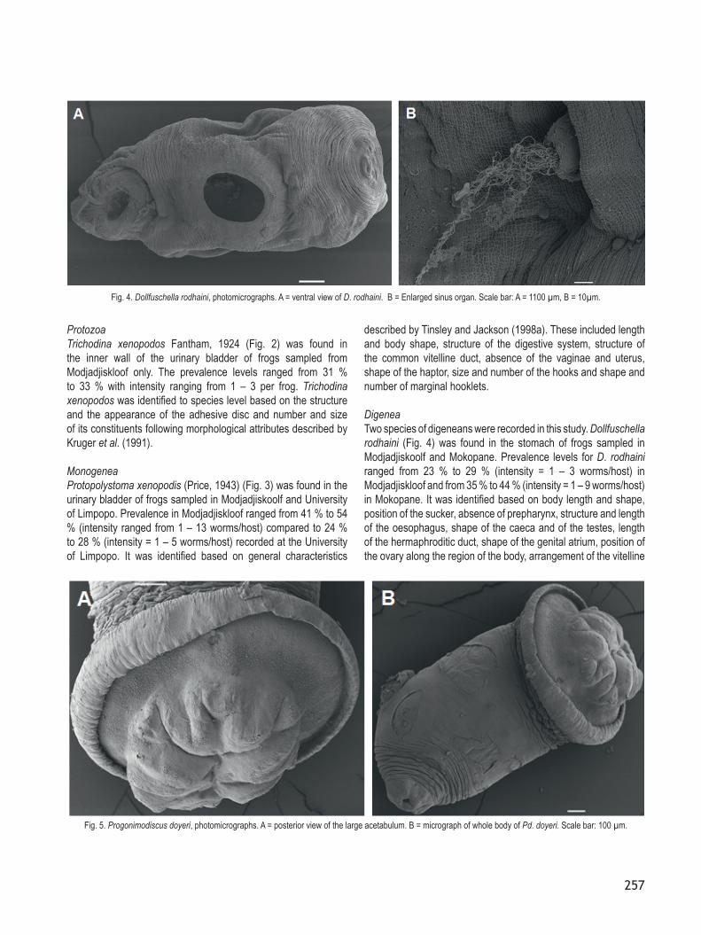
Fig. 3. Protopolystoma xenopodis, photomicrographs. A = ventral view. B = illustration suckers. Scale bar: 100 µm.
257
ProtozoaTrichodina xenopodos Fantham, 1924 (Fig. 2) was found in the inner wall of the urinary bladder of frogs sampled from Modjadjiskloof only. The prevalence levels ranged from 31 % to 33 % with intensity ranging from 1 – 3 per frog. Trichodina xenopodos was identified to species level based on the structure and the appearance of the adhesive disc and number and size of its constituents following morphological attributes described by Kruger et al. (1991).
MonogeneaProtopolystoma xenopodis (Price, 1943) (Fig. 3) was found in the urinary bladder of frogs sampled in Modjadjiskoolf and University of Limpopo. Prevalence in Modjadjiskloof ranged from 41 % to 54 % (intensity ranged from 1 – 13 worms/host) compared to 24 % to 28 % (intensity = 1 – 5 worms/host) recorded at the University of Limpopo. It was identified based on general characteristics
described by Tinsley and Jackson (1998a). These included length and body shape, structure of the digestive system, structure of the common vitelline duct, absence of the vaginae and uterus, shape of the haptor, size and number of the hooks and shape and number of marginal hooklets.
DigeneaTwo species of digeneans were recorded in this study. Dollfuschella rodhaini (Fig. 4) was found in the stomach of frogs sampled in Modjadjiskoolf and Mokopane. Prevalence levels for D. rodhaini ranged from 23 % to 29 % (intensity = 1 – 3 worms/host) in Modjadjiskloof and from 35 % to 44 % (intensity = 1 – 9 worms/host) in Mokopane. It was identified based on body length and shape, position of the sucker, absence of prepharynx, structure and length of the oesophagus, shape of the caeca and of the testes, length of the hermaphroditic duct, shape of the genital atrium, position of the ovary along the region of the body, arrangement of the vitelline
Fig. 4. Dollfuschella rodhaini, photomicrographs. A = ventral view of D. rodhaini. B = Enlarged sinus organ. Scale bar: A = 1100 µm, B = 10µm.
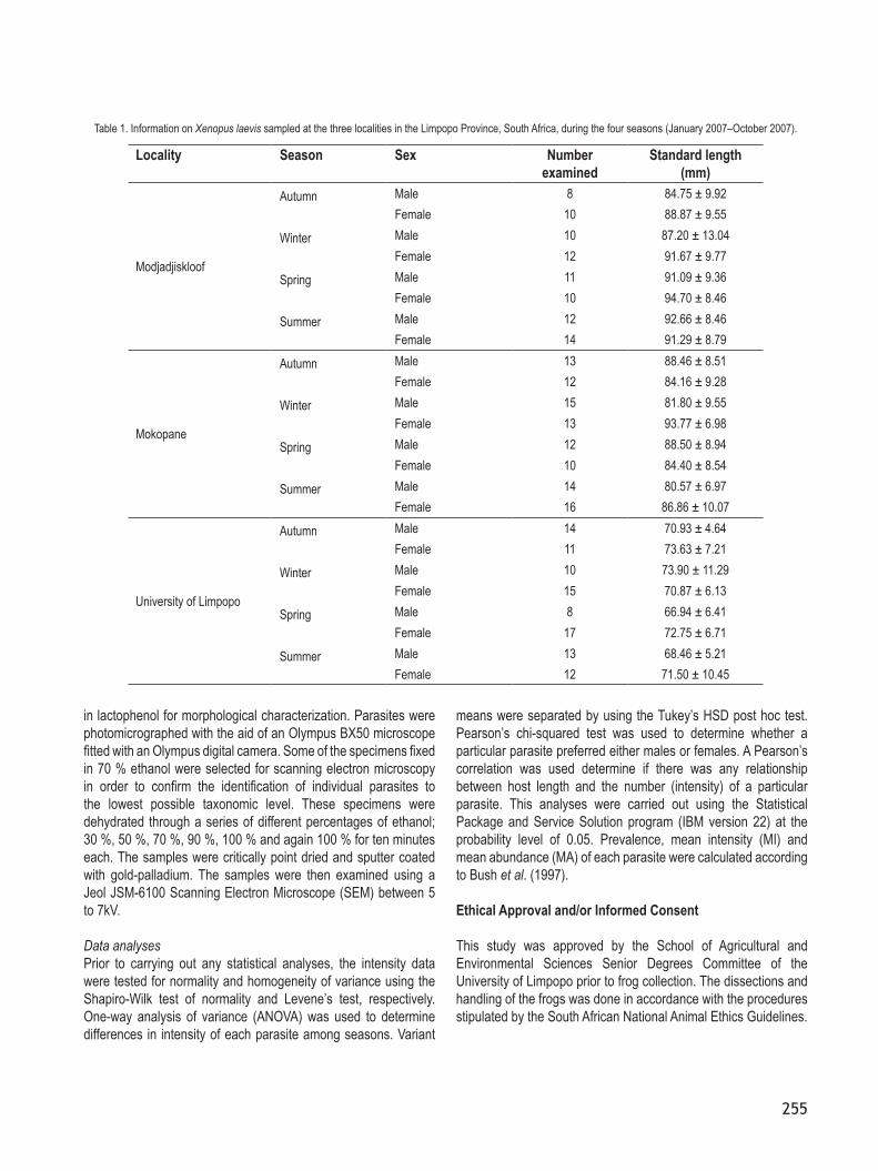
Fig. 5. Progonimodiscus doyeri, photomicrographs. A = posterior view of the large acetabulum. B = micrograph of whole body of Pd. doyeri. Scale bar: 100 µm.
258
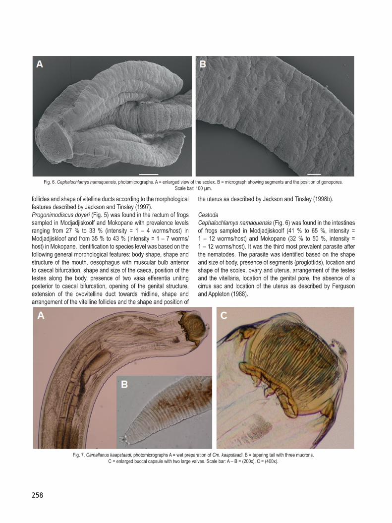
follicles and shape of vitelline ducts according to the morphological features described by Jackson and Tinsley (1997).Progonimodiscus doyeri (Fig. 5) was found in the rectum of frogs sampled in Modjadjiskoolf and Mokopane with prevalence levels ranging from 27 % to 33 % (intensity = 1 – 4 worms/host) in Modjadjiskloof and from 35 % to 43 % (intensity = 1 – 7 worms/host) in Mokopane. Identification to species level was based on the following general morphological features: body shape, shape and structure of the mouth, oesophagus with muscular bulb anterior to caecal bifurcation, shape and size of the caeca, position of the testes along the body, presence of two vasa efferentia uniting posterior to caecal bifurcation, opening of the genital structure, extension of the ovovitelline duct towards midline, shape and arrangement of the vitelline follicles and the shape and position of
the uterus as described by Jackson and Tinsley (1998b).
CestodaCephalochlamys namaquensis (Fig. 6) was found in the intestines of frogs sampled in Modjadjiskoolf (41 % to 65 %, intensity = 1 – 12 worms/host) and Mokopane (32 % to 50 %, intensity = 1 – 12 worms/host). It was the third most prevalent parasite after the nematodes. The parasite was identified based on the shape and size of body, presence of segments (proglottids), location and shape of the scolex, ovary and uterus, arrangement of the testes and the vitellaria, location of the genital pore, the absence of a cirrus sac and location of the uterus as described by Ferguson and Appleton (1988).
Fig. 6. Cephalochlamys namaquensis, photomicrographs. A = enlarged view of the scolex. B = micrograph showing segments and the position of gonopores. Scale bar: 100 µm.
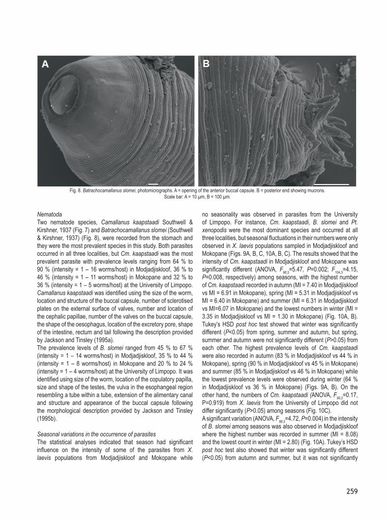
Fig. 7. Camallanus kaapstaadi, photomicrographs A = wet preparation of Cm. kaapstaadi. B = tapering tail with three mucrons. C = enlarged buccal capsule with two large valves. Scale bar: A – B = (200x), C = (400x).
259
NematodaTwo nematode species, Camallanus kaapstaadi Southwell & Kirshner, 1937 (Fig. 7) and Batrachocamallanus slomei (Southwell & Kirshner, 1937) (Fig. 8), were recorded from the stomach and they were the most prevalent species in this study. Both parasites occurred in all three localities, but Cm. kaapstaadi was the most prevalent parasite with prevalence levels ranging from 64 % to 90 % (intensity = 1 – 16 worms/host) in Modjadjiskloof, 36 % to 46 % (intensity = 1 – 11 worms/host) in Mokopane and 32 % to 36 % (intensity = 1 – 5 worms/host) at the University of Limpopo. Camallanus kaapstaadi was identified using the size of the worm, location and structure of the buccal capsule, number of sclerotised plates on the external surface of valves, number and location of the cephalic papillae, number of the valves on the buccal capsule, the shape of the oesophagus, location of the excretory pore, shape of the intestine, rectum and tail following the description provided by Jackson and Tinsley (1995a).The prevalence levels of B. slomei ranged from 45 % to 67 % (intensity = 1 – 14 worms/host) in Modjadjiskloof, 35 % to 44 % (intensity = 1 – 8 worms/host) in Mokopane and 20 % to 24 % (intensity = 1 – 4 worms/host) at the University of Limpopo. It was identified using size of the worm, location of the copulatory papilla, size and shape of the testes, the vulva in the esophangeal region resembling a tube within a tube, extension of the alimentary canal and structure and appearance of the buccal capsule following the morphological description provided by Jackson and Tinsley (1995b).
Seasonal variations in the occurrence of parasitesThe statistical analyses indicated that season had significant influence on the intensity of some of the parasites from X. laevis populations from Modjadjiskloof and Mokopane while
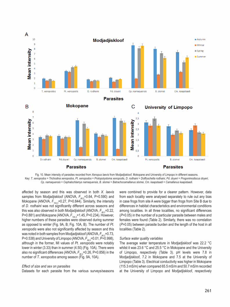
no seasonality was observed in parasites from the University of Limpopo. For instance, Cm. kaapstaadi, B. slomei and Pt. xenopodis were the most dominant species and occurred at all three localities, but seasonal fluctuations in their numbers were only observed in X. laevis populations sampled in Modjadjiskloof and Mokopane (Figs. 9A, B, C, 10A, B, C). The results showed that the intensity of Cm. kaapstaadi in Modjadjiskloof and Mokopane was significantly different (ANOVA, F86,3=5.47, P=0.002; F104,3=4.15, P=0.008, respectively) among seasons, with the highest number of Cm. kaapstaadi recorded in autumn (MI = 7.40 in Modjadjiskloof vs MI = 6.91 in Mokopane), spring (MI = 5.31 in Modjadjiskloof vs MI = 6.40 in Mokopane) and summer (MI = 6.31 in Modjadjiskloof vs MI=6.07 in Mokopane) and the lowest numbers in winter (MI = 3.35 in Modjadjiskloof vs MI = 1.30 in Mokopane) (Fig. 10A, B). Tukey’s HSD post hoc test showed that winter was significantly different (P<0.05) from spring, summer and autumn, but spring, summer and autumn were not significantly different (P>0.05) from each other. The highest prevalence levels of Cm. kaapstaadi were also recorded in autumn (83 % in Modjadjiskloof vs 44 % in Mokopane), spring (90 % in Modjadjiskloof vs 45 % in Mokopane) and summer (85 % in Modjadjiskloof vs 46 % in Mokopane) while the lowest prevalence levels were observed during winter (64 % in Modjadjiskloof vs 36 % in Mokopane) (Figs. 9A, B). On the other hand, the numbers of Cm. kaapstaadi (ANOVA, F99,3=0.17, P=0.919) from X. laevis from the University of Limpopo did not differ significantly (P>0.05) among seasons (Fig. 10C).A significant variation (ANOVA, F86,3=4.72, P=0.004) in the intensity of B. slomei among seasons was also observed in Modjadjiskloof where the highest number was recorded in summer (MI = 8.08) and the lowest count in winter (MI = 2.80) (Fig. 10A). Tukey’s HSD post hoc test also showed that winter was significantly different (P<0.05) from autumn and summer, but it was not significantly
Fig. 8. Batrachocamallanus slomei, photomicrographs. A = opening of the anterior buccal capsule. B = posterior end showing mucrons. Scale bar: A = 10 µm, B = 100 µm.

260
different (P>0.05) from spring. Higher prevalence levels were also recorded in summer (65 %) and autumn (66 %) while lower levels were found in spring (52 %) and winter (45 %) (Fig. 9A). On the other hand, the intensity of B. slomei from frogs collected in Mokopane was not significantly different (ANOVA, F104,3=0.39; P=0.759) among seasons, although lower mean intensity numbers were found during winter (MI = 3.50) compared to 4.09 in autumn, 3.87 in spring and 3.69 in summer (Fig. 10 ). Similarly, the lowest prevalence levels were recorded during winter (35 %) while the highest levels were noted during summer (43 %) (Fig. 9B). The intensity of B. slomei (ANOVA, F99,3=0.03, P=0.992) from X. laevis from the University of Limpopo also did not differ significantly (P>0.05) among seasons and neither was there any trend in prevalence levels among seasons (Figs. 9C, 10C).The intensity of Cp. namaquensis from frogs sampled both from the Modjadjiskloof and Mokopane localities also fluctuated
significantly (ANOVA, F86,3=6.87, P=0.00; ANOVA, F104,3=3.69, P=0.014, respectively) among seasons, with the highest intensity levels recorded during the spring-autumn period and the lowest intensity levels observed during winter (Fig. 10A,B). This was consistent with the trends observed in the prevalence levels. In Modjadjiskloof, for example, the highest prevalence level was recorded during summer (65 %) and the lowest was observed during winter (41 %) (Fig. 9A). Post hoc test confirmed that winter was significantly different (P<0.05) from autumn, spring and summer. In Mokopane, prevalence levels of Cp. namaquensis were also lower in winter (32 %) compared to summer (50 %) (Fig. 9B). Post hoc test also showed that winter was significantly different (P<0.05) from autumn and summer. However, no significant differences (P>0.05) were found between winter and spring as well as among autumn, spring and summer.On the other hand, the number of Pd. Doyeri was not significantly
Fig. 9. Prevalence of parasites recorded from Xenopus laevis from Modjadjiskloof, Mokopane and University of Limpopo in different seasons. Key: T. xenopodos = Trichodina xenopodos, Pt. xenopodos = Protopolystoma xenopodis, D. rodhaini = Dollfuschella rodhaini, Pd. doyeri = Progonimodiscus doyeri,
Cp. namaquensis = Cephalochlamys namaquensis, B. slomei = Batrachocamallanus slomei, Cm. kaapstaadi = Camallanus kaapstaadi.
261
affected by season and this was observed in both X. laevis samples from Modjadjiskloof (ANOVA, F86,3=0.64, P=0.590) and Mokopane (ANOVA, F104,3=0.27, P=0.844). Similarly, the intensity of D. rodhaini was not significantly different across seasons and this was also observed in both Modjadjiskloof (ANOVA, F86,3=0.22, P=0.881) and Mokopane (ANOVA, F104,3=1.45, P=0.234). However, higher numbers of these parasites were observed during summer as opposed to winter (Fig. 9A, B; Fig. 10A, B). The number of Pt. xenopodis were also not significantly affected by season and this was noted in both samples from Modjadjiskloof (ANOVA, F86,3=0.73, P=0.538) and University of Limpopo (ANOVA, F99,3=0.01, P=0.998), although in the former, MI values of Pt. xenopodis were notably lower in winter (3.33) than in summer (4.50) (Fig. 10A). There were also no significant differences (ANOVA, F86,3=0.26, P=0.856) in the number of T. xenopodos among season (Fig. 9A, 10A).
Effect of size and sex on parasitesDatasets for each parasite from the various surveys/seasons
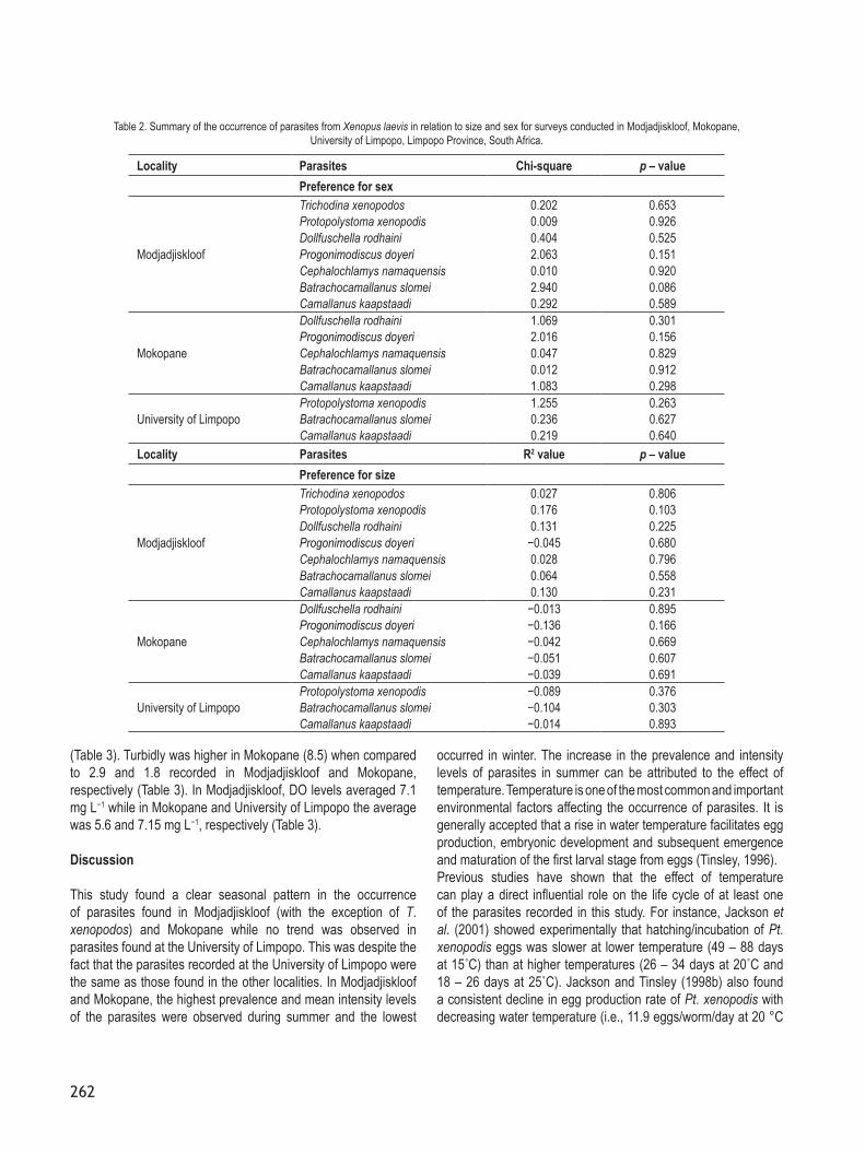
were combined to provide for a clearer pattern. However, data from each locality were analysed separately to rule out any bias in case frogs from site A were bigger than frogs from Site B due to differences in habitat characteristics and environmental conditions among localities. In all three localities, no significant differences (P>0.05) in the number of a particular parasite between males and females were found (Table 2). Similarly, there was no correlation (P>0.05) between parasite burden and the length of the host in all localities (Table 2).
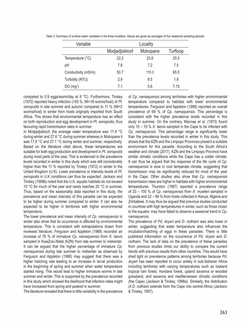
Surface water quality variablesThe average water temperature in Modjadjiskloof was 22.2 °C whilst it was 23.6 °C and 25.5 °C in Mokopane and the University of Limpopo, respectively (Table 3). pH levels were 7.8 in Modjadjiskloof, 7.2 in Mokopane and 7.5 at the University of Limpopo (Table 3). Electrical conductivity was higher in Mokopane (115.3 mS/m) when compared 65.5 mS/m and 50.7 mS/m recorded at the University of Limpopo and Modjadjiskloof, respectively
Fig. 10. Mean intensity of parasites recorded from Xenopus laevis from Modjadjiskloof, Mokopane and University of Limpopo in different seasons. Key: T. xenopodos = Trichodina xenopodos, Pt. xenopodos = Protopolystoma xenopodis, D. rodhaini = Dollfuschella rodhaini, Pd. doyeri = Progonimodiscus doyeri,
Cp. namaquensis = Cephalochlamys namaquensis, B. slomei = Batrachocamallanus slomei, Cm. kaapstaadi = Camallanus kaapstaadi.
262
(Table 3). Turbidly was higher in Mokopane (8.5) when compared to 2.9 and 1.8 recorded in Modjadjiskloof and Mokopane, respectively (Table 3). In Modjadjiskloof, DO levels averaged 7.1 mg L−1 while in Mokopane and University of Limpopo the average was 5.6 and 7.15 mg L−1, respectively (Table 3).
Discussion
This study found a clear seasonal pattern in the occurrence of parasites found in Modjadjiskloof (with the exception of T. xenopodos) and Mokopane while no trend was observed in parasites found at the University of Limpopo. This was despite the fact that the parasites recorded at the University of Limpopo were the same as those found in the other localities. In Modjadjiskloof and Mokopane, the highest prevalence and mean intensity levels of the parasites were observed during summer and the lowest
occurred in winter. The increase in the prevalence and intensity levels of parasites in summer can be attributed to the effect of temperature. Temperature is one of the most common and important environmental factors affecting the occurrence of parasites. It is generally accepted that a rise in water temperature facilitates egg production, embryonic development and subsequent emergence and maturation of the first larval stage from eggs (Tinsley, 1996). Previous studies have shown that the effect of temperature can play a direct influential role on the life cycle of at least one of the parasites recorded in this study. For instance, Jackson et al. (2001) showed experimentally that hatching/incubation of Pt. xenopodis eggs was slower at lower temperature (49 – 88 days at 15˚C) than at higher temperatures (26 – 34 days at 20˚C and 18 – 26 days at 25˚C). Jackson and Tinsley (1998b) also found a consistent decline in egg production rate of Pt. xenopodis with decreasing water temperature (i.e., 11.9 eggs/worm/day at 20 °C
Locality Parasites Chi-square p – valuePreference for sex
Modjadjiskloof
Trichodina xenopodos 0.202 0.653Protopolystoma xenopodis 0.009 0.926Dollfuschella rodhaini 0.404 0.525Progonimodiscus doyeri 2.063 0.151Cephalochlamys namaquensis 0.010 0.920Batrachocamallanus slomei 2.940 0.086Camallanus kaapstaadi 0.292 0.589
Mokopane
Dollfuschella rodhaini 1.069 0.301Progonimodiscus doyeri 2.016 0.156Cephalochlamys namaquensis 0.047 0.829Batrachocamallanus slomei 0.012 0.912Camallanus kaapstaadi 1.083 0.298
University of LimpopoProtopolystoma xenopodis 1.255 0.263Batrachocamallanus slomei 0.236 0.627Camallanus kaapstaadi 0.219 0.640
Locality Parasites R2 value p – valuePreference for size
Modjadjiskloof
Trichodina xenopodos 0.027 0.806Protopolystoma xenopodis 0.176 0.103Dollfuschella rodhaini 0.131 0.225Progonimodiscus doyeri −0.045 0.680Cephalochlamys namaquensis 0.028 0.796Batrachocamallanus slomei 0.064 0.558Camallanus kaapstaadi 0.130 0.231
Mokopane
Dollfuschella rodhaini −0.013 0.895Progonimodiscus doyeri −0.136 0.166Cephalochlamys namaquensis −0.042 0.669Batrachocamallanus slomei −0.051 0.607Camallanus kaapstaadi −0.039 0.691
University of LimpopoProtopolystoma xenopodis −0.089 0.376Batrachocamallanus slomei −0.104 0.303Camallanus kaapstaadi −0.014 0.893
Table 2. Summary of the occurrence of parasites from Xenopus laevis in relation to size and sex for surveys conducted in Modjadjiskloof, Mokopane, University of Limpopo, Limpopo Province, South Africa.
263
compared to 0.9 eggs/worm/day at 8 °C). Furthermore, Tinsley (1972) reported heavy infection (>50 %, MI=18 worms/host) of Pt. xenopodis in late summer and autumn compared to 11 % (MI=2 worms/host) in winter from hosts originally imported from South Africa. This shows that environmental temperature has an effect on both reproduction and egg development in Pt. xenopodis, thus favouring rapid transmission rates in summer.In Modjadjiskloof, the average water temperature was 17.4 °C during winter and 27.6 °C during summer whereas in Mokopane it was 17.8 °C and 27.1 °C during winter and summer, respectively. Based on the literature cited above, these temperatures are suitable for both egg production and development in Pt. xenopodis during most parts of the year. This is evidenced in the prevalence levels recorded in winter in this study which was still considerably higher than the 11 % reported by Tinsley (1972) in winter in the United Kingdom (U.K). Lower prevalence or intensity levels of Pt. xenopodis in U.K conditions can thus be expected. Jackson and Tinsley (1998b) noted that the U.K. aquatic habitats do not exceed 10 °C for much of the year and rarely reaches 20 °C in summer. Thus, based on the seasonality data reported in this study, the prevalence and mean intensity of this parasite can be expected to be higher during summer compared to winter. It can also be expected to be higher in territories with higher environmental temperature.The lower prevalence and mean intensity of Cp. namaquensis in winter also show that its occurrence is affected by environmental temperature. This is consistent with extrapolations drawn from reviewed literature. Ferguson and Appleton (1988) recorded an increase of 76 % of immature Cp. namaquensis from X. laevis sampled in KwaZulu-Natal (KZN) from late summer to midwinter. It can be argued that the higher percentage of immature Cp. namaquensis during late summer to midwinter as observed by Ferguson and Appleton (1988) may suggest that there was a higher hatching rate leading to an increase in larval production in the beginning of spring and summer when water temperature started rising. This would lead to higher immature worms in late summer and winter. This is supported by the prevalence recorded in this study which showed the likelihood that infection rates might have increased from spring and peaked in summer. The literature revealed that there is little variability in the prevalence
of Cp. namaquensis among territories with higher environmental temperature compared to habitats with lower environmental temperatures. Ferguson and Appleton (1988) reported an overall prevalence of 69 % of Cp. namaquensis. This percentage is consistent with the higher prevalence levels recorded in this study in summer. On the contrary, Macnae et al. (1973) found only 10 – 15 % X. laevis sampled in the Cape to be infected with Cp. namaquensis. This percentage range is significantly lower than the prevalence levels recorded in winter in this study. This shows that the KZN and the Limpopo Provinces present a suitable environment for this parasite. According to the South Africa’s weather and climate (2017), KZN and the Limpopo Province have similar climatic conditions while the Cape has a colder climate. It can thus be argued that the response of the life cycle of Cp. namaquensis is slow in cool temperate climate, suggesting that transmission may be significantly reduced for most of the year in the Cape. Other studies also show that Cp. namaquensis transmission rates are higher in habitats with higher environmental temperatures. Thurston (1967) reported a prevalence range of 33 – 100 % of Cp. namaquensis from X. muelleri sampled in Uganda and 22 – 89 % from hosts collected in Kenya, Nigeria and Zimbabwe. It may thus be argued that previous studies conducted in countries with high temperatures in winter, such as those closer to the equator, may have failed to observe a seasonal trend in Cp. namaquensis. The prevalence of Pd. doyeri and D. rodhaini was also lower in winter, suggesting that water temperature also influences the incubation/hatching of eggs in these parasites. There is little published information on the occurrence of Pd. doyeri and D. rodhaini. The lack of data on the prevalence of these parasites from previous studies limits our ability to compare the current trends with previous results from other countries. This would have shed light on prevalence patterns among territories because Pd. doyeri has been reported to occur widely in sub-Saharan Africa including territories with varying temperatures such as lowland tropical rain forest, montane forest, upland savanna or wooded grassland, arid savanna and mediterranean climatic conditions (the Cape) (Jackson & Tinsley, 1998a). Similarly, the distribution of D. rodhaini extends from the Cape into central Africa (Jackson & Tinsley, 1997).
Variable LocalityModjadjiskloof Mokopane Turfloop
Temperature (°C) 22.2 23.6 25.5pH 7.8 7.2 7.5Conductivity (mS/m) 50.7 115.3 65.5Turbidity (NTU) 2.9 8.5 1.8DO (mg-l) 7.1 5.6 7.15
Table 3. Summary of surface water variables in the three localities. Values are given as averages of four seasonal sampling periods.

264
The lower prevalence levels of the camallanid nematodes, Cm. kaapstaadi and B. slomei recorded in winter also suggest that their occurrence is temperature-dependent. The camallanid nematodes were the most prevalent parasites in the current study and they are commonly reported from X. laevis in South Africa. Svitin et al. (2018) also reported higher prevalence levels of these nematodes from different localities in South Africa. However, prevalence levels in these parasites seem to differ significantly among regions. For example, Svitin et al. (2018) reported that B. slomei infected 7 % of X. laevis on the western coast of KZN, 57 % in Hermanus, Western Cape, 83 % – 100 % in two localities in Limpopo, 100 % in North-West and 63 % – 100 % in three localities in Mpumalanga. Similarly, Cm. kaapstaadi infected 80 % of hosts in Mpumalanga and 60 % in the Western Cape (Svitin et al., 2018). It is interesting to note that the lowest prevalence in both parasites was found in the Western Cape, which has cooler temperatures (South Africa’s weather and climate, 2017). It can be speculated that lower temperatures can lead to low infection rates. This claim is supported by the present study in that lower prevalence levels in both nematodes were found in winter. There were no significant differences in the prevalence or intensity of T. xenopodos among seasons, which probably indicates that environmental temperature does not have a significant effect on its life cycle. This can be attributed to its life cycle and transmission strategy. Trichodina xenopodos occurs in the urinary bladder where it is protected from adverse environmental conditions by the host. It reproduces by binary fission and is known to survive for a short period outside the urinary bladder of its host (Kruger et al., 1991). Therefore, crowded hosts or close contact will facilitate transmission. According to Kruger et al. (1991), spontaneous transmission from one host to another may occur when hosts are crowded in a small habitat (e.g., tanks). In addition, cross-infection can occur during amplexus when male and female frogs are attached for extended periods (Kruger et al., 1991). This may thus explain why the prevalence and intensity T. xenopodos did not differ significantly among seasons. Additionally, it may also explain the low intensity levels observed in this study due to the relatively larger size of the habitat. The second important finding of this study was the significant variations in the diversity and intensity of parasites among the three localities. The higher diversity and intensity of parasites in Modjadjiskloof can be explained by the nature of the habitat. In Modjadjiskloof, the sampling locality was a natural mountain stream mainly surrounded by forests and vegetation and there are no anthropogenic activities in the vicinity of the stream. As a result, it is a natural water body in a relatively undisturbed area and it can be regarded as pristine. Conditions associated with undisturbed habitats facilitate the concentration of the first and second intermediate hosts (Ondračková et al., 2004; Mbokane et al., 2015), thus allowing for the highest parasite species richness and the highest overall transmission rate. Previous studies have also reported higher parasite diversity in X. laevis from undisturbed
habitats. Recently, Svitin et al. (2018) reported a higher level of parasite species diversity in X. laevis collected from dams in a mountain stream in the Mpumalanga Province. Svitin et al. (2018) attributed their findings to the pristine condition of the habitat and lack of anthropogenic activities in the surrounding area, which creates a better environment for the proliferation of parasites. In addition, these authors recorded higher levels of nematode diversity and infection at that locality which corresponded with higher numbers of other parasites such as cestodes, monogeneans and digeneans. This observation is similar to the results obtained in this study in that the nematodes were the most prevalent species and their higher numbers in Modjadjiskloof also corresponded with higher numbers in other parasites. Schoeman et al. (2019) also found few parasites in X. laevis from newly established habitats compared to a more diverse parasite community from natural water bodies. It is, therefore, clear that natural water bodies in undisturbed areas are ideal for parasites establishment. On the other hand, the lower prevalence and intensity levels of parasites from Mokopane can suggest that the passing of the stream or its close proximity to the mine and informal settlements has an effect on the environment. The poor diversity of parasites, and to a larger extent, failure to observe a seasonal trend in parasites recorded at University of Limpopo can be attributed to the disruption in the life cycle of the parasites. The presence of the intermediate hosts is crucial in the successful transmission of the parasites recorded at the University of Limpopo. The frogs were kept at a facility with constant water treatment or removal of waste which might have led to the decline in the intermediate hosts. In addition, the optimal conditions at the University of Limpopo ensured less temperature variations throughout the study, which can explain the lack of seasonal variations in the intensity of parasites that have been shown to be influenced by temperature changes. All the parasites recorded at the University of Limpopo are long-lived in their host and without new infections the prevalence and intensity levels will remain somewhat unchanged among seasons. For instance, Pt. xenopodis was reported to be present nine months post capture (Tinsley, 1972), with a maximum survival of more than two years (Jackson & Tinsley, 1988; Jackson & Tinsley, 2001a). Camallanus kaapstaadi together with other camallanid nematodes, Pt. xenopodis and Cp. namaquensis have all been found to survive for more than a year in laboratory-maintained hosts (Tinsley, 1996). It is, thus, probable that the frogs infected with parasites at the University of Limpopo may have acquired the parasites prior to being held captive. It is interesting to note that the diversity of parasites from X. laevis in South Africa seems to differ from that reported from other countries where X. laevis was introduced. Parasite diversity seem to be greater in places falling within the geographic native range of X. laevis. This is consistent with findings from Schoeman et al. (2019) who studied differences in parasite species diversity of X. laevis between native and invasive hosts and found lower parasite
265
diversity (Pt. xenopodis and C. namaquensis) in France, a place where X. laevis is considered an invasive species, compared to 21 different metazoan parasites species from South Africa. Lower parasite diversity (3 species, Pt. xenopodis, Opisthodiscus cf. nigrivasis (von Meheli, 1929), and one unidentified species) was also found in Portugal (Rodrigues, 2014). In Chile, Castillo et al. (2017) found only one parasite from X. laevis identified as belonging to the genus Contracaecum. In California, X. laevis was reported to be infected with Cp. namaquensis, G. gallieni, Pt. xenopodis, Clinostomum sp., Contracaecum spp., Eustrongylides sp., and Acanthocephalus sp. (Lafferty & Page, 1997; Kuperman et al., 2004). Schoeman et al. (2019) noted that although the South African and Californian invasive X. laevis parasite communities were much more diverse than in other areas, they represented parasites native to where hosts were collected. This observation seems to hold true for the Contracaecum sp. from Chile and California which have never been reported from X. laevis in its native range in South Africa. Castillo et al. (2017) mentioned that Contracaecum sp. is the first record of Anisakidae in amphibians, and suggested that the source could be other vertebrates (bird, fishes). This may also apply to the parasites reported in California which are not normally found in the host’s native range. The occurrence of Cp. namaquensis and Pt. xenopodis in California suggests that these parasites are capable of adapting and reproducing successfully in new environments. On the other hand, it is strange why Pt. xenopodis was not reported in Chile which has a more favorable climate for both the parasite and the host when it has apparently persisted in the U.K for many years under cold environmental conditions (Jackson & Tinsley 1998b; Tinsley & Jackson, 1998a). In the present study, there was no correlation between the number of parasites and the length of the host, which indicates that size did not have an influence on the intensity of the parasites. The hosts examined in this study were mainly adults and did not differ significantly from each other in terms of size. Perhaps it would have been expected that hosts of smaller size or juveniles may have had fewer parasites than adults due to the little space juveniles provide for colonisation or attachment and resources for parasites. It has also been shown that diet preferences vary among the different stages of X. laevis with frogs becoming more predatory or shifting to live feed as they transition from juveniles to adults (Schoonbee et al., 1992). Many of the aquatic invertebrates consumed by X. laevis are potential carriers of the parasite larvae, thus consumption of zooplanktons by adults will lead to higher parasitic infections. In addition, this study found that parasites did not have a preference for a particular sex. There was sufficient representation of both sexes collected in this study which makes our samples relatively unbiased and, therefore, a conclusion can be drawn that sex has no influence on the occurrence of the parasites found in this study. There are no previous studies that have investigated the effect of either size or sex of X. laevis on its parasites to compared our results with.
In conclusion, this study showed that the occurrence of the parasites from X. laevis was influenced by environmental temperature. It was also established that more diverse composition and high intensity of parasites from X. laevis was associated with undisturbed environments. The occurrence of the parasites was neither influenced by size nor sex of the host. To our knowledge, this report is the first to describe the effect of season on parasites from X. laevis in South Africa.
Conflict of Interest
Authors declare no conflict of interest.
Acknowledgements
This study was funded by the National Research Foundation of South Africa (NRF) (Grant No. 101054). A word of appreciation to Mr. Gavin Geldenhuys for the collection of the hosts in Modjadjiskloof and Mokopane and to Mr. Hendrik Hattingh for collection of hosts at the Animal Unit (UL). The authors would also like to express gratitude to the anonymous reviewers whose comments and suggestion greatly improved this paper.
Author Contributions
W.J.L-P. (University of Limpopo) was the supervisor of the project and J.T. (University of Limpopo) was the co-supervisor. E.M.M. (University of Limpopo) undertook field surveys, collected and analyzed data and also wrote the manuscript.
References
Bush, A.O., LAfferty, K.D., LOtz, J.M., shOstAK. A.W. (1997): Parasitology meets ecology on its own terms: Margolis et al. revisited. J. Parasit., 83: 575 ‒ 583CAnnAteLLA, D.C., De sá, r.O. (1993): Xenopus laevis as a model organism. Syst Biol., 42: 476–507CAstiLLO, C., LOBOs, G., GOnzáLez-ACuñA, D., MOrenO, L., GOnzáLez, Ce., LAnDAetA-Aqueveque, C. (2017): First parasitological study of the African clawed frog (Xenopus laevis, Amphibia) in Chile. Rev. Bras. Parasitol. Vet., 26(2): 243 – 247. DOI: 10.1590/S1984-29612017029COsGrOve, G.e., JAreD, D.W. (1974): Diseases and parasites of Xenopus, the clawed toad. In: AMBOrsKi, r.L., hOOD, M.A., MiLLer, r.r. (Eds). Proceedings of Gulf Coast Regional Symposium on Diseases of Aquatic Animals. Center for Wetlands Research, Louisiana State University, Baton Rouge, pp. 225 – 242.De BussChere, C., COurAnt, J., herreL, A., ruBeLO, r., rODDer, D., MeAsey, J., BACKeLJAu, t. (2016). Unequal contribution of native South African phylogeographic lineages to the invasion of the African clawed frog, Xenopus laevis, in Europe. PeerJ., DOI: 10.7717/peerj.1659

266
De viLLiers, f.A., MeAsey, G.J. (2017): Overland movement in African clawed frogs (Xenopus laevis): empirical dispersal data from within their native range. PeerJ., 5: 4039. DOI: 10.7717/peerj.4039eLKAn, e., MurrAy, s.r.W. (1952): A larval trematode infection of the lateral line system of the Toad Xenopus laevis (Daudin). Proceedings of the Zoological Society of London 122(1): 121 – 125. DOI: 10.1111/j.1469-7998.1952.tb06314.xespinOsA, h.e., GArCiA, p.L., De LeOn, G.p.p. (1996): Helminth community structure of Chirostoma attenuatum (Ostheichtyhyes: Atherinidae) in two Mexican lakes. Southwest. Nat. 41: 288 – 292ferGusOn, r.r., AppLetOn, C.C. (1988): Some aspects of the morphology, population structure and larval biology of Cephalochlamys namaquensis (Cestoda: Diphyllidea), a parasite of the clawed toad, Xenopus laevis. S. Afr. J. Zoo., 23(2): 117–123.Furman, B.L.S., Bewick, a.J., HarriSon, T.L., GreenBaum, e., Gvoždík, v., KusAMBA, C., evAns, B. (2015): Pan-African phylogeography of a model organism, the African clawed frog ‘‘Xenopus laevis’’. Mol. Ecol., 24: 909 – 925GurDOn, J.B. (1996): Introductory comments: Xenopus as a laboratory animal. In: tinsLey, r.C., KOBeL, h.r. (Eds) The Biology of Xenopus. Oxford University Press, Oxford, pp. 3 – 6GurDOn, J.B., hOpWOOD, n. (2000): The introduction of Xenopus laevis into developmental biology: of empire, pregnancy testing and ribosomal genes. Int. J. Dev. Biol., 44: 43 – 50hArris, p.D., tinsLey, r.C. (1987): The biology of Gyrdicotylus gallieni (Gyrodactylidea), an unusual viviparous monogenean from the African clawed toad, Xenopus laevis. J. Zool., 212(2): 325 – 346. DOI: 10.1111/j.1469-7998.1987.tb05993.xJACKsOn, h.C., tinsLey, r.C. (1988): The capacity for viable egg production by the monogenean Protopolystoma xenopodis in single and multiple infections. Int. J. Parasitol., 18: 585 – 589. DOI: 10.1016/0020-7519(88)90091-4JACKsOn, J.A., tinsLey r.C. (1995a): Evolutionary relationships, host range and geographical distribution of Camallanus Railliet & Henry, 1915 species (Nematoda: Camallaninae) from clawed toads of the genus Xenopus (Anura: Pipidae). Syst. Parasitol., 32: 1 ‒ 21JACKsOn, J.A., tinsLey, r.C. (1995b): Representatives of Batrachocamallanus n. g. (Nematoda: Procamallaninae) from Xenopus spp. (Anura: Pipidae): geographical distribution, host range and evolutionary relationships. Syst. Parasitol., 31: 159 ‒ 188JACKsOn, J.A., tinsLey, r.C. (1997): The taxonomic status, host range and geographical distribution of Dollfuschella Vercammen-Grandjean, 1960 (Digenea: Halipeginae) from Xenopus spp. (Anura: Pipidae). Syst. Parasitol., 36: 1 – 11JACKsOn, J.A., tinsLey, r.C. (1998a): Paramphistome digeneans from Xenopus species (Pipidae) in Africa: taxonomy, host-specificity and biogeography. Syst. Parasitol., 40: 143 – 160. DOI: 10.1023/A:1005936429562JACKsOn, J.A., tinsLey, r.C. (1998b): Effects of temperature
on oviposition rate in Protopolystoma xenopodis (Monogenea: Polystomatidae). Int. J. Parasitol., 28: 309 – 315. DOI: 10.1016/S0020-7519(97)00151-3JACKsOn, J.A., tinsLey, r.C. (2001a): Protopolystoma xenopodis (Monogenea) primary and secondary infections in Xenopus laevis. Parasitology 123: 455 – 463. DOI: https://doi.org/10.1017/S0031182001008745JACKsOn, J.A., tinsLey, r.C. (2001b): Host-specificity and distribution of cephalochlamydid cestodes: correlation with allopolyploid evolution of pipid anuran hosts. J. Zool., 254: 405 – 419. DOI: 10.1017/S0952836901000905JACKsOn, J.A., tinsLey, r.C., Du preez, L.h. (2001): Differentiation of two locally sympatric Protopolystoma (Monogenea: Polystomatidae) species by temperature-dependent larval development and survival. Int. J. Parasitol., 31: 815 – 421KinG, p.h., vAn As, J.G. (1997): Description of the adult and larval stages of Tylodelphys xenopi (Trematoda: Diplostomidae) from southern Africa. J. Parasitol., 83(2): 287 – 295KinG, p.h., vAn As, J.G. (2000): Morphology and life history of Petasiger variospinosus (Trematoda: Echinostomatidae) in the Free State, South Africa. J. Parasitol., 86(2): 312 – 318. DOI: 10.1645/0022-3395(2000)086[0312:MALHOP]2.0.CO;2KruGer, J., BAssOn, L., vAn As, J.G. (1991): Redescription of Trichodina xenopodos Fantham, 1924 (Ciliophora: Peritrichida), a urinary bladder parasite of Xenopus laevis laevis Daudin, 1802, with notes on transmission. Syst. Parasitol.,19: 43 – 50KruGer, n., Du preez, L. (2015): Reproductive strategies of the kangaroo leech, Marsupiobdella africana (Glossiphoniidae) Int. J. Parasitol. Parasites. Wildl. 4(1): 142 – 147. DOI: 10.1016/j.ijppaw.2015.01.005KuperMAn, B.i., MAtey, v.e., fisher, r.n., ervin, e., WArBurtOn, M., BAKhirevA, L., LehMAn, C.A. (2004): Parasites of the African clawed frog, Xenopus laevis, in Southern California, U.S.A. Comp. Parasitol., 71: 229 – 232. DOI: 10.1654/4112LAfferty, K.D., pAGe, C.J. (1997): Predation on the endangered tidewater goby, Eucyclogobius newberryi, by the introduced African clawed frog, Xenopus laevis, with notes on the frog’s parasites. Copeia., 1997: 589 – 592MACnAe, W., rOCK, L., MAKOWsKi, M. (1973): Platyhelminths from the South African clawed toad, or platanna (Xenopus laevis). J. Helminthol., 47: 199 – 235MBOKAne, e.M., therOn, J., Luus-pOWeLL, W. J. (2015): Seasonal occurrence of some larval stages of endoparasites in three cyprinids from the Nwanedi-Luphephe dams, the Limpopo River System, South Africa. Helminthologia., 52(3): 229 – 235. DOI: 10.1515/helmin-2015-0037MeAsey, G.J., DAvies, s.J. (2011): Struggling against domestic exotics at the southern end of Africa. FrogLog., 97: 28 – 30MeAsey, G.J., röDDer, D., Green, s., KOBAyAshi, r., LiLLO, f., LOBOs, G., reBeLO, G., thiriOn, J. (2012): Ongoing invasions of the African clawed frog, Xenopus laevis: a global review. Biol. Invasions., 14: 2255 – 2270. DOI: 10.1007/s10530-012-0227-8
267
MOrAveC, f., COsGrOve, G.e. (1982): Pseudocapillaroides xenopi gen, et sp. nov. from the skin of the South African clawed frog, Xenopus laevis Daud. Rev Zool African., 96: 129 – 137niGreLLi, r.e., MArAventAnO, L.W. (1944): Pericarditis in Xenopus laevis caused by Diplostomulum xenopi sp. nov., a larval strigeid. J. Parasitol., 30: 184 – 190ondračková, m., Šimková, A., GeLnAr, M., JurAJDA, p. (2004): Posthodiplostomum cuticola (Digenea: Diplostomatidae) in intermediate fish hosts: factors contributing to the parasite infection and prey selection by definitive bird host. Parasitology., 129: 761 – 770. DOI: 10.1017/S0031182004006456priCe, e.W. (1943): A new trematode of the genus Polystoma (Monogenea: Polystomatidae) from Xenopus laevis Daud. Proc. Helm. Soc. Wash., 10: 83 – 85pritChArD, M.h. (1964): Notes on four helminths from the clawed toad, Xenopus laevis (Daudin), in South Africa. Proc. helm. Soc. Wash., 31: 121 – 128rODriGues, r.A.e. (2014): Macroparasites of invasive Xenopus laevis (Amphibia: Anura): characterization and assessment of possible exchanges with native Pelophylax perezi in Oeiras streams, Portugal. Lisboa: University of Lisboa. (dissertation).sAnDOn, h. (1941): Studies on South endozoic ciliates. I. Paranyctotherus (Balantidium) kirbyi (Rodriquez) emend, gen. nov. from the rectum of the clawed toad, Xenopus laevis. S. Afr. Med. J., 6: 116 ‒ 127sChOOnBee, h.J., prinsLOO, J.f., nxiWen, J.G. (1992): Observations on the feeding habits of larvae, juvenile and adult stages of the African clawed frog, Xenopus laevis, in impoundments in Transkei. Water SA., 18: 227 ‒ 236sChOeMAn, A.L., KruGer, n., seCOnDi, J., Du preez, L.h. (2019): Repeated reduction in parasite diversity in invasive populations of Xenopus laevis: a global experiment in enemy release. Biol. Invasions., 21(4): 1323 – 1338. DOI: 10.1007/s10530-018-1902-1sOuth AfriCA’s WeAther AnD CLiMAte. (2017): South Africa gateway. Archived from the original on 1 December 2017. Retrieved 20 November 2017. https://southafrica-info.com/south-africa-weather-climate/sOuthWeLL, t., Kirshner, A. (1937): On some parasitic worms found in Xenopus laevis, the South African clawed toad. Ann. Trop. Med. Parasitol. 31: 245 ‒ 265svitin, r., sChOeMAn, A.L., Du preez, L.h. (2018): New information on morphology and molecular data of camallanid nematodes parasitising Xenopus laevis (Anura: Pipidae) in South Africa. Folia. Parasit., 65: 003. DOI: 10.14411/fp.2018.003theunissen, M., tieDt, L., Du preez, L.h. (2014): The morphology and attachment of Protopolystoma xenopodis (Monogenea: Polystomatidae) infecting the African clawed frog Xenopus laevis. Parasite., 21: 20. DOI: 10.1051/parasite/2014020
thurstOn, J.p. (1967): The morphology and life-cycle of Cephalochlamys namaquensis (Cohn, 1906) (Cestoda:Pseudophyllidea) from Xenopus muelleri and X. laevis. Parasitology 57: 187 ‒ 200tinsLey, r.C. (1971): The adaptation for attachment by the Polystomatidae (Monogenoidea). Comptes Rendus Multicolloque Européen Parasitologie 1: 65 – 68tinsLey, r.C. (1972): The adaptation for attachment by the Polystomatidae (Monogenoidea). C. r. Multicolloque euroParasit., 1: 65 – 68tinsLey, r.C., OWen, r.W. (1975): Studies on the biology of Protopolystoma xenopodis (Monogenoidea): the oncomiracidium and life-cycle. Parasitology., 71: 445 – 463tinsLey, r.C., OWen, r.W. (1979): The morphology and biology of Xenopodistomum xenopodis from the gall bladder of the African clawed toad, Xenopus laevis. J. Helminthol., 53: 307 – 316tinsLey, r.C., sWeetinG, r.A. (1974): Studies on the biology and taxonomy of Diplostomulum (Tylodelphylus) xenopodis from the African clawed toad, Xenopus laevis. J. Helminthol. 48: 247 – 263tinsLey, r., WhiteAr, M. (1980): The surface fauna of Xenopus skin. Proceedings of the Royal Society of Edinburgh Section B Biological Sciences., 79: 127 – 130tinsLey, r.C. (1981): The evidence from parasite relationships for the evolutionary status of Xenopus (Anura: Pipidae). Monit. zoof. ital. (N.s.) (Suppl.)., 15: 367 – 385tinsLey, r.C. (1995): Parasitic disease in amphibians: control by the regulation of worm burdens. Parasitology., 111: 153 – 78tinsLey, r.C. (1996): Parasites of Xenopus. In: tinsLey, r.C., KOBeL, h.r. (Eds) The Biology of Xenopus. Oxford, UK: Oxford University Press, pp. 233 – 261tinsLey, r.C., LOuMOnt, C., KOBeL, h.r. (1996): Geographical distribution and ecology. In: tinsLey, r.C., KOBeL, h.r. (Eds) The Biology of Xenopus. Oxford, UK: Clarendon Press, pp. 35 – 59tinsLey, r.C., JACKsOn, J.A. (1998a): Speciation of Protopolystoma Bychovsky, 1957 (Monogenea: Polystomatidae) in hosts of the genus Xenopus (Anura: Pipidae). Syst. Parasitol. 40: 93 – 141tinsLey, r.C., JACKsOn, J.A. (1998b): Correlation of parasite speciation and specificity with host evolutionary relationships. Int. J. Parasitol., 28(10): 1573 – 1582. DOI: 10.1016/S0020-7519(98)00085-XWADe, s.e. (1982): Capillaria xenopodis sp. n. (Nematoda:Trichuroidea) from the epidermis of the South African clawed frog (Xenopus laevis Daudin). Proceedings of the Helminthological Society of Washington., 49: 86 – 92WiLLiAMs, J.B. (1959): Preliminary note on the anatomy of a polystome from the bladder of Xenopus laevis Daud. J. Helm., 33: 207 – 208