Embed Size (px)
Citation preview

Vol. 50, No. 1JOURNAL OF VIROLOGY, Apr. 1984, p. 145-1540022-538X/84/040145-10$02.00/0Copyright © 1984, American Society for Microbiology
D,L-OL-Difluoromethylornithine Inhibits Human CytomegalovirusReplication
WADE GIBSON,* RICHARD VAN BREEMEN, ALAN FIELDS, ROBERT LAFEMINA, AND ALICE IRMIEREDepartment of Pharmacology and Experimental Therapeutics, The Johns Hopkins University School of Medicine,
Baltimore, Maryland 21205
Received 21 June 1983/Accepted 20 December 1983
D,L-a-Difluoromethylornithine (DFMO) is an inhibitor of ornithine decarboxylase, the first enzyme in thepolyamine biosynthetic pathway. Exposure of human foreskin fibroblast cells to DFMO before theirinfection with human strains of cytomegalovirus (CMV) resulted in a reduction in the amount of infectiousvirus produced. A 3-day exposure to the drug was required to elicit maximal antiviral effect. Cells exposedto DFMO at the time of infection produced normal amounts of infectious virus. Preexposure to the drug for1, 2, or 3 days before infection resulted in at least 10-, 100-, or 1,000-fold decreases, respectively, in theamount of infectious virus produced. This decrease paralleled the loss of intracellular spermidine and waspartially spared by the addition of exogenous putrescine, spermidine, or spermine (10 ,uM). When added 3days before infection, DFMO depressed production of herpes simplex virus and simian CMV, as well aswild-type and laboratory prototype strains of human CMV. Although some antiviral effect was observed at adrug concentration of 1 mM, 10 mM gave a stronger effect and was the amount routinely used. At 30 mMDFMO, growth of noninfected cells was slowed but not arrested. Studies to investigate the level at whichDFMO interferes with CMV replication showed that DFMO-treated, infected cells (i) exhibit a typicalCMV-specific cytopathic effect, (ii) synthesize both viral proteins and viral DNA, (iii) contain at least somecapsid forms, and (iv) shed greatly reduced amounts of virus particles into the growth medium. Since CMVvirions, like those of herpes simplex virus, contain the polyamines spermidine and spermine, and sinceDFMO essentially eliminates the pool of intracellular spermidine, the possibility is suggested that this drugmay exert its antiviral effect by interfering with virus assembly, perhaps at the level of DNA packaging orcapsid envelopment or both.
Cytomegalovirus (CMV) is an enveloped, DNA-contain-ing virus whose nucleic acid is replicated and encapsidatedin the cell nucleus. It belongs to the herpesvirus group and isdistinguished, among the five members of that group thatinfect humans, by having the largest genome (i.e., 150 x 106daltons). This virus is a significant pathogen of humans,causing birth defects and life-threatening complications inimmunosuppressed patients and appearing in close associa-tion with acquired immune deficiency syndrome and Kapo-si's sarcoma (2, 3, 5, 16). Efforts are in progress to developdrugs with antiviral effects on this agent, and several withpromise have been described. Perhaps most notable of theseto date is phosphonoformate, which selectively interfereswith viral DNA synthesis (12, 34). Another drug withantiviral activity (25, 30), D,L-a-difluoromethylornithine(DFMO), has been tested on CMV with mixed results (15,33) and is the topic of this paper.DFMO is an analog of the amino acid ornithine and
functions as an enzyme-activated, irreversible inhibitor ofornithine decarboxylase (ODC; EC 4.1.1.17), which con-verts ornithine to 1,4-diaminobutane (i.e., putrescine), thefirst step in polyamine biosynthesis (20, 23). Putrescine, inturn, is converted to the larger polyamines spermidine andspermine by the sequential addition of an aminopropyl groupto one amino terminus (yielding spermidine) and then to theother (yielding spermine). Thus, inhibition of ODC byDFMO blocks the first step in the polyamine biosyntheticpathway and results in a reduction of the intracellularconcentrations of these polycations. Infection of cell cul-tures with herpes simplex virus (HSV) also produces adecrease in ODC activity (9, 10, 21, 22), presumably as a
* Corresponding author.
consequence of the virus inhibition of host cell proteinsynthesis (6, 24, 28). An implication of this observation isthat HSV replication requires no more than the amounts ofintracellular polyamines available at the time of infection.Consistent with this possibility are reports that inhibitors ofputrescine synthesis (i.e., a-methylornithine and DFMO), aswell as spermidine and spermine synthesis [i.e., methyl-glyoxal-bis(guanylhydrazene)], are without effect on HSVreplication when added after infection (25, 30, 31, 33). Incontrast, ODC activity is markedly stimulated after infectionwith strains of human cytomegalovirus (HCMV) and contin-ues to increase for 20 to 30 h postinfection (14). Thisobservation, taken together with reports that inhibitors ofputrescine, spermidine, and spermine biosynthesis all inhibitproduction of HCMV (31-33), suggests a tighter couplingbetween polyamine biosynthesis and HCMV replication andmay indicate a requirement for greater amounts of poly-amines than are present in the cell at the time of infection.Although the function(s) of polyamines during herpesvirus
replication is not established, a role in assembly is suggestedby studies with HSV which have demonstrated that (i) thevirion contains both spermidine and spermine in a molarratio of 1.6 and (ii) these polyamines are compartmentalizedin the virion-spermine with the nucleocapsid and spermi-dine with the envelope (9, 10, 21). If intracellular polyaminesplay an essential role during virus replication, then theirdepletion after DFMO treatment would be expected toadversely affect virus production. This expectation is sup-ported by several studies reporting that DFMO inhibitsproduction of infectious HSV (25, 30) as well as CMV (33).In addition, cytochemical evidence has been presented (33)indicating that HCMV DNA synthesis and accumulation isinhibited in DFMO-treated, infected cells. Another study,
145
on August 17, 2018 by guest
http://jvi.asm.org/
Dow
nloaded from

146 GIBSON ET AL.
however, concluded that DFMO treatment had little or noeffect on either viral DNA synthesis or production of infec-tious virus (15). With the objective of resolving these appar-ent discrepancies, we initiated a series of experiments toexamine some of the variables that could influence theresults of such studies and investigate the level at whichDFMO interferes with CMV replication.
Results presented here show that DFMO has a strongantiviral effect on CMV, but only when added to cell culturesbefore infection. Observations that the drug had little, if any,effect on the synthesis of viral proteins and DNA or on theassembly of some capsid forms are discussed in view of thepossibility that DFMO may act by interfering with DNApackaging or nucleocapsid envelopment or both.
(These results were initially presented at the SeventhHerpesvirus Workshop held at Cold Spring Harbor Labora-tory, Cold Spring Harbor, N.Y., 31 August to 5 September1982.)
MATERIALS AND METHODSCells and viruses. Human foreskin fibroblast (HFF) cell
cultures were prepared as described previously (7) andmaintained in Dulbecco modified minimal essential medium(no. 430-2100; GIBCO Laboratories, Grand Island, N.Y.)supplemented with 10% fetal calf serum (Rehatuin F.S.;Reheis Chemical Co., Pheonix, Ariz., or Hyclone; SterileSystems, Inc., Logan, Utah). Most cell cultures were grownin 6-cm plastic petri dishes (no. 25010; Corning Glass Works,Corning, N.Y.) containing 5 ml of medium and incubated at37°C in an atmosphere of 5% C02-95% air. Cultures used forproduction of virus containing radiolabeled polyamines were
grown in 32-ounce (960 ml) glass bottles, flushed with amixture of5% CO-95% air, capped tightly, and incubated at370C.HSV type 1 (HSV-1) was the F prototype strain obtained
from Bernard Roizman, University of Chicago, Chicago, Ill.;the sources of HCMV strains AD169, Towne, and 751 and ofCMV strain Colburn (simian-like) have been described else-where (8). Multiplicities of infection used in these experi-ments were between 5 and 20. Virus titers were measured byeither plaque assay or endpoint dilution. Plaque assays weredone by infecting 6-cm petri cultures with serial 10-folddilutions of the preparation to be tested. After a 1-h period ofadsorption, the cell layer was covered with 5 ml of growthmedium containing 0.5% agarose. Cultures were incubatedfor 2 to 3 weeks, after which the plaques (recognized as fociof rounded cells) were counted with the aid of a microscope.Endpoint dilution titrations were done by infecting celllayers, at about 50% confluence in 96-well tissue cultureclusters (no. 3596; Costar, Cambridge, Mass.) (200 p.l ofmedium per well), with 20 of serial 10-fold dilutions of thesample. The titer of the original material was calculated asthe number of plaques observed at the highest dilutionshowing viral cytopathic effect multiplied by the dilutionfactor.
Virus purification. Virions were recovered from the mediaof infected cells by sedimentation in negative viscosity-positive density (glycerol-tartrate) gradients (1, 13, 29).Gradient solutions for preparations to be used in polyamineanalyses were buffered with sodium phosphate (0.05 M, pH7.4) rather than Tris-hydrochloride to avoid potential com-plications arising from the presence of polyamines in Tris.
Polyamine analyses. Polyamine analyses were done by themethod of Seiler and Weichmann (27) as modified by Dionand Herbst (4). Polyamines were extracted from cell andvirus pellets with perchloric acid, dansylated (dansyl chlo-ride, no. 21752; Pierce Chemical Co., Rockford, Ill.), and
separated from the aqueous reaction mixture with benzene.Dansylated polyamines were resolved by chromatographyon 250-p.m Silica Gel G plates (no. 5763; E. Merck AG,Darmstadt, Germany) with ethylacetate-cyclohexane (2:3,vol/vol). Plates were developed either three or seven (toresolve DFMO from slightly slower-moving spermine) timeswith 5 min of drying between each development, sprayedwith triethanolamine-isopropanol (1:4, vollvol), and driedovernight in the dark at reduced pressure. The plates werescanned with a Turner 111 fluorometer, and polyamineswere quantified by comparing their peak areas with standardcurves determined for each experiment.The amount of radioactivity in [3H]ornithine (Net 489;
New England Nuclear Corp., Boston, Mass.)-radiolabeledpolyamines was determined by scraping the appropriatefluorescent spot from the thin-layer plate, combining it with3 ml of scintillation cocktail (Spectrafluor, diluted withtoluene; Amersham Corp., Arlington Heights, Ill.), andmeasuring the radioactivity with a Searle Analytic ISO-CAP/300 scintillation spectrometer.
Polyacrylamide gel analysis. Proteins were separated in asodium dodecyl sulfate-containing, 7.5% polyacrylamide gelessentially as described by Laemmli (17). The gel wassubsequently stained with Coomassie brilliant blue, and therelative amount of protein in each band was determined fromabsorbance (540 nm) measurements made with an EC910scanning densitometer (E-C Apparatus Co., St. Petersburg,Fla.).DNA analysis. DNA samples were digested to completion
with BamHI (Bethesda Research Laboratories, Gaithers-burg, Md.) in 50 mM Tris-hydrochloride (pH 8.0)-10 mMMgCI2-50 mM NaCI-10 ,ug of bovine serum albumin in avolume of 50 [L1 and subjected to electrophoresis through a1.0% agarose gel for 15 h at 50 V in a buffer of 0.036 M Tris-hydrochloride-0.03 M NaH2PO4--0.001 M EDTA. Afterelectrophoresis, the gel was stained with ethidium bromide(200 p.g/ml) and photographed under UV light with Polaroidtype 57 film (18).
Electron microscopy. Cells growing in plastic petri disheswere rinsed with phosphate-buffered saline; fixed in 4%glutaraldehyde prepared in 0.1 M cacodylate buffer, pH 7.4(cacodylate buffer); washed in cacodylate buffer; postfixedin 1% osmium tetroxide prepared in cacodylate buffer; anddehydrated in ethanol. The preparations were then embed-ded in Epon-Araldite, sectioned, placed on Formvar-carbongrids, stained with uranyl acetate and lead citrate (26), andexamined with a Hitachi HU 11F electron microscope.
Protein determination. Protein determinations were doneby the procedure of Lowry et al. (19) with bovine serumalbumin as the standard.
Chemicals. DFMO (compound RMI 71,782 A) was a giftfrom Merrell-Dow Pharmaceuticals, Inc., Cincinnati, Ohio.Polyamine standards were obtained from Sigma ChemicalCo., St. Louis, Mo. Reagents used in the polyamine analyseswere of spectral grade.
RESULTS
Initial experiments in which DFMO was added at the timeof or after infection with CMV gave variable results, someshowing positive antiviral effect and others not. It becameapparent that, when the drug was added at the time ofinfection, a positive effect was seen only if the multiplicity ofinfection was very low (e.g., 10-2 or 10-3). An explanationof this observation was that the antiviral effect of DFMOrequired exposure of the cells to the drug for a period of timepreceding infection, presumably to deplete intracellular
J. VIROL.
on August 17, 2018 by guest
http://jvi.asm.org/
Dow
nloaded from
DFMO INHIBITS HCMV REPLICATION 147
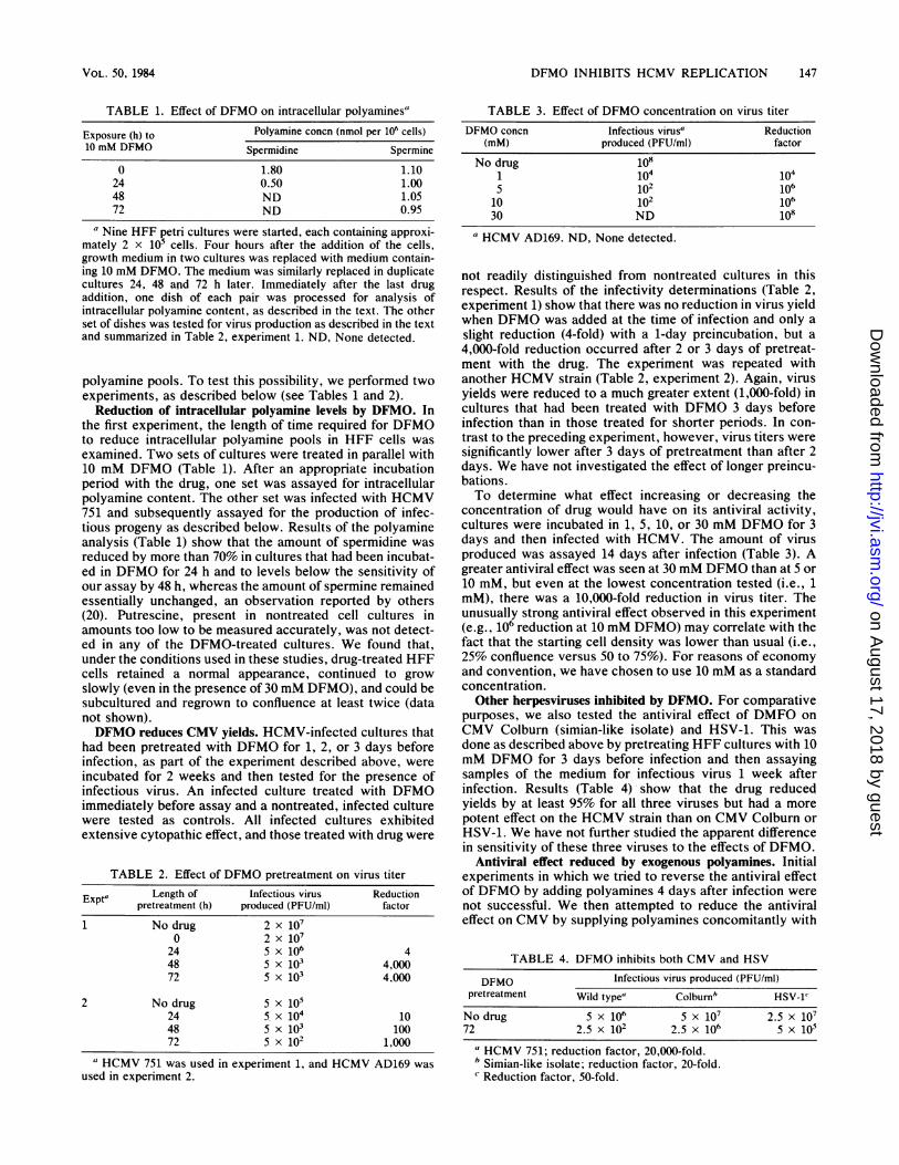
TABLE 1. Effect of DFMO on intracellular polyamines"
Exposure (h) to Polyamine concn (nmol per 106 cells)10 mM DFMO Spermidine Spermine
0 1.80 1.1024 0.50 1.0048 ND 1.0572 ND 0.95
a Nine HFF petri cultures were started, each containing approxi-mately 2 x 10⁵ cells. Four hours after the addition of the cells,growth medium in two cultures was replaced with medium contain-ing 10 mM DFMO. The medium was similarly replaced in duplicatecultures 24, 48 and 72 h later. Immediately after the last drugaddition, one dish of each pair was processed for analysis ofintracellular polyamine content, as described in the text. The otherset of dishes was tested for virus production as described in the textand summarized in Table 2, experiment 1. ND, None detected.
polyamine pools. To test this possibility, we performed twoexperiments, as described below (see Tables 1 and 2).Reduction of intracellular polyamine levels by DFMO. In
the first experiment, the length of time required for DFMOto reduce intracellular polyamine pools in HFF cells wasexamined. Two sets of cultures were treated in parallel with10 mM DFMO (Table 1). After an appropriate incubationperiod with the drug, one set was assayed for intracellularpolyamine content. The other set was infected with HCMV751 and subsequently assayed for the production of infec-tious progeny as described below. Results of the polyamineanalysis (Table 1) show that the amount of spermidine wasreduced by more than 70% in cultures that had been incubat-ed in DFMO for 24 h and to levels below the sensitivity ofour assay by 48 h, whereas the amount of spermine remainedessentially unchanged, an observation reported by others(20). Putrescine, present in nontreated cell cultures inamounts too low to be measured accurately, was not detect-ed in any of the DFMO-treated cultures. We found that,under the conditions used in these studies, drug-treated HFFcells retained a normal appearance, continued to growslowly (even in the presence of 30 mM DFMO), and could besubcultured and regrown to confluence at least twice (datanot shown).DFMO reduces CMV yields. HCMV-infected cultures that
had been pretreated with DFMO for 1, 2, or 3 days beforeinfection, as part of the experiment described above, wereincubated for 2 weeks and then tested for the presence ofinfectious virus. An infected culture treated with DFMOimmediately before assay and a nontreated, infected culturewere tested as controls. All infected cultures exhibitedextensive cytopathic effect, and those treated with drug were
TABLE 2. Effect of DFMO pretreatment on virus titerLength of Infectious virus ReductionExpt' pretreatment (h) produced (PFU/ml) factor
1 No drug 2 x 1070 2x 10⁷
24 5 x 10⁶ 448 5 x 10³ 4,00072 5 x 10³ 4,000
2 No drug 5 x 10524 5 x 10⁴ 1048 5 x 10³ 10072 5 x 10² 1,000
a HCMV 751 was used in experiment 1, and HCMV AD169 wasused in experiment 2.
TABLE 3. Effect of DFMO concentration on virus titer
DFMO concn Infectious virusa Reduction(mM) produced (PFU/mI) factor
No drug 1081 104 1045 102 10610 102 10630 ND 108
a HCMV AD169. ND, None detected.
not readily distinguished from nontreated cultures in thisrespect. Results of the infectivity determinations (Table 2,experiment 1) show that there was no reduction in virus yieldwhen DFMO was added at the time of infection and only aslight reduction (4-fold) with a 1-day preincubation, but a4,000-fold reduction occurred after 2 or 3 days of pretreat-ment with the drug. The experiment was repeated withanother HCMV strain (Table 2, experiment 2). Again, virusyields were reduced to a much greater extent (1,000-fold) incultures that had been treated with DFMO 3 days beforeinfection than in those treated for shorter periods. In con-trast to the preceding experiment, however, virus titers weresignificantly lower after 3 days of pretreatment than after 2days. We have not investigated the effect of longer preincu-bations.To determine what effect increasing or decreasing the
concentration of drug would have on its antiviral activity,cultures were incubated in 1, 5, 10, or 30 mM DFMO for 3days and then infected with HCMV. The amount of virusproduced was assayed 14 days after infection (Table 3). Agreater antiviral effect was seen at 30 mM DFMO than at 5 or10 mM, but even at the lowest concentration tested (i.e., 1mM), there was a 10,000-fold reduction in virus titer. Theunusually strong antiviral effect observed in this experiment(e.g., 106 reduction at 10 mM DFMO) may correlate with thefact that the starting cell density was lower than usual (i.e.,25% confluence versus 50 to 75%). For reasons of economyand convention, we have chosen to use 10 mM as a standardconcentration.Other herpesviruses inhibited by DFMO. For comparative
purposes, we also tested the antiviral effect of DMFO onCMV Colburn (simian-like isolate) and HSV-1. This wasdone as described above by pretreating HFF cultures with 10mM DFMO for 3 days before infection and then assayingsamples of the medium for infectious virus 1 week afterinfection. Results (Table 4) show that the drug reducedyields by at least 95% for all three viruses but had a morepotent effect on the HCMV strain than on CMV Colburn orHSV-1. We have not further studied the apparent differencein sensitivity of these three viruses to the effects of DFMO.
Antiviral effect reduced by exogenous polyamines. Initialexperiments in which we tried to reverse the antiviral effectof DFMO by adding polyamines 4 days after infection werenot successful. We then attempted to reduce the antiviraleffect on CMV by supplying polyamines concomitantly with
TABLE 4. DFMO inhibits both CMV and HSV
DFMO Infectious virus produced (PFU/ml)pretreatment Wild type' Colburnm HSV-1'
No drug 5 x 10⁶ 5 x 10⁷ 2.5 x 10772 2.5 x 10² 2.5 x 10⁶ 5 x 10⁵
a HCMV 751; reduction factor, 20,000-fold.b Simian-like isolate; reduction factor, 20-fold.Reduction factor, 50-fold.
VOL. 50, 1984
on August 17, 2018 by guest
http://jvi.asm.org/
Dow
nloaded from

148 GIBSON ET AL.
the drug. This was done by incubating cultures for 3 days inthe presence of 10 mM DFMO; 10 KM polyamines, i.e.,putrescine, spermidine, or spermine; 10 mM DFMO inaddition to 10 KM polyamines; or no drug. Pretreated cellswere then infected with HCMV, and the titer of progenyvirus was determined by plaque assay. The results of thisexperiment (Table 5) show that DFMO alone reduced virustiter by 1,000-fold. The presence of putrescine during pre-treatment with DFMO increased the amount of virus by 50-fold, to 5% of the control value. Addition of either spermi-dine or spermine along with the drug increased virus yield80-fold (i.e., 8% of control) above that of the culturecontaining DFMO alone. The presence of 10 pKM putrescine,spermidine, or spermine alone was without detectable effecton virus yield. The influence of these drug combinations onthe types of particles produced by the infected cells is shownin Fig. 1 and described below.
In a similar experiment in which intracellular polyaminelevels were measured, it was found that spermine was themost abundant species present and that its relative concen-tration among the different cultures did not vary appreciably(i.e., .20%; data not shown). These data also showed thatthe spermidine concentration in infected, nontreated cul-tures was approximately one-third that of spermine.DFMO reduces amount of extracellular particles. Since
extensive viral cytopathic effect was observed, even in thepresence of 30 mM DFMO, it was apparent that at leastsome virus products were being made. As a first step inattempting to determine how DFMO exerts its antiviraleffect on HCMV, we tested the extracellular medium for thepresence of virus particles. Media samples from the experi-ment described above were processed and subjected to rate-velocity centrifugation in glycerol-tartrate gradients, andabsorbance (280 nm) profiles were recorded as the gradientswere collected (as described in Materials and Methods).Figure 1 shows two types of extracellular virus particlesrecovered from HFF cells normally infected with HCMVAD169. These are referred to as noninfectious envelopedparticles (NIEPs [13]) and virions. In the context of this studyit should be emphasized that NIEPs and virions are nearlyindistinguishable in protein composition and architecture butdiffer in that NIEPs contain no DNA. Medium from theculture treated with DFMO contained neither type of parti-cle (Fig. 1, lower left). However, when putrescine, spermi-dine, or spermine was added together with DFMO, a virionpeak was observed. Furthermore, in the presence ofDFMO,exogenous polyamines altered the relative amounts ofNIEPs and virions. Putrescine had a stronger sparing effecton NIEPs than virions, whereas the opposite was true forspermidine and spermine. Comparison of the infectivities ofthese preparations (Table 5) with the amount of virionspresent in each (i.e., absorbance; Fig. 1) indicates that
TABLE 5. DFMO effect reduced by exogenous polyaminesVirus6 Protein Virus titer % of
Treatment' titer (PFU/ (mg per per mg of nontreatedml) dish) protein
None 1.5 x 10⁷ 3.6 4.2 x 10⁶ 100DFMO 1.0 x 10⁴ 2.3 4.4 x 10³ 0.1DFMO + Put. 5.7 x 10⁵ 2.6 2.2 x 10⁵ 5.2DFMO + Spd. 6.5 x 10⁵ 1.9 3.4 x 10⁵ 8.1DFMO + Sp. 5.5 x 10⁵ 1.5 3.7 x 10⁵ 8.8
"Put., Putrescine; Spd., spermidine; Sp., spermine.6 HCMV AD169.
MIIUJ U1JUUspu
DFMO+Sp.+ DFMO
SedimentationFIG. 1. Effect of DFMO on NIEP and virion production by
HCMV-infected cells. HFF cells were incubated for 3 days with 10mM DFMO or with 10 mM DFMO supplemented with either 10 ,uMputrescine, spermidine, or spermine and then infected with HCMVAD169. Fourteen days after infection, media were collected fromthe cultures, clarified by centrifugation at 1,500 x g at 4°C for 10min, layered above glycerol-tartrate gradients, and subjected tocentrifugation at 40,000 rpm at 4°C for 20 min in a Beckman SW41rotor. The gradients were monitored at 280 nm as they weredisplaced from the tubes with a model 185 density gradient fraction-ator and a model UA-5 absorbance monitor (ISCO, Lincoln, Neb.).Full-scale absorbance for all gradients was 0.2 optical density units.The positions of NIEPs and virions (VIR.) are indicated. Theprofiles shown are from infected cells that had been preincubatedand continued in the absence of drug (No DFMO), in the presence ofDFMO alone (+DFMO), in the presence of DFMO and putrescine(DFMO + Put.), in the presence of DFMO and spermidine (DFMO+ Spd.), or in the presence of DFMO and spermine (DFMO + Sp.).Shown here is a collage prepared from photocopies of the record-ings.
DFMO + Put.NIEPA VIR.
No DFMONIEP VIR.
nfMf(+ Ind
J. VIROL.
on August 17, 2018 by guest
http://jvi.asm.org/
Dow
nloaded from

DFMO INHIBITS HCMV REPLICATION 149
DFMO :X1- =
FIG. 2. Cells infected in the presence of DFMO produce viralDNA. HFF cells were pretreated with DFMO for 3 days or nottreated and then were infected (in presence or absence of drug,respectively) with HCMV 751. Infected cell DNA was extracted 6days postinfection and cleaved with BamHI restriction endonucle-ase. Such preparations from cells infected in the presence (+) orabsence (-) of DFMO were subjected to electrophoresis in a 1%agarose gel for 15 h at 50 V. A sample of strain 751 virion DNA was
compared in an adjacent channel. Shown here is a photograph of theresulting gel, stained with ethidium bromide.
cultures treated with spermine produced approximately theamount of virions expected from the infectivity measure-
ment (i.e., about 10% the amount of non-drug-treated con-
trol). The cultures treated with putrescine or spermidine,however, produced about five- and fourfold more particles,respectively, than was predicted from their infectivities.
Viral DNA made in DFMO-treated cells. We next lookedfor intracellular evidence of virus replication and began bytesting for viral DNA synthesis. This was done by preparingDNA from cultures infected with HCMV 751 in the presenceor absence of drug, cleaving the DNA with BamHI restric-tion endonuclease, and separating the resulting fragments ina 1% agarose slab gel (as described in Materials and Meth-ods). A photograph of the resulting gel, stained with ethid-ium bromide (Fig. 2), demonstrates that viral DNA waspresent in both DFMO-treated and nontreated cultures. Anadjacent channel containing DNA from purified HCMV 751virions, cleaved in parallel with the cellular preparations,
shows that the bands visualized are viral. Although we areunable to make precise quantitative comparisons betweenthese preparations, it is clear that, in the DFMO-treatedcultures, viral DNA is replicated and accumulates in sub-stantial amounts (e.g., .50% normal). On the basis of resultsof similar experiments in which the infected cell DNA waslabeled with 32p, (data not shown), we are confident that thebands shown here represent newly synthesized DNA ratherthan input molecules introduced by infection.
Viral proteins made in DFMO-treated cells. To determinewhether viral proteins were also being synthesized inDFMO-treated cells, we compared drug-treated and non-treated, infected cultures by gel electrophoresis. HCMVstrains Towne and 751 were both used. Seven days afterinfection, the cells were collected and processed for analysisby polyacrylamide gel electrophoresis (Fig. 3). A photographof the resulting gel after staining with Coomassie brilliantblue is shown in Fig. 3. Only the nuclear fractions, whichcontain most of the CMV protein mass (8), are shown. All ofthe infected-cell-specific proteins detected in the normallyinfected cells were also present in the DFMO-treated cells.Among these, the major capsid protein, 140,000-kilodaltonprotein, matrix protein, DNA-binding protein, and assemblyprotein are the most prominent. This figure also demon-strates that exposure to 10 mM DFMO does not appreciablyalter the pattern of nuclear proteins in noninfected cells.Although there is some variability among experiments, den-sitometric measurements made from such comparative gelsindicate that, relative to the amount of actin or major capsidprotein present, preparations from DFMO-treated, infectedcells contain more matrix protein than do those from non-treated, infected cells. Expression and accumulation of theseviral proteins was the same in cells treated with 30 mMDFMO (data not shown).Some virus particles are assembled in DFMO-treated cells.
Since viral DNA and proteins were both present in DFMO-treated cells, we became interested in the possibility that thisdrug may exert its inhibitory effect at the level of virusassembly. This possibility was explored by using an electronmicroscope to examine thin sections of drug-treated andnontreated, HCMV-infected cells. Electron micrographsprepared from these cultures (e.g., Fig. 4 and 5) showed thatDFMO-treated and nontreated cultures contained a compa-rable number of particles. Among those observed in non-treated cells were intranuclear capsids that were eitherempty or partially filled with densely stained material (e.g.,DNA) or that contained an apparent internal structure thatwas variable in configuration but often looked like a concen-tric ring about 15 nm thick or a string of six symmetricallydistributed beads, each 10 to 15 nm in diameter (see Fig. 5).In addition, enveloped capsids containing a densely stainedcenter were frequently observed in the cytoplasm (e.g., Fig.4, "Virion"; Fig. SE). The particles present in DFMO-treated cells appeared more uniform in structure. Fewparticles with darkly stained centers were seen (Fig. 4B); thenumber of enveloped particles in the cytoplasm was re-duced, and those that were observed did not have denselystained centers (e.g., Fig. 5F). Although these general obser-vations were characteristic of the cells examined, no attemptwas made to quantify the various particle types since theywere heterogeneous in appearance and we could not assesswith any certainty whether internal capsid structures repre-sented DNA or protein or both. In the course of this study,numerous cells were examined. The structures shown in Fig.4 and S are representative of those observed and werepresent in the majority of cells.
VOL. 50, 1984
on August 17, 2018 by guest
http://jvi.asm.org/
Dow
nloaded from

150 GIBSON ET AL.
Mock Towne 751-m
140K
X-4-Ma6-matrix
wo _ _DBP--Actin
Assembly
FIG. 3. Proteins present in DFMO-treated, HCMV-infectedHFF cells. HFF cells were incubated for 3 days in the presence (+)or absence (-) of DFMO. One culture each was then infected withHCMV (Towne or 751) or was left uninfected (Mock). Seven daysafter infection, the cells were scraped from the petri dishes into theculture medium, collected by centrifugation at 1,500 x g at 4°C for10 min, disrupted with Nonidet P-40 (0.5% in 0.15 M NaCI-0.04 Msodium phosphate buffer, pH 7.4; 0°C, 10 min), and separated intonuclear pellet and cytoplasmic supernatant fractions by centrifuga-tion as described above, and each fraction was solubilized inpreparation for analysis by gel electrophoresis. Aliquots of theresulting Nonidet P-40 nuclear fractions were subjected to electro-phoresis in a sodium dodecyl sulfate-containing, 7.5% polyacryl-amide gel. Shown here is a photograph of the Coomassie brilliantblue-stained gel after it had been dried onto a piece of filter paper.Five HCMV proteins and the cellular protein (Actin) are indicated inthe right-hand margin. CMV protein designations (molecular weight)(8): MCP, major capsid protein (153,000); 140K (140,000); Matrix,matrix-like protein (69,000); DBP, DNA-binding protein (53,000);Assembly, assembly protein (35,000).
HCMV virions contain polyamines. Finally, it was ofinterest to determine whether HCMV virions contain poly-amines. Since it has not yet been possible for us to measurethe polyamine content of HCMV virions directly (e.g.,dansylation-fluorometry or high-pressure liquid chromatog-raphy), we have used a radiolabeling approach in these initialexperiments. This was done by radiolabeling HFF cells with[3H]ornithine (5 p.Ci/ml) for 24 h and then infecting thecultures with HCMV strain AD169 or 751 or HSV-1. Radio-label remained in the culture medium throughout the infec-tion. When strong cytopathic effect was observed (i.e., 7 to10 days for HCMV and 2 to 3 days for HSV-1), virusparticles were collected from the maintenance media by rate-velocity sedimentation in glycerol-tartrate gradients andconcentrated by pelleting. Polyamines were acid extracted
from the pelleted virions, dansylated, and separated by thin-layer chromatography. Spermine and spermidine spots werelocated by fluorescence and scraped from the plate, and theirradioactivity was measured by scintillation spectrometry.All experiments were done at least twice (Table 6). HCMVAD169 and 751 virions contained the polyamines spermineand spermidine. No putrescine was detected. The ratio ofradiolabeled spermine to spermidine was calculated to beapproximately 2.3 (2.17 ± 0.27 for strain AD169 and 2.540.22 for strain 751).This ratio of spermine to spermidine is much higher than
that previously determined for HSV virions by radiolabeling(i.e., 0.3; see reference 9, Table 3). We considered thepossibility that this discrepancy may be due to experimentaldifferences between the two studies, such as cell type (i.e.,HFF versus HEp-2 cells), radiolabeling conditions (i.e.,continuous versus 18-h preinfection only), or virus recoveryregimen (i.e., glycerol-tartrate gradients and extracellularvirus versus dextran gradients and intracellular virus). Thiswas tested by analyzing HSV virions grown and purified inthe same way that we prepared HCMV virions. Results oftwo such experiments (Table 6) show that the HSV sperm-ine/spermidine ratio was the same as cited above (i.e., 0.3)and, therefore, not significantly affected by the differentexperimental conditions of this study.We note, without further comment, first, that the HCMV
polyamine ratio, calculated here from measurements ofradioactivity, is predicted to be a minimal estimate since thespecific activity of spermine is slower to increase than that ofspermidine. Second, the absolute ratio of spermine to sper-midine in HSV virions is approximately 0.6 when measureddirectly (9, 21), a value still three- to four-fold lower than thetheoretically underestimated ratio of approximately 2.3 de-termined for HCMV in the present experiments.
DISCUSSIONIn this report we have presented evidence that DFMO, an
enzyme-activated inhibitor of the polyamine biosyntheticenzyme ODC, exhibits a strong inhibitory effect on theproduction of infectious CMV by HFF cells. Although thisgeneral conclusion is in agreement with an earlier report byTyms and Williamson (33), specific details differ significantlyand warrant further discussion.
First, with respect to the time of drug addition, we foundthat, when sufficiently high multiplicities of infection wereused to ensure infection of every cell, a 2- to 3-day preincu-bation of the cell culture with DFMO was necessary to
TABLE 6. Virions of HCMV contain polyamines
Strain and expt Radioactivity (cpm)ano. Spermine Spermidine Ratio Mean t SEM
HCMV AD169 2.17 t 0.271 2,634 1,413 1.862b 5,441 2,579 2.113 3,980 1,574 2.53
HCMV 751 2.54 ± 0.221 1,956 708 2.76
14,714 6,352 2.32
HSV-1 0.34 ± 0.041 865 2,208 0.392b 5,166 17,141 0.30a Radiolabel was supplied to cells as [3H]ornithine.b Done together as part of the same experiment.
J. VIROL.
on August 17, 2018 by guest
http://jvi.asm.org/
Dow
nloaded from

DFMO INHIBITS HCMV REPLICATION 151~~~~~~~~~,>. .Virion ;s
Cyto. v$
t~i ts
A
.
-4-,~~~~~~~~~~~~~~~~~~~~~~~~~~~~~~~*
bA.
FIG. 4. DFMO-treated, HCMV-infected cells contain virus particles. HFF cell cultures, either nontreated or grown continuously in thepresence of 10 mM DFMO added 3 days before infection, were infected with HCMV AD169. Nine days later, the cell layers were rinsed withphosphate-buffered saline, fixed, scraped from the petri dish, and processed for thin sectioning and analysis by electron microscopy asdescribed in the text. Shown here are representative thin sections of cells infected in the absence (A) or presence (B) of DFMO. Capsids thatappear empty (1), partially filled with dense-staining material (2), and contain internal beadlike structures (3) were seen in the nucleus (Nuc.)of cultures lacking DFMO; enveloped particles with a densely stained core (Virion) were present in the cytoplasm (Cyto.). Capsids with abeaded internal structure (3) were present in the nucleus of DFMO-treated cultures, and empty, apparently coated particles (4) were seenwithin cytoplasmic invaginations into the nucleus. Arrows indicate nuclear membrane. Bar, 200 nm. Representative capsid forms from suchcells are shown in greater detail in Fig. 5.
confer the antiviral effect. This observation is consistentwith the earlier findings of Tuomi et al. (30), who showedthat the inhibitory effect of DFMO on Semliki Forest virus(enveloped, RNA-containing virion) and HSV-2 occurredonly when infection followed a 3-day incubation with thedrug. In light of the consistency of these results from studiesin which different cells (i.e., human or monkey) and viruses(i.e., HCMV, Semliki Forest virus, and HSV-2) were used, itis not clear why in some studies DFMO and D,L-a-methylor-nithine produced an antiviral effect without preincubation(31, 33) whereas in others DFMO failed to confer an antiviraleffect even with a 3-day preincubation (15). It seems unlikelythat these differences were due to the cells or virus, since allstudies with HCMV used diploid human fibroblast cells andall but one used CMV AD169. A more plausible explanation,therefore, may be differences in the culture conditions (e.g.,cell density, serum composition, and concentration), whichwe also believe to be responsible for variability in the extentof the antiviral effect observed in the present studies. Sincethe antiviral effect correlated with depletion of the intracellu-lar spermidine pool, it would be informative to know howthis value changed in the other studies.
Second, experiments to investigate the level at whichDFMO interferes with the production of infectious HCMVshowed that the drug did not spare the cells from typical viralcytopathic effect, nor did it block either viral DNA or proteinsynthesis. Further, since at least empty capsids (i.e., partial-ly or nonstained center) were present in the drug-treatedcells (Fig. 4), it is apparent that the level of inhibition is not
capsid assembly per se. We did find, however, that the drugtreatment greatly reduced the number of mature particles(e.g., enveloped with densely stained core) both within thecell and released into the medium. These observations aremore compatible with the antiviral effect of DFMO beingdirected against a late event in the virus replication cycle,such as DNA packaging or capsid envelopment or both, thanwith an early event, as suggested elsewhere (33).
Since CMV virions, like those of HSV (9, 10, 21), containthe polyamines spermidine and spermine (Table 6) and sinceDFMO essentially eliminates the intracellular pool of sper-midine (Table 1), the possibility that this drug exerts itsantiviral effect by interfering with virus assembly is mecha-nistically attractive. If polyamines serve as counterions toneutralize the electronegativity of virion constituents duringassembly, then at least two steps in the maturation pathwaymight be particularly sensitive to a decrease in their avail-ability. The first is DNA packaging, in which nucleotidephosphates would require neutralization to permit encapsi-dation, and the second is nucleocapsid envelopment, duringwhich phosphate charges of the tegument proteins coatingthe capsid (W. Gibson and A. Irmiere, in CMV: Pathogene-sis and Prevention of Human Infection. March of DimesBirth Defects, original article series, vol. 19, in press),envelope phospholipids (W. Gibson, unpublished observa-tions), or other electronegative virion constituents mayrequire neutralization.
If the inhibitory effect of DFMO were on DNA packagingalone, then one would not expect the production of particles
VOL. 50, 1984
1.
"4-
A
P" ...I2; .
4.1
on August 17, 2018 by guest
http://jvi.asm.org/
Dow
nloaded from

152 GIBSON ET AL.
..
.2S
C
4
*<.: r
S..
. 55...wXN. r' v,
.t:....
..
'..
.&
0
.:..~~~~~~~~~~~~~~~~~~~~~~~~~..:.
..4S
FIG. 5. Intranuclear HCMV capsid forms and enveloped cytoplasmic particles present in DFMO-treated and nontreated HFF cells.Shown here are high-magnification pictures of virus particles in nontreated (A and B) and DFMO-treated (C and D) cells, prepared asdescribed in the legend to Fig. 4. (A) Three particles with densely stained centers of the type designated 2 in Fig. 4, one with a ring-shaped in-ternal structure of the type designated 3 in Fig. 4 (lower right-hand corner), one with less clearly organized internal material (lower left-handcorner), and adjacent to it, an empty capsid of the type designated 1 in Fig. 4. (B) Particle containing an internal structure, beaded inappearance. Arrows indicate coincidence of beads with capsid vertices. Many of the particles in drug-treated cells contained ring-shapedinternal structures of the type designated 3 in Fig. 4, but other organizations were also observed (C and D). (E) Enveloped capsid with adensely stained center (e.g., virion) within a cytoplasmic tubule (arrow indicates tubule membrane). (F) Enveloped particle with a weaklystained center (e.g., NIEP [13]) also within a cytoplasmic tubule (see arrow). All micrographs are the same magnification; bar, ca. 100 nm.
J. VIROL.
4*e
tAlkEkt, ?4-,,,i,-
I
T '. ..;.
on August 17, 2018 by guest
http://jvi.asm.org/
Dow
nloaded from

DFMO INHIBITS HCMV REPLICATION 153
lacking DNA to be interrupted. This prediction is supportedby experiments in which we have shown that the productionof virion-like NIEPs, which contain all of the virion proteinsbut no DNA, is unaffected by inhibitors of DNA synthesisthat abolish virion production (Gibson and Irmiere, in press).Our finding that DFMO treatment blocks the production ofNIEPs as well as virions (Fig. 1) is, therefore, taken asevidence for an additional, or different, level of inhibition,such as envelopment. In this connection, we have beeninterested in determining whether one of the putative assem-bly intermediates (e.g., intranuclear A and B capsids orcytoplasmic C capsids [7]) may accumulate in DFMO-treated, infected cells, thereby providing an indication of thestep blocked. Since recovery of such particles from non-treated, HCMV-infected cells has proven difficult, theseexperiments are being done with strain CMV Colburn,previously shown (7) to produce both intracellular andextracellular particles in good yield during a normal infec-tion. It is anticipated that DFMO will be useful in studyingaspects of virus replication and assembly that are dependentupon cellular polyamines and are not otherwise directlyaccessible.Although DFMO inhibits the growth of CMV (Tables 2
and 4; 33), HSV (Table 4; 30), and Semliki Forest virus (30)in cell culture, it remains to be determined whether it will beof value as an antiviral agent in humans. The need forpreexposure and high concentrations to achieve an antiviraleffect with DFMO (in vitro), coupled with its generalizedsuppression of cell proliferation, would tend to argue againstthis possibility. Despite these potential limitations, there arefeatures of this drug that make its clinical evaluation as anantiviral agent seem attractive. First, DFMO is well tolerat-ed in humans (11). Second, since DFMO acts on a host cellprotein rather than on one encoded by the viral genome, theprobability of a drug-resistant variant or mutant virus emerg-ing is expected to be low. Third, unlike many chemothera-peutic agents used to inhibit DNA viruses, DFMO does notinterfere directly with DNA replication. Thus, concerns arereduced about it promoting DNA replication-mediated muta-genesis, transformation, or integration of viral DNA into thehost chromosome. Fourth, although DFMO inhibits theproduction of infectious virus and thereby the spread ofinfection, it does not spare the initially infected cell fromdeath. Therefore, the possibility of perpetuating the virus ina drug-mediated latent form is reduced. Finally, becauseDFMO alters the intracellular environment by inhibiting acellular enzyme, it is expected to interfere with a broadspectrum of viruses that contain cellular polyamines orotherwise depend upon their availability during replication.
ACKNOWLEDGMENTSWe thank Paul Lietman for catalyzing this study, Peter McCann,
Merrell Research Center, for his generosity in providing DFMO,Ken Bott for kindly loaning us his scanning fluorometer, and theCore Electron Microscopy Laboratory, supported by Public HealthService grant HD 06268 from the National Institutes of Health toThe Johns Hopkins University Population Center, for assistancewith electron microscopy. We also thank Susan Goehring forexcellent technical assistance and Louise Flannery for typing themanuscript.
R.v.B., A.F. and A.I. are Predoctoral Fellows in the AnticancerDrug Development (R.v.B., A.F.) and Biochemistry, Cellular andMolecular Biology (A.I.) Training Programs and were supported byPublic Health Service grants CA 09243 and GM 07445, respectively,from the National Institutes of Health. These studies were aided byresearch grants Al 13718 and Al 16959 from the National Institutesof Health.
LITERATURE CITED
1. Barzilai, R., L. H. Lazarus, and N. Goldblum. 1972. Viscosity-density gradient for purification of foot-and-mouth disease vi-rus. Arch. Gesamte Virusforsch. 36:141-146.
2. Boldogh, I., E. Beth, E.-S. Huang, S. K. Kyalwazi, and G.Giraldo. 1981. Kaposi's sarcoma. IV. Detection of CMV DNA,CMV RNA and CMNA in tumor biopsies. Int. J. Cancer28:469-474.
3. Centers for Disease Control. 1982. Update on Kaposi's sarcomaand opportunistic infections in previously healthy persons-United States. Morbid. Mortal. Weekly Rep. 31:294-301.
4. Dion, A. S., and E. J. Herbst. 1970. Polyamine changes duringdevelopment of Drosophila melanogaster. Ann. N.Y. Acad. Sci.171:723-734.
5. Drew, W. L., L. Mintz, R. C. Miner, M. Sands, and B. Ketterer.1981. Prevalence of cytomegalovirus infection in homosexualmen. J. Infect. Dis. 143:188-192.
6. Fenwick, M. L., and M. J. Walker. 1978. Suppression of thesynthesis of cellular macromolecules by herpes simplex virus. J.Gen. Virol. 41:37-51.
7. Gibson, W. 1981. Structural and nonstructural proteins of strainColburn cytomegalovirus. Virology 111:516-537.
8. Gibson, W. 1983. Protein counterparts of human and simiancytomegaloviruses. Virology 128:391-406.
9. Gibson, W., and B. Roizman. 1971. Compartmentalization ofspermine and spermidine in the herpes simplex virion. Proc.Natl. Acad. Sci. U.S.A. 68:2818-2821.
10. Gibson, W., and B. Roizman. 1973. The structural role andmetabolic involvement of polyamines with herpes simplex vi-rus, p. 123-135. In D. H. Russell (ed.), Polyamines in normaland neoplastic growth. Raven Press, Inc., New York.
11. Haegele, K., R. Alken, J. Grove, P. Schechter, and J. Koch-Weser. 1981. Kinetics of a-difluoromethylornithine: an irrevers-ible inhibitor of ornithine decarboxylase. Clin. Pharmacol.Ther. 30:210-217.
12. Helgstrand, E., B. Eriksson, N. G. Johansson, B. Lannero, A.Larsson, A. Misiorny, J. 0. Noren, B. Sjoberg, K. Stenberg, G.Stening, G. Stridh, B. Oberg, S. Alenius, and L. Philipson. 1978.Trisodium phosphonoformate, a new antiviral compound. Sci-ence 201:819-821.
13. Irmiere, A., and W. Gibson. 1983. Isolation and characterizationof a noninfectious virion-like particle released from cells infect-ed with human strains of cytomegalovirus. Virology 130:118-133.
14. Isom, H. C. 1979. Stimulation of ornithine decarboxylase byhuman cytomegalovirus. J. Gen. Virol. 42:265-278.
15. Isom, H. C., and A. E. Pegg. 1979. Inhibition of cytomegalovi-rus-stimulated human cell ornithine decarboxylase by alpha-difluoromethylornithine. Biochem. Biophys. Acta 564:402-413.
16. Kaposi, M. 1872. Idiopathisches multiples pigmentsarkom derHaut. Arch. Dermatol. Syph. 4:265-273.
17. Laemmli, U. K. 1970. Cleavage of structural proteins during theassembly of the head of bacteriophage T4. Nature (London)277:680-684.
18. LaFemina, R. L., and G. S. Hayward. 1983. Replicative forms ofhuman cytomegalovirus DNA with joined termini are found inpermissively infected human cells but not in non-permissiveBALB/c-3T3 mouse cells. J. Gen. Virol. 64:373-389.
19. Lowry, 0. H., N. J. Rosebrough, A. L. Farr, and R. J. Randall.1951. Protein measurement with the Folin phenol reagent. J.Biol. Chem. 193:265-275.
20. Mamont, P. S., M.-C. Duchesne, J. Grove, and P. Bey. 1978.Anti-proliferative properties of D,L-alpha-difluoromethyl orni-thine in cultured cells. A consequence of the irreversibleinhibition of ornithine decarboxylase. Biochem. Biophys. Res.Commun. 81:58-66.
21. McCormick, F. 1978. Polyamine turnover and leakage duringinfection of HeLa and L-cells with herpes simplex virus type 1.Virology. 91:496-503.
22. McCormick, F., and A. A. Newton. 1975. Polyamine metabolismin cells infected with herpes simplex virus. J. Gen. Virol. 27:25-33.
VOL. 50, 1984
on August 17, 2018 by guest
http://jvi.asm.org/
Dow
nloaded from

154 GIBSON ET AL.
23. Metcalf, B. W., P. Bey, C. Danzin, M. J. Jung, P. Casara, andJ. P. Vevert. 1978. Catalytic irreversible inhibition of mammali-an ornithine decarboxylase (E.C.4.1.1.17) by substrate andproduct analogues. J. Am. Chem. Soc. 100:2551-2553.
24. Nishioka, Y., and S. Silverstein. 1978. Requirement of proteinsynthesis for the degredation of host mRNA in Friend erythro-leukemia cells infected with herpes simplex virus type 1. J.Virol. 27:619-627.
25. Raina, A., K. Tuomi, and R.-L. Pajula. 1981. Synthesis ofmacromolecules and viruses in cells treated with inhibitors ofpolyamines synthesis. Adv. Polyamine Res. 3:163-173.
26. Reynolds, E. S. 1963. The use of lead citrate at high pH as an
electron-opaque stain in electron microscopy. J. Cell Biol.17:208-212.
27. Seiler, N., and M. Weichmann. 1967. Die Mikrobestimmung von
Spermin und Spermidin als 1-Dimethylamino-Naphthalin-5-Sul-fonsaure-Derivative. Z. Phys. Chem. 348:1285-1290.
28. Sydiskis, R. J., and B. Roizman. 1966. Polysomes and proteinsynthesis in cells infected with a DNA virus. Science 153:76-78.
29. Talbot, P., and J. Almeida. 1977. Human cytomegalovirus:purification of enveloped virions and dense bodies. J. Gen.Virol. 36:345-349.
30. Tuomi, K., R. Mantyjarvi, and A. Raina. 1980. Inhibition ofSemliki Forest and herpes simplex virus production in alphadi-fluoromethylornithine-treated cells: reversal by polyamines.FEBS Lett. 121:292-294.
31. Tyms, A. S., E. Scamans, and J. D. Williamson. 1979. Polyaminemetabolism in MRC5 cells infected with different herpesviruses.Biochem. Biophys. Res. Commun. 86:312-318.
32. Tyms, A. S., and J. D. Williamson. 1980. Polyamine metabolismin MRC-5 cells infected with human cytomegalovirus. J. Gen.Virol. 48:183-191.
33. Tyms, A. S., and J. D. Williamson. 1982. Inhibitors of polyaminebiosynthesis block human cytomegalovirus replication. Nature(London) 297:690-691.
34. Wahren, B., and B. Oberg. 1980. Reversible inhibition ofcytomegalovirus replication by phosphonoformate. Intervi-rology 14:7-15.
J. VIROL.
on August 17, 2018 by guest
http://jvi.asm.org/
Dow
nloaded from


![a-Difluoromethylornithine Alters Calcium Signaling in ... · [CANCER RESEARCH 52, 6782-6789, December 15, 1992] a-Difluoromethylornithine Alters Calcium Signaling in Platelet-derived](https://img.pdfslide.net/doc/110x75/60695047b56b137949449037/a-difluoromethylornithine-alters-calcium-signaling-in-cancer-research-52-6782-6789.jpg)