Embed Size (px)
Citation preview
Dg
Sa
b
a
ARRAA
KC��AB
1
srinwaaea(hesdtL[ff
0d
Mutation Research 716 (2011) 10– 19
Contents lists available at ScienceDirect
Mutation Research/Fundamental and MolecularMechanisms of Mutagenesis
jou rna l h omepa g e: www.elsev ier .com/ locate /molmutCo mm uni t y ad d ress : www.elsev ier .com/ locate /mutres
NA damage response signaling in lung adenocarcinoma A549 cells followingamma and carbon beam irradiation
omnath Ghosha, Himanshi Naranga,∗, Asitikantha Sarmab, Malini Krishnaa
Radiation Biology and Health Sciences Division, Bhabha Atomic Research Centre, Trombay, Mumbai 400 085, IndiaRadiation Biology Laboratory, Inter University Accelerator Centre, Aruna Asaf Ali Marg, New Delhi 110 067, India
r t i c l e i n f o
rticle history:eceived 24 March 2011eceived in revised form 22 July 2011ccepted 26 July 2011vailable online 3 August 2011
ey words:arbon
a b s t r a c t
Carbon beams (5.16 MeV/u, LET = 290 keV/�m) are high linear energy transfer (LET) radiation character-ized by higher relative biological effectiveness than low LET radiation. The aim of the current study wasto determine the signaling differences between �-rays and carbon ion-irradiation. A549 cells were irra-diated with 1 Gy carbon or �-rays. Carbon beam was found to be three times more cytotoxic than �-raysdespite the fact that the numbers of �-H2AX foci were same. Percentage of cells showing ATM/ATR fociwere more with �-rays however number of foci per cell were more in case of carbon irradiation. LargeBRCA1 foci were found in all carbon irradiated cells unlike �-rays irradiated cells and prosurvival ERK
-Rays-H2AXTMRCA1
pathway was activated after �-rays irradiation but not carbon. The noteworthy finding of this study isthe early phase apoptosis induction by carbon ions. In the present study in A549 lung adenocarcinoma,authors conclude that despite activation of same repair molecules such as ATM and BRCA1, differences inlow and high LET damage responses might be due to their distinct macromolecular complexes rather thantheir individual activation and the activation of cytoplasmic pathways such as ERK, whether it applies to
e fur
all the cell lines need to b. Introduction
Biological consequences of high-energy charged particle expo-ure are of crucial importance in the clinic, space exploration andisk assessments [1]. Track structure of charged-particle radiations known to be a critical determinant of its biological effective-ess [2–4]. Energy deposition by HZE ions is highly heterogeneous,ith a localized contribution along the trajectory of every particle
nd lateral diffusion of energetic electrons (i.e., �-rays, the targettom electrons ionized by the incident HZE ion and emitted at highnergy) many microns from the path of the ions. These particlesre therefore densely ionizing (high-LET) along the primary tracke.g., the track followed by the incident heavy ion); and also theyave a low-LET component because of the high-energy electronsjected by ions as they traverse tissue. Biophysical models havehown that energy-deposition events by high-LET radiation pro-uce different DNA lesions, including complex DNA breaks, andhat qualitative difference between high-LET radiation and low-ET radiation affects both the induction and repair of DNA damage
4–8]. Because of the effective cell killing many clinicians nowavor charged particle beam therapy over conventional photonsor tumors that are radio resistant and when the survival of nearby∗ Corresponding author. Tel.: +91 22 2559 5310; fax: +91 22 2550 5151.E-mail address: [email protected] (H. Narang).
027-5107/$ – see front matter © 2011 Elsevier B.V. All rights reserved.oi:10.1016/j.mrfmmm.2011.07.015
ther explored.© 2011 Elsevier B.V. All rights reserved.
cells cannot be compromised e.g. in treatment of lung cancer [9,10].Although charged particle beams like carbon etc. are currentlybeing used for the treatment, the signaling pathways activated andtheir variance from �-rays irradiation is not well understood.
DNA, the main target for radiation induced cell killing, upondamage, triggers a signaling network that senses different typesof damage and coordinates responses which include activation oftranscription, cell cycle control, apoptosis, senescence, and DNArepair processes [11]. Hundreds of proteins are recruited in timedependent manner in the vicinity of a DSB leading to formationof a signaling-repair complex which can be visualized as foci andare therefore termed as radiation induced foci (RIF). One of thecomponents of RIF is �-H2AX that is immediately phosphorylatedupon irradiation, can be visualized in situ by immunostaining witha �-H2AX specific antibody and measured quantitatively [12–14].Mathematical methods has been developed to analyze flow cytom-etry data to describe the kinetics of DNA damage response proteinactivation after exposure to X-rays and high energy Iron nuclei [15].Other proteins like ATM (ataxia telangiectasia mutated), ATR (ATMand Rad3-related) and BRCA1, also function in complexes that canbe seen as foci. There are reports that ATM responds differentlyto DSBs induced by high-LET and low-LET radiations [16,17]. The
signal is transduced downstream by proteins such as Chk1/Chk2and p53 that trigger off DNA repair, cycle arrest or apoptosis. Thecytoplasmic MAPK pathways, namely ERK and JNK, have also beenlinked to the DNA damage response and ATM-mediated signaling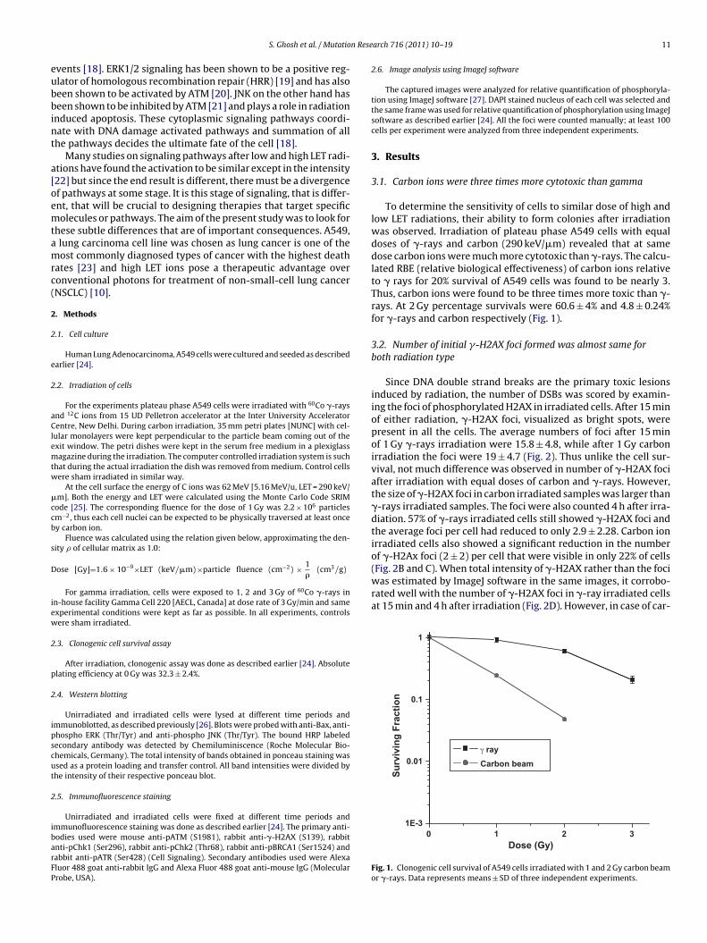
n Rese
eubbint
a[oemtamrc(
2
2
e
2
aClemtw
�ccb
s
D
iew
2
p
2
ipscut
2
ibarFP
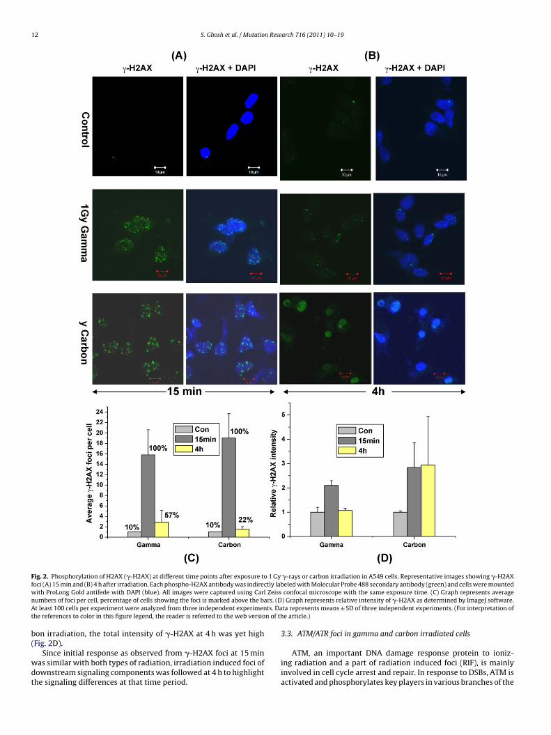
(Fig. 2B and C). When total intensity of �-H2AX rather than the fociwas estimated by ImageJ software in the same images, it corrobo-rated well with the number of �-H2AX foci in �-ray irradiated cellsat 15 min and 4 h after irradiation (Fig. 2D). However, in case of car-
1
0.1
0.01γ rayCarbon beam
Surv
ivin
g Fr
actio
n
1E-3
S. Ghosh et al. / Mutatio
vents [18]. ERK1/2 signaling has been shown to be a positive reg-lator of homologous recombination repair (HRR) [19] and has alsoeen shown to be activated by ATM [20]. JNK on the other hand haseen shown to be inhibited by ATM [21] and plays a role in radiation
nduced apoptosis. These cytoplasmic signaling pathways coordi-ate with DNA damage activated pathways and summation of allhe pathways decides the ultimate fate of the cell [18].
Many studies on signaling pathways after low and high LET radi-tions have found the activation to be similar except in the intensity22] but since the end result is different, there must be a divergencef pathways at some stage. It is this stage of signaling, that is differ-nt, that will be crucial to designing therapies that target specificolecules or pathways. The aim of the present study was to look for
hese subtle differences that are of important consequences. A549, lung carcinoma cell line was chosen as lung cancer is one of theost commonly diagnosed types of cancer with the highest death
ates [23] and high LET ions pose a therapeutic advantage overonventional photons for treatment of non-small-cell lung cancerNSCLC) [10].
. Methods
.1. Cell culture
Human Lung Adenocarcinoma, A549 cells were cultured and seeded as describedarlier [24].
.2. Irradiation of cells
For the experiments plateau phase A549 cells were irradiated with 60Co �-raysnd 12C ions from 15 UD Pelletron accelerator at the Inter University Acceleratorentre, New Delhi. During carbon irradiation, 35 mm petri plates [NUNC] with cel-
ular monolayers were kept perpendicular to the particle beam coming out of thexit window. The petri dishes were kept in the serum free medium in a plexiglassagazine during the irradiation. The computer controlled irradiation system is such
hat during the actual irradiation the dish was removed from medium. Control cellsere sham irradiated in similar way.
At the cell surface the energy of C ions was 62 MeV [5.16 MeV/u, LET = 290 keV/m]. Both the energy and LET were calculated using the Monte Carlo Code SRIM
ode [25]. The corresponding fluence for the dose of 1 Gy was 2.2 × 10⁶ particlesm−2, thus each cell nuclei can be expected to be physically traversed at least oncey carbon ion.
Fluence was calculated using the relation given below, approximating the den-ity � of cellular matrix as 1.0:
ose [Gy]=1.6 × 10⁻⁹×LET (keV/�m) ×particle fluence (cm−2) × 1�
(cm3/g)
For gamma irradiation, cells were exposed to 1, 2 and 3 Gy of 60Co �-rays inn-house facility Gamma Cell 220 [AECL, Canada] at dose rate of 3 Gy/min and samexperimental conditions were kept as far as possible. In all experiments, controlsere sham irradiated.
.3. Clonogenic cell survival assay
After irradiation, clonogenic assay was done as described earlier [24]. Absolutelating efficiency at 0 Gy was 32.3 ± 2.4%.
.4. Western blotting
Unirradiated and irradiated cells were lysed at different time periods andmmunoblotted, as described previously [26]. Blots were probed with anti-Bax, anti-hospho ERK (Thr/Tyr) and anti-phospho JNK (Thr/Tyr). The bound HRP labeledecondary antibody was detected by Chemiluminiscence (Roche Molecular Bio-hemicals, Germany). The total intensity of bands obtained in ponceau staining wassed as a protein loading and transfer control. All band intensities were divided byhe intensity of their respective ponceau blot.
.5. Immunofluorescence staining
Unirradiated and irradiated cells were fixed at different time periods andmmunofluorescence staining was done as described earlier [24]. The primary anti-
odies used were mouse anti-pATM (S1981), rabbit anti-�-H2AX (S139), rabbitnti-pChk1 (Ser296), rabbit anti-pChk2 (Thr68), rabbit anti-pBRCA1 (Ser1524) andabbit anti-pATR (Ser428) (Cell Signaling). Secondary antibodies used were Alexaluor 488 goat anti-rabbit IgG and Alexa Fluor 488 goat anti-mouse IgG (Molecularrobe, USA).arch 716 (2011) 10– 19 11
2.6. Image analysis using ImageJ software
The captured images were analyzed for relative quantification of phosphoryla-tion using ImageJ software [27]. DAPI stained nucleus of each cell was selected andthe same frame was used for relative quantification of phosphorylation using ImageJsoftware as described earlier [24]. All the foci were counted manually; at least 100cells per experiment were analyzed from three independent experiments.
3. Results
3.1. Carbon ions were three times more cytotoxic than gamma
To determine the sensitivity of cells to similar dose of high andlow LET radiations, their ability to form colonies after irradiationwas observed. Irradiation of plateau phase A549 cells with equaldoses of �-rays and carbon (290 keV/�m) revealed that at samedose carbon ions were much more cytotoxic than �-rays. The calcu-lated RBE (relative biological effectiveness) of carbon ions relativeto � rays for 20% survival of A549 cells was found to be nearly 3.Thus, carbon ions were found to be three times more toxic than �-rays. At 2 Gy percentage survivals were 60.6 ± 4% and 4.8 ± 0.24%for �-rays and carbon respectively (Fig. 1).
3.2. Number of initial �-H2AX foci formed was almost same forboth radiation type
Since DNA double strand breaks are the primary toxic lesionsinduced by radiation, the number of DSBs was scored by examin-ing the foci of phosphorylated H2AX in irradiated cells. After 15 minof either radiation, �-H2AX foci, visualized as bright spots, werepresent in all the cells. The average numbers of foci after 15 minof 1 Gy �-rays irradiation were 15.8 ± 4.8, while after 1 Gy carbonirradiation the foci were 19 ± 4.7 (Fig. 2). Thus unlike the cell sur-vival, not much difference was observed in number of �-H2AX fociafter irradiation with equal doses of carbon and �-rays. However,the size of �-H2AX foci in carbon irradiated samples was larger than�-rays irradiated samples. The foci were also counted 4 h after irra-diation. 57% of �-rays irradiated cells still showed �-H2AX foci andthe average foci per cell had reduced to only 2.9 ± 2.28. Carbon ionirradiated cells also showed a significant reduction in the numberof �-H2Ax foci (2 ± 2) per cell that were visible in only 22% of cells
3210Dose (Gy)
Fig. 1. Clonogenic cell survival of A549 cells irradiated with 1 and 2 Gy carbon beamor �-rays. Data represents means ± SD of three independent experiments.
12 S. Ghosh et al. / Mutation Research 716 (2011) 10– 19
Fig. 2. Phosphorylation of H2AX (�-H2AX) at different time points after exposure to 1 Gy �-rays or carbon irradiation in A549 cells. Representative images showing �-H2AXfoci (A) 15 min and (B) 4 h after irradiation. Each phospho-H2AX antibody was indirectly labeled with Molecular Probe 488 secondary antibody (green) and cells were mountedw Zeissn rs. (DA ts. Dat n of th
b(
wdt
ith ProLong Gold antifede with DAPI (blue). All images were captured using Carlumbers of foci per cell, percentage of cells showing the foci is marked above the bat least 100 cells per experiment were analyzed from three independent experimen
he references to color in this figure legend, the reader is referred to the web versio
on irradiation, the total intensity of �-H2AX at 4 h was yet highFig. 2D).
Since initial response as observed from �-H2AX foci at 15 minas similar with both types of radiation, irradiation induced foci ofownstream signaling components was followed at 4 h to highlighthe signaling differences at that time period.
confocal microscope with the same exposure time. (C) Graph represents average) Graph represents relative intensity of �-H2AX as determined by ImageJ software.ta represents means ± SD of three independent experiments. (For interpretation ofe article.)
3.3. ATM/ATR foci in gamma and carbon irradiated cells
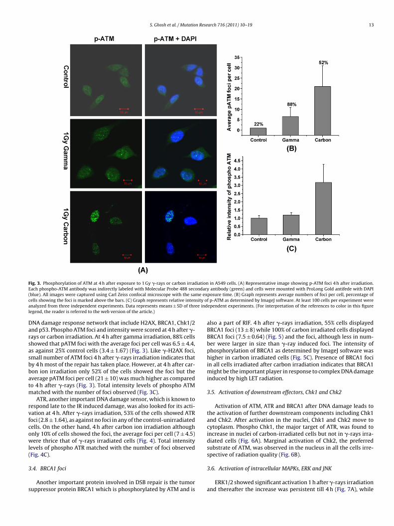
ATM, an important DNA damage response protein to ioniz-ing radiation and a part of radiation induced foci (RIF), is mainlyinvolved in cell cycle arrest and repair. In response to DSBs, ATM isactivated and phosphorylates key players in various branches of the
S. Ghosh et al. / Mutation Research 716 (2011) 10– 19 13
Fig. 3. Phosphorylation of ATM at 4 h after exposure to 1 Gy �-rays or carbon irradiation in A549 cells. (A) Representative image showing p-ATM foci 4 h after irradiation.Each phospho-ATM antibody was indirectly labeled with Molecular Probe 488 secondary antibody (green) and cells were mounted with ProLong Gold antifede with DAPI( me exc sity oa ee indl
Darsasbbatm
rvfcowl(
3
s
blue). All images were captured using Carl Zeiss confocal microscope with the saells showing the foci is marked above the bars. (C) Graph represents relative intennalyzed from three independent experiments. Data represents means ± SD of thregend, the reader is referred to the web version of the article.)
NA damage response network that include H2AX, BRCA1, Chk1/2nd p53. Phospho ATM foci and intensity were scored at 4 h after �-ays or carbon irradiation. At 4 h after gamma irradiation, 88% cellshowed that pATM foci with the average foci per cell was 6.5 ± 4.4,s against 25% control cells (3.4 ± 1.67) (Fig. 3). Like �-H2AX foci,mall number of ATM foci 4 h after �-rays irradiation indicates thaty 4 h most of the repair has taken place. However, at 4 h after car-on ion irradiation only 52% of the cells showed the foci but theverage pATM foci per cell (21 ± 10) was much higher as comparedo 4 h after �-rays (Fig. 3). Total intensity levels of phospho ATM
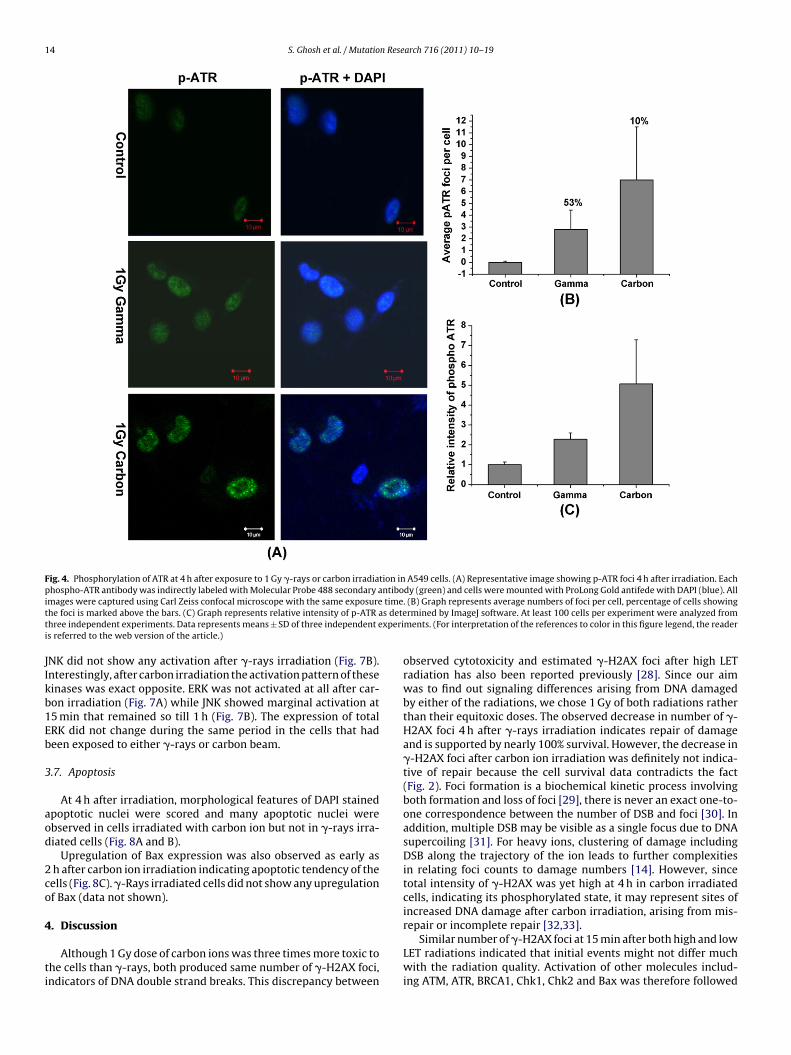
atched with the number of foci observed (Fig. 3C).ATR, another important DNA damage sensor, which is known to
espond late to the IR induced damage, was also looked for its acti-ation at 4 h. After �-rays irradiation, 53% of the cells showed ATRoci (2.8 ± 1.64), as against no foci in any of the control-unirradiatedells. On the other hand, 4 h after carbon ion irradiation althoughnly 10% of cells showed the foci, the average foci per cell (7 ± 4.5)ere thrice that of �-rays irradiated cells (Fig. 4). Total intensity
evels of phospho ATR matched with the number of foci observedFig. 4C).
.4. BRCA1 foci
Another important protein involved in DSB repair is the tumoruppressor protein BRCA1 which is phosphorylated by ATM and is
posure time. (B) Graph represents average numbers of foci per cell, percentage off p-ATM as determined by ImageJ software. At least 100 cells per experiment wereependent experiments. (For interpretation of the references to color in this figure
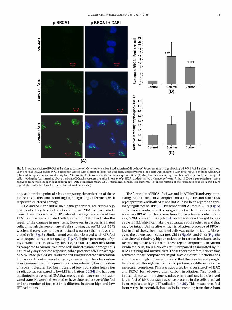
also a part of RIF. 4 h after �-rays irradiation, 55% cells displayedBRCA1 foci (13 ± 8) while 100% of carbon irradiated cells displayedBRCA1 foci (7.5 ± 0.64) (Fig. 5) and the foci, although less in num-ber were larger in size than �-ray induced foci. The intensity ofphosphorylation of BRCA1 as determined by ImageJ software washigher in carbon irradiated cells (Fig. 5C). Presence of BRCA1 fociin all cells irradiated after carbon irradiation indicates that BRCA1might be the important player in response to complex DNA damageinduced by high LET radiation.
3.5. Activation of downstream effectors, Chk1 and Chk2
Activation of ATM, ATR and BRCA1 after DNA damage leads tothe activation of further downstream components including Chk1and Chk2. After activation in the nuclei, Chk1 and Chk2 move tocytoplasm. Phospho Chk1, the major target of ATR, was found toincrease in nuclei of carbon-irradiated cells but not in �-rays irra-diated cells (Fig. 6A). Marginal activation of Chk2, the preferredsubstrate of ATM, was observed in the nucleus in all the cells irre-spective of radiation quality (Fig. 6B).
3.6. Activation of intracellular MAPKs, ERK and JNK
ERK1/2 showed significant activation 1 h after �-rays irradiationand thereafter the increase was persistent till 4 h (Fig. 7A), while
14 S. Ghosh et al. / Mutation Research 716 (2011) 10– 19
Fig. 4. Phosphorylation of ATR at 4 h after exposure to 1 Gy �-rays or carbon irradiation in A549 cells. (A) Representative image showing p-ATR foci 4 h after irradiation. Eachphospho-ATR antibody was indirectly labeled with Molecular Probe 488 secondary antibody (green) and cells were mounted with ProLong Gold antifede with DAPI (blue). Alli timet s detet xperii
JIkb1Eb
3
aod
2co
4
ti
mages were captured using Carl Zeiss confocal microscope with the same exposurehe foci is marked above the bars. (C) Graph represents relative intensity of p-ATR ahree independent experiments. Data represents means ± SD of three independent es referred to the web version of the article.)
NK did not show any activation after �-rays irradiation (Fig. 7B).nterestingly, after carbon irradiation the activation pattern of theseinases was exact opposite. ERK was not activated at all after car-on irradiation (Fig. 7A) while JNK showed marginal activation at5 min that remained so till 1 h (Fig. 7B). The expression of totalRK did not change during the same period in the cells that hadeen exposed to either �-rays or carbon beam.
.7. Apoptosis
At 4 h after irradiation, morphological features of DAPI stainedpoptotic nuclei were scored and many apoptotic nuclei werebserved in cells irradiated with carbon ion but not in �-rays irra-iated cells (Fig. 8A and B).
Upregulation of Bax expression was also observed as early as h after carbon ion irradiation indicating apoptotic tendency of theells (Fig. 8C). �-Rays irradiated cells did not show any upregulationf Bax (data not shown).
. Discussion
Although 1 Gy dose of carbon ions was three times more toxic tohe cells than �-rays, both produced same number of �-H2AX foci,ndicators of DNA double strand breaks. This discrepancy between
. (B) Graph represents average numbers of foci per cell, percentage of cells showingrmined by ImageJ software. At least 100 cells per experiment were analyzed from
ments. (For interpretation of the references to color in this figure legend, the reader
observed cytotoxicity and estimated �-H2AX foci after high LETradiation has also been reported previously [28]. Since our aimwas to find out signaling differences arising from DNA damagedby either of the radiations, we chose 1 Gy of both radiations ratherthan their equitoxic doses. The observed decrease in number of �-H2AX foci 4 h after �-rays irradiation indicates repair of damageand is supported by nearly 100% survival. However, the decrease in�-H2AX foci after carbon ion irradiation was definitely not indica-tive of repair because the cell survival data contradicts the fact(Fig. 2). Foci formation is a biochemical kinetic process involvingboth formation and loss of foci [29], there is never an exact one-to-one correspondence between the number of DSB and foci [30]. Inaddition, multiple DSB may be visible as a single focus due to DNAsupercoiling [31]. For heavy ions, clustering of damage includingDSB along the trajectory of the ion leads to further complexitiesin relating foci counts to damage numbers [14]. However, sincetotal intensity of �-H2AX was yet high at 4 h in carbon irradiatedcells, indicating its phosphorylated state, it may represent sites ofincreased DNA damage after carbon irradiation, arising from mis-repair or incomplete repair [32,33].
Similar number of �-H2AX foci at 15 min after both high and lowLET radiations indicated that initial events might not differ muchwith the radiation quality. Activation of other molecules includ-ing ATM, ATR, BRCA1, Chk1, Chk2 and Bax was therefore followed
S. Ghosh et al. / Mutation Research 716 (2011) 10– 19 15
Fig. 5. Phosphorylation of BRCA1 at 4 h after exposure to 1 Gy �-rays or carbon irradiation in A549 cells. (A) Representative image showing p-BRCA1 foci 4 h after irradiation.Each phospho-BRCA1 antibody was indirectly labeled with Molecular Probe 488 secondary antibody (green) and cells were mounted with ProLong Gold antifede with DAPI(blue). All images were captured using Carl Zeiss confocal microscope with the same exposure time. (B) Graph represents average numbers of foci per cell, percentage ofc ity of
a ee indl
omr
ubArcwdwranAiioiavaL
ells showing the foci is marked above the bars. (C) Graph represents relative intensnalyzed from three independent experiments. Data represents means ± SD of thregend, the reader is referred to the web version of the article.)
nly at later time point of 4 h as comparing the activation of theseolecules at this time could highlight signaling differences with
espect to clustered damage.ATM and ATR, the initial DNA damage sensors, are critical reg-
lators of cell cycle checkpoints and repair. ATM has particularlyeen shown to respond to IR induced damage. Presence of fewTM foci in �-rays irradiated cells 4 h after irradiation indicates theepair of the damage in most cells. However, in carbon irradiatedells, although the percentage of cells showing the pATM foci (55%)as less, the average number of foci/cell was more than �-rays irra-iated cells (Fig. 3). Similar trend was also observed with ATR fociith respect to radiation quality (Fig. 4). Higher percentage of �-
ays irradiated cells showing the ATM/ATR foci 4 h after irradiations compared to carbon irradiated cells indicates more homogenousature of �-rays induced responses while presence of lesser averageTM/ATR foci per �-rays irradiated cell as against carbon irradiation
ndicates efficient repair after �-rays irradiation. This observations in agreement with the previous studies where higher activationf repair molecules had been observed few hours after heavy ionrradiation as compared to low LET irradiation [22,34] and has been
ttributed to unrepaired DNA that keeps the damage sensors in acti-ated state. However, these studies have shown that size of the focind the number of foci at 24 h is different between high and lowET radiations.p-BRCA1 as determined by ImageJ software. At least 100 cells per experiment wereependent experiments. (For interpretation of the references to color in this figure
The formation of BRCA1 foci was unlike ATM/ATR and very inter-esting. BRCA1 exists in a complex containing ATM and other DSBrepair proteins and both ATM and BRCA1 have been regarded as pri-mary regulators of HRR [35]. Presence of BRCA1 foci in ∼55% (Fig. 5)of the �-rays irradiated cells is in agreement with the previous stud-ies where BRCA1 foci have been found to be activated only in cellsin S, G2/M phases of the cycle [34] and therefore is thought to playa role in HRR which can take the advantage of the other strand thatmay be intact. Unlike after �-rays irradiation, presence of BRCA1foci in all of the carbon irradiated cells was quite intriguing. More-over, the downstream substrates, Chk1 (Fig. 6A) and Chk2 (Fig. 6B)also showed relatively higher activation in carbon irradiated cells.Despite higher activation of all these repair components in carbonirradiated cells, their DNA was still unrepaired as indicated by �-H2AX staining and survival data. The authors therefore, believe thatactivated repair components might have different functionalitiesafter low and high LET radiations and that this functionality mightbe imparted through association of proteins in different macro-molecular complexes. This was supported by larger size of �-H2AXand BRCA1 foci observed after carbon irradiation. This result is
in accordance with previous studies where authors had observedlarger foci of DNA damage response proteins in the cells that hadbeen exposed to high LET radiation [14,36]. This means that focifrom �-rays in essentially have a distinct meaning from those from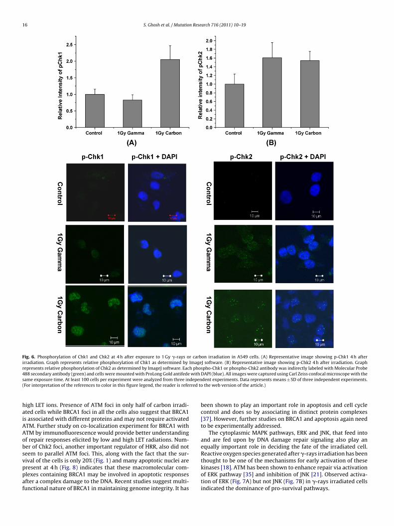
16 S. Ghosh et al. / Mutation Research 716 (2011) 10– 19
Fig. 6. Phosphorylation of Chk1 and Chk2 at 4 h after exposure to 1 Gy �-rays or carbon irradiation in A549 cells. (A) Representative image showing p-Chk1 4 h afterirradiation. Graph represents relative phosphorylation of Chk1 as determined by ImageJ software. (B) Representative image showing p-Chk2 4 h after irradiation. Graphrepresents relative phosphorylation of Chk2 as determined by ImageJ software. Each phospho-Chk1 or phospho-Chk2 antibody was indirectly labeled with Molecular Probe488 secondary antibody (green) and cells were mounted with ProLong Gold antifede with DAPI (blue). All images were captured using Carl Zeiss confocal microscope with thes depen( rred to
haiAAobsvppaf
ame exposure time. At least 100 cells per experiment were analyzed from three inFor interpretation of the references to color in this figure legend, the reader is refe
igh LET ions. Presence of ATM foci in only half of carbon irradi-ted cells while BRCA1 foci in all the cells also suggest that BRCA1s associated with different proteins and may not require activatedTM. Further study on co-localization experiment for BRCA1 withTM by immunofluorescence would provide better understandingf repair responses elicited by low and high LET radiations. Num-er of Chk2 foci, another important regulator of HRR, also did noteem to parallel ATM foci. This, along with the fact that the sur-ival of the cells is only 20% (Fig. 1) and many apoptotic nuclei are
resent at 4 h (Fig. 8) indicates that these macromolecular com-lexes containing BRCA1 may be involved in apoptotic responsesfter a complex damage to the DNA. Recent studies suggest multi-unctional nature of BRCA1 in maintaining genome integrity. It hasdent experiments. Data represents means ± SD of three independent experiments. the web version of the article.)
been shown to play an important role in apoptosis and cell cyclecontrol and does so by associating in distinct protein complexes[37]. However, further studies on BRCA1 and apoptosis again needto be experimentally addressed.
The cytoplasmic MAPK pathways, ERK and JNK, that feed intoand are fed upon by DNA damage repair signaling also play anequally important role in deciding the fate of the irradiated cell.Reactive oxygen species generated after �-rays irradiation has beenthought to be one of the mechanisms for early activation of these
kinases [18]. ATM has been shown to enhance repair via activationof ERK pathway [35] and inhibition of JNK [21]. Observed activa-tion of ERK (Fig. 7A) but not JNK (Fig. 7B) in �-rays irradiated cellsindicated the dominance of pro-survival pathways.
S. Ghosh et al. / Mutation Research 716 (2011) 10– 19 17
Fig. 7. Phosphorylation of ERK (pERK) and JNK (pJNK) in A549 cells at different time periods (h) after exposure to 1 Gy dose of �-rays or carbon ion irradiation. Treated anduntreated cells were immunoblotted and detected using anti-phospho ERK (panel A) or anti phospho JNK (panel B). One representative image of three independent experimentsis shown here. Bar graph represents the relative pERK (panel A) and pJNK (panel B) band intensities plotted against time. The intensities of pERK/pJNK were obtained afterdividing them with the respective loading control intensities and have been normalized for comparison. Data represents means ± SEM of three independent experiments.
Fig. 8. Apoptosis in carbon irradiated A549 cells. (A) Apoptotic nuclei of A549 cells were visualized and quantified by using DAPI staining, 4 h after carbon or �-raysirradiation (1 Gy). (B) Graph showing the percentage of cells with apoptotic nuclei. At least 100 cells per experiment were analyzed from three independent experiments. (C)Representative immunoblot image depicting upregulation of Bax, observed at various time periods (2, 3, 4 and 6 h) after carbon irradiation (1 Gy).
1 n Rese
pnoriJBvabwSwd
5
llritpwD
C
A
DatDwoLwRr
R
[
[
[
[
[
[
[
[
[
[
[
[
[
[
[
[
[
[
[
[
[
[
[
[
[
[
[
8 S. Ghosh et al. / Mutatio
Since, with high LET radiation yield of ROS is very low as com-ared to low LET radiation [38–40], the early activation of ERK wasot expected with high LET radiation. However, complete absencef ERK activation even hours after high LET radiation suggests thatepair pathways are neither feeding into nor being fed upon byntracellular cytoprotective signaling. However, the pro-apoptoticNK pathway was found to be activated after high LET radiation.RCA1 induced apoptotic responses have also been shown to beia activation of JNK [41]. Thus, after carbon irradiation, DNA dam-ge induced activation of repair components are being fed intoy cytotoxic signaling rather than cytoprotective responses andas evident in the form of early induction of apoptosis (Fig. 8).
ince, cellular decisions are summation of all the activated path-ays, the final response after high LET radiation tips towardseath.
. Conclusions
In summary, our results suggest that the subtle differenceseading to different outcomes due to radiation quality seem toie in different macromolecular complexes of crucial DNA damageesponse proteins and activation of other intracellular pathwaysn A549 lung adenocarcinoma cell line. Analysis of functionality ofhese macromolecular complexes as a whole rather than individualroteins and holistic study involving intracellular survival path-ays could help in revealing the mechanistic details of complexNA damage responses.
onflict of interest
The authors declare that there are no conflicts of interest.
cknowledgments
This work was funded by Board of Research in Nuclear Sciences,epartment of Atomic Energy [DAE], Government of India, through
project sanctioned to one of the authors, A. Sarma, bearing sanc-ion number 2007/37/37/BRNS. All the authors are thankful to theirector, IUAC, New Delhi for providing radiation facility for theork. We would also like to extend our thanks to all the members
f Pelletron group of IUAC and Harminder Kaur, Radiation Biologyaboratory, IUAC for their sincere help during irradiation. Authorsould like to thank Mr. Manjoor Ali and Mr. Paresh Khadilkar,B&HSD, BARC for their help in Confocal Microscopy and Lab workespectively.
eferences
[1] F.A. Cucinotta, M. Durante, Cancer risk from exposure to galactic cosmic rays:implications for space exploration by human beings, Lancet Oncol. 7 (2006)431–435.
[2] F.A. Cucinotta, H. Wu, M.R. Shavers, K. George, Radiation dosimetry and bio-physical models of space radiation effects, Gravit. Space Biol. Bull. 16 (2003)11–18.
[3] D.T. Goodhead, The initial physical damage produced by ionizing radiations,Int. J. Radiat. Biol. 56 (1989) 623–634.
[4] F.A. Cucinotta, H. Nikjoo, D.T. Goodhead, Model for radial dependence of fre-quency distributions for energy imparted in nanometer volumes from HZEparticles, Radiat. Res. 153 (2000) 459–468.
[5] E.A. Blakely, A. Kronenberg, Heavy-ion radiobiology: new approaches to delin-eate mechanisms underlying enhanced biological effectiveness, Radiat. Res.150 (1998) S126–S145.
[6] F.A. Cucinotta, R. Katz, J.W. Wilson, Radial distribution of electron spectra fromhigh-energy ions, Radiat. Environ. Biophys. 37 (1998) 259–265.
[7] B.M. Sutherland, P.V. Bennett, O. Sidorkina, J. Laval, Clustered DNA damagesinduced in isolated DNA and in human cells by low doses of ionizing radiation,
Proc. Natl. Acad. Sci. U.S.A. 97 (2000) 103–108.[8] B. Rydberg, B. Cooper, P.K. Cooper, W.R. Holley, A. Chatterjee, Dose-dependentmisrejoining of radiation-induced DNA double-strand breaks in human fibrob-lasts: experimental and theoretical study for high- and low-LET radiation,Radiat. Res. 163 (2005) 526–534.
[
arch 716 (2011) 10– 19
[9] D. Schulz-Ertner, A. Nikoghosyan, C. Thilmann, T. Haberer, O. Jakel, C. Karger, G.Kraft, M. Wannenmacher, J. Debus, Results of carbon ion radiotherapy in 152patients, Int. J. Radiat. Oncol. Biol. Phys. 58 (2004) 631–640.
10] T. Miyamoto, N. Yamamoto, H. Nishimura, M. Koto, H. Tsujii, J.E. Mizoe, T.Kamada, H. Kato, S. Yamada, S. Morita, K. Yoshikawa, S. Kandatsu, T. Fujisawa,Carbon ion radiotherapy for stage I non-small cell lung cancer, Radiother. Oncol.66 (2003) 127–140.
11] S.P. Jackson, Sensing and repairing DNA double-strand breaks, Carcinogenesis23 (2002) 687–696.
12] E.P. Rogakou, D.R. Pilch, A.H. Orr, V.S. Ivanova, W.M. Bonner, DNA double-stranded breaks induce histone H2AX phosphorylation on serine 139, J. Biol.Chem. 273 (1998) 5858–5868.
13] O. Fernandez-Capetillo, A. Lee, M. Nussenzweig, A. Nussenzweig, H2AX: thehistone guardian of the genome, DNA Repair (Amst.) 3 (2004) 959–967.
14] N. Desai, E. Davis, P. O’Neill, M. Durante, F.A. Cucinotta, H. Wu, Immunoflu-orescence detection of clustered gamma-H2AX foci induced by HZE-particleradiation, Radiat. Res. 164 (2005) 518–522.
15] L.J. Chappell, M.K. Whalen, S. Gurai, A. Ponomarev, F.A. Cucinotta, J.M. Pluth,Analysis of flow cytometry DNA damage response protein activation kineticsafter exposure to X rays and high-energy iron nuclei, Radiat. Res. 174 (2010)691–702.
16] M.K. Whalen, S.K. Gurai, H. Zahed-Kargaran, J.M. Pluth, Specific ATM-mediatedphosphorylation dependent on radiation quality, Radiat. Res. 170 (2008)353–364.
17] R. Ugenskiene, K. Prise, M. Folkard, J. Lekki, Z. Stachura, M. Zazula, J. Stachura,Dose response and kinetics of foci disappearance following exposure to high-and low-LET ionizing radiation, Int. J. Radiat. Biol. 85 (2009) 872–882.
18] K. Valerie, A. Yacoub, M.P. Hagan, D.T. Curiel, P.B. Fisher, S. Grant, P. Dent,Radiation-induced cell signaling: inside-out and outside-in, Mol. Cancer Ther.6 (2007) 789–801.
19] S.E. Golding, E. Rosenberg, S. Neill, P. Dent, L.F. Povirk, K. Valerie, Extracel-lular signal-related kinase positively regulates ataxia telangiectasia mutated,homologous recombination repair, and the DNA damage response, Cancer Res.67 (2007) 1046–1053.
20] K.E. Keating, N. Gueven, D. Watters, H.P. Rodemann, M.F. Lavin, Transcrip-tional downregulation of ATM by EGF is defective in ataxia-telangiectasia cellsexpressing mutant protein, Oncogene 20 (2001) 4281–4290.
21] N. Weizman, Y. Shiloh, A. Barzilai, Contribution of the ATM protein tomaintaining cellular homeostasis evidenced by continuous activation ofthe AP-1 pathway in ATM-deficient brains, J. Biol. Chem. 278 (2003)6741–6747.
22] A. Asaithamby, N. Uematsu, A. Chatterjee, M.D. Story, S. Burma, D.J. Chen, Repairof HZE-particle-induced DNA double-strand breaks in normal human fibrob-lasts, Radiat. Res. 169 (2008) 437–446.
23] A. Jemal, R. Siegel, E. Ward, Y. Hao, J. Xu, T. Murray, M.J. Thun, Cancer statistics,CA Cancer J. Clin. 58 (2008) 71–96.
24] S. Ghosh, N.N. Bhat, S. Santra, R.G. Thomas, S.K. Gupta, R.K. Choudhury, M.Krishna, Low energy proton beam induces efficient cell killing in A549 lungadenocarcinoma cells, Cancer Invest. 28 (2010) 615–622.
25] J.F. Ziegler, M.D. Ziegler, J.P. Biersack, SRIM – The stopping and range of ions inmatter, Nucl. Instrum. Meth. B 268 (2010) 1818–1823.
26] H. Narang, M. Krishna, Effect of nitric oxide donor and gamma irradiation onMAPK signaling in murine peritoneal macrophages, J. Cell. Biochem. 103 (2008)576–587.
27] W.S. Rasband, ImageJ, U.S. National Institutes of Health, Bethesda, USA,1997–2006, http://rsb.info.nih.gov/ij/.
28] K.M. Prise, M. Pinto, H.C. Newman, B.D. Michael, A review of studies of ioniz-ing radiation-induced double-strand break clustering, Radiat. Res. 156 (2001)572–576.
29] F.A. Cucinotta, J.M. Pluth, J.A. Anderson, J.V. Harper, P. O’Neill, Biochemicalkinetics model of DSB repair and induction of gamma-H2AX foci by non-homologous end joining, Radiat. Res. 169 (2008) 214–222.
30] S. Ghosh, H. Narang, A. Sarma, H. Kaur, M. Krishna, Activation of DNA damageresponse signaling in lung adenocarcinoma A549 cells following oxygen beamirradiation, Mutat. Res. 723 (2011) 190–198.
31] A.L. Ponomarev, S.V. Costes, F.A. Cucinotta, Stochastic properties of radiation-induced DSB: DSB distributions in large scale chromatin loops the HPRT geneand within the visible volumes of DNA repair foci, Int. J. Radiat. Biol. 84 (2008)916–929.
32] E.R. Foster, J.A. Downs, Histone H2A phosphorylation in DNA double-strandbreak repair, FEBS J. 272 (2005) 3231–3240.
33] N.F. Lowndes, G.W. Toh, DNA repair: the importance of phosphorylating histoneH2AX, Curr. Biol. 15 (2005) R99–R102.
34] K.H. Karlsson, B. Stenerlow, Focus formation of DNA repair proteins in normaland repair-deficient cells irradiated with high-LET ions, Radiat. Res. 161 (2004)517–527.
35] S.E. Golding, E. Rosenberg, A. Khalil, A. McEwen, M. Holmes, S. Neill, L.F. Povirk,K. Valerie, Double strand break repair by homologous recombination is regu-lated by cell cycle-independent signaling via ATM in human glioma cells, J. Biol.Chem. 279 (2004) 15402–15410.
36] A.L. Ponomarev, J. Huff, F.A. Cucinotta, The analysis of the densely populated
patterns of radiation-induced foci by a stochastic, Monte Carlo model of DNAdouble-strand breaks induction by heavy ions, Int. J. Radiat. Biol. 86 (2010)507–515.37] C.X. Deng, BRCA1: cell cycle checkpoint genetic instability, DNA damageresponse and cancer evolution, Nucleic Acids Res. 34 (2006) 1416–1426.

n Rese
[
[
S. Ghosh et al. / Mutatio
38] A. Kuppermann, Diffusion kinetics in radiation chemistry: an assessment, in:R.D. Cooper, R.W. Wood (Eds.), Physical Mechanisms in Radiation Biology,Springfield, Virginia, 1974, pp. 155–183.
39] D.E. Watt, Absolute biological effectiveness of neutrons and photons, Radiat.Prot. Dosim. 23 (1988) 63–67.
[
[
arch 716 (2011) 10– 19 19
40] M. Spotheim-Maurizot, M. Charlier, R. Sabattier, DNA radiolysis by fast neu-trons, Int. J. Radiat. Biol. 57 (1990) 301–313.
41] S. Lafarge, V. Sylvain, M. Ferrara, Y.J. Bignon, Inhibition of BRCA1 leads toincreased chemoresistance to microtubule-interfering agents, an effect thatinvolves the JNK pathway, Oncogene 20 (2001) 6597–6606.