Embed Size (px)
Citation preview
JOURNAL OF CLINICAL MICROBIOLOGY, Dec. 1991, p. 2752-2757 Vol. 29, No. 120095-1137/91/122752-06$02.00/0Copyright © 1991, American Society for Microbiology
DNA Fingerprinting of Enterococcus faecium by Pulsed-Field GelElectrophoresis May Be a Useful Epidemiologic ToolABRAHAM G. MIRANDA,1'2 KAVINDRA V. SINGH,' AND BARBARA E. MURRAY1 2*
Center for Infectious Diseases' and Department of Internal Medicine,2 University ofTexas Medical School at Houston, Houston, Texas 77030
Received 19 June 1991/Accepted 27 August 1991
Pulsed-field gel electrophoresis was used to compare 34 isolates of Enterococcus faecium from six differentgeographic locations. This procedure generated an average of 13 discernible fragment bands per isolate (range,10 to 19 fragment bands) of 34 to 485 kb. The resulting restriction endonuclease digestion patterns were quiteheterogeneous and were able to differentiate 27 of 34 isolates from each other, as defined by one or moremismatched fragment bands. Five patterns were shared by two or more isolates, and each set of isolates withmatching patterns (shared pattern) originated in the same medical center, suggesting a common epidemiologicbackground, including highly penicillin resistant isolates in Richmond and Philadelphia. We conclude thatpulsed-field gel electrophoresis of DNA digested with low-frequency-cleavage restriction enzymes offers arelatively simple method of comparing E. faecium for the purpose of epidemiologic study.
Since its discovery as a human pathogen around the turnof the century, members of the genus Enterococcus havedistinguished themselves from other gram-positive cocci bymultiple antibiotic resistances, distinct biochemical proper-ties, and controversial virulence potential in certain mixedinfections (18). The role of enterococci in urinary tractinfections (17) and infective endocarditis (16) is well estab-lished, and it appears that infections with these organismsare on the rise in the nosocomial setting (15, 17). While themajority of clinical enterococcal isolates are Enterococcusfaecalis, a significant percentage (10 to 15%) are Enterococ-cus faecium, a species which is generally more inherentlyresistant to antibiotics, including penicillin, ampicillin, someaminoglycosides, and imipenem (18). The clinical implica-tions of infection with an organism which may be moredifficult to treat is compounded by the apparent nosocomialtransmission of this pathogen. Patients have been reportedto contract bacteremia and meningitis by this mode oftransmission (7, 18, 29). In addition, recent reports describethe recovery of more highly penicillin- and ampicillin-resis-tant clinical isolates in certain centers (5, 7, 25, 26). Thus,further characterization of E. faecium is an important objec-tive. Investigations into the source and mechanism of spreadof enterococci, however, have been hindered by the lack ofreliable typing schemes that are able to identify particularstrains beyond the species level. Methodologies that havebeen used include antibiotic resistance patterns, biochemicalreactions (10), bacteriocin typing (13), phage typing (6), totalplasmid content (31), and serologic typing (27). Most of thesetechniques are not sufficiently sensitive to distinguish dif-ferent strains and can require considerable amounts of time,material, and expertise to perform. In addition, with some ofthese techniques, factors such as plasmid loss and various invivo and/or in vitro conditions, which can change the phe-notypic characteristics of certain organisms (21), could ren-der variable results. We and other investigators have suc-cessfully used pulsed-field gel electrophoresis (PFGE) orelectrophoretic devices with similar principles to make com-
* Corresponding author.
parative chromosomal DNA analyses of E. faecalis (19, 20),Escherichia coli (3), Pseudomonas aeruginosa (2), Staphy-lococcus aureus (11), and other organisms (1, 2). The advan-tage of PFGE is its ability to separate large DNA fragments(i.e., from 10 kb to 1.5 Mb) such as those generated bylow-frequency-cleavage restriction endonuclease digestion(RED) of whole chromosomes. This technique yields REDpatterns which consist of relatively few, generally wellseparated fragment bands and which are much less ambigu-ous than the patterns generated by conventional electropho-resis (23). The purpose of this study was to determinewhether PFGE could be used for the comparison of isolatesof E. faecium and to determine the characteristics of theresulting RED patterns among isolates from the same andfrom distant geographic areas.
MATERIALS AND METHODS
Bacterial strains. Thirty-four isolates of E. faecium fromdiverse geographic locations were used for this analysis; 11other isolates sent to us were subsequently identified asspecies other than E. faecium (see below) and were thereforenot used. The distribution of isolates was as follows: fiveisolates collected by the Centers for Disease Control (Atlan-ta, Ga.), origin unknown, kindly provided by Richard R.Facklam; four isolates from New England Deaconess Hos-pital (Boston, Mass.), kindly provided by George M. Elio-poulos; four isolates from University of Wisconsin Hospitalsand Clinics (Madison, Wis.), kindly provided by Carol A.Spiegel; six isolates from The Medical College of Pennsyl-vania (Philadelphia, Pa.), kindly provided by Caroline C.Johnson; seven isolates from Rancho Los Amigos MedicalCenter (Downey, Calif.), kindly provided by Francisco L.Sapico; and eight isolates from Medical College of Virginia(Richmond), kindly provided by Harry P. Dalton. Strainswere requested from the latter three individuals because ofprior reports of highly penicillin- and ampicillin-resistant E.faecium in their hospitals (5, 7, 26).
The control strains used for identification were as follows:(i) E. faecium GE-1, kindly provided by George M. Eliopou-los; (ii) Enterococcus gallinarum SS-1228, Enterococcus
2752
on June 29, 2018 by guesthttp://jcm
.asm.org/
Dow
nloaded from
DNA FINGERPRINTING OF E. FAECIUM BY PFGE 2753
solitarius SS-1277, and Enterococcus mundtài SS-1232,kindly provided by Richard R. Facklam; and (iii) E. mundtiiATCC 43186, kindly provided by Kathryn L. Ruoff. E. coliMG1655 was used as a control for molecular size determi-nation (4).
Strain identification. The identification of all isolates wasperformed by a combination of methods. Our primary iden-tification scheme was one based on conventional biochemi-cal reactions as described by Facklam (8) and Facklam andCarey (9) and as modified by Facklam and Collins (10); wealso used the commercial API Rapid Strep System (AnalytabProducts, Plainview, N.Y.) for identification of isolates. Thebiochemical tests that were performed, in addition to theAPI tests, were the fermentation of mannitol, sorbitol (Fish-er, Fair Lawn, N.J.), sucrose (J. T. Baker, Phillipsburg,N.J.), raffinose (Difco, Detroit, Mich.), lactose (BBL, Cock-eysville, Md.), and sorbose (Sigma, St. Louis, Mo.); abilityto grow in 6.5% NaCl (J. T. Baker); tolerance to 0.04%tellurite, pyruvate utilization, and deamination of arginine(Sigma); Gram stain and group D serologic testing (Difco) bythe autoclave extraction and capillary precipitin method (9);and pigment production on tryptic soy agar and motilitydetermination on motility media (BBL). Also tested werecatalase production (EM Science, Gibbstown, N.J.) andP-lactamase production by the nitrocefin reaction (Glaxo,Research Triangle Park, N.C.). Todd-Hewitt broth was usedfor the carbohydrate fermentation media (Difco). TheVoges-Proskauer tests; pyrrolidonylarylamidase activity;hydrolysis of esculin and sodium hippurate; and the fermen-tation of trehalose, inulin, L-arabinose, and ribose weredirectly interpreted from the API strip.
Antibiotic disk susceptibility (BBL) testing was performedon tryptic soy agar (Difco) by using ampicillin (10 ,ug),chloramphenicol (30 ,ug), erythromycin (15 ,ug), tetracycline(30 ,ug), and vancomycin (30 ,ug) disks. Susceptible, inter-mediate, and resistant isolates were defined by disk zonesize according to the Zone Size Interpretative Chart includedin the manufacturer's instructions. High-level gentamicinresistance was determined by the ability to grow on brainheart infusion agar containing 2000 ,ug of gentamicin (Scher-ing, Bloomfield, N.J.) per ml. The MICs of penicillin G(Marsam, Cherry Hill, N.J.) were also determined by theagar dilution method (30). Isolates for which the MICs were256,ug/ml or above were considered to be highly resistant topenicillin G.DNA analysis. Genomic DNA was prepared from all E.
faecium isolates by previously described methods (19, 28).Briefly, an overnight culture grown in 5 ml of Todd-Hewittor brain heart infusion broth (Difco) was centrifuged, andthe packed cells were suspended in 5 ml of PIV solution(1 M NaCl, 10 mM Tris-HCl [pH 7.6]). A total of 750 pdl ofthis suspension was mixed with 250 ,ul of 3.2% agarose(Incert Agarose; FMC, Rockland, Maine) and pipetted intosmall rectangular molds (final agarose concentration, 0.8%).The cells suspended in the agarose plugs were then lysedovernight at 37°C by using a solution containing 6 mMTris-HCl (pH 7.6), 1 M NaCi, 100 mM EDTA (pH 7.5), 0.5%Brij 58, 0.2% deoxycholate, 0.5% sodium lauroyl sarcosine(Sarkosyl), 20,ug of RNase (DNase free) per ml, and 1 mg oflysozyme per ml. Next, the lysis solution was replaced witha solution containing proteinase K (50 ktg/ml), 1% Sarkosyl,and 0.5 M EDTA (pH 9 to 9.5); and the plugs were furtherincubated overnight at 50°C with slow shaking. The plugswere then washed three times (30 min each time at 37°C)with TE solution (10 mM Tris-HCI [pH 7.5], 1 mM EDTA)
and stored in fresh TE solution at 4°C until and after theiruse.
All E. faecium DNAs were digested with the restrictionenzyme SmaI (recognition sequence, CCCGGG), and theDNA of the size control E. coli MG1655 was digested withNotI (recognition sequence, GCGGCCGC). Lambda con-catemers (New England BioLabs, Beverly, Mass.) were alsoused as size controls in some gels. The enzymes wereobtained from Boehringer-Mannheim (Indianapolis, Ind.) orNew England BioLabs. The digestions were performed byplacing a 4- to 5-mm slice of each plug in 225 pl of lxrestriction buffer solution, and approximately 20 to 40 U ofthe respective enzyme was added. Incubations were asfollows: (i) SmaI, 25°C for 4 to 20 h; (ii) NotI, 37°C for 12 to20 h. After incubation, the plugs were washed and equili-brated with TE solution for 1 h at 37°C. The TE solution wassubsequently discarded, and the plugs were then melted at55 to 60°C and pipetted into the wells of a 1.2% SeaplaqueGTG agarose gel (FMC) (in 0.5x TBE buffer [0.089 MTris-HCl, 0.089 M boric acid, 0.0025 M EDTA). Electropho-resis was performed with the Contoured-Clamped Homoge-neous Electric Field apparatus (CHEF-DRII; Bio-Rad) byusing ramped pulse times beginning with 5 s and ending with35 s at 200 V for 32 to 40 h. For selected isolates, DNA wasalso run with a fixed pulse time of 5 s in order to discern thelower-molecular-weight fragments. The gels were thenstained with ethidium bromide for 30 min and destained indistilled water for 12 to 20 h before they were photographedwith UV radiation.
RESULTS
Chromosomal patterns of isolates. A unique pattern wasarbitrarily defined in this study as any RED pattern gener-ated with a ramped pulse time of 5 to 35 s which varied fromanother pattern by one or more clearly visible fragmentbands, or simply, any nonidentical RED pattern (ramped at5 to 35 s) constituted a pattern of its own. This notwithstand-ing, our experience suggests that RED patterns with differ-ences in only a few fragment bands would be expected torepresent closely related isolates of a single strain (19, 20).By using a ramped pulse time of 5 to 35 s, the bandsvisualized best were those corresponding to the higher-molecular-weight fragments (range, 200 to 500 kb). Bydecreasing the pulse time to a 5-s fixed interval, the lower-molecular-weight fragments became more visible (range, 30to 190 kb) at the expense of those with higher molecularweights, which segregated poorly or not at all.
In the present study, all but 1 of the 34 isolates analyzeddisplayed evaluable RED patterns by PFGE on the firstattempt. The remaining isolate was evaluable after a newplug was made. The range of clearly visible fragment bandson each gel when a ramped pulse time of 5 to 35 s (manyisolates were repeated on different gels) was used was 10 to19 fragment bands, with an average of 13 to 14 fragmentbands visible per isolate. In all, 27 distinct RED patternswere seen, 5 ofwhich were shared by two or three members,which segregated the organisms by geographic locale. TheRED patterns which were shared (designated with the suffixSP) contained organisms isolated in the same medical centerand, in one case, from the same patient (Fig. 1A, lanes b, c,and d, pattern MCV-SP1, and Table 1). Shared RED pat-terns were seen in isolates from Richmond, Va. (MCV-SP1and MCV-SP2); Philadelphia, Pa. (MCP-SP1); Downey,Calif. (RLA-SP1); and Boston, Mass. (NEDH-SP1) (data notshown, except for the isolate from Richmond in Fig. 1A).
VOL. 29, 1991
on June 29, 2018 by guesthttp://jcm
.asm.org/
Dow
nloaded from
2754 MIRANDA ET AL.
A a b c d e f g h
1000 -
3 6 6 -
252 -
192-
157 -135 -108a -
34 -14 -
B a b c d e f g h
15710890
3 429
14
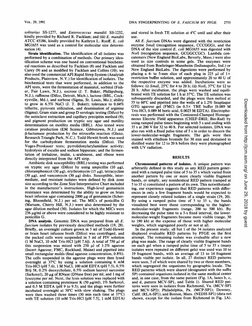
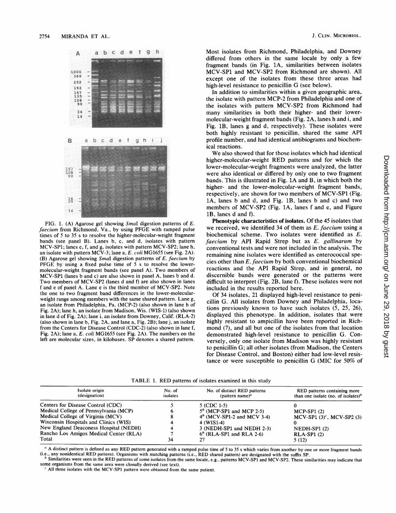
FIG. 1. (A) Agarose gel showing SmaI digestion patterns of E.faecium from Richmond, Va., by using PFGE with ramped pulsetimes of 5 to 35 s to resolve the higher-molecular-weight fragmentbands (see panel B). Lanes b, c, and d, isolates with patternMCV-SP1; lanes e, f, and g, isolates with pattern MCV-SP2; lane h,an isolate with pattern MCV-3; lane a, E. coli MG1655 (see Fig. 2A).(B) Agarose gel showing SmaI digestion patterns of E. faecium byPFGE by using a fixed pulse time of 5 s to resolve the lower-molecular-weight fragment bands (see panel A). Two members ofMCV-SP1 (lanes b and c) are also shown in panel A, lanes b and d.Two members of MCV-SP2 (lanes d and f) are also shown in lanesf and e of panel A. Lane e is the third member of MCV-SP2. Notethe one to two fragment band differences in the lower-molecular-weight range among members with the same shared pattern. Lane g,an isolate from Philadelphia, Pa. (MCP-2) (also shown in lane h ofFig. 2A); lane h, an isolate from Madison, Wis. (WIS-1) (also shownin lane d of Fig. 2A); lane i, an isolate from Downey, Calif. (RLA-2)(also shown in lane b, Fig. 2A, and lane a, Fig. 2B); lane j, an isolatefrom the Centers for Disease Control (CDC-2) (also shown in lane f,Fig. 2A); lane a, E. coli MG1655 (see Fig. 2A). The numbers on theleft are molecular sizes, in kilobases. SP denotes a shared pattern.
Most isolates from Richmond, Philadelphia, and Downeydiffered from others in the same locale by only a fewfragment bands (in Fig. 1A, similarities between isolatesMCV-SP1 and MCV-SP2 from Richmond are shown). Allexcept one of the isolates from these three areas hadhigh-level resistance to penicillin G (see below).
In addition to similarities within a given geographic area,the isolate with pattern MCP-2 from Philadelphia and one ofthe isolates with pattern MCV-SP2 from Richmond hadmany similarities in both their higher- and their lower-molecular-weight fragment bands (Fig. 2A, lanes h and i, andFig. 1B, lanes g and d, respectively). These isolates wereboth highly resistant to penicillin, shared the same APIprofile number, and had identical antibiograms and biochem-ical reactions.We also showed that for those isolates which had identical
higher-molecular-weight RED patterns and for which thelower-molecular-weight fragments were analyzed, the latterwere also identical or differed by only one to two fragmentbands. This is illustrated in Fig. lA and B, in which both thehigher- and the lower-molecular-weight fragment bands,respectively, are shown for two members of MCV-SP1 (Fig.1A, lanes b and d, and Fig. 1B, lanes b and c) and twomembers of MCV-SP2 (Fig. 1A, lanes f and e, and Figure1B, lanes d and i).Phenotypic characteristics of isolates. Of the 45 isolates that
we received, we identified 34 of them as E. faecium using a
biochemical scheme. Two isolates were identified as E.faecium by API Rapid Strep but as E. gallinarum byconventional tests and were not included in the analysis. Theremaining nine isolates were identified as enterococcal spe-cies other than E. faecium by both conventional biochemicalreactions and the API Rapid Strep, and in general, no
discernible bands were generated or the patterns were
difficult to interpret (Fig. 2B, lane f). These isolates were notincluded in the results reported here.Of 34 isolates, 21 displayed high-level resistance to peni-
cillin G. All isolates from Downey and Philadelphia, loca-tions previously known to have such isolates (5, 25, 26),displayed this phenotype. In addition, isolates that were
highly resistant to ampicillin have been reported in Rich-mond (7), and all but one of the isolates from that locationdemonstrated high-level resistance to penicillin G. Con-versely, only one isolate from Madison was highly resistantto penicillin G; all other isolates (from Madison, the Centersfor Disease Control, and Boston) either had low-level resis-tance or were susceptible to penicillin G (MIC for 50% of
TABLE 1. RED patterns of isolates examined in this study
Isolate origin No. of No. of distinct RED patterns RED patterns containing more(designation) isolates (pattern name)' than one isolate (no. of isolates)'
Centers for Disease Control (CDC) 5 5 (CDC 1-5) 0Medical College of Pennsylvania (MCP) 6 5b (MCP-SP1 and MCP 2-5) MCP-SP1 (2)Medical College of Virginia (MCV) 8 4b (MCV-SP1-2 and MCV 3-4) MCV-SP1 (3)c, MCV-SP2 (3)Wisconsin Hospitals and Clinics (WIS) 4 4 (WIS1-4) 0New England Deaconess Hospital (NEDH) 4 3 (NEDH-SP1 and NEDH 2-3) NEDH-SP1 (2)Rancho Los Amigos Medical Center (RLA) 7 6b (RLA-SP1 and RLA 2-6) RLA-SP1 (2)Total 34 27 5 (12)
a A distinct pattern is defined as any RED pattern generated with a ramped pulse time of 5 to 35 s which varies from another by one or more fragment bands(i.e., any nonidentical RED pattern). Organisms with matching patterns (i.e., RED shared pattern) are designated with the suffix SP.
b Similarities were seen in the RED patterns of some isolates from the same locale, e.g., patterns MCV-SP1 and MCV-SP2. These similarities may indicate thatsome organisms from the same area were clonally derived (see text).
' All three isolates with the MCV-SP1 pattern were obtained from the same patient.
:-,
J. CLIN. MICROBIOL.
j
on June 29, 2018 by guesthttp://jcm
.asm.org/
Dow
nloaded from
DNA FINGERPRINTING OF E. FAECIUM BY PFGE 2755
A a b c d e f g hhe.'Iim!ll
1000
366
252
192
157
135
108
90
34
14
B a b c d e f g h i
- 339.5291
242.5
- 194
- 145.5
- 97
- 48.5
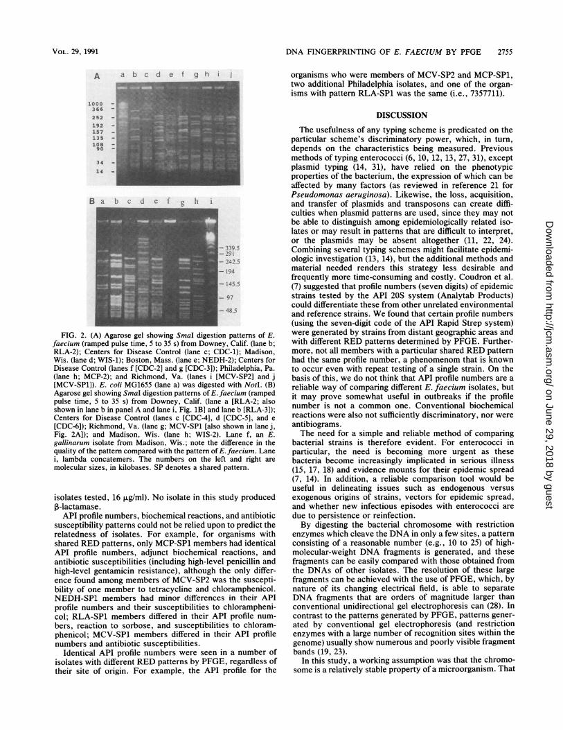
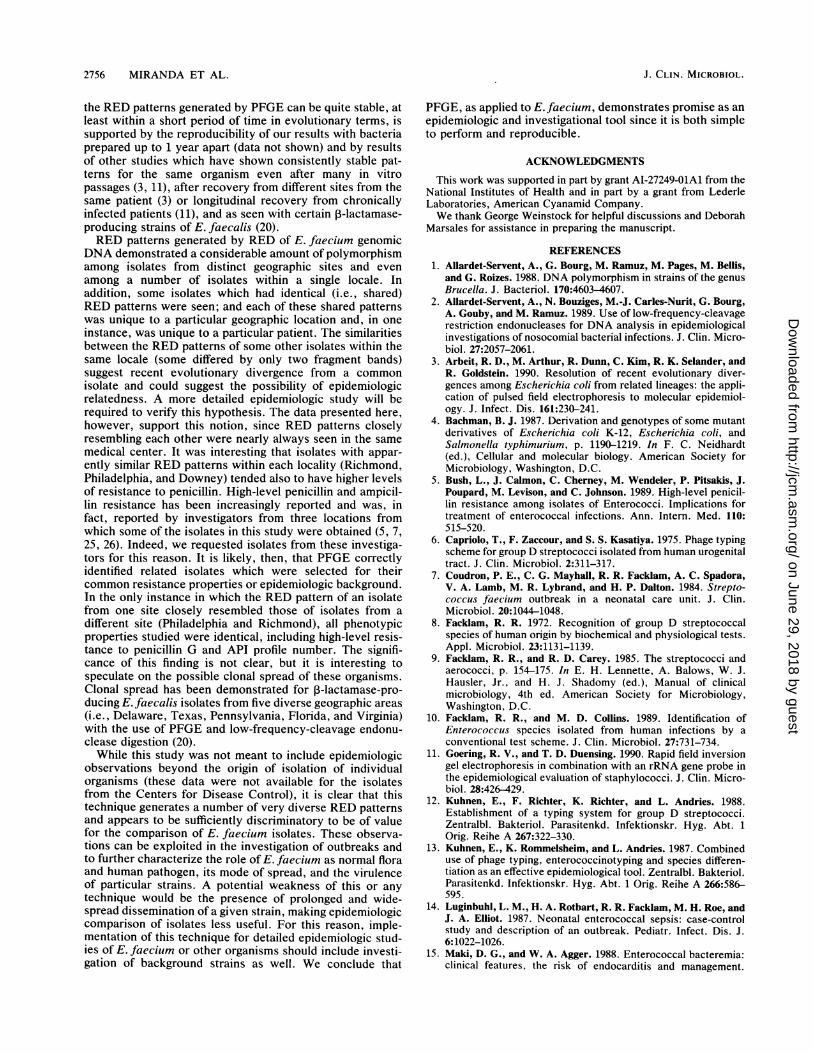
FIG. 2. (A) Agarose gel showing SmaI digestion patterns of E.faecium (ramped pulse time, 5 to 35 s) from Downey, Calif. (lane b;RLA-2); Centers for Disease Control (lane c; CDC-1); Madison,Wis. (lane d; WIS-1); Boston, Mass. (lane e; NEDH-2); Centers forDisease Control (lanes f [CDC-2] and g [CDC-3]); Philadelphia, Pa.(lane h; MCP-2); and Richmond, Va. (lanes i [MCV-SP2] and j[MCV-SP1]). E. coli MG1655 (lane a) was digested with NotI. (B)Agarose gel showing SmaI digestion patterns of E. faecium (rampedpulse time, 5 to 35 s) from Downey, Calif. (lane a [RLA-2; alsoshown in lane b in panel A and lane i, Fig. 1B] and lane b [RLA-3]);Centers for Disease Control (lanes c [CDC-4], d [CDC-5], and e
[CDC-6]); Richmond, Va. (lane g; MCV-SP1 [also shown in lane j,Fig. 2A]); and Madison, Wis. (lane h; WIS-2). Lane f, an E.gallinarum isolate from Madison, Wis.; note the difference in thequality of the pattern compared with the pattern of E. faecium. Lanei, lambda concatemers. The numbers on the left and right are
molecular sizes, in kilobases. SP denotes a shared pattern.
isolates tested, 16 ,tg/ml). No isolate in this study produced[B-lactamase.API profile numbers, biochemical reactions, and antibiotic
susceptibility patterns could not be relied upon to predict therelatedness of isolates. For example, for organisms withshared RED patterns, only MCP-SP1 members had identicalAPI profile numbers, adjunct biochemical reactions, andantibiotic susceptibilities (including high-level penicillin andhigh-level gentamicin resistance), although the only differ-ence found among members of MCV-SP2 was the suscepti-bility of one member to tetracycline and chloramphenicol.NEDH-SP1 members had minor differences in their APIprofile numbers and their susceptibilities to chlorampheni-col; RLA-SP1 members differed in their API profile num-bers, reaction to sorbose, and susceptibilities to chloram-phenicol; MCV-SP1 members differed in their API profilenumbers and antibiotic susceptibilities.
Identical API profile numbers were seen in a number ofisolates with different RED patterns by PFGE, regardless oftheir site of origin. For example, the API profile for the
organisms who were members of MCV-SP2 and MCP-SP1,two additional Philadelphia isolates, and one of the organ-isms with pattern RLA-SP1 was the same (i.e., 7357711).
DISCUSSION
The usefulness of any typing scheme is predicated on theparticular scheme's discriminatory power, which, in turn,depends on the characteristics being measured. Previousmethods of typing enterococci (6, 10, 12, 13, 27, 31), exceptplasmid typing (14, 31), have relied on the phenotypicproperties of the bacterium, the expression of which can beaffected by many factors (as reviewed in reference 21 forPseudomonas aeruginosa). Likewise, the loss, acquisition,and transfer of plasmids and transposons can create diffi-culties when plasmid patterns are used, since they may notbe able to distinguish among epidemiologically related iso-lates or may result in patterns that are difficult to interpret,or the plasmids may be absent altogether (11, 22, 24).Combining several typing schemes might facilitate epidemi-ologic investigation (13, 14), but the additional methods andmaterial needed renders this strategy less desirable andfrequently more time-consuming and costly. Coudron et al.(7) suggested that profile numbers (seven digits) of epidemicstrains tested by the API 20S system (Analytab Products)could differentiate these from other unrelated environmentaland reference strains. We found that certain profile numbers(using the seven-digit code of the API Rapid Strep system)were generated by strains from distant geographic areas andwith different RED patterns determined by PFGE. Further-more, not all members with a particular shared RED patternhad the same profile number, a phenomenom that is knownto occur even with repeat testing of a single strain. On thebasis of this, we do not think that API profile numbers are areliable way of comparing different E. faecium isolates, butit may prove somewhat useful in outbreaks if the profilenumber is not a common one. Conventional biochemicalreactions were also not sufficiently discriminatory, nor wereantibiograms.The need for a simple and reliable method of comparing
bacterial strains is therefore evident. For enterococci inparticular, the need is becoming more urgent as thesebacteria become increasingly implicated in serious illness(15, 17, 18) and evidence mounts for their epidemic spread(7, 14). In addition, a reliable comparison tool would beuseful in delineating issues such as endogenous versusexogenous origins of strains, vectors for epidemic spread,and whether new infectious episodes with enterococci aredue to persistence or reinfection.By digesting the bacterial chromosome with restriction
enzymes which cleave the DNA in only a few sites, a patternconsisting of a reasonable number (e.g., 10 to 25) of high-molecular-weight DNA fragments is generated, and thesefragments can be easily compared with those obtained fromthe DNAs of other isolates. The resolution of these largefragments can be achieved with the use of PFGE, which, bynature of its changing electrical field, is able to separateDNA fragments that are orders of magnitude larger thanconventional unidirectional gel electrophoresis can (28). Incontrast to the patterns generated by PFGE, patterns gener-ated by conventional gel electrophoresis (and restrictionenzymes with a large number of recognition sites within thegenome) usually show numerous and poorly visible fragmentbands (19, 23).
In this study, a working assumption was that the chromo-some is a relatively stable property of a microorganism. That
VOL. 29, 1991
on June 29, 2018 by guesthttp://jcm
.asm.org/
Dow
nloaded from
2756 MIRANDA ET AL.
the RED patterns generated by PFGE can be quite stable, atleast within a short period of time in evolutionary terms, issupported by the reproducibility of our results with bacteriaprepared up to 1 year apart (data not shown) and by resultsof other studies which have shown consistently stable pat-terns for the same organism even after many in vitropassages (3, 11), after recovery from different sites from thesame patient (3) or longitudinal recovery from chronicallyinfected patients (11), and as seen with certain ,-lactamase-producing strains of E. faecalis (20).RED patterns generated by RED of E. faecium genomic
DNA demonstrated a considerable amount of polymorphismamong isolates from distinct geographic sites and evenamong a number of isolates within a single locale. Inaddition, some isolates which had identical (i.e., shared)RED patterns were seen; and each of these shared patternswas unique to a particular geographic location and, in oneinstance, was unique to a particular patient. The similaritiesbetween the RED patterns of some other isolates within thesame locale (some differed by only two fragment bands)suggest recent evolutionary divergence from a commonisolate and could suggest the possibility of epidemiologicrelatedness. A more detailed epidemiologic study will berequired to verify this hypothesis. The data presented here,however, support this notion, since RED patterns closelyresembling each other were nearly always seen in the samemedical center. It was interesting that isolates with appar-ently similar RED patterns within each locality (Richmond,Philadelphia, and Downey) tended also to have higher levelsof resistance to penicillin. High-level penicillin and ampicil-lin resistance has been increasingly reported and was, infact, reported by investigators from three locations fromwhich some of the isolates in this study were obtained (5, 7,25, 26). Indeed, we requested isolates from these investiga-tors for this reason. It is likely, then, that PFGE correctlyidentified related isolates which were selected for theircommon resistance properties or epidemiologic background.In the only instance in which the RED pattern of an isolatefrom one site closely resembled those of isolates from adifferent site (Philadelphia and Richmond), all phenotypicproperties studied were identical, including high-level resis-tance to penicillin G and API profile number. The signifi-cance of this finding is not clear, but it is interesting tospeculate on the possible clonal spread of these organisms.Clonal spread has been demonstrated for P-lactamase-pro-ducing E. faecalis isolates from five diverse geographic areas(i.e., Delaware, Texas, Pennsylvania, Florida, and Virginia)with the use of PFGE and low-frequency-cleavage endonu-clease digestion (20).While this study was not meant to include epidemiologic
observations beyond the origin of isolation of individualorganisms (these data were not available for the isolatesfrom the Centers for Disease Control), it is clear that thistechnique generates a number of very diverse RED patternsand appears to be sufficiently discriminatory to be of valuefor the comparison of E. faecium isolates. These observa-tions can be exploited in the investigation of outbreaks andto further characterize the role of E. faecium as normal floraand human pathogen, its mode of spread, and the virulenceof particular strains. A potential weakness of this or anytechnique would be the presence of prolonged and wide-spread dissemination of a given strain, making epidemiologiccomparison of isolates less useful. For this reason, imple-mentation of this technique for detailed epidemiologic stud-ies of E. faecium or other organisms should include investi-gation of background strains as well. We conclude that
PFGE, as applied to E. faecium, demonstrates promise as anepidemiologic and investigational tool since it is both simpleto perform and reproducible.
ACKNOWLEDGMENTS
This work was supported in part by grant AI-27249-OlA1 from theNational Institutes of Health and in part by a grant from LederleLaboratories, American Cyanamid Company.We thank George Weinstock for helpful discussions and Deborah
Marsales for assistance in preparing the manuscript.
REFERENCES1. Allardet-Servent, A., G. Bourg, M. Ramuz, M. Pages, M. Bellis,
and G. Roizes. 1988. DNA polymorphism in strains of the genusBrucella. J. Bacteriol. 170:4603-4607.
2. Allardet-Servent, A., N. Bouziges, M.-J. Carles-Nurit, G. Bourg,A. Gouby, and M. Ramuz. 1989. Use of low-frequency-cleavagerestriction endonucleases for DNA analysis in epidemiologicalinvestigations of nosocomial bacterial infections. J. Clin. Micro-biol. 27:2057-2061.
3. Arbeit, R. D., M. Arthur, R. Dunn, C. Kim, R. K. Selander, andR. Goldstein. 1990. Resolution of recent evolutionary diver-gences among Escherichia coli from related lineages: the appli-cation of pulsed field electrophoresis to molecular epidemiol-ogy. J. Infect. Dis. 161:230-241.
4. Bachman, B. J. 1987. Derivation and genotypes of some mutantderivatives of Escherichia coli K-12, Escherichia coli, andSalmonella typhimurium, p. 1190-1219. In F. C. Neidhardt(ed.), Cellular and molecular biology. American Society forMicrobiology, Washington, D.C.
5. Bush, L., J. Calmon, C. Cherney, M. Wendeler, P. Pitsakis, J.Poupard, M. Levison, and C. Johnson. 1989. High-level penicil-lin resistance among isolates of Enterococci. Implications fortreatment of enterococcal infections. Ann. Intern. Med. 110:515-520.
6. Capriolo, T., F. Zaccour, and S. S. Kasatiya. 1975. Phage typingscheme for group D streptococci isolated from human urogenitaltract. J. Clin. Microbiol. 2:311-317.
7. Coudron, P. E., C. G. Mayhall, R. R. Facklam, A. C. Spadora,V. A. Lamb, M. R. Lybrand, and H. P. Dalton. 1984. Strepto-coccus faecium outbreak in a neonatal care unit. J. Clin.Microbiol. 20:1044-1048.
8. Facklam, R. R. 1972. Recognition of group D streptococcalspecies of human origin by biochemical and physiological tests.Appl. Microbiol. 23:1131-1139.
9. Facklam, R. R., and R. D. Carey. 1985. The streptococci andaerococci, p. 154-175. In E. H. Lennette, A. Balows, W. J.Hausler, Jr., and H. J. Shadomy (ed.), Manual of clinicalmicrobiology, 4th ed. American Society for Microbiology,Washington, D.C.
10. Facklam, R. R., and M. D. Collins. 1989. Identification ofEnterococcus species isolated from human infections by aconventional test scheme. J. Clin. Microbiol. 27:731-734.
11. Goering, R. V., and T. D. Duensing. 1990. Rapid field inversiongel electrophoresis in combination with an rRNA gene probe inthe epidemiological evaluation of staphylococci. J. Clin. Micro-biol. 28:426-429.
12. Kuhnen, E., F. Richter, K. Richter, and L. Andries. 1988.Establishment of a typing system for group D streptococci.Zentralbl. Bakteriol. Parasitenkd. Infektionskr. Hyg. Abt. 1Orig. Reihe A 267:322-330.
13. Kuhnen, E., K. Rommelsheim, and L. Andries. 1987. Combineduse of phage typing, enterococcinotyping and species differen-tiation as an effective epidemiological tool. Zentralbl. Bakteriol.Parasitenkd. Infektionskr. Hyg. Abt. 1 Orig. Reihe A 266:586-595.
14. Luginbuhl, L. M., H. A. Rotbart, R. R. Facklam, M. H. Roe, andJ. A. Elliot. 1987. Neonatal enterococcal sepsis: case-controlstudy and description of an outbreak. Pediatr. Infect. Dis. J.6:1022-1026.
15. Maki, D. G., and W. A. Agger. 1988. Enterococcal bacteremia:clinical features, the risk of endocarditis and management.
J. CLIN. MICROBIOL.
on June 29, 2018 by guesthttp://jcm
.asm.org/
Dow
nloaded from
DNA FlNGERPRINTING OF E. FAECIUM BY PFGE 2757
Medicine 67:248-269.16. Mandell, G. L., D. Kaye, M. E. Levinson, and E. W. Hook. 1970.
Enterococcal endocarditis: an analysis of 38 patients observedat the New York Hospital-Cornell Medical Center. Arch. In-tern. Med. 125:248-264.
17. Morrison, A. J., Jr., and R. P. Wenzel. 1986. Nosocomialurinary tract infections due to enterococcus: ten years' experi-ence at a university hospital. Arch. Intern. Med. 146:1549-1551.
18. Murray, B. E. 1990. The life and times of the enterococcus.Clin. Microbiol. Rev. 3:46-65.
19. Murray, B. E., K. V. Singh, J. D. Heath, B. R. Sharma, andG. M. Weinstock. 1990. Comparison of genomic DNA of dif-ferent enterococcal isolates using restriction endonucleaseswith infrequent recognition sites. J. Clin. Microbiol. 28:2059-2063.
20. Murray, B. E., K. V. Singh, S. M. Markowitz, H. A. Lopardo,J. E. Patterson, M. J. Zervos, E. Rubeglio, G. M. Eliopoulos,L. B. Rice, F. W. Goldstein, S. G. Jenkins, G. M. Caputo, R.Nasnas, L. S. Moore, E. S. Wong, and G. Weinstock. 1991.Evidence for clonal spread of a single strain of P-lactamase-producing Enterococcusfaecalis to six hospitals in five states. J.Infect. Dis. 163:780-785.
21. Ogle, J. W., J. Michael-Janda, D. E. Woods, and M. L. Vasil.1987. Characterization and use of a DNA probe as an epidemi-ological marker for Pseudomonas aeruginosa. J. Infect. Dis.155:119-126.
22. Platt, D. J., J. S. Chesham, and K. G. Kirstinsson. 1986.R-plasmid transfer in vivo: a prospective study. J. Med. Micro-biol. 21:325-330.
23. Renaud, F., J. Freney, J. Etienne, M. Bes, Y. Brun, O. Barsotti,
S. Andre, and J. Fleurette. 1988. Restriction endonucleaseanalysis of Staphylococcus epidermidis DNA may be a usefulepidemiological marker. J. Clin. Microbiol. 26:1729-1734.
24. Rubens, C. E., W. E. Farrar, Jr., Z. A. McGee, and W.Schaffner. 1981. Evolution of a plasmid mediating resistance tomultiple antimicrobial agents during a prolonged epidemic ofnosocomial infections. J. Infect. Dis. 143:170-181.
25. Rupar, D., M. Fisher, and H. Fletcher. 1989. Emergence ofisolates resistant to ampicillin. Am. J. Dis. Child. 143:1033-1037.
26. Sapico, F., H. Canawati, V. Ginunas, D. Gilmore, J. Montgom-erie, W. Tuddenham, and R. Facklam. 1989. Enterococci highlyresistant to penicillin and ampicillin: an emerging clinical prob-lem? J. Clin. Microbiol. 27:2091-2095.
27. Sharpe, M. D., and P. M. F. Shattock. 1952. The serologicaltyping of group D streptococci associated with outbreaks ofneonatal diarrhoea. J. Gen. Microbiol. 6:150-165.
28. Smith, C. L., and C. R. Cantor. 1987. Purification, specificfragmentation, and separation of large DNA molecules. Meth-ods Enzymol. 155:499-567.
29. Warren, R. E. 1988. Difficult streptococci. J. Hosp. Infect.11(Suppl. A):352-357.
30. Washington, J., Il. 1985. Susceptibility tests: agar dilution, p.967-971. In E. H. Lennette, A. Balows, W. J. Hausler Jr., andH. J. Shadomy (ed.), Manual of clinical microbiology, 4th ed.American Society for Microbiology, Washington, D.C.
31. Zervos, M. J., C. A. Kauffman, P. M. Therasse, A. G. Bergman,T. S. Mikesell, and D. R. Schaberg. 1987. Nosocomial infectionby gentamicin-resistant Streptococcus faecalis. Ann. Intern.Med. 106:687-691.
VOL. 29, 1991
on June 29, 2018 by guesthttp://jcm
.asm.org/
Dow
nloaded from