Embed Size (px)
Citation preview
DNA in a Celland how is it arranged!
Mike Clark, M.D.
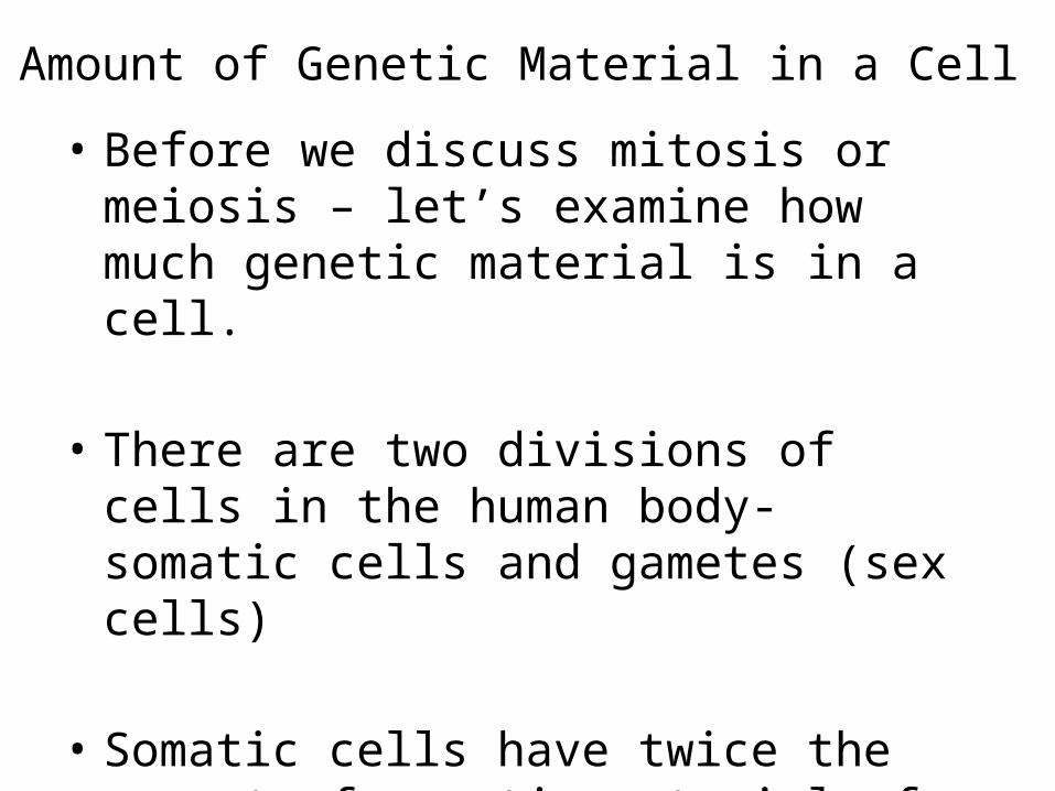
Amount of Genetic Material in a Cell• Before we discuss mitosis or meiosis – let’s
examine how much genetic material is in a cell.
• There are two divisions of cells in the human body- somatic cells and gametes (sex cells)
• Somatic cells have twice the amount of genetic material of the gametes
Somatic Cells• Somatic cells are all the human body cells
except the gametes
• Somatic cells have 46 pieces of genetic material (46 DNA pieces) – organized into 23 pairs
• 23 pieces (one set) of the genetic material was given to your cells by your mother and the other 23 pieces (the other set) was given to you by your father
How do we identify a set of Genetic Material
• Most authors say that we have 46 chromosomes – which is true – but if you notice I use the term genetic material rather than chromosome because remember that a chromosome is a certain folding pattern of the DNA – thus not always is the DNA folded into the chromosome fold – as a matter of fact most of the time it is not folded into the chromosome fold.
DNA Folding in a Cell• The average human cell nucleus has a diameter
of approximately 5 micrometers (5 x 10 -6 meter or 5 millionths of a meter)
• If we stretch out the DNA in a somatic cell in the human – it is approximately 2 meters in length – so you can see we need a heck of a good folding system for the DNA. The DNA must be folded good so it can be read properly.
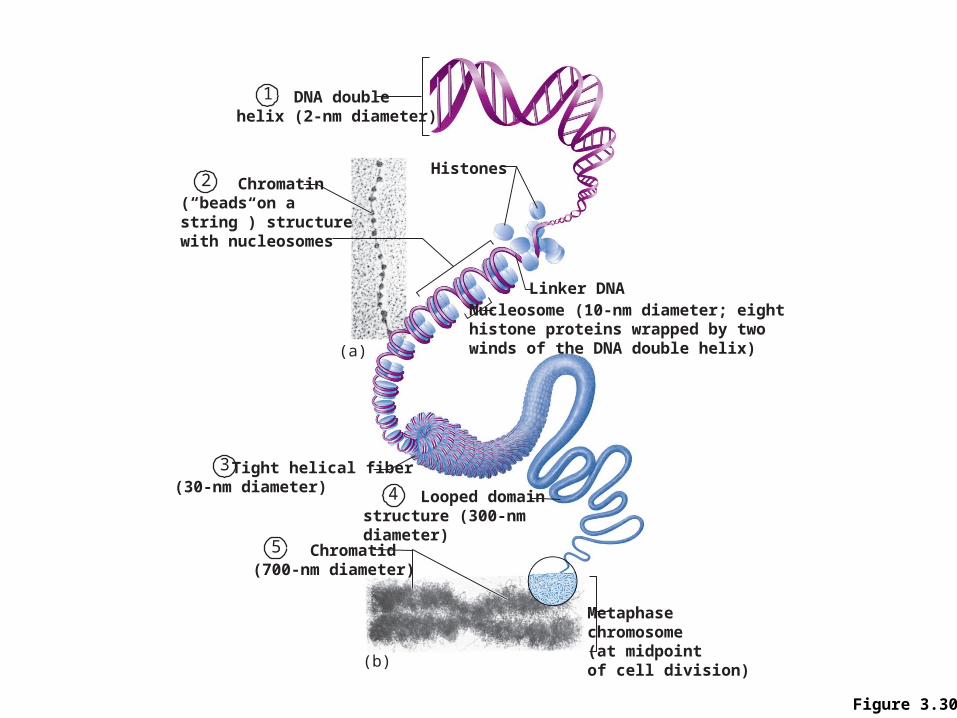
Figure 3.30
Metaphasechromosome(at midpointof cell division)
Nucleosome (10-nm diameter; eight histone proteins wrapped by two winds of the DNA double helix)
Linker DNA
Histones
(a)
(b)
1 DNA doublehelix (2-nm diameter)
2 Chromatin(“beads on a string”) structurewith nucleosomes
3 Tight helical fiber(30-nm diameter)
5 Chromatid(700-nm diameter)
4 Looped domain structure (300-nm diameter)
Examination of Genetic Material• The chromosome is thick enough to be seen
using the light microscope• The genetic material (DNA) is folded into the
chromosome fold during the M-phase (mitosis or meiosis) of the cell divisional cycle.
• The genetic material folds from the loop domain fold (too thin to be seen under light microscope) to the chromosome fold during the phases of prophase and metaphase – which are subdivisions of the cell’s M-phase
• These prophase and metaphase stages will be discussed in the lecture on mitosis
How are the different chromosomes distinguished?
• Human chromosomes must be determined one from another because each chromosome carries a different set of vital genes – lose a chromosome and lose those genes – thus lose the ability to make those necessary proteins
• Scientist have assigned the various chromosomes certain numbers
• Chromosomes are numbered 1 – 23• The 23rd chromosomes are the sex chromosomes (X
versus Y) – the first 22 chromosomes are termed “autosomes”
Sets of Chromosomes in Human Cells
• A karyotype is an ordered display of the pairs of chromosomes from a cell
• The two chromosomes in each pair are called homologous chromosomes, or homologs – non-identical are termed “heterologous pairs”
• Chromosomes in a homologous pair are the same length and carry genes controlling the same inherited characters
• A different chromosomes are identified and classified in accordance with their individual (1) lengths (2) centromere positions and (3) unique staining patterns
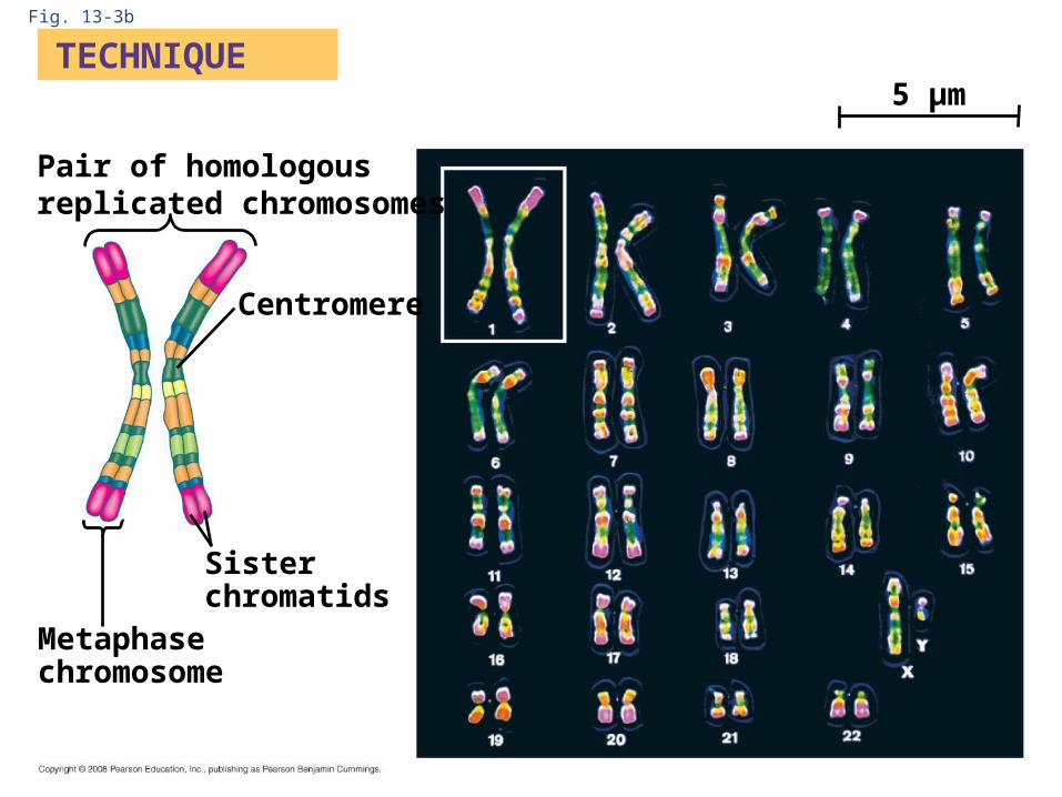
Fig. 13-3b
TECHNIQUE
Pair of homologousreplicated chromosomes
Centromere
Sisterchromatids
Metaphasechromosome
5 µm
Ploidy
• Ploidy refers to a full set of chromosomes – a full set can also be represented by the letter n
• Thus one ploidy would relate to one complete set of chromosomes (23) with one representative chromosome from each of the 23 different types – a representative from chromosome one, a representative from chromosome two – as so on till a representative from all 23.
• A somatic cell is diploid (2n) with two full sets of genetic material (one set from dad and one from mom)
• A gamete has ½ the amount of genetic material as a somatic cell – it is termed haploid (1n)
Karyotype
• A karyotype is an ordered display of the pairs of chromosomes from a cell
• The two chromosomes in each pair are called homologous chromosomes, or homologs
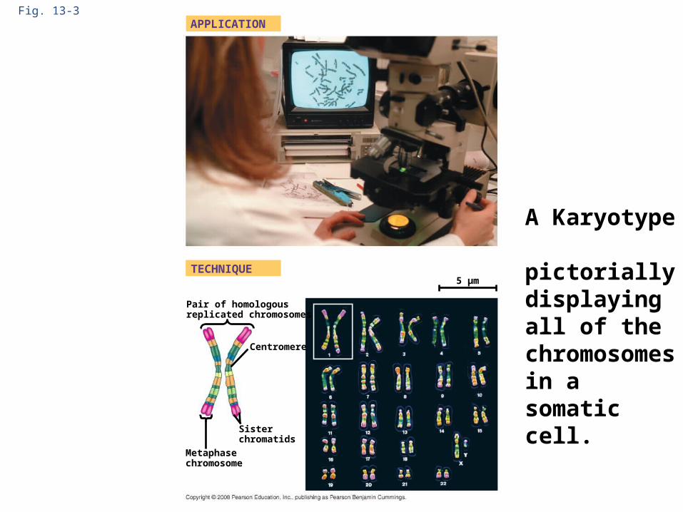
Fig. 13-3APPLICATION
TECHNIQUE
Pair of homologousreplicated chromosomes
5 µm
Centromere
Sisterchromatids
Metaphasechromosome
A Karyotype pictorially displayingall of the chromosomes in a somatic cell.

Fig. 13-3b
TECHNIQUE
Pair of homologousreplicated chromosomes
Centromere
Sisterchromatids
Metaphasechromosome
5 µm
Discussion of a Chromosome/Chromatid
• There are two forms of a chromosome• The singlet non-attached type chromosome• The doublet (attached) type –the
chromosome/chromatid form• Both forms are termed a chromosome
The doublet type – a chromosome/chromatid
• During the S-phase in the cell divisional cycle –DNA is duplicated (as previously discussed)
• Immediately after the S-phase (Synthesis phase) in the cell divisional cycle – there is twice as much DNA – each chromosome is duplicated
• The new duplicate and the original stay temporarily attached to one another–thus two pieces of DNA remain temporarily attached
• Each attached piece of DNA is termed a “chromatid” more especially “sister chromatids”
• The two attached chromatids are termed one chromosome
AdenineThymineCytosineGuanine Old (template) strand
Two new strands (leading and lagging)synthesized in opposite directions
DNA polymerase
DNA polymerase
Laggingstrand
Leading strand
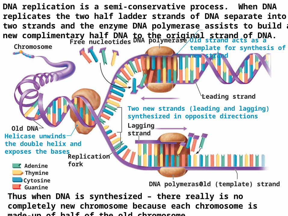
Free nucleotides Old strand acts as a template for synthesis of new strandChromosome
Helicase unwindsthe double helix andexposes the bases
Old DNA
Replicationfork
DNA replication is a semi-conservative process. When DNA replicates the two half ladder strands of DNA separate into two strands and the enzyme DNA polymerase assists to build a new complimentary half DNA to the original strand of DNA.
Thus when DNA is synthesized – there really is no completely new chromosome because each chromosome is made-up of half of the old chromosome.
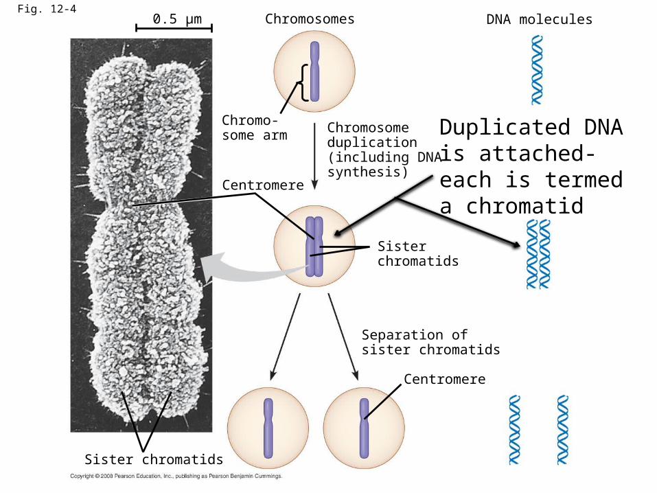
Fig. 12-40.5 µm Chromosomes
Chromosomeduplication(including DNAsynthesis)
Chromo-some arm
Centromere
Sisterchromatids
DNA molecules
Separation ofsister chromatids
Centromere
Sister chromatids
Duplicated DNA is attached- each is termed a chromatid
• Each attached piece of DNA is termed a “chromatid” more especially “ a sister chromatid” – this is the doublet form of DNA
• The two attached chromatids are termed one chromosome
• When the two doublets break apart (separate) each of the now split doublets are termed a chromosome – but a singlet version
• Thus the term chromatid/chromosome and the term chromosome are a matter of semantics
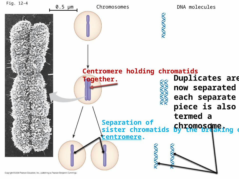
Fig. 12-40.5 µm Chromosomes DNA molecules
Separation ofsister chromatids by the breaking of the centromere.
Duplicates are now separatedeach separate piece is also termed a chromosome.
Centromere holding chromatidsTogether.