Embed Size (px)
Citation preview

Mutation Research 509 (2002) 79–91
DNA repair during organogenesis
Robert K. Vinson, Barbara F. Hales∗Department of Pharmacology and Therapeutics, McGill University, 3655 Promenade Sir-William-Osler,
Montreal, Que., Canada H3G-1Y6
Abstract
DNA damage caused by genotoxic agents can impact on virtually any cellular process due to its ability to affect geneexpression and subsequent gene products. The importance of repairing damaged DNA is evidenced by the variety of DNArepair pathways that have evolved in all living organisms, and the human syndromes caused by a lack of this repair ability.This review focuses on the expression and activity of DNA repair pathways during mammalian organogenesis, and the roleof these pathways in ensuring the stability of the conceptal genome. DNA repair capacity may play a role also in the responseof the conceptus to genotoxic agents that may induce malformations; the consequences of exposure to a genotoxic agentduring organogenesis depend on the extent of the damage and on the ability of the embryo to respond by repairing DNA orarresting cell division. The four main repair pathways (nucleotide excision repair, base excision repair, mismatch repair, andrecombination repair) are expressed to various degrees during organogenesis, as are members of the genotoxic stress-activatedcell cycle checkpoint pathways. Developmental-stage-specific alterations in transcript levels, protein levels, as well as activity,indicate that the regulation of DNA repair pathways during development is complex. The importance of DNA repair pathwaysin endogenous damage control is illustrated by the sensitivity of development to their disruption if some of these genes aremutated. Furthermore, the conceptus has a limited capacity to alter DNA repair responses following exposure to genotoxicagents.© 2002 Elsevier Science B.V. All rights reserved.
Keywords: DNA repair; DNA damage; Organogenesis; Embryo; Teratogen; Cell cycle checkpoint
1. Introduction
DNA repair is crucial for all living organisms. It isestimated that each human cell experiences 2× 10⁴
lesions per day due to normal cellular metabolism[1]. Endogenous metabolites, such as lipid peroxides[2] and reactive nitrogen species[3] create as broad a
Abbreviations: BER, base excision repair; FISH, fluorescentin situ hybridization; GD, gestational day; IR, ionizing radiation;MMR, mismatch repair; NER, nucleotide excision repair; NHEJ,non-homologous end-joining; RCR, recombination repair; UNG,uracil-DNA glycosylase; UVR, ultraviolet radiation
∗ Corresponding author. Tel.:+1-514-398-3610;fax: +1-514-398-7120.E-mail address: [email protected] (B.F. Hales).
spectrum of genotoxic lesions as do external agents.It is currently impossible to determine how manychemical compounds, both naturally occurring andman-made, are genotoxic. Solar ultraviolet radiation(UVR; [4]), cigarette smoke[5], and air pollution[6–8], as well as nitrates[9–11], dyes[12–14], andthe heterocyclic amines (reviewed in[15]) foundin human diets, are just a few of the agents shownto cause significant genotoxic damage at exposurescommonly encountered during a person’s lifetime.Exposure to genotoxic agents at any stage of the lifecycle, from gamete formation to fertilization, fromthe early cleavage stage embryo to organogenesis orfetal or postnatal life, may impact on the genomicintegrity and fate of the organism. The focus of this
0027-5107/02/$ – see front matter © 2002 Elsevier Science B.V. All rights reserved.PII: S0027-5107(02)00223-3

80 R.K. Vinson, B.F. Hales / Mutation Research 509 (2002) 79–91
review is on the role and regulation of DNA repairduring mammalian development.
To cope with the onslaught of genotoxic stresscaused both by endogenous and exogenous agents,organisms have evolved cellular pathways that recog-nize and repair specific types of damage. DNA repairenzymes are found in all organisms examined to date[16]. Conservation of DNA repair pathways, fromthe most primitive unicellular to the most complexmulticellular organisms, indicates the absolute neces-sity for specific DNA repair pathways (reviewed in[17]). The four main DNA repair pathways in mam-malian cells, nucleotide excision repair (NER), baseexcision repair (BER), mismatch repair (MMR), andrecombination repair (RCR) (reviewed in[18–21])are involved not only in the response to exogenousgenotoxic damage, but also in fundamental processesduring development. Many null-mutant animals lack-ing specific DNA repair enzymes have compromisedcellular functions, and abnormal cell proliferationand tissue growth, resulting in embryolethality and/orphenotypic alterations (reviewed in[22]). It is theseobservations, beginning with a report in 1993 thatnull-animals lacking ERCC-1 died before weaning[23], that began the search for the role of DNA re-pair systems during development, especially duringorganogenesis. Rapid cell proliferation, active genetranscription, and a high rate of DNA metabolism, allrequired while the major organ systems are forming,would be expected to increase the risk of genotoxicinsult damaging the conceptal genome. In partic-ular, elucidation of the role of DNA repair duringorganogenesis, when the conceptus is highly suscep-tible to genotoxic insult, would help in understandingthe deleterious effects seen by genotoxic teratogens.Characterization of the role of DNA repair proteinsduring mammalian development (e.g. in null-mutantanimals, as reviewed in[22]) has also brought insightsinto the role of repair pathways in adult organisms inwhich DNA damage is triggered by endogenous orexogenous agents.
Massive DNA replication and rapid cell prolif-eration occur during development. The cell cycleis much shorter in embryonic cells compared toadult cells: gastrulation-stage (gestational day (GD)8.5–9.5) rat embryonic cells divide every 3–7.5 h[24]. Co-ordination of the cell cycle is a key aspectin the response to DNA damage, with the goal of
allowing DNA repair to take place before DNA syn-thesis and cell division occur. DNA damage inductionof cell cycle checkpoint arrest ensures that the DNArepair machinery has an opportunity to repair thedamage and prevent further insult from occurring dueto replication of damaged DNA.
2. DNA repair activity during development
The ability of germ cells and slowly proliferatingor terminally differentiated tissues to repair DNAdamage is frequently assessed by measuring unsched-uled DNA synthesis (also known as DNA repairreplication). Unscheduled DNA synthesis is inducedby damaged DNA even in the early pronuclear stageof the zygote. Exposure to DNA repair inhibitors inthe pre-DNA replication period potentiated the chro-mosome aberrations induced by ionizing radiation(IR) of either mature spermatozoa or oocytes[25].While the 1-cell embryo is transcriptionally active,expressing genes particularly in the male pronucleus,these transcripts are translated poorly[26], suggest-ing that the ability of the zygote to repair DNA isdependent on DNA repair enzymes derived fromthe oocyte. Thus, maternal deficiency in DNA re-pair enzymes affects the early embryo; for example,a maternal deficiency in the DNA MMR enzyme,Pms2, led to the formation of unrepaired replicationerrors during early cleavage divisions[27]. How-ever, not all DNA damage is repaired in early em-bryos. Cleavage stage human embryos have a highincidence of postzygotic chromosomal mosaicism,including aneuploidy and polyploidy, as revealed bymulticolor fluorescence in situ hybridization (FISH)analysis[28]. Chromosomal rearrangements or im-balances in chromosome number contribute substan-tially to the causes of spontaneous abortions, infer-tility and mental retardation[29]. Such large-scalechromosomal abnormalities, also frequently seen incancer cells, may be due to deficiencies in RCR[30–32].
A number of studies have examined DNA repairactivity in the mouse or rat conceptus at a variety oftime points during post-cleavage stage development.Sister chromatid exchange frequencies were five timeshigher in mouse preimplantation embryos than in anypostimplantation stages[33]. Studies have shown that

R.K. Vinson, B.F. Hales / Mutation Research 509 (2002) 79–91 81
the capacity for excision repair of UVR damaged DNAin mouse embryonic primary cell cultures depends onthe gestational stage[34]. Others have assessed DNArepair in the whole conceptus during organogenesis,in limb buds, or in yolk sac primary cell cultures afterexposure to IR[35], N-methyl-N-nitrosourea[35,36],methylmethane sulfonate[35], phenytoin [37] orUVR [38]. A difficulty in assessing DNA repair ca-pacity in postimplantation embryos has been the needto differentiate between DNA repair and DNA repli-cation in rapidly dividing cells; unfortunately it isnot straightforward to inhibit “scheduled” DNA syn-thesis at these stages of development because mostof the drugs used, such as hydroxyurea, are terato-genic during organogenesis[39]. While most studieshave examined alkylation type damage repaired bythe NER pathway, the activity of some BER en-zymes has been examined at specific times duringdevelopment, e.g. uracil-DNA glycosylase (UNG) inthe mouse[40,41]. These studies indicate that repaircapabilities appear to vary widely with the develop-mental age of the conceptus. Tissue-specific activitypatterns exist, indicative of highly regulated repairpathways.
3. DNA damage-dependent cell cyclecheckpoint control during development
All four major mammalian DNA repair pathways(NER, BER, MMR, and RCR), as well as genotoxicstress sensors, interact with components of the cellcheckpoint machinery. These interactions act mainlyto slow cell cycle progression or cell division untilthe damage is repaired. However, some DNA damagesensors elicit a cell checkpoint that may then activatethe apoptotic cell death pathway. While the roles ofcell cycle components that are regulated in responseto genotoxic stress have been studied extensively incancer models, less is known about their regulationor activity during development. Analysis of cleavagestage human embryos by FISH has suggested that cellcycle checkpoints may not operate during early cleav-age development[28]. P53, a tumor suppressor andcell cycle checkpoint gene, plays an important role inembryonic development. Recent studies have shownthat there exists p53-dependent cross-talk between themale and female pronuclei, and that p53 is involved
in an S-phase-dependent checkpoint which suppressesthe rate of pronuclear DNA synthesis[42].
Evidence is accumulating to suggest that theconceptal DNA damage checkpoint response isdevelopmental-stage-specific.Xenopus embryos re-moved damaged cells by apoptosis when irradiatedbefore, but not after, the midblastula transition[43];DNA damage during the rapid, early cleavage, cell di-visions resulted in a synchronous apoptotic responsethat was delayed until the onset of gastrulation[44].In Drosophila embryos, during cleavage cycles, DNAdamage delayed anaphase chromosome separation;during post-blastoderm cycles, irradiation delayedentry into mitosis[45].
Other studies have examined the expression ofgenotoxic stress-activated genes which interact withthe cell cycle machinery during mammalian embry-onic development. GADD45 expression was foundto be high in various tissues in the rat embryo dur-ing late stages of development[46]. Chen and Lee[47] found ubiquitous expression of ATM (AtaxiaTelangiectasia mutated) during mouse development,with particularly high mRNA levels on GD 10.5 and13.5 in the nervous system and lung. Soares et al.[48] determined that the expression pattern of ATMin GD 10–16.5 mouse embryos was consistent witha role for ATM in genome maintenance during celldivision, as the highest levels of expression werefound in areas undergoing rapid cell division. Thep53 gene, a downstream target of ATM, is highlyexpressed at both the transcript and protein levels inmouse embryos up to midgestation, a time of rapidcell proliferation (reviewed in[49,50]). However,there is evidence that the transcriptional activationof p53 target genes does not occur in all tissues, de-spite the presence of high levels of p53 protein[51].Thus, regulation of the DNA repair response duringdevelopment, at least in so far as it is p53-dependent,has an additional component not found in adultorganisms.
The response of the mammalian embryo to DNAdamage, such as IR, is also cell lineage specific.Cells of embryonic origin undergo ATM- andp53-dependent apoptosis following IR, with no acti-vation of cell cycle checkpoints, whereas extraembry-onic cells do not die[52]. The window of sensitivitycorresponds to the start of gastrulation (GD 5.5)through to the beginning of neurulation (GD 8.5), indi-

82 R.K. Vinson, B.F. Hales / Mutation Research 509 (2002) 79–91
cating that a specific period of development is depen-dent on ATM activity to cope with genotoxic stress.ATM may be required to eliminate damaged cellsbefore they have time to divide to form “abnormal”daughter cells. This insight gives a glimpse into therole checkpoint arrest plays in ensuring normal de-velopment. Another hint at the role of cell cyclecheckpoints during development is that null-mutantmice lackingATR [53] or Chk1 [54] do not surviveto birth.
Fig. 1. Expression profiles of DNA repair and genotoxic stress response genes during mid-organogenesis in the rat conceptus. Data arefrom [64,65]. (∗), significantly different from GD 10.
4. DNA repair and cell cycle checkpoint geneexpression during development
DNA repair/genotoxic stress response expres-sion profile data are now available for various cells[55–59]. Several studies have examined the expres-sion of individual DNA repair genes (e.g.[60,61]),as well as the coordinate expression of multiplegenes[46,62–65]in different embryonic tissues dur-ing development. These studies demonstrate very
R.K. Vinson, B.F. Hales / Mutation Research 509 (2002) 79–91 83
particular tissue expression patterns. A number ofstudies have demonstrated that DNA repair geneexpression is temporally regulated during the life-time of an organism; some examples of genes withtime/developmental-stage-specific expression patternsinclude APE/Ref-1 [66,67], ATM [68], MPG [69],and UNG [70]. The temporal regulation of DNArepair gene transcripts suggests that there may bespecific requirements for specific DNA repair en-zymes during certain events. Most of the DNA repairgenes studied are expressed in the conceptus duringmid-organogenesis, with a number at or near the limitof detection[64,65] (Fig. 1). Furthermore, the expres-sion profiles of virtually all repair genes are similar inboth the yolk sac and embryo proper on GD 10 and11; only on GD 12 does the embryo display increasedtranscript levels for many of the genes examined,compared both to GD 10 and 11 embryo as well asto GD 12 yolk sac values. The close relationship be-tween embryo and yolk sac gene expression on GD10 and 11 argues for a similar requirement to repairdamage at these time points, with increased repairnecessary in the GD 12 embryo. The need for DNArepair during organogenesis may be driven by theongoing rapid cell proliferation and coincides withthe switch from anaerobic metabolism to an oxidativeone, which may result in increased genotoxic stress.An overview of the expression profile of DNA repairgenes at various stages of development (e.g. as inFig. 1) provides insight into the status of various DNArepair pathways in the conceptus; if DNA repair geneexpression is transcriptionally regulated, transcript de-ficiencies that are stage and tissue-specific may indi-cate potential “bottlenecks” in the repair process, andreveal critical windows of susceptibility to genotoxicagents.
While alterations in gene expression do not neces-sarily imply a change in DNA repair capability, thisis one of the key mechanisms by which DNA repairactivity can be modified. Some of the repair enzymesexamined during development are involved also inother cellular pathways, which might explain thedevelopmental-stage and potential tissue-specificity(embryo versus yolk sac, perhaps organ specificity)of their expression during organogenesis. However,the majority of genes examined are involved primar-ily in DNA repair, and thus it is possible that thegene expression pattern may ultimately determine
developmental repair status and susceptibility togenotoxic agents during organogenesis.
5. Requirement for DNA repair gene productsduring development as evidenced by nullmutation mice
The requirement for DNA repair gene productsduring development is best evidenced by the pheno-types observed among null animals lacking key repairgenes (Table 1; reviewed in[22]). A number of theanimals lacking DNA repair enzymes are not viable,with pre-implantation death being a common result;much of the embryonic loss occurs around implan-tation, demonstrating the absolute requirement forrepair ability when the embryonic cells begin to pro-liferate rapidly, forming specific tissues (reviewed in[22]). Among the mouse strains with targeted muta-tions in genes for BER, mutations in DNA polymerase�, AP endonuclease (Apex), and DNA ligase 1 wereembryonic lethal, whereas mutations in many of theglycosylases resulted in viable mice with no obviousphenotype[22]. Interestingly, even some of the het-erozygotes may have elevated sensitivity to stress, astheApex heterozygote mice showed elevated levels ofmarkers for oxidative stress which were restored bydietary supplementation with antioxidants[71].
Mutations in many of the genes required for NERresult in viable but growth retarded mice, with theexception of XPD, which was early embryolethal[22]. Mutations in mismatch repair genes result prin-cipally in viable mice but in a number of these strainsmeiosis is defective, resulting in sterility[22]. Incontrast, many of the genes involved in the repair ofDNA strand breaks and recombination are essentialfor normal embryo development, as evidenced by thenumber of strains in which targeted mutations areembryolethal[22]. Interestingly, a heterodimeric en-donuclease involved in homologous recombination,Ercc1-XPF, was essential for targeted gene replace-ment in mouse embryonic stem cells[72].
Mutations in some of the cell cycle checkpoint path-way genes also result in mice with an altered pheno-type. Absence or inactivation of ATM results in AtaxiaTelangiectasia, a disease characterized by neuronal de-generation, immunodeficiency, genetic instability, pre-mature aging and a predisposition to cancer (reviewed

84 R.K. Vinson, B.F. Hales / Mutation Research 509 (2002) 79–91
Table 1Differential requirements for DNA repair gene products during development, as evidenced by null mutation micea
Gene; gene product Pathway Developmental phenotype
Polβ; DNA polymerase� BER Embryonic lethal after GD 10.5Apex; AP endonuclease BER Embryonic lethal by GD 5.5 or 9.5Lig1; DNA ligase 1 BER Embryonic lethal by GD 15.5–16.5; defect in erythropoiesisUNG; uracil-DNA glycosylase BER Viable; develop normallyXpa; xeroderma pigmentosum-group A NER Viable; develop normallyXpd; xeroderma pigmentosum-group D NER Viable; growth retardation; symptoms of agingCSB; cockayne Syndrome-group B NER Develop normally; minor postnatal growth retardation and
neurological defectsPms2; mismatch repair protein Pms2 MMR Viable; develop normally but males infertileXrcc1; Xrcc1 protein BER Embryonic lethal; GD 7.5 embryos abnormalXrcc4; Xrcc4 protein NHEJ Embryonic lethal by GD 16.5; growth retardationATM; ataxia telengiectasia Mutated Checkpoint Growth retardation, neurological abnormalities; infertileATR; ataxia telengiectasia related Checkpoint Embryonic lethal by GD 8.5NBS1; Nijmegen breakage syndrome Checkpoint Viable; microcephaly; immunodeficientP53; p53 protein Checkpoint Most viable; increased exencephaly in females; incisor
fusion; ocular abnormalitiesa This table is a partial summary of the mouse strains with targeted mutations to DNA repair genes that are discussed in this review. For
more complete information on the transgenic mice deficient in DNA repair genes please see reference[22]; other references are providedin the text.
in [73]). Gamma-irradiation of mouse embryos re-sults in ATM-dependent apoptosis only of postmitoticpopulations in the developing nervous system[74].Mutations in NBS1 (Nijmegen breakage syndrome)result in microcephaly, immune deficiency, and a highincidence of cancer[75,76]. While p53 null mice areviable, a substantial fraction of the female embryosare exencephalic; other defects include upper incisorfusion, ocular abnormalities and hindlimb polydactyly[77,78]. The p53-deficient fibroblasts were defectivein global and transcription-coupled NER[79].
An additional clue to the role of DNA repair en-zymes during development can be obtained from thedouble-knockout null-mutant animals; some of thesemutations were embryolethal, whereas the individualsingle gene knockout animals were viable[80,81].Examples of double null-mutant animals that diedduring organogenesis include ATM and DNA-PKcs[81], ATM and PARP-1[80], Rad51d and Rad51L3[82]. Interestingly, mice lacking both XPA (Xero-derma pigmentosum-group A) and CSB (Cockaynesyndrome-group B) did not die but displayed severegrowth retardation, ataxia, and motor dysfunctionduring early postnatal development, suggesting thatthese genes have additive roles in nervous system de-velopment[83]. In contrast to these synergistic or ad-ditive effects, p53 deficiency rescuedmdm-2-deficient
mice [84,85], and partially rescuedRad51-deficient[86] and Brca1 and Brca2-deficent mice[87,88].ATM-deficiency prevented apoptosis in the develop-ing nervous system of DNA ligase IV-deficient mice,but not the deficits in immune differentiation[89].Thus, double mutant animals for DNA damage re-sponsive genes, in particular, demonstrate how essen-tial these pathways are during organogenesis, not onlyto sense damage due to exogenous agents but also tocoordinate DNA replication during rapid cell division.Therefore, the role of DNA repair enzymes duringdevelopment must be to: (i) counteract endogenousgenotoxic agents arising naturally due to extensiveDNA metabolism; (ii) actively participate in rapidDNA metabolism due to tremendous cellular pro-liferation; or (iii) participate in non-repair pathwayscritical for proper tissue growth and development.
Null-mutant animals that lack DNA repair orgenotoxic stress response genes but are still viableare being used to elucidate the mechanisms of ter-atogenesis. The p53 null mice have been used toexamine the mechanisms of action of several terato-gens[90–95]. Most studies have reported a role forp53 as a teratological suppressor gene, as evidencedby the enhanced susceptibility of null mutationmice to chemical (benzo[�]pyrene and cyclophos-phamide) and physical (ionizing radiation) teratogens.

R.K. Vinson, B.F. Hales / Mutation Research 509 (2002) 79–91 85
Paradoxically, however, the absence of p53 pro-tected against eye defects induced by 2-chloro-2-deo-xyadenosine[92]. Hall and Lane[96] argued that oneof the functions of p53 is as a “guardian of the babies”.
6. Response of the embryo to genotoxicagents—role of DNA repair
It has been estimated that 25–30% of all birth de-fects are due to gene mutations and chromosomal ab-normalities [97], both of which can be induced bygenotoxic stress. While the cause of 60–70% of birthdefects is not established, 25% (8/32) of known hu-man teratogens are mutagenic[98]. It is not surprisingthat exposure to genotoxic agents during developmentleads to adverse outcomes. The process of creating acomplete, multicellular organism from a single, fer-tilized egg requires a complex, interwoven pattern ofgene expression and regulation. Any conditions thatinterfere with gene function could lead to dramaticconsequences in the developing embryo, in which celldivision times and windows of gene expression arerapid and transitory.
Some of the genotoxic teratogens which have beenexamined in experimental models have been reviewedby Bishop et al.[98] and are detailed inTable 2. An-ticancer drugs (such as busulfan, cyclophosphamide,cytarabine, and 6-mercaptopurine) are both genotoxicand teratogenic. Furthermore, developmental abnor-malities in rodents exposed to alkylating mutagens areoften similar to human malformations of unknown ae-tiology [105]. Many teratogens have equivocal activityas mutagens or DNA damaging agents[98]. However,
Table 2Studies demonstrating the genotoxic effects of teratogens during development
Genotoxic teratogen Developmental age (GD) Organism Reference(s)
Benzo[�]pyrene 9.5 Mouse [99]Cyclophosphamide 9 Mouse [100]Dioxin 12 Mouse [101]Ethylmethane sulfonate 9–12 Mouse [102]Methotrexate 10–12 Rat [41]∗Methylmethane sulfonate 10.5–12.5 Mouse [35]∗N-Methyl-N-nitrosourea 10–11;10.5–12.5 Mouse [35]∗; [36]∗; [103]Phenytoin 12 Mouse [37]∗Pyrimethamine 13 Mouse [14]Thalidomide 8–12 Rabbit [104]
Those which examined DNA repair activity are indicated by an asterisk.
such well-studied teratogens as phenytoin[106] orthalidomide[104], which are not directly genotoxic,have been demonstrated to cause DNA damage viathe induction of reactive oxygen species and oxidativestress. The teratogenicity of at least some of thesecompounds appears to be dependent on bioactivationby prostaglandin H synthase to electrophilic, reactive,intermediates[107]. Ethanol induces H2O2 and O2
−production in the fetus[108]; both are potent ROSthat create oxidative damage to DNA. Inhibition ofDNA repair enzymes by hydroxyurea, 5-fluorouracil,or caffeine may also play a role in the mechanism ofaction of teratogens (e.g.[109,25]).
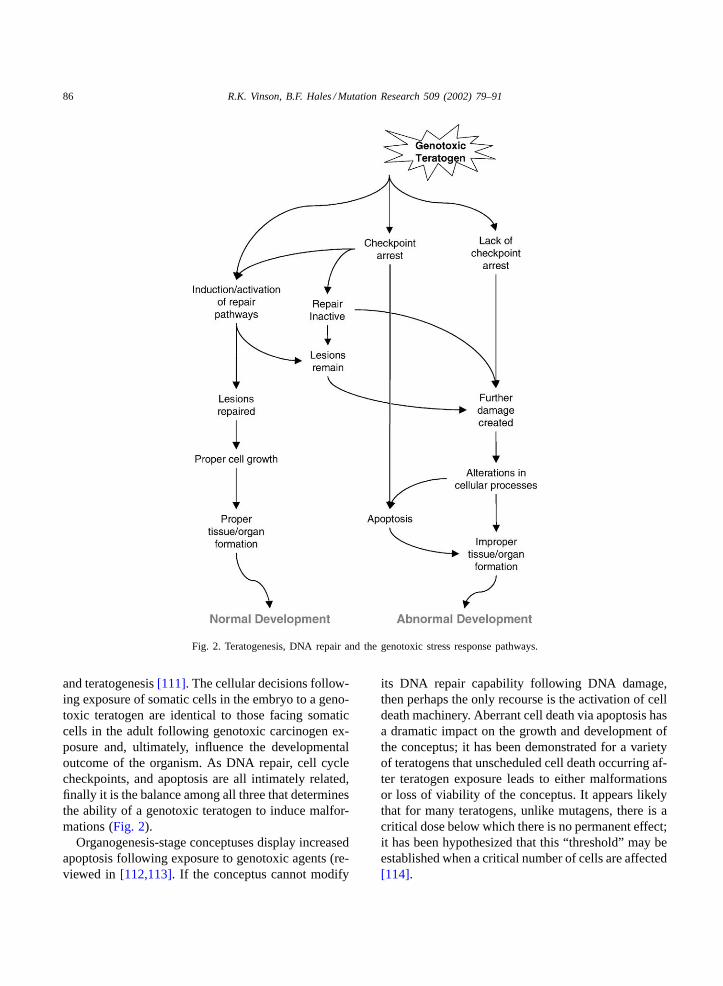
There exist three fundamental choices for a cell onceits genome is damaged (Fig. 2). The first is to removethe lesions from the genome and to return to as closea pre-exposure condition as possible. The second op-tion is to continue functioning in spite of the damage;this may result in further damage and alterations ingene transcription and DNA replication. This almostcertainly impacts on cellular metabolism, and can leadto carcinogenesis or cell death. Finally, the cell canchoose to activate its apoptotic cascade, leading to celldeath. The choice between these three pathways cangreatly affect the functioning of the whole organism;a single genotoxic molecule, creating a single DNAlesion in a single cell, has the ability to trigger cellularevents leading to cell death and potentially the deathof the whole organism. Many teratogens, such as hy-perthermia and cyclophosphamide, induce changes incell cycle kinetics, leading to excessive apoptotic celldeath[110].
Studies as early as 1977 noted the similarity bet-ween DNA damage-mediated chemical carcinogenesis
86 R.K. Vinson, B.F. Hales / Mutation Research 509 (2002) 79–91
Fig. 2. Teratogenesis, DNA repair and the genotoxic stress response pathways.
and teratogenesis[111]. The cellular decisions follow-ing exposure of somatic cells in the embryo to a geno-toxic teratogen are identical to those facing somaticcells in the adult following genotoxic carcinogen ex-posure and, ultimately, influence the developmentaloutcome of the organism. As DNA repair, cell cyclecheckpoints, and apoptosis are all intimately related,finally it is the balance among all three that determinesthe ability of a genotoxic teratogen to induce malfor-mations (Fig. 2).
Organogenesis-stage conceptuses display increasedapoptosis following exposure to genotoxic agents (re-viewed in[112,113]. If the conceptus cannot modify
its DNA repair capability following DNA damage,then perhaps the only recourse is the activation of celldeath machinery. Aberrant cell death via apoptosis hasa dramatic impact on the growth and development ofthe conceptus; it has been demonstrated for a varietyof teratogens that unscheduled cell death occurring af-ter teratogen exposure leads to either malformationsor loss of viability of the conceptus. It appears likelythat for many teratogens, unlike mutagens, there is acritical dose below which there is no permanent effect;it has been hypothesized that this “threshold” may beestablished when a critical number of cells are affected[114].

R.K. Vinson, B.F. Hales / Mutation Research 509 (2002) 79–91 87
One of the most striking results found followingexposure of the organogenesis-stage conceptus togenotoxic teratogens is the near absence of responseof DNA repair pathways at the gene, protein, or ac-tivity levels. Of the four repair pathways and thegenotoxic stress checkpoints examined during devel-opment, transcript levels for only GADD45[62] andUNG [41] increased following genotoxic stress byeither methylmercury or methotrexate, respectively.This lack of induction of DNA repair or checkpointgenes demonstrates the rigidity of the developmen-tal gene expression program. Although, DNA repairpathways are only one part of a complex system ofbiochemical pathways that can respond to teratogens,other cellular responses to genotoxic damage will notresolve the fundamental problem, the need to repairthe genotoxic lesions.
7. Summary
The expression of DNA repair enzymes may makethe difference between embryonic survival and deathfollowing genotoxic insult during development. Theremay be different requirements for members of eachpathway in the conceptus. In addition, many DNArepair enzymes function in other DNA metabolismpathways that may be crucial during development.
The conclusions from studies published to date per-mit the formulation of an initial global view of DNArepair gene expression in the organogenesis-stage ratconceptus. Many holes exist in our current knowl-edge, however, particularly with respect to the precisedevelopmental role of several DNA repair genes.With the increased sensitivity of molecular biologytools, it will become easier to examine levels of geno-toxic stress, as well as the responses to that stress,in order to define the role of DNA repair systemsduring organogenesis. Elucidation of these path-ways during development is essential to understandthe effect of exposure to genotoxic agents duringdevelopment.
Acknowledgements
The research conducted in our lab was supportedby the Canadian Institutes of Health Research.
References
[1] B.N. Ames, M.K. Shigenaga, Oxidants are a majorcontributor to aging, Ann. N.Y. Acad. Sci. 663 (1992) 85–96.
[2] P.C. Burcham, Internal hazards: baseline DNA damage byendogenous products of normal metabolism, Mutat. Res.443 (1999) 11–36.
[3] D.A. Wink, K.S. Kasprzak, C.M. Maragos, R.K. Elespuru,M. Misra, T.M. Dunams, T.A. Cebula, W.H. Koch, A.W.Andrews, J.S. Allen, L.K. Keefe, DNA deaminating abilityand genotoxicity of nitric oxide and its progenitors, Science254 (1994) 1001–1003.
[4] H.R. Griffiths, P. Mistry, K.E. Herbert, J. Lunec, Molecularand cellular effects of ultraviolet light-induced genotoxicity,Crit. Rev. Clin. Lab. Sci. 35 (1998) 189–237.
[5] W.A. Pryor, Cigarette smoke radicals and the role offree radicals in chemical carcinogenicity, Environ. Health.Perspect. 105 (Suppl 4) (1997) 875–882.
[6] Y.A. Courtois, S. Min, C. Lachenal, J.M. Jacquot-Deschamps, F. Callais, B. Festy, Genotoxicity of organicextracts from atmospheric particles, Ann. N. Y. Acad. Sci.534 (1988) 724–740.
[7] W.L. Hsiao, Z.Y. Mo, M. Fang, X.M. Shi, F. Wang,Cytotoxicity of PM(2.5) and PM(2.5–10) ambient airpollutants assessed by the MTT and the Comet assays,Mutat. Res. 471 (2000) 45–55.
[8] A. Don Porto Carero, P.H. Hoet, L. Verschaeve, G. Schoeters,B. Nemery, Genotoxic effects of carbon black particles,diesel exhaust particles, and urban air particulates and theirextracts on a human alveolar epithelial cell line (A549)and a human monocytic cell line (THP-1), Environ. Mol.Mutagen. 37 (2001) 155–163.
[9] B.L. Rubenchik, N.D. Osinkovskaya, V.M. Mikhailenko,M.A. Furman, T.M. Boim, The carcinogenic danger of nitritepollution of the environment, J. Environ. Pathol. Toxicol.Oncol. 10 (1990) 290–296.
[10] M.N. Routledge, F.J. Mirsky, D.A. Wink, L.K. Keefer, A.Dipple, Nitrite-induced mutations in a forward mutationassay: influence of nitrite concentration and pH, Mutat. Res.322 (1994) 341–346.
[11] C.J. Wang, H.P. Huang, T.H. Tseng, Y.L. Lin, S.J. Shiow,N-Nitroso-N-(3-keto-1,2-butanediol)-3′-nitrotyramine: a newgenotoxic agent derived from the reaction of tyrosine andglucose in the presence of sodium nitrite, Arch. Toxicol. 70(1995) 10–15.
[12] A.A. Lakdawalla, M.S. Netrawali, Mutagenicity, comut-agenicity, and antimutagenicity of erythrosine (FD and Cred 3), a food dye, in the Ames/Salmonella assay, Mutat.Res. 204 (1988) 131–139.
[13] P. Moller, H. Wallin, N. Grunnet, L. Risom, L.E. Knudsen,DNA damage in isolated rat hepatocytes exposed to C.I.pigment orange 5 and C.I. pigment yellow 12 by the alkalinecomet assay, Teratog. Carcinog. Mutagen. 18 (1998) 9–16.
[14] S. Tsuda, Y. Kosaka, N. Matsusaka, Y.F. Sasaki, Detectionof pyrimethamine-induced DNA damage in mouse embryoand maternal organs by the modified alkaline single cell gelelectrophoresis assay, Mutat. Res. 415 (1998) 69–77.
88 R.K. Vinson, B.F. Hales / Mutation Research 509 (2002) 79–91
[15] T. Sugimura, Nutrition and dietary carcinogens, Carcino-genesis 21 (2000) 387–395.
[16] V. Brendel, L. Brocchieri, S.J. Sandler, A.J. Clark, S. Karlin,Evolutionary comparisons of RecA-like proteins across allmajor kingdoms of living organisms, J. Mol. Evol. 44 (1997)528–541.
[17] E.M. Taylor, A.R. Lehmann, Conservation of eukaryoticDNA repair mechanisms, Int. J. Radiat. Biol. 74 (1998)277–286.
[18] P. Modrich, Mismatch repair in replication fidelity, geneticrecombination, and cancer biology, Annu. Rev. Biochem. 65(1996) 101–133.
[19] R.D. Wood, DNA repair in eukaryotes, Annu. Rev. Biochem.65 (1996) 135–167.
[20] C.J. Norbury, I.D. Hickson, Cellular responses to DNAdamage, Annu. Rev. Pharmacol. Toxicol. 41 (2001) 367–401.
[21] A. Ronen, B.W. Glickman, Human DNA repair genes,Environ. Mol. Mutagen. 37 (2001) 241–283.
[22] E.C. Friedberg, L.B. Meira, Database of mouse strainscarrying targeted mutations in genes affected cellularresponses to DNA damage Version 4, Mutat. Res. 459 (2000)243–274.
[23] J. McWhir, J. Selfridge, D.J. Harrison, S. Squires, D.S.Melton, Mice with DNA repair gene (ERCC-1) deficiencyhave elevated levels of p53, liver nuclear abnormalities anddie before weaning, Nat. Genet. 5 (1993) 217–224.
[24] A. MacAuley, Z. Werb, P.E. Mirkes, Characterization ofthe unusually rapid cell cycles during rat gastrulation,Development 117 (1993) 873–883.
[25] Y. Matsuda, I. Tobari, Repair capacity of fertilized mouseeggs for X-ray damage induced in sperm and mature oocytes,Mutat. Res. 210 (1989) 35–47.
[26] R.M. Schultz, The molecular foundations of the maternalto zygotic transition in the preimplantation embryo, HumanReprod. Update 8 (2002) 323–331.
[27] V.E. Gurtu, S. Verma, A.H. Grossmann, R.M. Liskay, W.C.Skarnes, S.M. Baker, Maternal effect for DNA mismatchrepair in the mouse, Genetics 160 (2002) 271–277.
[28] R.H. Harrison, H.C. Kuo, P.N. Scriven, A.H. Handyside,C.M. Ogilvie, Lack of cell cycle checkpoints in humancleavage stage embryos revealed by a clonal pattern ofchromosomal mosaicism analysed by sequential multicolourFISH, Zygote 8 (2000) 217–224.
[29] D.E. McFadden, J.M. Friedman, Chromosome abnormalitiesin human beings, Mutat. Res. 396 (1997) 129–140.
[30] S.P. Jackson, Sensing and repairing DNA double-strandbreaks, Carcinogenesis 23 (2002) 687–696.
[31] D.O. Ferguson, F.W. Alt, DNA double strand break repairand chromosomal translocation: lessons from animal models,Oncogene 20 (2001) 5572–5579.
[32] P. Pfeiffer, W. Goedecke, G. Obe, Mechanisms of DNAdouble-strand break repair and their potential to inducechromosomal aberrations, Mutagenesis 15 (2000) 289–302.
[33] S. el-Hage, S.M. Singh, A 5-fold reduction insister-chromatid exchange following implantation of mouseembryos is not directly related to the expression of
embryonic genes responsible for oxygen radical metabolism,Mutat. Res. 232 (1990) 217–226.
[34] L. Peleg, E. Raz, R. Ben-Ishai, Changing capacity for DNAexcision repair in mouse embryonic cells in vitro, Exp. CellRes. 104 (1977) 301–307.
[35] S. Jirakulsomchok, K.L. Yielding, DNA damage and repairin mouse embryos following treatment transplacentally withmethylnitrosourea and methylmethanesulfonate, Teratog.Carcinog. Mutagen. 4 (1984) 523–536.
[36] R. Loch-Caruso, C.S. Baxter, DNA damage and repair inembryonic mouse limbs exposed to teratogenic doses ofmethylnitrosourea, Mutat. Res. 131 (1984) 147–155.
[37] L. Liu, P.G. Wells, DNA oxidation as a potential molecularmechanism mediating drug-induced birth defects: phenytoinand structurally related teratogens initiate the formation of8-hydroxy-2′-deoxyguanosine in vitro and in vivo in murinematernal hepatic and embryonic tissues, Free Radic. Biol.Med. 19 (1995) 639–648.
[38] J.J. Latimer, M.L. Hultner, J.E. Cleaver, R.A. Pedersen,Elevated DNA excision repair capacity in the extraembryonicmesoderm of the midgestation mouse embryo, Exp. Cell.Res. 228 (1996) 19–28.
[39] J. Timson, Hydroxyurea, Mutat. Res. 32 (1975) 115–132.[40] Y. Weng, M.A. Sirover, Developmental regulation of the
base excision repair enzyme uracil DNA glycosylase in therat, Mutat. Res. 293 (1993) 133–141.
[41] R.K. Vinson, B.F. Hales, Expression and activity of the DNArepair enzyme uracil DNA glycosylase during organogenesisin the rat conceptus and following methotrexate exposure invitro, Biochem. Pharmacol. 64 (2002) 711–721.
[42] T. Shimura, M. Inoue, M. Taga, K. Shiraishi, N. Uematsu,N. Takei, Z.M. Yuan, T. Shinohara, O. Niwa, p53-DependentS-phase damage checkpoint and pronuclear cross talk inmouse zygotes with X-irradiated sperm, Mol. Cell. Biol. 22(2002) 2220–2228.
[43] C.V. Finkielstein, A.L. Lewellyn, J.L. Maller, The midblas-tula transition in Xenopus embryos activates multiplepathways to prevent apoptosis in response to DNA damage,Proc. Natl. Acad. Sci. U.S.A. 98 (2001) 1006–1011.
[44] J. Greenwood, V. Costanzo, K. Robertson, C. Hensey, J.Gautier, Responses to DNA damage inXenopus: cell deathor cell cycle arrest, Novartis Found Symp. 237 (2001) 221–230.
[45] T.T. Su, J. Walker, J. Stumpff, Activating the DNA damagecheckpoint in a developmental context, Curr. Biol. 10 (2000)119–126.
[46] W.D. Rees, S.M. Hay, N.C. Fontanier-Razzaq, C. Amtipatis,D.N. Harries, Expression of the growth arrest genes (GASand GADD) changes during organogenesis in the rat fetus,J. Nutr. 129 (1999) 1532–1536.
[47] G. Chen, E.Y.-H.P. Lee, The product of the ATM gene is a370-kDa nuclear phosphoprotein, J. Biol. Chem. 271 (1996)33693–33697.
[48] H.D. Soares, J.I. Morgan, P.J. McKinnon, ATM expressionpatterns suggest a contribution from the peripheralnervous system to the phenotype of Ataxia-Telangiectasia,Neuroscience 86 (1998) 1045–1054.

R.K. Vinson, B.F. Hales / Mutation Research 509 (2002) 79–91 89
[49] P. Schmid, A. Lorenz, H. Hameister, M. Montenarh,Expression of p53 during mouse embryogenesis, Develo-pment 113 (1991) 857–865.
[50] J. Choi, L.A. Donehower, p53 in embryonic development:maintaining a fine balance, Cell Mol. Life Sci. 55 (1999)38–47.
[51] E.A. Komarova, M.V. Chernov, R. Franks, K. Wang, G.Armin, C.R. Zelnick, D.M. Chin, S.S. Bacus, G.R. Stark,A.V. Gudkov, Transgenic mice with p53-responsive lacZ:p53 activity varies dramatically during normal developmentand determines radiation and drug sensitivity in vivo, EMBOJ. 16 (1997) 1391–1400.
[52] B.S. Heyer, A. MacAuley, O. Behrendtsen, Z. Werb,Hypersensitivity to DNA damage leads to increasedapoptosis during early mouse development, Genes Dev. 14(2000) 2072–2084.
[53] A. de Klein, M. Muijtjens, R. van Os, Y. Verhoeven, B. Smit,A.M. Carr, A.R. Lehmann, J.H.J. Hoeijmakers, Targeteddisruption of the cell-cycle checkpoint gene ATR leads toearly embryonic lethality in mice, Curr. Biol. 10 (2000)479–482.
[54] H. Takai, K. Tominaga, N. Motoyama, Y.A. Minamishima,H. Nagahama, T. Tsukiyama, K. Ikeda, K. Nakayama, M.Nakanishi, K.-I. Nakayama, Aberrant cell cycle checkpointfunction and early embryonic death in Chk1(−/−) mice,Genes Dev. 14 (2000) 1439–1447.
[55] S.M. Keyse, The induction of gene expression in mammaliancells by radiation, Semin. Cancer Biol. 4 (1993) 119–128.
[56] S.A. Amundson, M. Bittner, Y. Chen, J. Trent, P.Meltzer, A.J. Fornace Jr., Fluorescent cDNA microarrayhybridization reveals complexity and heterogeneity ofcellular genotoxic stress responses, Oncogene 18 (1999)3666–3672.
[57] S.A. Jelinsky, L.D. Samson, Global response ofSacch-aromyces cerevisiae to an alkylating agent, Proc. Natl. Acad.Sci. U.S.A. 96 (1999) 1486–1491.
[58] D.P. Harkin, P.A. Hall, Measuring a cell’s response to stress:the p53 pathway, Genome Biol. 1 (2000) 105.
[59] S.A. Amundson, M. Bittner, P. Meltzer, J. Trent, A.J.Fornace Jr., Physiological function as regulation of largetranscriptional programs: the cellular response to genotoxicstress, Comp. Biochem. Physiol. B. Biochem. Mol. Biol.129 (2001) 703–710.
[60] M. Hubank, L. Mayne, Expression of the excision repairgene, ERCC3 (excision repair cross-complementing) duringmouse development, Brain Res. Dev. Brain Res. 81 (1994)66–76.
[61] N.K. Kim, S.H. Lee, T.J. Sohn, R. Roy, S. Mitra,H.M. Chung, J.J. Ko, K.Y. Cha, Spatial expression of aDNA repair gene,N-methylpurine-DNA glycosylase (MPG)during development in mice, Anticancer Res. 20 (2000)3037–3043.
[62] Y.C. Ou, S.A. Thompson, S.C. Kirchner, T.J. Kavanagh,E.M. Faustman, Induction of growth arrest and DNAdamage-inducible genes Gadd45 and Gadd153 in primaryrodent embryonic cells following exposure to methyl-mercury, Toxicol. Appl. Pharm. 147 (1997) 31–38.
[63] W. Harrouk, A. Codrington, R. Vinson, B. Robaire, B.F.Hales, Paternal exposure to cyclophosphamide induces DNAdamage and alters the expression of DNA repair genes inthe rat preimplantation embryo, Mutation Res. DNA Repair461 (2000) 229–241.
[64] R.K. Vinson, B.F. Hales, Nucleotide excision repair geneexpression in the rat conceptus during organogenesis, Mutat.Res. 486 (2001) 113–123.
[65] R.K. Vinson, B.F. Hales, Expression of base excision,mismatch, and recombination repair genes in the organo-genesis-stage rat conceptus and effects of exposure to a geno-toxic teratogen, 4-hydroperoxycyclophosphamide, Tera-tology 64 (2001) 283–291.
[66] Y. Ono, M. Watanabe, Y. Inoue, T. Ohmoto, K. Akiyama,K. Tsutsui, S. Seki, Developmental expression of APEXnuclease, a multifunctional DNA repair enzyme, in mousebrains, Brain Res. Dev. Brain Res. 86 (1995) 1–6.
[67] T.M. Wilson, S.A. Rivkees, W.A. Deutsch, M.R.Kelley, Differential expression of the apurinic/apyrimidinicendonuclease (APE/ref-1) multifunctional DNA baseexcision repair gene during fetal development and in adultrat brain and testis, Mutat. Res. 362 (1996) 237–248.
[68] G. Chen, E.Y.-H.P. Lee, The product of the ATM gene is a370-kDa nuclear phosphoprotein, J. Biol. Chem. 271 (1996)33693–33697.
[69] N.K. Kim, S.H. Lee, T.J. Sohn, R. Roy, S. Mitra,H.M. Chung, J.J. Ko, K.Y. Cha, Spatial expression of aDNA repair gene,N-methylpurine-DNA glycosylase (MPG)during development in mice, Anticancer Res. 20 (2000)3037–3043.
[70] H. Nilsen, K.S. Steinsbekk, M. Otterlei, G. Slupphaug, P.A.Aas, H.E. Krokan, Analysis of uracil-DNA glycosylasesfrom the murineUNG gene reveals differential expressionin tissues and in embryonic development and a subcellularsorting pattern that differs from the human homologues,Nucl. Acids Res. 28 (2000) 2277–2285.
[71] L.B. Meira, S. Devaraj, G.E. Kisby, D.K. Burns, R.L.Daniel, R.E. Hammer, S. Grundy, I. Jialal, E.C. Friedberg,Heterozygosity for the mouse Apex gene results inphenotypes associated with oxidative stress, Cancer Res. 61(2001) 5552–5557.
[72] L.J. Niedernhofer, J. Essers, G. Weeda, B. Beverloo, J. deWit, M. Muijtjens, H. Odijk, J.H. Hoeijmakers, R. Kanaar,The structure-specific endonuclease Ercc1-XPF is requiredfor targeted gene replacement in embryonic stem cells,EMBO J. 20 (2001) 6540–6549.
[73] G. Rotman, Y. Shiloh, ATM: from gene to function, Hum.Mol. Genet. 7 (1998) 1555–1563.
[74] Y. Lee, M.J. Chong, P.J. McKinnon, Ataxia Telangiectasiamutated-dependent apoptosis after genotoxic stress in thedeveloping nervous system is determined by cellulardifferentiation status, J. Neurosci. 21 (2001) 6687–6693.
[75] J. Kang, R.T. Bronson, Y. Xu, Targeted disruption of NBS1reveals its roles in mouse development and DNA repair,EMBO J. 21 (2002) 1447–1455.
[76] J. Zhu, S. Petersen, L. Tessarollo, A. Nussenzweig, Targeteddisruption of the Nijmegen breakage syndrome gene NBS1

90 R.K. Vinson, B.F. Hales / Mutation Research 509 (2002) 79–91
leads to early embryonic lethality in mice, Curr. Biol. 11(2001) 105–109.
[77] J.F. Armstrong, M.H. Kaufman, D.J. Harrison, A.R.Clarke, High-frequency developmental abnormalities inp53-deficient mice, Curr. Biol. 5 (1995) 931–936.
[78] V.P. Sah, L.D. Attardi, G.J. Mulligan, B.O. Williams, R.T.Bronson, T. Jacks, A subset of p53-deficient embryos exhibitexencephaly, Nat. Genet. 10 (1995) 175–180.
[79] J.P. Therrien, R. Drouin, C. Baril, E.A. Drobetsky, Humancells compromised for p53 function exhibit defective globaland transcription-coupled nucleotide excision repair, whereascells compromised for pRb function are defective only inglobal repair, Proc. Natl. Acad. Sci. U.S.A. 96 (1999) 15038–15043.
[80] J. Ménissier-de Murcia, M. Mark, O. Wendling, A.Wynshaw-Boris, G. de Murcia, Early embryonic lethalityin PARP-1 ATM double-mutant mice suggests a functionalsynergy in cell proliferation during development, Mol. Cell.Biol. 21 (2001) 1828–1832.
[81] K.E. Gurley, C.J. Kemp, Synthetic lethality betweenmutation in ATM and DNA-PKcs during murine embryo-genesis, Curr. Biol. 11 (2001) 191–194.
[82] D.L. Pittman, J.C. Schimenti, Midgestation lethality inmice deficient for the RecA-related gene Rad51d/Rad51l3,Genesis 26 (2000) 167–173.
[83] M. Murai, Y. Enokido, N. Inamura, M. Yoshino, Y. Nakatsu,G.T. van der Horst, J.H. Hoeijmakers, K. Tanaka, H.Hatanaka, Early postnatal ataxia and abnormal cerebellardevelopment in mice lacking Xeroderma pigmentosumGroup A and Cockayne syndrome Group B DNA repairgenes, Proc. Natl. Acad. Sci. U.S.A. 98 (2001) 13379–13384.
[84] R. Montes de Oca Luna, D.S. Wagner, G. Lozano, Rescueof early embryonic lethality in mdm2-deficient mice bydeletion of p53, Nature 378 (1995) 203–206.
[85] S.N. Jones, A.E. Roe, L.A. Donehower, A. Bradley, Rescueof embryonic lethality in Mdm2-deficient mice by absenceof p53, Nature 378 (1995) 206–208.
[86] D.S. Lim, P. Hasty, A mutation in mouse rad51 results inan early embryonic lethal that is suppressed by a mutationin p53, Mol. Cell. Biol. 16 (1996) 7133–7143.
[87] J. Brugarolas, T. Jacks, Double indemnity—p53 BRCA andcancer: p53 mutation partially rescues developmental arrestin Brca1 and Brca2 null mice suggesting a role for familialbreast cancer genes in DNA damage repair, Nat. Med. 3(1997) 721–722.
[88] R. Hakem, J.L. de la Pompa, A. Elia, J. Potter, T.W. Mak,Partial rescue of Brca1 (5–6) early embryonic lethality byp53 or p21 null mutation, Nat. Genet. 16 (1997) 298–302.
[89] Y. Lee, D.E. Barnes, T. Lindahl, P.J. McKinnon, Defectiveneurogenesis resulting from DNA ligase IV deficiencyrequires ATM, Genes Dev. 14 (2000) 2576–2580.
[90] C.J. Nicol, M.L. Harrison, R.R. Laposa, I.L. Gimelshtein,P.G. Wells, A teratologic suppressor role forp53 inbenzo[�]pyrene-treated transgenicp53-deficient mice, Nat.Genet. 10 (1995) 181–187.
[91] T. Norimura, S. Nomoto, M. Katsuki, Y. Gondo, S. Kondo,p53-Dependent apoptosis suppresses radiation-inducedteratogenesis, Nat. Med. 2 (1996) 577–580.
[92] J.A. Wubah, M.M. Ibrahim, X. Gao, D. Nguyen, M.M.Pisano, T.B. Knudsen, Teratogen-induced eye defectsmediated by p53-dependent apoptosis, Curr. Biol. 6 (1996)60–69.
[93] S.A. Moallem, B.F. Hales, The role of p53 and celldeath by apoptosis and necrosis in 4-hydroperoxycyclo-phosphamide-induced limb malformations, Development125 (1998) 3225–3234.
[94] J. Frenkel, D. Sherman, A. Fein, D. Schwartz, N. Almog,A. Kapon, N. Goldfinger, V. Rotter, Accentuated apoptosisin normally developing p53 knockout mouse embryosfollowing genotoxic stress, Oncogene 18 (1999) 2901–2907.
[95] B. Wang, Involvement of p53-dependent apoptosis inradiation teratogenesis and in the radioadaptive response inthe late organogenesis of mice, J. Radiat. Res. (Tokyo) 42(2001) 1–10.
[96] P.A. Hall, D.P. Lane, Tumor suppressors: a developing rolefor p53? Curr. Biol. 7 (1997) R144–R147.
[97] D.A. Beckman, R.L. Brent, Mechanisms of teratogenesis,Ann. Rev. Pharmacol. Toxicol. 24 (1984) 483–500.
[98] J.B. Bishop, K.L. Witt, R.A. Sloane, Genetic toxicities ofhuman teratogens, Mutat. Res. 396 (1997) 9–43.
[99] L.M. Winn, P.G. Wells, Evidence for embryonic prostaglan-din H synthase-catalyzed bioactivation and reactive oxygenspecies-mediated oxidation of cellular macromolecules inphenytoin and benzo[�]pyrene teratogenesis, Free Radic.Biol. Med. 22 (1997) 607–621.
[100] V.V. Murthy, B.A. Becker, W.J. Steele, Effects of dosage,phenobarbital, and 2-diethylaminoethyl-2,2-diphenylvalerateon the binding of cyclophosphamide and-or its metabolitesto the DNA, RNA and protein of the embryo and liver inpregnant mice, Cancer Res. 33 (1973) 664–670.
[101] E.A. Hassoun, D. Bagchi, S.J. Stohs, Evidence of2,3,7,8-tetrachlorodibenzo-p-dioxin (TCDD)-induced tissuedamage in fetal and placental tissues and changes in amnioticfluid lipid metabolites of pregnant CF1 mice, Toxicol. Lett.76 (1995) 245–250.
[102] T. Platzek, G. Bochert, U. Rahm, Embryotoxicity inducedby alkylating agents: 8. DNA adduct formation inducedby ethylmethanesulfonate in mouse embryos, Teratog.Carcinog. Mutagen. 14 (1994) 65–73.
[103] G. Bochert, T. Platzek, U. Rahm, D. Neubert, Embryo-toxicity induced by alkylating agents: 6. DNA adductformation induced by methylnitrosourea in mouse embryos,Arch. Toxicol. 65 (1991) 390–395.
[104] T. Parman, M.J. Wiley, P.G. Wells, Free radical-mediatedoxidative DNA damage in the mechanism of thalidomideteratogenicity, Nat. Med. 5 (1999) 582–585.
[105] W.M. Generoso, A.G. Shourbaji, W.W. Piegorsch, J.B.Bishop, Developmental response of zygotes exposed tosimilar mutagens, Mutat. Res. 250 (1991) 439–446.
[106] L. Liu, P.G. Wells, DNA oxidation as a potential molecularmechanism mediating drug-induced birth defects: phenytoinand structurally related teratogens initiate the formation of8-hydroxy-2’-deoxyguanosine in vitro and in vivo in murinehepatic and embryonic tissues, Free Radic. Biol. Med. 19(1995) 639–648.
R.K. Vinson, B.F. Hales / Mutation Research 509 (2002) 79–91 91
[107] P.G. Wells, P.M. Kim, R.R. Laposa, C.J. Nicol, T. Parman,L.M. Winn, Oxidative damage in chemical teratogenesis,Mutat. Res. 396 (1997) 65–78.
[108] G.I. Henderson, J.J. Chen, S. Schenker, Ethanol, oxidativestress, reactive aldehydes, and the fetus, Front. Biosci. 4(1999) 541–550.
[109] R.G. Skalko, P.D. Poche, T.E. Kwasigroch, The toxicologyof chemical interactions during pregnancy in the mouse:caffeine and phenytoin, Toxicology 30 (1984) 7–16.
[110] A. Brill, A. Torchinsky, H. Carp, V. Toder, The role ofapoptosis in normal and abnormal embryonic development,J. Assist. Reprod. Genet. 16 (1999) 512–519.
[111] T. Nomura, Similarity of the mechanism of chemicalcarcinogen-initiated teratogenesis and carcinogenesis inmice, Cancer Res. 37 (1977) 969–973.
[112] T.B. Knudsen, Cell death, in: R.J. Kavlock, G.P. Daston(Eds.), Drug Toxicity in Embryonic Development I, Springer,Berlin, 1997, pp. 211–244.
[113] P.E. Mirkes, 2001 Warkany lecture: to die or not to die,the role of apoptosis in normal and abnormal mammaliandevelopment, Teratology 65 (2002) 228–239.
[114] L.R. Ferguson, J.H. Ford, Overlap between mutagens andteratogens, Mutat. Res. 396 (1997) 1–8.