Embed Size (px)
Citation preview

DNA Replication in Prokaryotes
and Eukaryotes
1. Overall mechanism
2. Roles of Polymerases & other proteins
3. More mechanism: Initiation and
Termination
4. Mitochondrial DNA replication

DNA replication is semi-conservative, i.e., each
daughter duplex molecule contains one new strand
and one old.

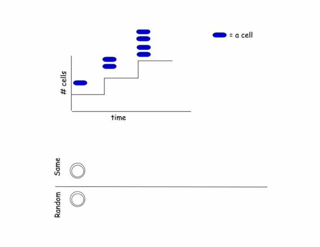
Does DNA
replication begin at
the same site in
every replication
cycle?
Electron microscope image of an E. coli
chromosome being
replicated.
Structure (theta, θ) suggests replication
started in only one place
on this chromosome. Fig. 20.9

Does DNA replication begin at the
same site in every replication cycle?
Experiment:
1. Pulse-label a synchronized cell population during successive rounds of DNA replication with two different isotopes, one that changes the density of newly synthesized DNA (15N), and one that makes it radioactive (32P).
2. DNA is then isolated, sheared, and separated by CsCl density gradient ultra-centrifugation.
3. Radioactivity (32P) in the DNAs of different densities is counted.

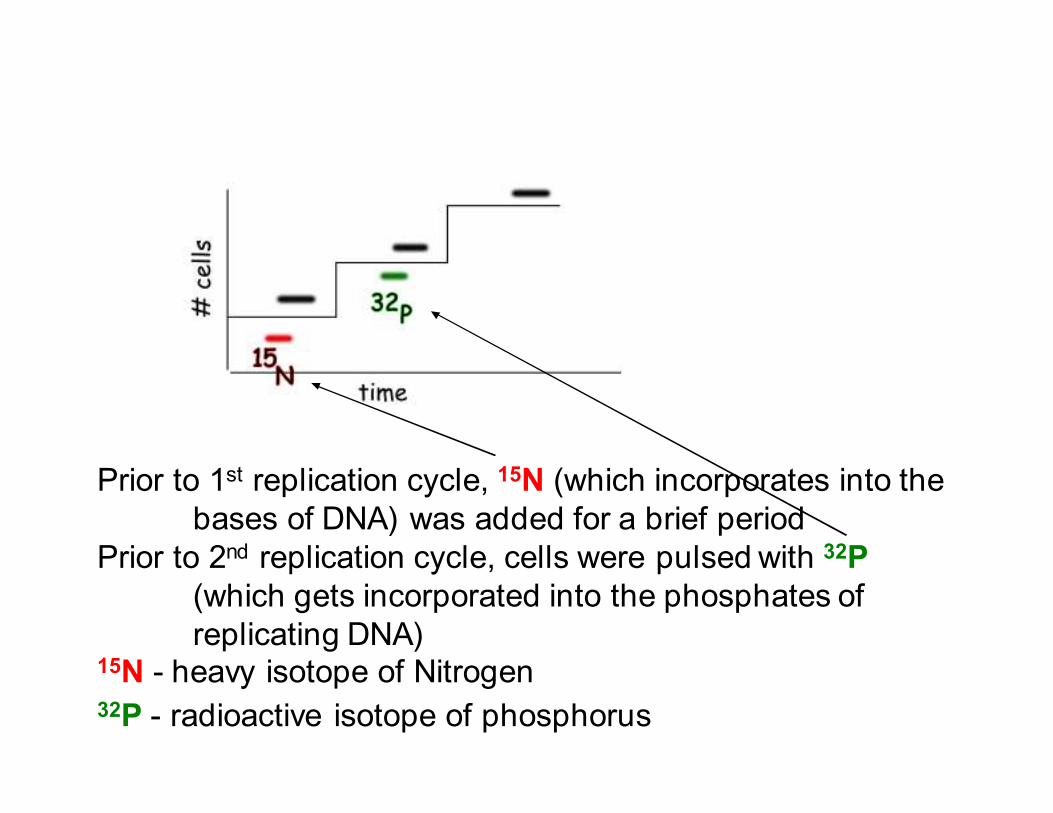
1st
Prior to 1st replication cycle, 15N (which incorporates into the
bases of DNA) was added for a brief period
Prior to 2nd replication cycle, cells were pulsed with 32P
(which gets incorporated into the phosphates of
replicating DNA) 15N - heavy isotope of Nitrogen 32P - radioactive isotope of phosphorus


DNA is isolated,
sheared into
fragments, and
separated by
CsCl-density gradient
centrifugation.


Blow up of the last
2 rows of DNA in
the previous slide
(i.e., labeled DNA,
and labeled, sheared DNA).
Labeled DNA
Labeled,
sheared DNA
Same Origin
Random
Origins

Conclusion:
Replication of bacterial chromosome
starts at the same place every time
Result:
~50% (the most possible) of the
incorporated 32P was in the same
DNA that was shifted by 15N
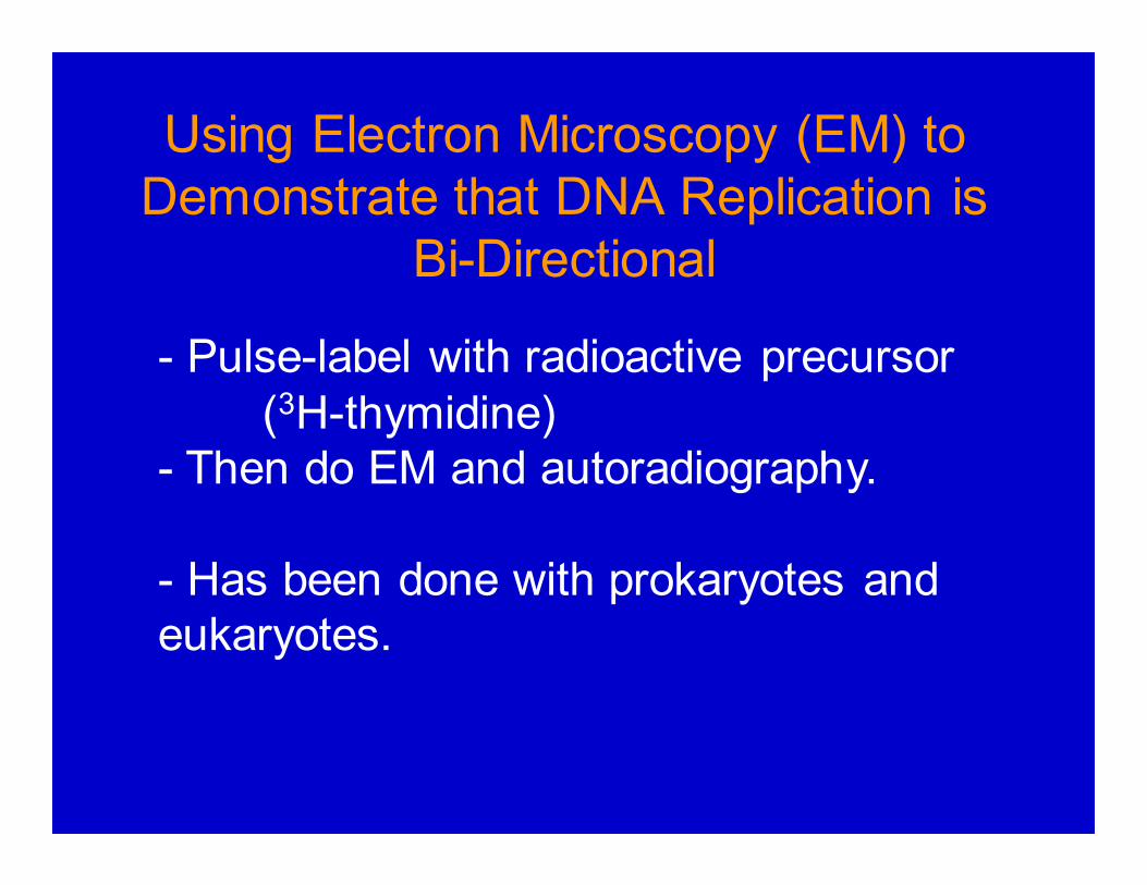
Using Electron Microscopy (EM) to
Demonstrate that DNA Replication is
Bi-Directional
- Pulse-label with radioactive precursor
(3H-thymidine)
- Then do EM and autoradiography.
- Has been done with prokaryotes and
eukaryotes.

Conclusion: eukaryotic origins also replicate bi-
directionally!
Drosophila cells were labeled with a pulse of highly
radioactive precursor, followed by a pulse of lower
radioactive precursor; then replication bubbles were
viewed by EM and autoradiography.
Fig. 20.12 in Weaver

Another way to
see that DNA
replication is
Bi-directional -- Cleave
replicating
SV40 viral DNA
with a
restriction enzyme that
cuts it once.
Similar to Fig. 21.2 in Weaver 4

Organism # of replicons Average
length of
replicon
Velocity of
fork
movement
Escherichia coli (bacteria) 1 4200 kb 50,000bp/min
Saccharomyces cerevisiae(yeast)
500 40 kb 3,600 bp/min
Drosophila melanogaster(fruit fly)
3,500 40 kb 2,600 bp/min
Xenopus laevis (frog) 15,000 200 kb 500 bp/minMus musculus (mouse) 25,000 150 kb 2,200 bp
/minHomo sapiens 10,000 to
100,000Š 300 kb
Replicon - DNA replicated from a single origin
Eukaryotes have many replication origins.
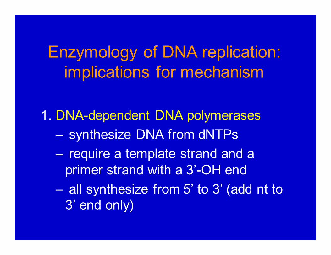
Enzymology of DNA replication:
implications for mechanism
1. DNA-dependent DNA polymerases
– synthesize DNA from dNTPs
– require a template strand and a
primer strand with a 3’-OH end
– all synthesize from 5’ to 3’ (add nt to
3’ end only)

Movie – DNA polymerization
Note: what happens to the P-P?

Comparison of E.coli DNA Polymerases I and III
1 subunit
10 subunits

Proofreading Activity
Insertion of the wrong nucleotide causes the DNA
polymerase to stall, and then the 3’-to-5’ exonuclease
activity removes the mispaired A nt. The polymerase then
continues adding nts to the primer.
Fig. 20.15 in Weaver 4

If DNA polymerases only synthesize 5’ to 3’, how
does the replication fork move directionally?

• Lagging strand synthesized as small
(~100-1000 bp) fragments - “Okazaki
fragments” .
• Okazaki fragments begin as very short 6-
15 nt RNA primers synthesized by primase.
2. Primase - RNA polymerase that
synthesizes the RNA primers (11-12 nt that
start with pppAG) for both lagging and
leading strand synthesis
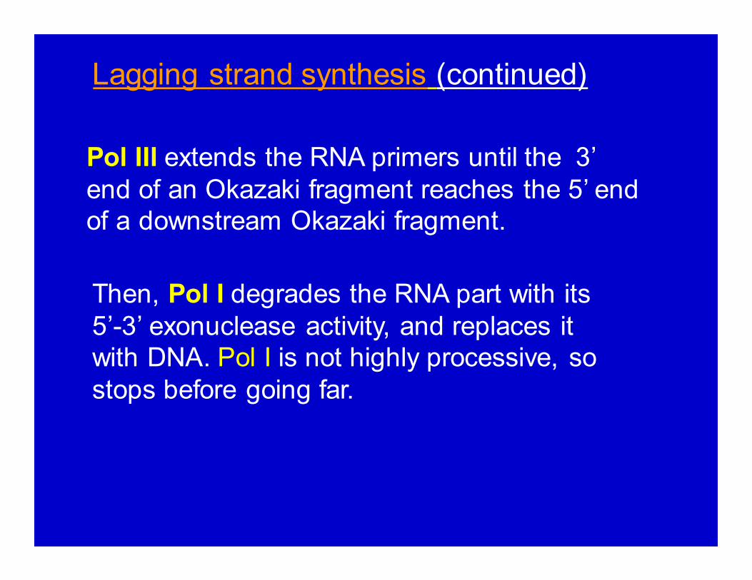
Pol III extends the RNA primers until the 3’
end of an Okazaki fragment reaches the 5’ end
of a downstream Okazaki fragment.
Lagging strand synthesis (continued)
Then, Pol I degrades the RNA part with its
5’-3’ exonuclease activity, and replaces it
with DNA. Pol I is not highly processive, so
stops before going far.

At this stage, Lagging strand is a series of DNA
fragments (without gaps).
Fragments stitched together covalently by
DNA Ligase.
3. DNA Ligase - joins the 5’ phosphate of
one DNA molecule to the 3’ OH of another,
using energy in the form of NAD
(prokaryotes) or ATP (eukaryotes). It
prefers substrates that are double-
stranded, with only one strand needing
ligation, and lacking gaps.

Lig ase w ill jo in the se tw o G--G--A--T--C--C--T--T--G--A--T--C--C
| | | | | | | | | | | | |
C--C--T--A--G G--A--A--C--T--A--G--G
Lig ase w ill NO T jo in thesetwo .
G--G--A--T--C--C--T--T--G--A--T--C--C
| | | | | | | | | | | |
C--C--T--A--G C--A--A--C--T--A--G--G
Lig ase w ill NO T jo in these
two .
G--G--A--T--C--C--T--T--G--A--T--C--C
| | | | | | | | | | | |
C--C--T--A--A G--A--A--C--T--A--G--G
Lig ase w ill NO T jo in thesetwo .
G--G--A--T--C--C--T--T--G--A--T--C--C
| | | | | | | | | | | |
C--C--T--A--G G--T--A--C--T--A--G--G
Lig ase w ill NO T jo in these
two . C--C--T--A--G C--T--A--C--T--A--G--G
DNA Ligase Substrate Specificity

2
1
+ AMP 3'
P AMP
P
AMP +
HO
3' P
5'
Ligase
N A D
1 2
1
3' N M N
HO P
3' 5'
P
Ligase
NAD NMN +AMP
Mechanism of Prokaryotic DNA Ligase
Ligase cleaves NAD and
attaches to AMP.
Ligase-AMP binds and
attaches to 5’ end of DNA #1 via the AMP.
The 3’OH of DNA #2
reacts with the
phosphodiester shown, displacing the AMP-
ligase.
AMP & ligase separate.
(Euk. DNA ligase uses ATP as AMP donor)

Movie - Bidirectional
Replication: Leading and
lagging strand synthesis
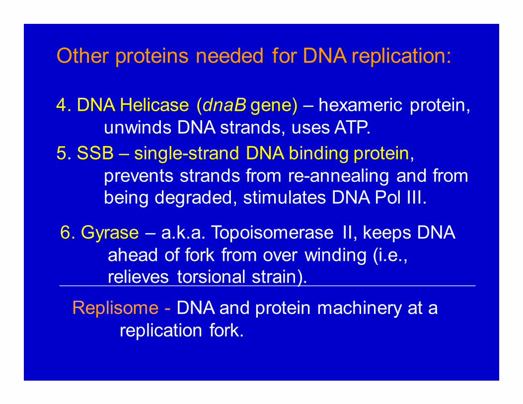
Replisome - DNA and protein machinery at a
replication fork.
Other proteins needed for DNA replication:
4. DNA Helicase (dnaB gene) – hexameric protein,
unwinds DNA strands, uses ATP.
5. SSB – single-strand DNA binding protein,
prevents strands from re-annealing and from
being degraded, stimulates DNA Pol III.
6. Gyrase – a.k.a. Topoisomerase II, keeps DNA
ahead of fork from over winding (i.e.,
relieves torsional strain).

DNA Helicase (dnaB gene) Assay
Fig. 20.21 in Weaver

Helicase – the movie

Replication Causes DNA to
Supercoil

Rubber Band Model
of Supercoiling DNA
DNA Gyrase relaxes positive
supercoils by breaking and
rejoining both DNA strands.


![[PPT]Role of DNA - Elizabeth Rose · Web viewcopyright cmassengale DNA Replication As the 2 DNA strands open at the origin, Replication Bubbles form Prokaryotes (bacteria) have a single](https://img.pdfslide.net/doc/110x75/5aa6232f7f8b9a7c1a8e557b/pptrole-of-dna-elizabeth-rose-viewcopyright-cmassengale-dna-replication-as-the.jpg)