Embed Size (px)
Citation preview
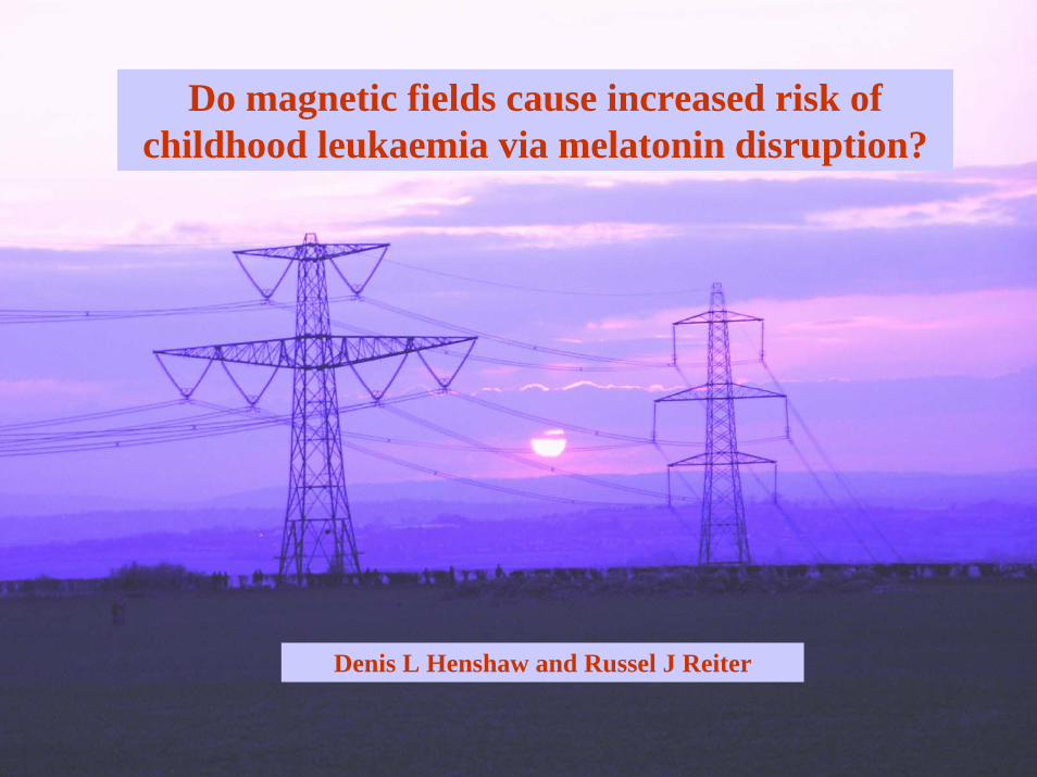
Denis L Henshaw and Russel J Reiter
Do magnetic fields cause increased risk of childhood leukaemia via melatonin disruption?
The authors
Denis L HenshawH. H. Wills Physics Laboratory, University of Bristol
Tyndall AvenueBristol, BS8 1TL, UK
and
Russel J ReiterDepartment of Cellular and Structural Biology
University of Texas Medical Center7703 Floyd Curl Drive
San AntonioTX 78284-7762, USA
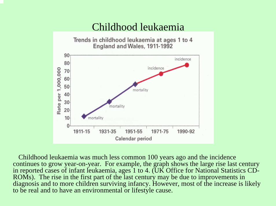
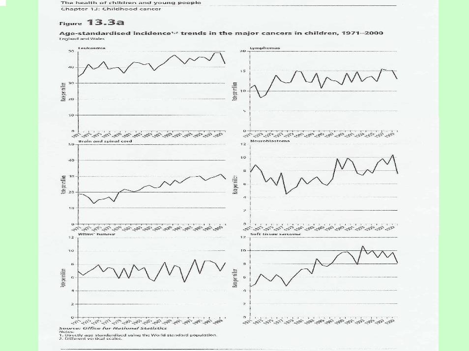
Childhood leukaemia
Childhood leukaemia was much less common 100 years ago and the incidence continues to grow year-on-year. For example, the graph shows the large rise last century in reported cases of infant leukaemia, ages 1 to 4. (UK Office for National Statistics CD-ROMs). The rise in the first part of the last century may be due to improvements in diagnosis and to more children surviving infancy. However, most of the increase is likely to be real and to have an environmental or lifestyle cause.
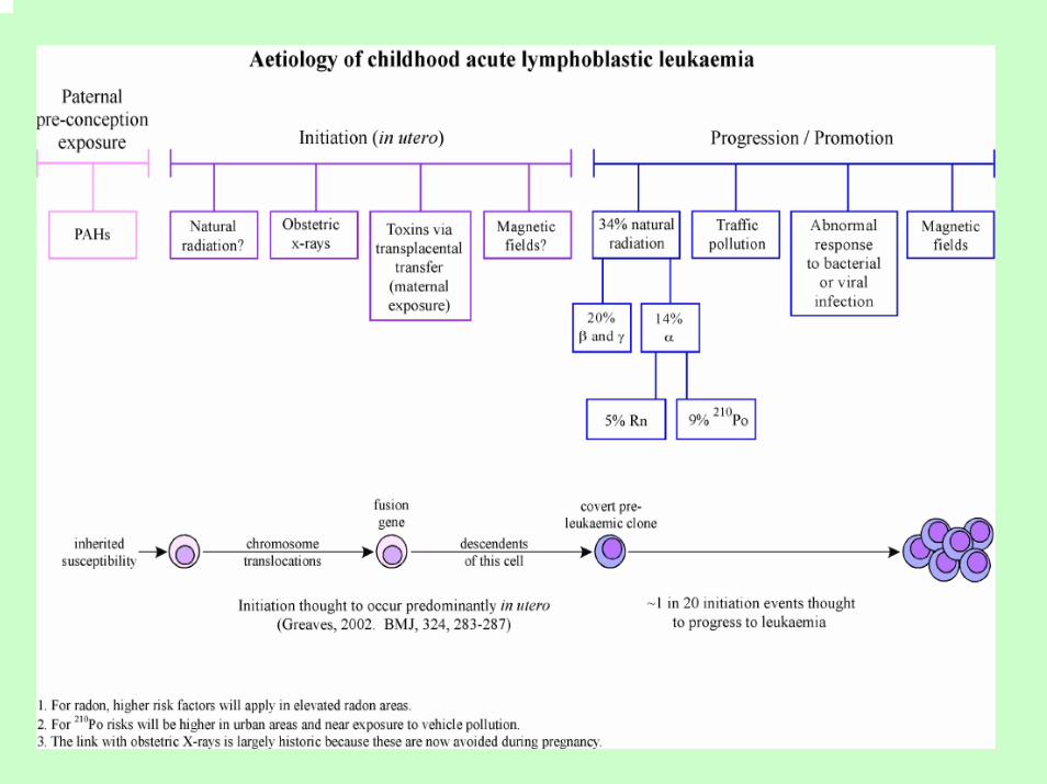
The hypothesis in outline
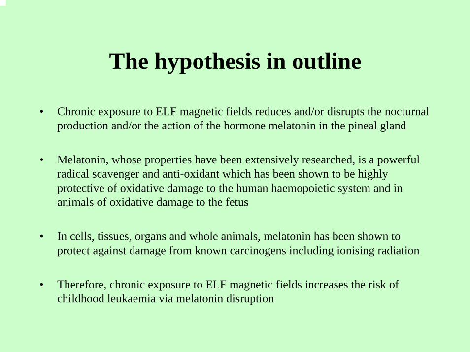
• Chronic exposure to ELF magnetic fields reduces and/or disrupts the nocturnal production and/or the action of the hormone melatonin in the pineal gland
• Melatonin, whose properties have been extensively researched, is a powerful radical scavenger and anti-oxidant which has been shown to be highly protective of oxidative damage to the human haemopoietic system and in animals of oxidative damage to the fetus
• In cells, tissues, organs and whole animals, melatonin has been shown to protect against damage from known carcinogens including ionising radiation
• Therefore, chronic exposure to ELF magnetic fields increases the risk of childhood leukaemia via melatonin disruption
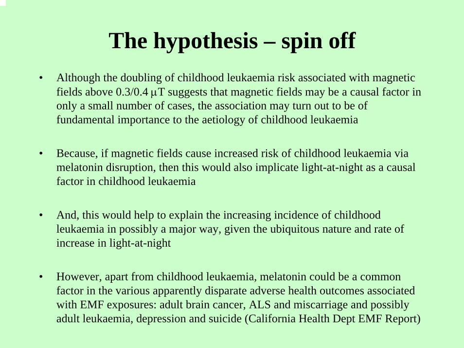
The hypothesis – spin off• Although the doubling of childhood leukaemia risk associated with magnetic
fields above 0.3/0.4 µT suggests that magnetic fields may be a causal factor in only a small number of cases, the association may turn out to be of fundamental importance to the aetiology of childhood leukaemia
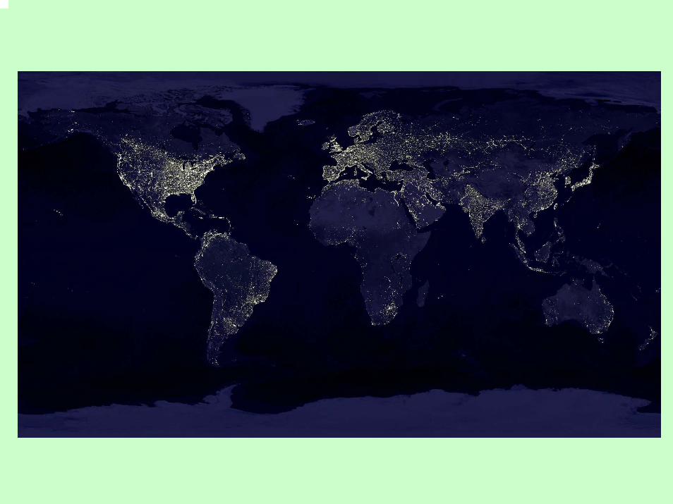
• Because, if magnetic fields cause increased risk of childhood leukaemia via melatonin disruption, then this would also implicate light-at-night as a causal factor in childhood leukaemia
• And, this would help to explain the increasing incidence of childhood leukaemia in possibly a major way, given the ubiquitous nature and rate of increase in light-at-night
• However, apart from childhood leukaemia, melatonin could be a common factor in the various apparently disparate adverse health outcomes associated with EMF exposures: adult brain cancer, ALS and miscarriage and possibly adult leukaemia, depression and suicide (California Health Dept EMF Report)
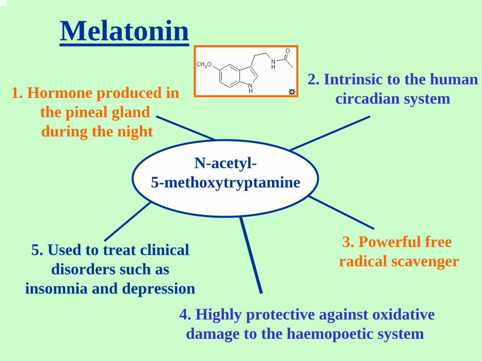
Melatonin
5. Used to treat clinical disorders such as
insomnia and depression
1. Hormone produced in the pineal gland during the night
3. Powerful free radical scavenger
2. Intrinsic to the humancircadian system
4. Highly protective against oxidativedamage to the haemopoetic system
N-acetyl-5-methoxytryptamine
Melatonin continued…
• Among its many properties, melatonin is a powerful radical scavenger and antioxidant, more effective than either vitamins C or E. The hormone has been found to protect nuclear DNA, tissues and organs against oxidative damage from known carcinogens including radiation.
• Melatonin is therefore a powerful natural anti-cancer agent.
• The so-called “Melatonin Hypothesis” has been discussed in relation to the significant rise in incidence of breast cancer in industrialised countries in recent decades (Stevens 1987).
• The risk is said to arise from reduced production of nocturnal melatonin brought about by exposure to light-at-night (LAN) from domestic as well as street lighting and magnetic fields associated with the electricity supply.
Melatonin contd
• Strong support for LAN affecting breast cancer risk has come from experiments in animals. In rats, exposure to constant light leads to rapid development of mammary gland tumours
• Support in humans comes from the observation of reduced hormone-related cancer rates in the blind and partially sighted and increased breast cancer rates in nightshift workers (e.g. Hahn 1991, Feychting et al. 1998, Hansen 2001)
Reduced cancer rates in the blind and partially sighted
• Hahn, R. A., 1991. Profound bilateral blindness and the incidence of breast cancer. Epidemiology, 2, 208-210.
• Feychting, M. Österlund, B. and Ahlbom, A., 1998. Reduced cancer incidence among the blind. Epidemiology, 9, 490-494.
• Verkasalo, P. K., Pukkala, E., Stevens, R. G., Ojamo, M. and Rudanko, S-L., 1999. Inverse association between breast cancer incidence and degree of visual impairment in Finland. British Journal of Cancer, 80 (9), 1459-1460.
• Hansen, J., 2001. Light at Night, Shiftwork, and Breast Cancer Risk. Journal of the National Cancer Institute, 93 (No. 20), 1513-1515.
• Swerdlow, A., 2003. Shift work and breast cancer: A critical review of the epidemiological evidence. Research Report 132, ISBN 071762708X, HSE Books.
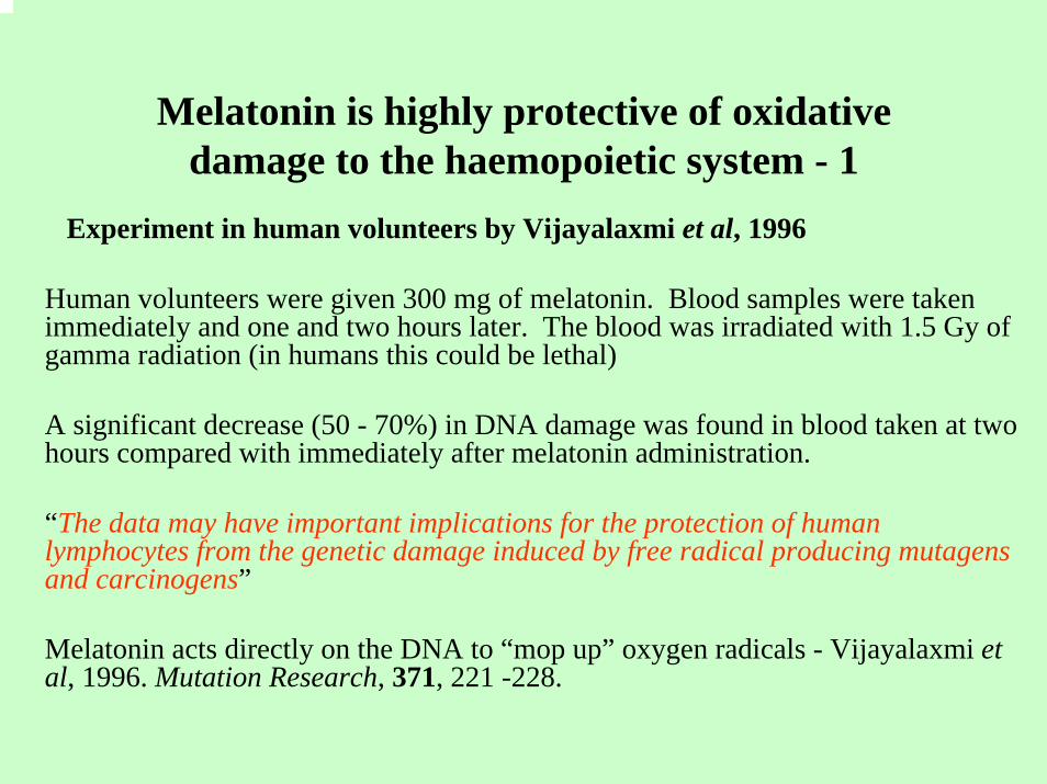
Melatonin is highly protective of oxidative damage to the haemopoietic system - 1
Experiment in human volunteers by Vijayalaxmi et al, 1996
Human volunteers were given 300 mg of melatonin. Blood samples were taken immediately and one and two hours later. The blood was irradiated with 1.5 Gy of gamma radiation (in humans this could be lethal)
A significant decrease (50 - 70%) in DNA damage was found in blood taken at two hours compared with immediately after melatonin administration.
“The data may have important implications for the protection of human lymphocytes from the genetic damage induced by free radical producing mutagens and carcinogens”
Melatonin acts directly on the DNA to “mop up” oxygen radicals - Vijayalaxmi et al, 1996. Mutation Research, 371, 221 -228.
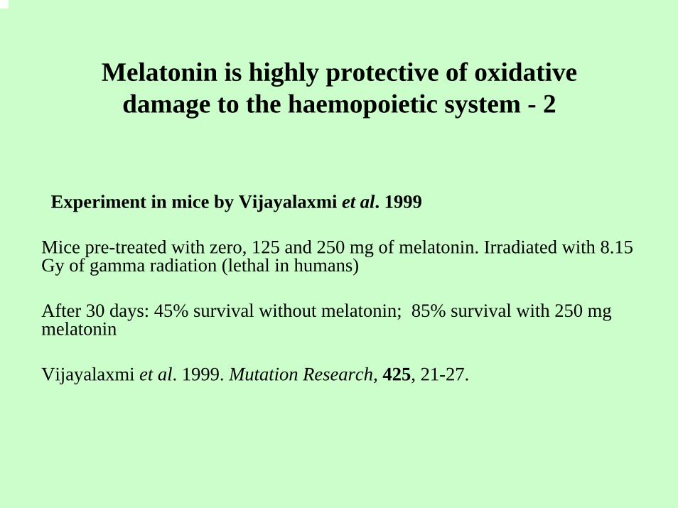
Melatonin is highly protective of oxidative damage to the haemopoietic system - 2
Experiment in mice by Vijayalaxmi et al. 1999
Mice pre-treated with zero, 125 and 250 mg of melatonin. Irradiated with 8.15 Gy of gamma radiation (lethal in humans)
After 30 days: 45% survival without melatonin; 85% survival with 250 mg melatonin
Vijayalaxmi et al. 1999. Mutation Research, 425, 21-27.
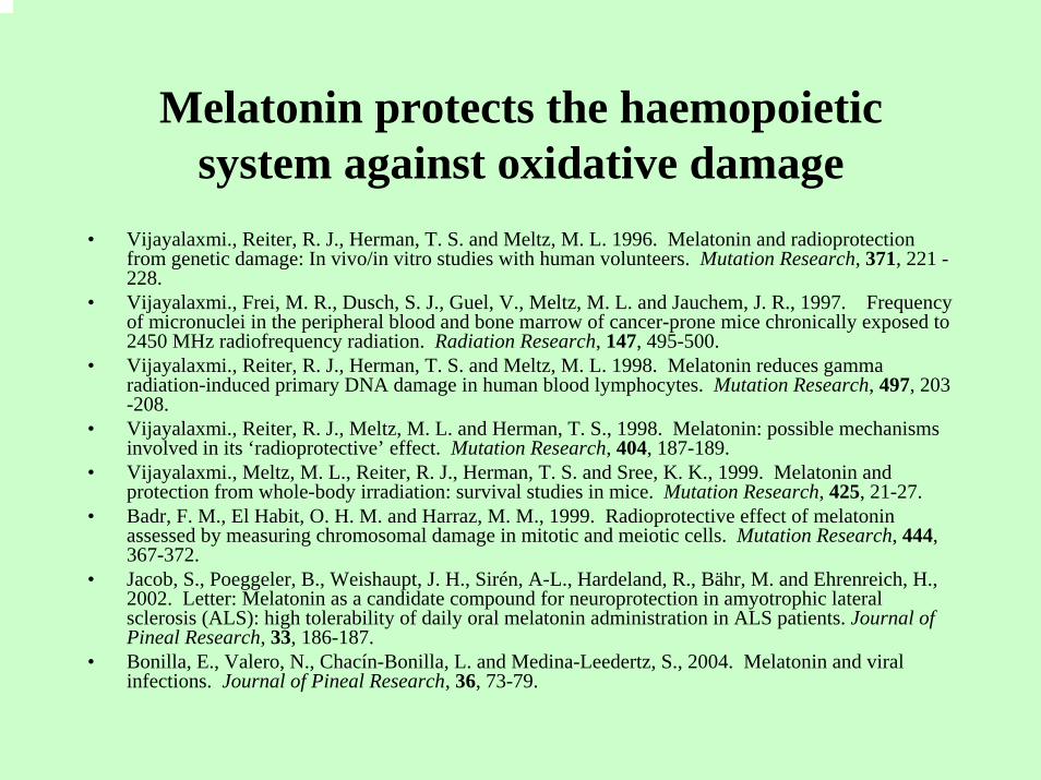
Melatonin protects the haemopoietic system against oxidative damage
• Vijayalaxmi., Reiter, R. J., Herman, T. S. and Meltz, M. L. 1996. Melatonin and radioprotection from genetic damage: In vivo/in vitro studies with human volunteers. Mutation Research, 371, 221 -228.
• Vijayalaxmi., Frei, M. R., Dusch, S. J., Guel, V., Meltz, M. L. and Jauchem, J. R., 1997. Frequency of micronuclei in the peripheral blood and bone marrow of cancer-prone mice chronically exposed to 2450 MHz radiofrequency radiation. Radiation Research, 147, 495-500.
• Vijayalaxmi., Reiter, R. J., Herman, T. S. and Meltz, M. L. 1998. Melatonin reduces gamma radiation-induced primary DNA damage in human blood lymphocytes. Mutation Research, 497, 203 -208.
• Vijayalaxmi., Reiter, R. J., Meltz, M. L. and Herman, T. S., 1998. Melatonin: possible mechanismsinvolved in its ‘radioprotective’ effect. Mutation Research, 404, 187-189.
• Vijayalaxmi., Meltz, M. L., Reiter, R. J., Herman, T. S. and Sree, K. K., 1999. Melatonin and protection from whole-body irradiation: survival studies in mice. Mutation Research, 425, 21-27.
• Badr, F. M., El Habit, O. H. M. and Harraz, M. M., 1999. Radioprotective effect of melatonin assessed by measuring chromosomal damage in mitotic and meiotic cells. Mutation Research, 444, 367-372.
• Jacob, S., Poeggeler, B., Weishaupt, J. H., Sirén, A-L., Hardeland, R., Bähr, M. and Ehrenreich, H., 2002. Letter: Melatonin as a candidate compound for neuroprotection in amyotrophic lateral sclerosis (ALS): high tolerability of daily oral melatonin administration in ALS patients. Journal of Pineal Research, 33, 186-187.
• Bonilla, E., Valero, N., Chacín-Bonilla, L. and Medina-Leedertz, S., 2004. Melatonin and viral infections. Journal of Pineal Research, 36, 73-79.
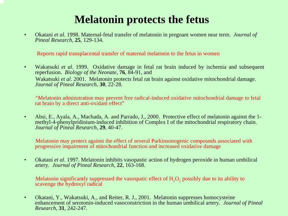
Melatonin protects the fetus• Okatani et al. 1998. Maternal-fetal transfer of melatonin in pregnant women near term. Journal of
Pineal Research, 25, 129-134.
Reports rapid transplacental transfer of maternal melatonin to the fetus in women
• Wakatsuki et al. 1999. Oxidative damage in fetal rat brain induced by ischemia and subsequent reperfusion. Biology of the Neonate, 76, 84-91, and Wakatsuki et al. 2001. Melatonin protects fetal rat brain against oxidative mitochondrial damage. Journal of Pineal Research, 30, 22-28.
“Melatonin adminstration may prevent free radical-induced oxidative mitochondrial damage to fetal rat brain by a direct anti-oxidant effect”
• Absi, E., Ayala, A., Machada, A. and Parrado, J., 2000. Protective effect of melatonin against the 1-methyl-4-phenylpridinium-induced inhibition of Complex I of the mitochondrial respiratory chain. Journal of Pineal Research, 29, 40-47.
Melatonin may protect against the effect of several Parkinsonogenic compounds associated with progressive impairment of mitochondrial function and increased oxidative damage
• Okatani et al. 1997. Melatonin inhibits vasopastic action of hydrogen peroxide in human umbilical artery. Journal of Pineal Research, 22, 163-168.
Melatonin significantly suppressed the vasospatic effect of H2O2 possibly due to its ability to scavenge the hydroxyl radical
• Okatani, Y., Wakatsuki, A., and Reiter, R. J., 2001. Melatonin suppresses homocysteineenhancement of serotonin-induced vasoconstriction in the human umbilical artery. Journal of Pineal Research, 31, 242-247.
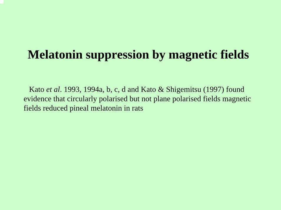
Melatonin suppression by magnetic fields
Kato et al. 1993, 1994a, b, c, d and Kato & Shigemitsu (1997) found evidence that circularly polarised but not plane polarised fields magnetic fields reduced pineal melatonin in rats
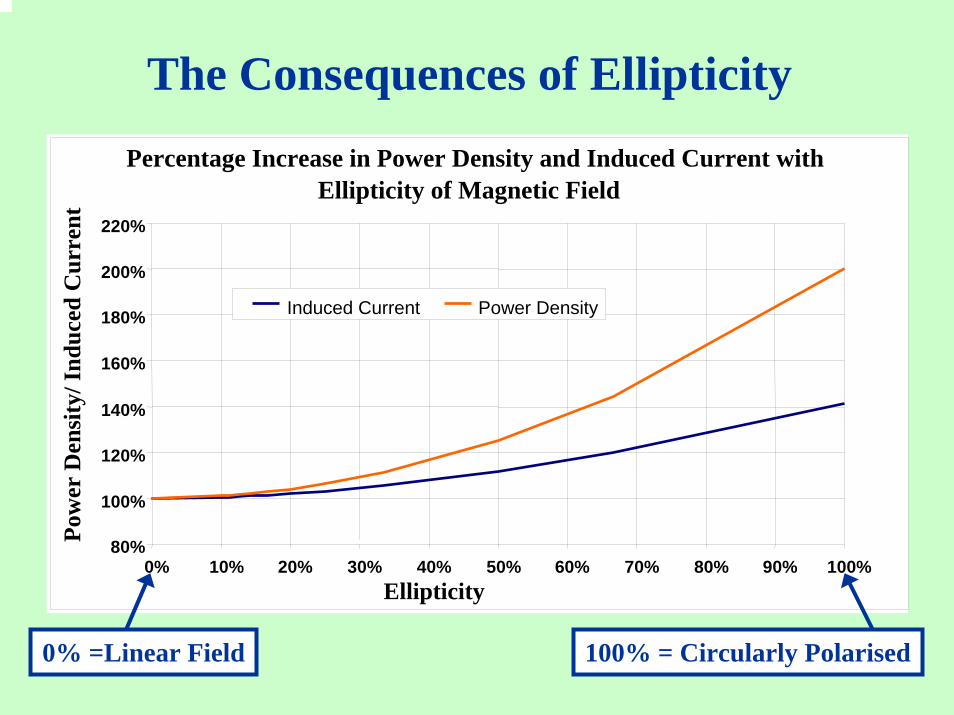
The Consequences of EllipticityPercentage Increase in Power Density and Induced Current with
Ellipticity of Magnetic Field
80%
100%
120%
140%
160%
180%
200%
220%
0% 10% 20% 30% 40% 50% 60% 70% 80% 90% 100%Ellipticity
Pow
er D
ensi
ty/ I
nduc
ed C
urre
nt
Induced Current Power Density
100% = Circularly Polarised0% =Linear Field
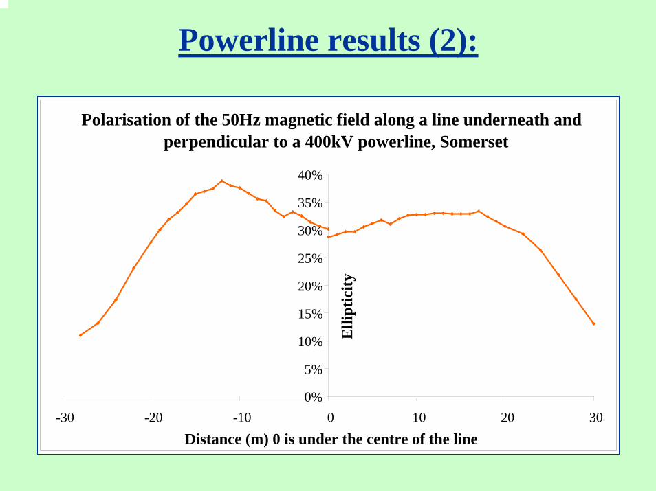
Powerline results (2):
Polarisation of the 50Hz magnetic field along a line underneath and perpendicular to a 400kV powerline, Somerset
0%
5%
10%
15%
20%
25%
30%
35%
40%
-30 -20 -10 0 10 20 30Distance (m) 0 is under the centre of the line
Elli
ptic
ity
Melatonin suppression by magnetic fieldsI – short term volunteer exposures
• There have been a number of studies of effects of magnetic fields on pineal melatonin production in volunteers exposed for relatively short periods.Melatonin levels in the body are assayed either from blood samples (serum melatonin) and/or from the melatonin metabolite 9-hydroxymelatonin sulphate (6-OHMS) in urine
• These volunteer studies have at best provided possible evidence of pineal melatonin suppression only after several days exposure
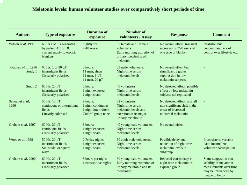
Melatonin levels: human volunteer studies over comparatively short periods of time
Authors Type of exposure Duration of exposure
Number of volunteers / Assay Response Comment
Wilson et al, 1990 60 Hz EMF’s generated by pulsed AC or DC current supply to electric blankets
nightly for7-10 weeks
32 female and 10 male volunteers. Early morning excretion of urinary metabolite of melatonin
No overall effect: transient increases in 7/28 users of one type of blanket
Realistic, but concomitant lack of control over lifestyle etc.
Graham et al, 1996Study 1
60 Hz, 1 or 20 µT intermittent fieldsCircularly polarised
8 hours:11 men, sham11 men, 1 µT11 men, 20 µT
33 male volunteers. Night-time serum melatonin levels
No overall effect but significantly grater suppression in low melatonin subjects.
-
Study 2 60 Hz, 20 µTintermittent fieldsCircularly polarised
8 hours:1 night exposed1 night sham
40 volunteers.Night-time serum melatonin levels.
No detected effect: possible effect on low melatonin subjects not replicated
Selmaoui et al,1996
50 Hz, 10 µT continuous or intermittent fieldsLinearly polarised
9 hours:1 night continuous1 night intermittentControl group none
32 volunteers. Night-time serum melatonin levels and excretion of its major urinary metabolite
No detected effect: a small non-significant shift in the onset of increased nocturnal melatonin
-
Graham et al, 1997 60 Hz, 20 µTcontinuous fields Circularly polarised
8 hours: 1 night exposed1 night sham
40 young male volunteers. Night-time serum melatonin levels
No overall effect -
Wood et al, 1998 50 Hz, 20 µTintermittent fieldsSinusoidal or square wave
2 Friday nights:1 night exposed1 night sham
30 adult male volunteers. Night-time serum melatonin levels
Possible delay and reduction of night-time melatonin levels in subgroup
Inconsistent, variable data: incomplete volunteer participation
Graham et al, 2000 60 Hz, 20 µT intermittent fieldsCircularly polarised
8 hours per night:4 consecutive nights
30 young male volunteers. Early morning excretion of urinary melatonin and its metabolite
Reduced consistency in night time melatonin in exposed group
Some suggestion that stability of melatonin measurements over time may be influenced by magnetic fields
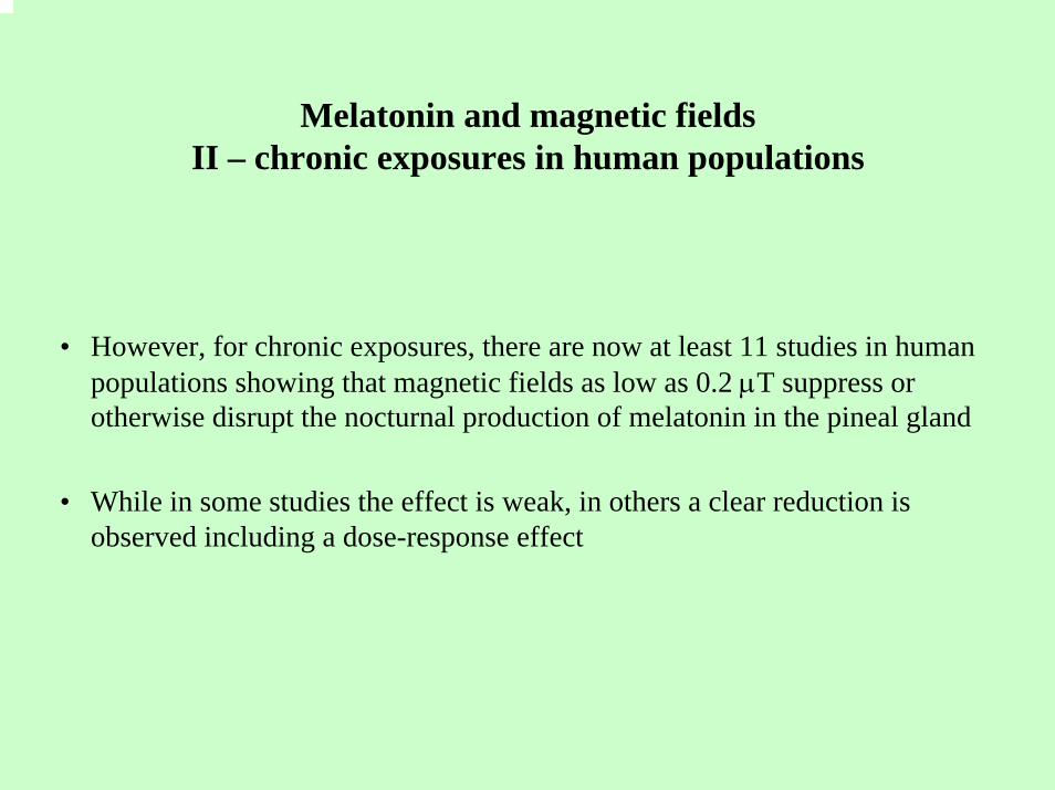
Melatonin and magnetic fieldsII – chronic exposures in human populations
• However, for chronic exposures, there are now at least 11 studies in human populations showing that magnetic fields as low as 0.2 µT suppress or otherwise disrupt the nocturnal production of melatonin in the pineal gland
• While in some studies the effect is weak, in others a clear reduction is observed including a dose-response effect
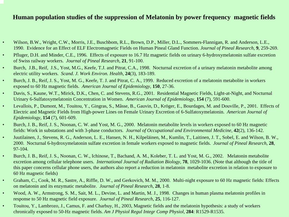
Human population studies of the suppression of Melatonin by power frequency magnetic fields
• Wilson, B.W., Wright, C.W., Morris, J.E., Buschbom, R.L., Brown, D.P., Miller, D.L., Sommers-Flannigan, R. and Anderson, L.E., 1990. Evidence for an Effect of ELF Electromagnetic Fields on Human Pineal Gland Function. Journal of Pineal Research, 9, 259-269.
• Pfluger, D.H. and Minder, C.E., 1996. Effects of exposure to 16.7 Hz magnetic fields on urinary 6-hydroxymelatonin sulfate excretion of Swiss railway workers. Journal of Pineal Research, 21, 91-100.
• Burch, J.B., Reif, J.S., Yost, M.G., Keefe, T.J. and Pitrat, C.A., 1998. Nocturnal excretion of a urinary melatonin metabolite among electric utility workers. Scand. J. Work Environ. Health, 24(3), 183-189.
• Burch, J. B., Reif, J. S., Yost, M. G., Keefe, T. J. and Pitrat, C. A., 1999. Reduced excretion of a melatonin metabolite in workers exposed to 60 Hz magnetic fields. American Journal of Epidemiology, 150, 27-36.
• Davis, S., Kaune, W.T., Mirick, D.K., Chen, C. and Stevens, R.G., 2001. Residential Magnetic Fields, Light-at-Night, and Nocturnal Urinary 6-Sulfatoxymelatonin Concentration in Women. American Journal of Epidemiology, 154 (7), 591-600.
• Levallois, P., Dumont, M., Touitou, Y., Gingras, S., Mâsse, B., Gauvin, D., Kröger, E., Bourdages, M. and Douville, P., 2001. Effects of Electric and Magnetic Fields from High-power Lines on Female Urinary Excretion of 6-Sulfatoxymelatonin. American Journal of Epidemiology, 154 (7), 601-609.
• Burch, J. B., Reif, J. S., Noonan, C. W. and Yost, M. G., 2000. Melatonin metabolite levels in workers exposed to 60 Hz magnetic fields: Work in substations and with 3-phase conductors. Journal of Occupational and Environmental Medicine, 42(2), 136-142.
• Juutilainen, J., Stevens, R. G., Anderson, L. E., Hansen, N. H., Kilpeläinen, M., Kumlin, T., Laitinen, J. T., Sobel, E. and Wilson, B. W., 2000. Nocturnal 6-hydroxymelatonin sulfate excretion in female workers exposed to magnetic fields. Journal of Pineal Research, 28, 97-104.
• Burch, J. B., Reif, J. S., Noonan, C. W., Ichinose, T., Bachand, A. M., Koleber, T. L. and Yost, M. G., 2002. Melatonin metabolite excretion among cellular telephone users. International Journal of Radiation Biology, 78, 1029-1036. (Note that although the title of this paper concerns cellular phone users, the authors also report a reduction in melatonin metabolite excretion in relation to exposure to 60 Hz magnetic fields)
• Graham, C., Cook, M. R., Sastre, A., Riffle, D. W., and Gerkovich, M. M., 2000. Multi-night exposure to 60 Hz magnetic fields: Effects on melatonin and its enzymatic metabolite. Journal of Pineal Research, 28, 1-8.
• Wood, A. W., Armstrong, S. M., Sait, M. L., Devine, L. and Martin, M. J., 1998. Changes in human plasma melatonin profiles in response to 50 Hz magnetic field exposure. Journal of Pineal Research, 25, 116-127.
• Touitou, Y., Lambrozo, J., Camus, F. and Charbuy, H., 2003, Magnetic fields and the melatonin hypothesis: a study of workers chronically exposed to 50-Hz magnetic fields. Am J Physiol Regul Integr Comp Physiol, 284: R1529-R1535.
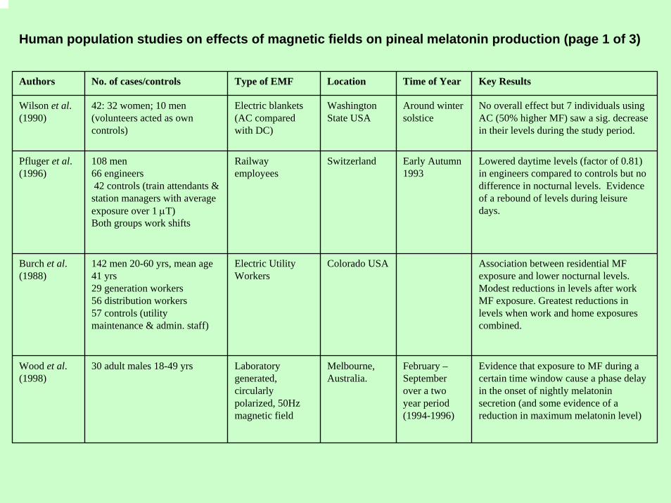
Human population studies on effects of magnetic fields on pineal melatonin production (page 1 of 3)
Authors No. of cases/controls Type of EMF Location Time of Year Key Results
Wilson et al. (1990)
42: 32 women; 10 men(volunteers acted as own controls)
Electric blankets (AC compared with DC)
Washington State USA
Around winter solstice
No overall effect but 7 individuals using AC (50% higher MF) saw a sig. decrease in their levels during the study period.
Pfluger et al. (1996)
108 men66 engineers42 controls (train attendants &
station managers with average exposure over 1 µT)Both groups work shifts
Railway employees
Switzerland Early Autumn 1993
Lowered daytime levels (factor of 0.81) in engineers compared to controls but no difference in nocturnal levels. Evidence of a rebound of levels during leisure days.
Burch et al. (1988)
142 men 20-60 yrs, mean age 41 yrs29 generation workers56 distribution workers57 controls (utility maintenance & admin. staff)
Electric Utility Workers
Colorado USA Association between residential MF exposure and lower nocturnal levels. Modest reductions in levels after work MF exposure. Greatest reductions in levels when work and home exposures combined.
Wood et al. (1998)
30 adult males 18-49 yrs Laboratory generated, circularly polarized, 50Hz magnetic field
Melbourne, Australia.
February –September over a two year period (1994-1996)
Evidence that exposure to MF during a certain time window cause a phase delay in the onset of nightly melatonin secretion (and some evidence of a reduction in maximum melatonin level)
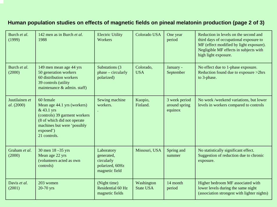
Human population studies on effects of magnetic fields on pineal melatonin production (page 2 of 3)
Burch et al. (1999)
142 men as in Burch et al. 1988
Electric Utility Workers
Colorado USA One year period
Reduction in levels on the second and third days of occupational exposure to MF (effect modified by light exposure). Negligible MF effects in subjects with high light exposure.
Burch et al. (2000)
149 men mean age 44 yrs50 generation workers60 distribution workers39 controls (utility maintenance & admin. staff)
Substations (3 phase – circularly polarized)
Colorado, USA
January -September
No effect due to 1-phase exposure. Reduction found due to exposure >2hrs to 3-phase.
Juutilainen et al. (2000)
60 femaleMean age 44.1 yrs (workers)& 43.1 yrs(controls) 39 garment workers (8 of which did not operate machines but were ‘possibly exposed’)21 controls.
Sewing machine workers..
Kuopio, Finland.
3 week period around spring equinox
No week /weekend variations, but lower levels in workers compared to controls
Graham et al. (2000)
30 men 18 –35 yrsMean age 22 yrs(volunteers acted as own controls)
Laboratory generated, circularly polarized, 60Hz magnetic field
Missouri, USA Spring and summer
No statistically significant effect.Suggestion of reduction due to chronic exposure.
Davis et al. (2001)
203 women 20-70 yrs
(Night time) Residential 60 Hz magnetic fields
Washington State USA
14 month period
Higher bedroom MF associated with lower levels during the same night (association strongest with lighter nights)
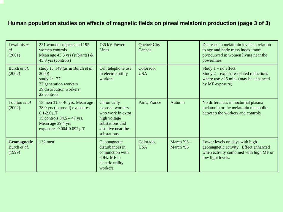
Human population studies on effects of magnetic fields on pineal melatonin production (page 3 of 3)
Levallois et al.(2001)
221 women subjects and 195 women controlsMean age 45.5 yrs (subjects) &45.8 yrs (controls)
735 kV Power Lines
Quebec City Canada.
Decrease in melatonin levels in relation to age and body mass index, more pronounced in women living near the powerlines.
Burch et al. (2002)
study 1: 149 (as in Burch et al. 2000)study 2: 7722 generation workers29 distribution workers23 controls
Cell telephone use in electric utility workers
Colorado, USA
Study 1 – no effect.Study 2 – exposure-related reductions where use >25 mins (may be enhanced by MF exposure)
Touitou et al (2002).
15 men 31.5- 46 yrs. Mean age 38.0 yrs (exposed) exposures 0.1-2.6 µT15 controls 34.5 – 47 yrs. Mean age 39.4 yrsexposures 0.004-0.092 µT
Chronically exposed workers who work in extra high voltage substations and also live near the substations
Paris, France Autumn No differences in nocturnal plasma melatonin or the melatonin metabolite between the workers and controls.
GeomagneticBurch et al. (1999)
132 men Geomagnetic disturbances in conjunction with 60Hz MF in electric utility workers
Colorado, USA
March ’95 –March ‘96
Lower levels on days with high geomagnetic activity. Effect enhanced when activity combined with high MF or low light levels.
Inhibition of the action of melatonin
• Experiments in vitro have shown that magnetic fields as low as 1.2 µT inhibit the ability of melatonin to suppress the growth of MCF-7 breast cancer cells at physiologically melatonin concentrations 10-11 to 10-9 molar (Ishido et al. 2001 and four other independent studies)
• Magnetic fields as low as 1.2 µT also suppress the anti-proliferative action Tamoxifen (Harland & Liburdy 1997, Harland et al. 1999, Blackman et al. 2001)
• Reiter (1998) pointed out that the role of the retina in coupling magnetic fields to the pineal was unproven. He pointed out that while it was assumed that melatonin synthesis is suppressed by magnetic fields, the reduction in serum melatonin could be a result of an increased uptake and utilisation as a free radical scavenger
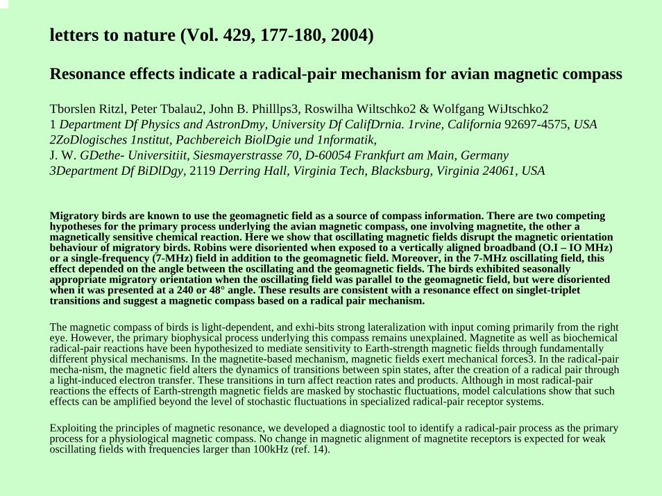
letters to nature (Vol. 429, 177-180, 2004)
Resonance effects indicate a radical-pair mechanism for avian magnetic compass
Tborslen Ritzl, Peter Tbalau2, John B. Philllps3, Roswilha Wiltschko2 & Wolfgang WiJtschko21 Department Df Physics and AstronDmy, University Df CalifDrnia. 1rvine, California 92697-4575, USA2ZoDlogisches 1nstitut, Pachbereich BiolDgie und 1nformatik,J. W. GDethe- Universitiit, Siesmayerstrasse 70, D-60054 Frankfurt am Main, Germany3Department Df BiDlDgy, 2119 Derring Hall, Virginia Tech, Blacksburg, Virginia 24061, USA
Migratory birds are known to use the geomagnetic field as a source of compass information. There are two competing hypotheses for the primary process underlying the avian magnetic compass, one involving magnetite, the other a magnetically sensitive chemical reaction. Here we show that oscillating magnetic fields disrupt the magnetic orientation behaviour of migratory birds. Robins were disoriented when exposed to a vertically aligned broadband (O.I – IO MHz) or a single-frequency (7-MHz) field in addition to the geomagnetic field. Moreover, in the 7-MHz oscillating field, this effect depended on the angle between the oscillating and the geomagnetic fields. The birds exhibited seasonally appropriate migratory orientation when the oscillating field was parallel to the geomagnetic field, but were disoriented when it was presented at a 240 or 48° angle. These results are consistent with a resonance effect on singlet-triplet transitions and suggest a magnetic compass based on a radical pair mechanism.
The magnetic compass of birds is light-dependent, and exhi-bits strong lateralization with input coming primarily from the right eye. However, the primary biophysical process underlying this compass remains unexplained. Magnetite as well as biochemical radical-pair reactions have been hypothesized to mediate sensitivity to Earth-strength magnetic fields through fundamentally different physical mechanisms. In the magnetite-based mechanism, magnetic fields exert mechanical forces3. In the radical-pair mecha-nism, the magnetic field alters the dynamics of transitions between spin states, after the creation of a radical pair through a light-induced electron transfer. These transitions in turn affect reaction rates and products. Although in most radical-pair reactions the effects of Earth-strength magnetic fields are masked by stochastic fluctuations, model calculations show that such effects can be amplified beyond the level of stochastic fluctuations in specialized radical-pair receptor systems.
Exploiting the principles of magnetic resonance, we developed a diagnostic tool to identify a radical-pair process as the primary process for a physiological magnetic compass. No change in magnetic alignment of magnetite receptors is expected for weak oscillating fields with frequencies larger than 100kHz (ref. 14).
Testing the hypotheses
• More carefully designed studies of pineal melatonin disruption in human populations chronically exposed to ELF (and indeed RF) magnetic fields/EMF
• Special attention should be given to polarised fields
• Studies of the protectiveness of melatonin on haemopoietic stem cells in vivo in the presence of ELF magnetic fields – cf the experiments of Ishido et al. (2001) and others
• Studies of the effects of melatonin disruption on human fetal development
• Does melatonin disruption affect the risk of miscarriage?
Summary
• Hypothesis: Exposure to ELF magnetic fields increases the risk of childhood leukaemia via melatonin disruption
• If this hypothesis is correct this would also implicate light-at-night as a causal factor in childhood leukaemia
• This would help to explain the increasing incidence of childhoodleukaemia in possibly a major way, given the ubiquitous nature and rate of increase in light-at-night
• However, apart from childhood leukaemia, melatonin could be a common factor in the various apparently disparate adverse healthoutcomes associated with EMF exposures: adult brain cancer, ALS and miscarriage and possibly adult leukaemia, depression and suicide(California Health Dept EMF Report, June 2002)
Acknowledgements
The research at Bristol University is supported by
CHILDREN with LEUKAEMIA and the Department of Health