Embed Size (px)
Citation preview
E4 I CUTIS® WWW.MDEDGE.COM/DERMATOLOGY
CASE LETTER
To the Editor:Sweet syndrome (SS), also known as acute febrile neutro-philic dermatosis, is an uncommon inflammatory skin dis-order characterized by sudden onset of fever, leukocytosis, neutrophilia, and tender erythematous papules or plaques or both. Skin biopsy usually reveals extensive infiltration of neutrophils into the epidermis and dermis.1-3 Although rare, cases of eosinophil-rich SS have been reported in patients with drug-induced and malignancy-associated SS.4,5 We report a case of a patient with classical SS with dermal eosinophilic infiltration.
An 80-year-old Hispanic man presented with abrupt onset of a rash on the posterior scalp, left ear, back, and hands of 5 days’ duration. The lesions were painful and had progressed to the point of impairing hand grip. The patient’s medical history included a reported common cold the week prior, hyperlipidemia, and hypertension, for which he took metoprolol, simvastatin, aspirin, and clopi-dogrel. He denied oral lesions and medication changes. He was afebrile and did not experience dietary changes, weight loss, or fatigue. He recently returned from travel to the Dominican Republic.
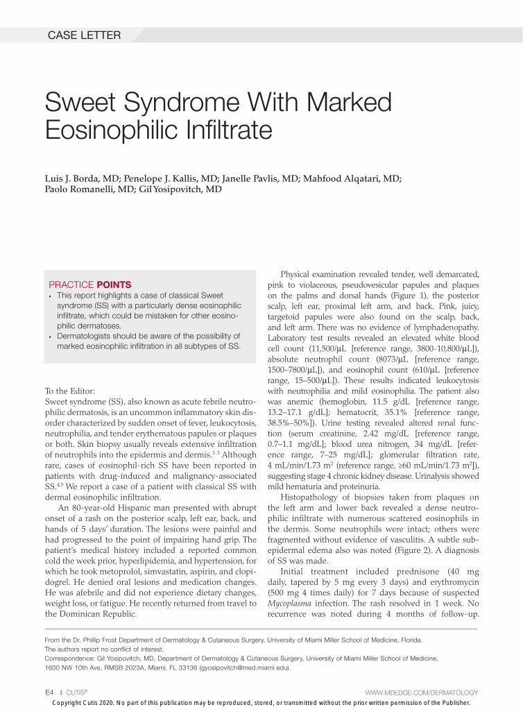
Physical examination revealed tender, well demarcated, pink to violaceous, pseudovesicular papules and plaques on the palms and dorsal hands (Figure 1), the posterior scalp, left ear, proximal left arm, and back. Pink, juicy, targetoid papules were also found on the scalp, back, and left arm. There was no evidence of lymphadenopathy. Laboratory test results revealed an elevated white blood cell count (11,500/µL [reference range, 3800-10,800/µL]), absolute neutrophil count (8073/µL [reference range, 1500–7800/µL]), and eosinophil count (610/µL [reference range, 15–500/µL]). These results indicated leukocytosis with neutrophilia and mild eosinophilia. The patient also was anemic (hemoglobin, 11.5 g/dL [reference range, 13.2–17.1 g/dL]; hematocrit, 35.1% [reference range, 38.5%–50%]). Urine testing revealed altered renal func-tion (serum creatinine, 2.42 mg/dL [reference range, 0.7–1.1 mg/dL]; blood urea nitrogen, 34 mg/dL [refer-ence range, 7–25 mg/dL]; glomerular filtration rate, 4 mL/min/1.73 m2 (reference range, ≥60 mL/min/1.73 m2]), suggesting stage 4 chronic kidney disease. Urinalysis showed mild hematuria and proteinuria.
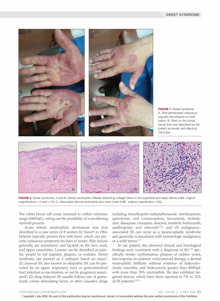
Histopathology of biopsies taken from plaques on the left arm and lower back revealed a dense neutro-philic infiltrate with numerous scattered eosinophils in the dermis. Some neutrophils were intact; others were fragmented without evidence of vasculitis. A subtle sub-epidermal edema also was noted (Figure 2). A diagnosis of SS was made.
Initial treatment included prednisone (40 mg daily, tapered by 5 mg every 3 days) and erythromycin (500 mg 4 times daily) for 7 days because of suspected Mycoplasma infection. The rash resolved in 1 week. No recurrence was noted during 4 months of follow-up.
Sweet Syndrome With Marked Eosinophilic Infiltrate
Luis J. Borda, MD; Penelope J. Kallis, MD; Janelle Pavlis, MD; Mahfood Alqatari, MD; Paolo Romanelli, MD; Gil Yosipovitch, MD
From the Dr. Phillip Frost Department of Dermatology & Cutaneous Surgery, University of Miami Miller School of Medicine, Florida.The authors report no conflict of interest.Correspondence: Gil Yosipovitch, MD, Department of Dermatology & Cutaneous Surgery, University of Miami Miller School of Medicine, 1600 NW 10th Ave, RMSB 2023A, Miami, FL 33136 ([email protected]).
PRACTICE POINTS• This report highlights a case of classical Sweet
syndrome (SS) with a particularly dense eosinophilic infiltrate, which could be mistaken for other eosino-philic dermatoses.
• Dermatologists should be aware of the possibility of marked eosinophilic infiltration in all subtypes of SS.
CUTIS
Do
not c
opy
Copyright Cutis 2020. No part of this publication may be reproduced, stored, or transmitted without the prior written permission of the Publisher.
SWEET SYNDROME
VOL. 105 NO. 5 I MAY 2020 E5WWW.MDEDGE.COM/DERMATOLOGY
The white blood cell count returned to within reference range (8400/µL), ruling out the possibility of a smoldering myeloid process.
Acute febrile neutrophilic dermatosis was first described in a case series of 8 women by Sweet6 in 1964. Patients typically present first with fever, which can pre-cede cutaneous symptoms for days or weeks. Skin lesions generally are asymmetric and located on the face, neck, and upper extremities. Lesions can be described as pain-ful, purple to red papules, plaques, or nodules. Sweet syndrome can present as 3 subtypes based on cause7: (1) classical SS, also known as idiopathic SS, can be pre-ceded by an upper respiratory tract or gastrointestinal tract infection or vaccination, or can be pregnancy associ-ated2; (2) drug-induced SS usually follows use of granu-locyte colony-stimulating factor, or other causative drugs
including trimethoprim-sulfamethoxazole, nitrofurantoin, quinolones, oral contraceptives, furosemide, hydrala-zine, diazepam, clozapine, abacavir, imatinib, bortezomib, azathioprine, and celecoxib2,3,8; and (3) malignancy- associated SS can occur as a paraneoplastic syndrome and generally is associated with hematologic malignancy or a solid tumor.1,9
In our patient, the observed clinical and histological findings were consistent with a diagnosis of SS,2,10 spe-cifically tender erythematous plaques of sudden onset, fast response to systemic corticosteroid therapy, a dermal neutrophilic infiltrate without evidence of leukocyto-clastic vasculitis, and leukocytosis greater than 8000/µL with more than 70% neutrophils. He also exhibited tar-getoid lesions, which have been reported in 7% to 12% of SS patients.10,11
FIGURE 1. Sweet syndrome. A, Well-demarcated violaceous papules and plaques on both palms. B, Rash on the dorsal hands that was described by the patient as tender and affecting hand grip.
FIGURE 2. Sweet syndrome. A and B, Dense neutrophilic infiltrate dissecting collagen fibers in the superficial and deep dermis (H&E, original magnifications ×4 and ×10). C, Associated dermal eosinophils also were noted (H&E, original magnification ×20).
A B
CBA
CUTIS
Do
not c
opy
Copyright Cutis 2020. No part of this publication may be reproduced, stored, or transmitted without the prior written permission of the Publisher.
SWEET SYNDROME
E6 I CUTIS® WWW.MDEDGE.COM/DERMATOLOGY
The predominant cells involved in the dermis of SS lesions are mature neutrophils; however, eosinophils have been observed in small numbers within dermal infil-trates in skin lesions of patients with either classical SS or drug-induced dermatosis.2 In 2 studies of cases of SS (N=73 and N=31), eosinophils were reported in 35% and 41% of skin biopsies, respectively.4,5 Nevertheless, cases with dense eosinophilic infiltrates are rare. Furthermore, Masuda et al12 reported a case of eosinophil-rich SS in a 29-year-old woman after treatment of an upper respira-tory tract infection with an antibiotic, and Soon et al13 described an eosinophil-rich case of SS in the setting of new-onset enteropathy-associated T-cell lymphoma.
Our patient was considered to have classical SS because he had an episode of an upper respiratory tract infection 1 week prior to onset of clinical manifestations. The histologic finding of numerous eosinophils in our case was unusual for idiopathic SS. This finding might suggest a drug hypersensitivity reaction, but the lack of any change in the patient’s long-term medication list and the lack of any other episodes made a diagnosis of drug-induced SS less likely in our patient.
Eosinophilic dermatosis of hematologic malignancy is a rare cutaneous condition in which nodules, pruritic papules, and vesicles arise in patients with a hemato-logic malignancy, such as chronic lymphocytic leukemia and mantle cell lymphoma,13 in which a deep perivas-cular lymphocytic infiltrate and numerous eosinophils are observed. Malignancy was ruled out in our patient because of the lack of characteristic abnormalities in blood testing, the fast response to corticosteroid therapy, and the lack of recurrence posttreatment or additional systemic concerns.
The typical pathology findings of SS consist of mature neutrophils found in the dermis without evidence of leukocytoclastic vasculitis. Eosinophil-rich infiltration,
however rare, has been reported in SS. This report high-lights a case of classical SS with a particularly dense eosinophilic infiltrate, which could be mistaken for other eosinophilic dermatoses. Dermatologists should be aware of the possibility of marked eosinophilic infiltration in all subtypes of this disorder.
REFERENCES’ 1. Herbert-Cohen D, Jour G, Saul T. Sweet’s syndrome. J Emerg Med.
2015;49:e95-e97. 2. Cohen PR. Sweet’s syndrome—a comprehensive review of an acute
febrile neutrophilic dermatosis. Orphanet J Rare Dis. 2007;2:34. 3. Villarreal-Villarreal CD, Ocampo-Candiani J, Villarreal-Martínez A.
Sweet syndrome: a review and update. Actas Dermosifiliogr. 2016;107:369-378.
4. Rochael MC, Pantaleão L, Vilar EA, et al. Sweet’s syndrome: study of 73 cases, emphasizing histopathological findings. An Bras Dermatol. 2011;86:702-707.
5. Ratzinger G, Burgdorf W, Zelger BG, et al. Acute febrile neutrophilic dermatosis: a histopathologic study of 31 cases with review of literature. Am J Dermatopathol. 2007;29:125-133.
6. Sweet RD. An acute febrile neutrophilic dermatosis. Br J Dermatol. 1964;76:349-356.
7. Cohen PR, Kurzrock R. Sweet’s syndrome revisited: a review of disease concepts. Int J Dermatol. 2003;42:761-778.
8. Polimeni G, Cardillo R, Garaffo E, et al. Allopurinol-induced Sweet’s syndrome. Int J Immunopathol Pharmacol. 2016;29:329-332.
9. Paydas S. Sweet’s syndrome: a revisit for hematologists and oncologists. Crit Rev Oncol Hematol. 2013;86:85-95.
10. Amouri M, Masmoudi A, Ammar M, et al. Sweet’s syndrome: a ret-rospective study of 90 cases from a tertiary care center. Int J Dermatol. 2016;55:1033-1039.
11. Marcoval J, Martín-Callizo C, Valentí-Medina F, et al. Sweet syndrome: long-term follow-up of 138 patients. Clin Exp Dermatol. 2016;41:741-746.
12. Masuda T, Abe Y, Arata J, et al. Acute febrile neutrophilic dermatosis (Sweet’s syndrome) associated with extreme infiltration of eosinophils. J Dermatol. 1994;21:341-346.
13. Soon CW, Kirsch IR, Connolly AJ, et al. Eosinophil-rich acute febrile neutrophilic dermatosis in a patient with enteropathy-associated T-cell lymphoma, type 1. Am J Dermatopathol. 2016;38:704-708.
CUTIS
Do
not c
opy
Copyright Cutis 2020. No part of this publication may be reproduced, stored, or transmitted without the prior written permission of the Publisher.


![Sample-Do Not Copy...Sample-Do Not Copy ... 1) [] []](https://img.pdfslide.net/doc/110x75/5f212217e1013b59055687b3/-sample-do-not-copy-sample-do-not-copy-1-.jpg)