Embed Size (px)
Citation preview
BRAINA JOURNAL OF NEUROLOGY
Taking both sides: do unilateral anterior temporallobe lesions disrupt semantic memory?Matthew A. Lambon Ralph,1 Lisa Cipolotti,2,3 Facundo Manes4 and Karalyn Patterson5
1 University of Manchester, Manchester, UK
2 National Hospital for Neurology and Neurosurgery, London, UK
3 Dipartimento di Psicologia, Universita’ degli Studi di Palermo, Italy
4 Favoloro University and Institute of Cognitive Neurology, Buenos Aires, Argentina
5 MRC Cognition and Brain Sciences Unit, and Department of Clinical Neurosciences, University of Cambridge, Cambridge, UK
Correspondence to: Prof. M.A. Lambon Ralph,
Neuroscience and Aphasia Research Unit,
School of Psychological Sciences,
University of Manchester,
Zochonis Building,
Brunswick Street, Manchester,
M13 9PL, UK
E-mail: [email protected]
The most selective disorder of central conceptual knowledge arises in semantic dementia, a degenerative condition associated
with bilateral atrophy of the inferior and polar regions of the temporal lobes. Likewise, semantic impairment in both herpes
simplex virus encephalitis and Alzheimer’s disease is typically associated with bilateral, anterior temporal pathology. These
findings suggest that conceptual representations are supported via an interconnected, bilateral, anterior temporal network
and that it may take damage to both sides to produce an unequivocal deficit of central semantic memory. We tested and
supported this hypothesis by investigating a case series of 20 patients with unilateral temporal damage (following vascular
accident or resection for tumour or epilepsy), utilizing a test battery that is sensitive to semantic impairment in semantic
dementia. Only 1/20 of the cases, with a unilateral left lesion, exhibited even a mild impairment on the receptive semantic
measures. On the expressive semantic tests of naming and fluency, average performance was worse in the left- than
right-unilateral cases, but even in this domain, only one left-lesion case had scores consistently more than two standard
deviations below control means. These results fit with recent parallel explorations of semantic function using repeti-
tive transcranial magnetic stimulation as well as functional imaging in stroke aphasic and neurologically intact
participants. The evidence suggests that both left and right anterior temporal lobe regions contribute to the representation of
semantic memory and together may form a relatively damage-resistant, robust system for this critical aspect of higher cognition.
Keywords: cognitive neuropsychology; language; semantic memory
Abbreviations: ATL = anterior temporal lobe
doi:10.1093/brain/awq264 Brain 2010: 133; 3243–3255 | 3243
Received April 9, 2010. Revised July 29, 2010. Accepted August 2, 2010
� The Author (2010). Published by Oxford University Press on behalf of the Guarantors of Brain. All rights reserved.
For Permissions, please email: [email protected]
Dow
nloaded from https://academ
ic.oup.com/brain/article-abstract/133/11/3243/316521 by guest on 03 April 2019
IntroductionThe evidence to be presented in this article concerns the organ-
ization and neural basis of conceptual knowledge or semantic
memory. As recently as 25–30 years ago, this issue might scarcely
have been considered a legitimate research topic: according to
Fodor, for example, a cognitive capacity as central as semantic
memory would entail an ‘approximation to universal connectivity’
in the brain and hence have ‘no stable neural architecture’ (Fodor,
1983, p. 119). Since that time, however, research in behavioural
neurology/neuropsychology has established that relatively select-
ive disorders of semantic memory can result from consistently
located brain lesions, which does suggest a stable neural architec-
ture. This is not to claim that consensus has been reached on
precisely what or where that architecture might be, only that it
may exist and be worth pursuing.
There are, of course, many facets to this issue and to summarize
all of these and the current state of knowledge about them is
clearly beyond the scope of any single study. We will therefore
side-step many unresolved questions and make the following three
assumptions that seem relatively uncontentious: (i) even if seman-
tic memory can be disrupted by focal lesions, it is a cognitive
capacity that depends not on one single brain region but rather
on a network of regions (Martin, 2007; Patterson et al., 2007;
Pobric et al., 2010b); (ii) many of the components of this network
are located in the temporal lobe (Binder et al., 2009); and
(iii) despite the well-established fact of left-hemisphere language
lateralization in the majority of people, the conceptual/semantic
network includes both left and right temporal lobes (Tranel et al.,
1997; Lambon Ralph et al., 2009; Visser et al., 2010).
The specific question asked by the current study is whether a
unilateral lesion to either the left or right anterior temporal lobe
(ATL) is sufficient to impair semantic memory, or whether such an
impairment requires significant bilateral damage. This question is
largely unanswered because the principal aetiologies known to
produce semantic disorders, such as Alzheimer’s disease, semantic
dementia and herpes simplex virus encephalitis, typically involve
bilateral temporal damage. These conditions therefore shed inad-
equate light on the distribution of the semantic network across the
left and right temporal lobes. In Alzheimer’s disease, atrophy and
hypometabolism are usually fairly left–right symmetrical (Nestor
et al., 2006). In early semantic dementia, degeneration can be
highly asymmetrical but, by the time that most cases come to
neuropsychological and neuroradiological attention, the temporal
lobe atrophy and hypometabolism are clearly bilateral (Studholme
et al., 2004; Nestor et al., 2006). Similarly, the temporal damage
caused by herpes simplex virus encephalitis—which is usually more
medial than lateral—is often asymmetrical but rarely unilateral,
at least in patients who present with semantic as well as episodic
memory deficits (Lambon Ralph et al., 2007; Noppeney et al.,
2007).
A further ‘bounding’ of our question is a restriction to unilateral
lesions including the ATL, by which we mean essentially anterior
to primary auditory cortex. What is the reason for this specifica-
tion? In all of the conditions mentioned above, the bilateral
temporal lobe damage is more anterior than posterior. What
aetiologies produce unilateral ATL abnormalities? The answer is,
first of all, not many and secondly that the ones that do (resection
for temporal lobe epilepsy, tumour resection or occasional vascular
accident) are imperfect singular sources of evidence for a variety
of reasons (see ‘Discussion’ section). Consequently, in this study,
we included patients from all of these aetiologies to determine
whether the pattern of findings applied across the board.
The design of our study is easily summarized. We identified
patients with a unilateral temporal-lobe lesion, either restricted
to or at least including the ATL, as a consequence of one
of these three aetiologies (resection for epilepsy, resection for
tumour or vascular accident). We then administered a battery of
semantic tests in regular use for research on semantic dementia,
the aetiology associated with the most striking and most selective
disorder of semantic memory. The nature of performance on these
semantic tests by patients with unilateral temporal lesions will form
the basis for our conclusions as to whether it ‘takes both sides’.
For the purpose of comparison, we will present these new data
alongside previously published results from a set of patients with
semantic dementia with a range of severities (Bozeat et al., 2000).
No attempt was made to match the patients in these two cohorts,
and therefore the patients with semantic dementia do not form a
proper component of this new study. The semantic dementia data
can, however, offer a ‘yardstick’ picture of how an indubitable
semantic disorder affects performance on the same set of tests.
Materials and methods
ParticipantsMost of the unilateral patients (n = 18) were recruited from the
Cambridge Cognitive Neuroscience Research Panel (a database of vol-
unteers with focal brain lesions). Two additional cases were recruited
from the Neuropsychology Department of the National Hospital for
Neurology and Neurosurgery (London). Appropriate patients were
defined as those who were more than 1 year post-onset and had a
unilateral temporal lobe lesion in or extending into the anterior half of
the temporal lobe (defined as rostral to Heschel’s gyrus). With this set
of criteria, we recruited 11 patients with left and 9 with right temporal
lobe damage. Basic demographic and clinical information is summar-
ized in Table 1. We did not select according to aetiology and
the recruited patients covered a range of neurological/neurosurgical
disorders including resection for tumour, abscess and temporal lobe
epilepsy, plus several cases of clipped arteriovenous malformation
and haemorrhage. The patients spanned a broad range of ages
(23–68 years; mean = 43.8 years) and years post-onset (1–5 years;
mean = 2.36 years). There was no difference in the representation of
the two sexes (male:female = 9:11).
Scans were performed for all patients and interpreted by a neurolo-
gist with experience in structural neuroimaging, who was blind to the
experimental results (F.M.). Structural scanning for all 20 patients is
shown in Fig. 1A and B. Where high-resolution research MRI scans
were available, lesions were traced using MRIcro (Rorden and Brett,
2000; http://www.sph.sc.edu/comd/rorden/mricro.html) and normal-
ized to a standard template using statistical parametric mapping-5
software (Wellcome Department of Imaging Neuroscience, London,
England; www.fil.ion.ucl.ac.uk) with cost-function masking to mask
the lesion from the calculation of the normalization parameters
3244 | Brain 2010: 133; 3243–3255 M. A. Lambon Ralph et al.
Dow
nloaded from https://academ
ic.oup.com/brain/article-abstract/133/11/3243/316521 by guest on 03 April 2019
(Brett et al., 2001). If only CT scans were available all lesion locations
were determined (by F.M.) and transposed onto templates according
to the procedure described by Damasio (1995). The remaining patients
had clinical magnetic resonance scans and representative slices are
included in Fig. 1. For comparative purposes, similar axial slices from
a patient with semantic dementia are shown in Fig. 1C; in contrast
to the scans in Fig. 1A and B, these reveal a decidedly bilateral distri-
bution. Another difference between patients with semantic dementia
and the unilateral cases is the variation across the latter in selectivity
and affinity to the temporopolar region. The damage in semantic de-
mentia (Fig. 1C) has a strong focus in the anterior and polar aspects of
the ATL, particularly inferior and lateral. Of the aetiologies included in
the patient sample for the current study, the closest to semantic de-
mentia in terms of focus and location of damage (albeit unilateral) is
resection for temporal lobe epilepsy. The lesions of some of the
tumour and abscess cases also extended into the same anterior, infer-
olateral aspects of the ATL. The centre of gravity for the remaining,
principally vascular, cases tended to be somewhat more superior and
caudal, although always (by criterion) with some ATL involvement.
The contrastive semantic dementia data (which, although not dir-
ectly and formally compared, are provided to give a clear background
picture of how this bilateral temporal lobe group typically perform on
the same neuropsychological measures) and the normative data were
first reported by Bozeat and colleagues (2000). In that study,
10 patients with semantic dementia (age range: 49–78 years;
mean = 61.0 years) were recruited according to standard inclusion and
exclusion criteria for the disorder. The study also reported the normative
data for the range of assessments included in the Cambridge Semantic
Battery (described in detail below). These data were collected from 31
neurologically intact control participants [18 females, 13 males; age
range 54–82; mean = 68.5 years (standard deviation, SD = 7.1); educa-
tion mean = 11.6 years (SD = 1.4)]. Performance on other measures was
compared with the published normative data with each test.
Neuropsychological assessment
Background assessments
The following subtests of the Wechsler Adult Intelligence Scale
(WAIS-R) were administered: (i) non-verbal: picture completion, pic-
ture arrangement, block design; and (ii) verbal: digit span, vocabulary,
arithmetic and similarities. The Raven’s Coloured Progressive Matrices
(Raven, 1962) is a well-known forced-choice pattern matching test
that provides a measure of non-semantic problem-solving ability.
The National Adult Reading Test (Nelson and Willison, 1991) consists
of 50 low-frequency printed words with atypical spelling-sound
correspondences that are presented to the participant for reading
aloud. A person must know these words to pronounce them correctly,
otherwise the ‘c’ in a word like ‘cellist’ might be pronounced like an ‘s’
(as in ‘cell’) and the ‘c’ in ‘facade’ might be pronounced like a ‘k’
(as in ‘arcade’). The test therefore provides a commonly used estimate
of pre-morbid (verbal) IQ.
The Short Recognition Memory Test and the Recognition Memory
Test (Warrington, 1984, 1996) were administered. Both tests come in
two parts: (i) printed words; and (ii) photographs of the faces of un-
known people. The Short Recognition Memory Test, consisting of 25
items, was administered to the majority of cases in this study. Only
two patients were given the Recognition Memory Test, consisting of
50 items (see Table 3). In each part, the participant first looks at the
sequence of 25 (or 50) items and makes a judgement about whether
each word or face seems pleasant or unpleasant. At the end of the
‘study’ list for each part, a series of 25 (or 50) pairs of words or faces
is presented, each pair consisting of one item from the study list and
one unrelated and unstudied foil. The participant is asked to choose
the previously seen member of each pair.
The Visual Object and Space Perception battery (Warrington and
James, 1991) assesses various aspects of visual and spatial processing
Table 1 Basic demographic and clinical information
Patient Aetiology Sex Age Education (years) Years post onset Handedness
Left temporal lobe damage
ASM Tumour resection Male 57 n/a 2 Right
LH Resection for TLE Male 25 13 2 Right
PT Abscess drain Female 50 11 3 Right
CP Haemorrhage Female 24 15 1 Right
DW Tumour resection Female 62 13 3 Right
JL CVA Male 51 10 3.5 Right
MJ Resection for TLE Male 43 12 3 Right
NC Tumour resection Female 66 n/a 3 Right
AL Tumour resection Female 68 13 2 Left
SE Resection for TLE Female 52 13 4 Right
ASA Aneurysm Female 56 11 1.5 Right
Right temporal lobe damage
SH AVM Male 36 n/a 1 Right
AW Tumour resection Male 35 10 1.5 Right
FH Cavenoma Female 40 16 – Right
MM Epilepsy-related lesion Male 31 13 – Right
SO AVM Female 39 n/a 1 Right
EL Haemorrhage Female 23 11 2.5 Right
GB Resection for TLE Male 34 11 5 Right
MC Abscess drained Female 39 11 2 Right
TD Resection for TLE Male 44 11 1.5 Right
AVM = arteriovenous malformation, CVA = cerebrovascular accident, n/a = not available, TLE = temporal lobe epilepsy.
Unilateral ATL lesions generate minimal semantic impairment Brain 2010: 133; 3243–3255 | 3245
Dow
nloaded from https://academ
ic.oup.com/brain/article-abstract/133/11/3243/316521 by guest on 03 April 2019
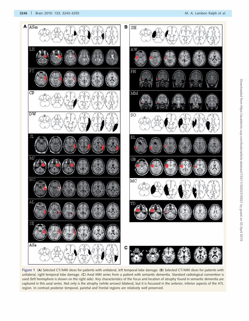
Figure 1 (A) Selected CT/MRI slices for patients with unilateral, left temporal lobe damage. (B) Selected CT/MRI slices for patients with
unilateral, right temporal lobe damage. (C) Axial MRI series from a patient with semantic dementia. Standard radiological convention is
used (left hemisphere is shown on the right side). Key characteristics of the focus and location of atrophy found in semantic dementia are
captured in this axial series. Not only is the atrophy (white arrows) bilateral, but it is focussed in the anterior, inferior aspects of the ATL
region. In contrast posterior temporal, parietal and frontal regions are relatively well preserved.
3246 | Brain 2010: 133; 3243–3255 M. A. Lambon Ralph et al.
Dow
nloaded from https://academ
ic.oup.com/brain/article-abstract/133/11/3243/316521 by guest on 03 April 2019
of objects and patterns. Two subtests from the Visual Object and
Space Perception battery were administered: (i) cube analysis, which
tests the ability to infer how many cubes there must be in a 3D ar-
rangement of cubes from a 2D picture of the arrangement and
(ii) object decision, which assesses the participant’s ability to recognize
the silhouette of a real object presented in the company of three foils
that are silhouettes of nonsense objects.
Semantic assessments
The experimental core of the study was a series of eight tests, four
expressive (requiring self-generated spoken responses) and four recep-
tive (i.e. requiring only a semantic judgement as indicated by pointing
or repeating). We had previously collected data on these same eight
tests for a group of 10 patients with semantic dementia as well as a
large group of normal controls to provide normative data (see Bozeat
et al., 2000). The same battery of tasks has been used successfully in
comparative case series investigations in order to reveal the similarities
and differences between various semantically impaired patient groups
(e.g. Jefferies and Lambon Ralph, 2006; Corbett et al., 2009). This
indicates that the battery has sufficient sensitivity not only to grade the
degree of semantic impairment in each patient but also to reveal quali-
tatively different types of semantic impairment.
One expressive test known to be highly sensitive to semantic im-
pairment (Hodges and Patterson, 1995) is category fluency, in which
the person is asked to produce as many exemplars of a semantic cat-
egory as possible in one minute; our version of this test uses six dif-
ferent categories, three natural groups (animals, birds, fruit) and three
artefacts (household items, tools, vehicles). We also administered a
letter fluency test, which forms a useful contrast to category fluency
because it has all of the same executive and monitoring components
but minimal semantic demands: letters of the alphabet replace seman-
tic categories as the cue for word retrieval (words starting with F, A or
S). The other two expressive tests consisted of two picture naming
tasks, one relatively easy (the Cambridge 64-item naming test:
Bozeat et al., 2000; Adlam et al., 2006) and one graded and much
harder (the 30-item Graded Naming Test: McKenna and Warrington,
1983). In both cases, the participant is shown each item in the test as
a line drawing of a familiar (or in the case of the items towards the
end of the Graded Naming Test, less familiar but still known) object
and asked to supply its name. Performance of the patients was com-
pared to the published norms for this psychometrically graded test.
The first receptive test was an easy one, in which the stimuli are the
same 64 items comprising the easier of the two naming tests described
above. Each item’s name is spoken to the participant as he or she
looks at an array of 10 pictures of objects, the target and nine other
objects from the same semantic category, and is asked to point to the
target. Two harder receptive semantic tests are the picture and word
versions of the Camel and Cactus Test (Bozeat et al., 2000; Adlam
et al., 2006), a semantic association test in which the participant must
select one of four pictures (or written words) that is related to a target
picture (or word). In each case, the four response choices belong to a
different category from the target and to the same category as each
other. For example, the choices for the target ‘duck’ are all places in
the natural world; one has to know that ducks are usually to be found
on a ‘lake’ (the correct choice) rather than on a ‘mountain’, in a
‘desert’ or on an ‘iceberg’ (the three foils). The final receptive semantic
test was the graded synonym judgement test (Warrington et al.,
1998), consisting of two spoken-word choices for each of 50
spoken-word targets, for example “Does ‘marquee’ mean the same
as ‘tent’ or ‘palace’?” The participant repeats his or her chosen word
response. For this latter psychometrically graded test, the patients’
performance was compared against the published norms.
Results
Background neuropsychologyThe results of the background assessments are summarized in
Tables 2 and 3. In all tables and figures, the patients are ordered
by descending scores on the category fluency test. Performance of
the patients with left temporal lobe damage (Table 2) was gener-
ally normal across the various cognitive domains, including execu-
tive skills, visuospatial perception and components of the Wechsler
Adult Intelligence Scale. Scores on the recognition memory tests
varied somewhat more, with half of the patients exhibiting weak
(5–10th percentile) or abnormal (�5th percentile) performance on
one or both subtests (Patients PT, JL, MJ, NC, SE and ASA). The
same pattern characterized the patients with right temporal lobe
damage (Table 3). Performance on tests other than recognition
memory was normal (except for Patient SO’s score on the cube
analysis subtest of the Visual Object and Space Perception bat-
tery). Again, around half of the right-sided patients demonstrated
weak or abnormal performance on at least one part of the recog-
nition memory test (Patients SH, FH, TD, MC and SO). There was
no apparent material specificity of the kind that one might predict
if anticipating an association of the left hemisphere with verbal
material and the right with non-verbal stimuli. In summary, at
least as reflected by the background tests administered, the
most likely consequence of the unilateral temporal lesions in
these patients was impaired episodic recognition memory for
recently encountered words and/or faces.
Expressive semantic assessmentFigure 2A summarizes the individual and group results across the
four measures of expressive semantic skill. Although these patients
with unilateral temporal lobe lesions were not (as already ex-
plained) matched in any formal way to the previously published
group of 10 patients with semantic dementia (Bozeat et al., 2000),
the 20 cases in the present study and the 10 in the semantic
dementia investigation were all assessed on the same semantic
tests. For comparison purposes, therefore, we have plotted the
semantic dementia data alongside those from the current cohort,
again as both averaged scores and as individual case scores (once
again ordered by performance in category fluency). As can be
seen, the semantic dementia cases covered the range from mild
(Patients JP and WM) to severe (Patients IF and JW), although the
difficult Graded Naming Test was sensitive enough to detect
abnormality in even the mildest semantic dementia cases.
At the ‘group’ level, two major aspects of the results are re-
vealed in Fig. 2A. Firstly, average scores for both of the unilateral
groups were higher—in three of the four tests, substantially
higher—than average scores for the semantic dementia case
series. Secondly, on these expressive tasks, the average scores
for patients with right temporal lesions were higher than averages
for their left-temporal counterparts (category: fluency
[t(18) = 2.96, P = 0.008]; letter fluency [t(18) = 3.8, P = 0.001];
64-item naming [t(18) = 2.6, P = 0.02]; Graded Naming Test
[t(18) = 3.3, P = 0.004]). This is probably not a reflection of overall
Unilateral ATL lesions generate minimal semantic impairment Brain 2010: 133; 3243–3255 | 3247
Dow
nloaded from https://academ
ic.oup.com/brain/article-abstract/133/11/3243/316521 by guest on 03 April 2019
Tab
le3
Bac
kgro
und
neu
ropsy
cholo
gic
alas
sess
men
t(p
atie
nts
wit
hri
ght
tem
pora
llo
be
dam
age)
SHA
WFH
MM
TD
ELG
BM
CSO
NA
RT
IQ123
107
110
120
97
90
107
82
105
WR
MT
Face
s25
(90%
)24
(75%
)38/5
0a
(5%
)50/5
0a
(75%
)24
(75%
)24
(75%
)25
(90%
)23
(50%
)23
(50%
)
Word
s23
(5–1
0%
)25
(90%
)44/5
0a
(50%
)42/5
0a
(25%
)22
(_5%
)24
(25%
)25
(50–9
0%
)23
(10%
)21
(10%
)
RC
PM
34
(95%
)32
(90–9
5%
)N
TN
T32
(90–9
5%
)32
(90–9
5%
)30
(90%
)30
(90%
)32
(90–9
5%
)
VO
SPC
ut-
off
Obje
ctdec
isio
n14
17
20
19
20
16
20
19
20
19
Cube
anal
ysis
610
910
10
10
10
10
10
5
WA
IS-R
(sca
led
score
s)
Pic
ture
com
ple
tion
811
11
12
11
910
12
11
Pic
ture
arra
ngem
ent
78
13
13
75
77
8
Blo
ckdes
ign
14
12
15
14
12
15
98
9
Dig
itsp
an15
78
16
13
816
613
Voca
bula
ry12
911
12
98
98
10
Arith
met
ic12
810
14
14
10
12
68
Sim
ilarities
16
812
11
10
10
10
12
13
aFo
rth
ese
two
pat
ients
the
long-f
orm
of
the
reco
gnitio
nm
emory
test
was
adm
inis
tere
d.
Abnorm
alsc
ore
sar
esh
ow
nin
embold
ened
font.
NA
RT
=N
atio
nal
Adult
Rea
din
gTes
t;N
T=
not
test
ed;
RC
PM
=R
aven
’sC
olo
ure
dPro
gre
ssiv
eM
atrice
s;V
OSP
=V
isual
Obje
ctan
dSp
atia
lPer
ception
bat
tery
;W
AIS
-R=
Wec
hsl
erA
dult
Inte
lligen
ceSc
ale;
WR
MT
=W
arringto
nR
ecognitio
nM
emory
Tes
t(s
hort
form
).
Tab
le2
Bac
kgro
und
neu
ropsy
cholo
gic
alas
sess
men
t(p
atie
nts
wit
hle
ftte
mpora
llo
be
dam
age)
ASM
LHPT
CP
DW
JLM
JN
CA
LSE
ASA
NA
RT
IQ121
98
80
108
121
103
101
113
87
116
97
RM
TFa
ces
22
(25–5
0%
)25
(90%
)20
(10%
)25
(90%
)25
(90%
)21
(10%
)23
(50%
)24
(75%
)22
(10-2
5%
)22
(25–5
0%
)19
(5%
)
Word
s21
(10–2
5%
)23
(50%
)18
(_5%
)24
(25%
)25
(90%
)21
(25%
)23
(5–1
0%
)21
(10%
)21
(25%
)20
(5–1
0%
)20
(5–1
0%
)
RC
PM
27
(50–7
5%
)31
(90–9
5%
)22
(25–5
0%
)34
(95%
)29
(75–9
0%
)31
(90–9
5%
)28
(75–9
0%
)28
(75–9
0%
)29
(90%
)33
(95%
)31
(90–9
5%
)
VO
SPC
ut-
off
Obje
ctdec
isio
n14
20
18
19
18
14
19
19
20
18
18
17
Cube
anal
ysis
69
10
99
99
10
10
810
10
WA
IS-R
(sca
led
score
s)
Pic
ture
com
ple
tion
10
76
57
810
NT
87
9
Pic
ture
arra
ngem
ent
11
66
12
10
713
NT
78
5
Blo
ckdes
ign
10
14
712
76
9N
T6
910
Dig
itsp
an13
12
77
10
77
NT
75
4
Voca
bula
ry7
74
11
12
710
NT
11
10
6
Arith
met
ic7
94
11
11
711
NT
411
6
Sim
ilarities
810
610
13
512
NT
98
5
3248 | Brain 2010: 133; 3243–3255 M. A. Lambon Ralph et al.
Dow
nloaded from https://academ
ic.oup.com/brain/article-abstract/133/11/3243/316521 by guest on 03 April 2019
Graded Naming Test
0
5
10
15
20
25
30
ASM L
H
PT CP
DW JL M
J
NC
AL SE
ASA SH AW FH MM TD EL
GB
MC SO JP
WM SL JC AT DS
JH DC IF JW L
eft
Rig
ht SD
Acc
urac
y
Left temporal lobe damage Right temporal lobe damage Semantic dementia Mean
64-item naming test
0
8
16
24
32
40
48
56
64
ASM L
H
PT CP
DW JL M
J
NC
AL SE
ASA SH AW FH MM TD EL
GB
MC SO JP
WM SL JC AT DS
JH DC IF JW L
eft
Rig
ht SD
Acc
urac
y
Left temporal lobe damage Right temporal lobe damage Semantic dementia Mean
Category fluency
0
30
60
90
120
150
ASM L
H
PT CP
DW JL M
J
NC
AL SE
ASA SH AW FH MM TD EL
GB
MC SO JP
WM SL JC AT DS
JH DC IF JW L
eft
Rig
ht SD
Acc
urac
y
Left temporal lobe damage Right temporal lobe damage Semantic dementia Mean
Letter fluency
0
10
20
30
40
50
60
ASM L
H
PT CP
DW JL M
J
NC
AL SE
ASA SH AW FH MM TD EL
GB
MC SO JP
WM SL JC AT DS
JH DC IF JW L
eft
Rig
ht SD
Left temporal lobe damage Right temporal lobe damage Semantic dementia Mean
Acc
urac
y
A
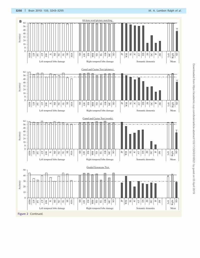
Figure 2 (A) Summary of expressive tasks. (B) Summary of receptive semantic tasks. The full horizontal line denotes the
control-participant mean performance on each test. The dashed line shows the cut-off score for each assessment (defined as
2 SD below the control mean).
Unilateral ATL lesions generate minimal semantic impairment Brain 2010: 133; 3243–3255 | 3249
Dow
nloaded from https://academ
ic.oup.com/brain/article-abstract/133/11/3243/316521 by guest on 03 April 2019
64-item word-picture matching
0
8
16
24
32
40
48
56
64
ASM L
H
PT CP
DW JL M
J
NC
AL SE
ASA SH AW FH MM TD EL
GB
MC SO JP
WM SL JC AT DS
JH DC IF JW L
eft
Rig
ht SD
Acc
urac
y
Left temporal lobe damage Right temporal lobe damage Semantic dementia Mean
Camel and Cactus Test (pictures)
0
8
16
24
32
40
48
56
64
ASM L
H
PT CP
DW JL M
J
NC
AL SE
ASA SH AW FH MM TD EL
GB
MC SO JP
WM SL JC AT DS
JH DC IF JW L
eft
Rig
ht SD
Acc
urac
y
Left temporal lobe damage Right temporal lobe damage Semantic dementia Mean
Camel and Cactus Test (words)
0
8
16
24
32
40
48
56
64
ASM L
H
PT CP
DW JL M
J
NC
AL SE
ASA SH AW FH MM TD EL
GB
MC SO JP
WM SL JC AT DS
JH DC IF JW L
eft
Rig
ht SD
Acc
urac
y
Left temporal lobe damage Right temporal lobe damage Semantic dementia Mean
Graded Synonyms Test
0
10
20
30
40
50
ASM L
H
PT CP
DW JL M
J
NC
AL SE
ASA SH AW FH MM TD EL
GB
MC SO JP
WM SL JC AT DS
JH DC IF JW L
eft
Rig
ht SD
Left temporal lobe damage Right temporal lobe damage Semantic dementia Mean
Acc
urac
y
B
Figure 2 Continued.
3250 | Brain 2010: 133; 3243–3255 M. A. Lambon Ralph et al.
Dow
nloaded from https://academ
ic.oup.com/brain/article-abstract/133/11/3243/316521 by guest on 03 April 2019
severity given that performance on the receptive semantic tasks,
which are admittedly easier tasks, turned out to be well matched
for the two unilateral groups. Instead, this result probably reflects
a phenomenon that we have reported before: for patients with
semantic dementia with relatively asymmetric left4right versus
right4left distributions of bilateral temporal lobe atrophy, compar-
able degrees of comprehension impairment in the two groups are
associated with more severe expressive (naming) deficits for the
left4right subset (Lambon Ralph et al., 2001). The relationship
between that finding and the present study is considered in more
detail in the ‘Discussion’ section. We also compared each patient
group against the participants who provided the normative data
during the original formulation of the semantic battery (Bozeat
et al., 2000). As would be expected from viewing Fig. 2A, the
right unilateral cases were, on average, as good as these control
participants on the 64-item naming and letter fluency tasks, whilst
the small difference on the category fluency test was
non-significant [t(38)51]. Although the degree of anomia in the
left unilateral group was not in the same league as that observed
in semantic dementia, the naming accuracy for this group was
significantly lower than the control participants [all measures:
t(40)42.23, all P50.05].
The ‘individual’ scores from the unilateral cases for the expres-
sive semantic assessments followed a similar left versus right pat-
tern. On the category fluency task (which is especially demanding
of the semantic system as well as several other cognitive domains),
three of the unilateral left cases had scores at the normal cut-off
(Patients DW, JL, MJ) and four below it (Patients NC, AL, SE,
ASA). In comparison, for the right-sided cases, only Patient MC
fell at the cut-off point and Patient SO just below it. On the letter
fluency tasks one left-sided case scored at the cut-off (Patient NC)
and four below (Patients JL, MJ, AL, ASA) whereas all of the
right-sided cases performed within 2 SD of the normal mean.
The same was true for naming on the 64-item and Graded
Naming tests: all right-sided cases were in the normal range but
about half of the left-sided cases performed at or below the
cut-off score.
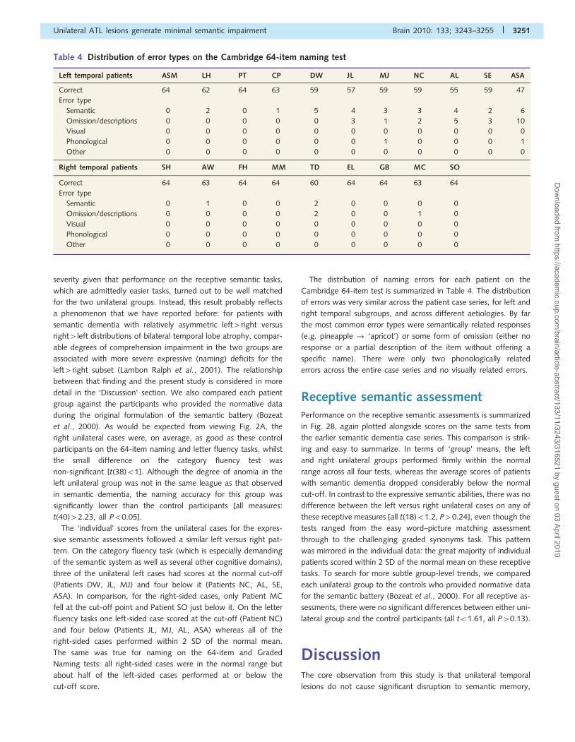
The distribution of naming errors for each patient on the
Cambridge 64-item test is summarized in Table 4. The distribution
of errors was very similar across the patient case series, for left and
right temporal subgroups, and across different aetiologies. By far
the most common error types were semantically related responses
(e.g. pineapple ! ‘apricot’) or some form of omission (either no
response or a partial description of the item without offering a
specific name). There were only two phonologically related
errors across the entire case series and no visually related errors.
Receptive semantic assessmentPerformance on the receptive semantic assessments is summarized
in Fig. 2B, again plotted alongside scores on the same tests from
the earlier semantic dementia case series. This comparison is strik-
ing and easy to summarize. In terms of ‘group’ means, the left
and right unilateral groups performed firmly within the normal
range across all four tests, whereas the average scores of patients
with semantic dementia dropped considerably below the normal
cut-off. In contrast to the expressive semantic abilities, there was no
difference between the left versus right unilateral cases on any of
these receptive measures [all t(18)51.2, P40.24], even though the
tests ranged from the easy word–picture matching assessment
through to the challenging graded synonyms task. This pattern
was mirrored in the individual data: the great majority of individual
patients scored within 2 SD of the normal mean on these receptive
tasks. To search for more subtle group-level trends, we compared
each unilateral group to the controls who provided normative data
for the semantic battery (Bozeat et al., 2000). For all receptive as-
sessments, there were no significant differences between either uni-
lateral group and the control participants (all t51.61, all P40.13).
DiscussionThe core observation from this study is that unilateral temporal
lesions do not cause significant disruption to semantic memory,
Table 4 Distribution of error types on the Cambridge 64-item naming test
Left temporal patients ASM LH PT CP DW JL MJ NC AL SE ASA
Correct 64 62 64 63 59 57 59 59 55 59 47
Error type
Semantic 0 2 0 1 5 4 3 3 4 2 6
Omission/descriptions 0 0 0 0 0 3 1 2 5 3 10
Visual 0 0 0 0 0 0 0 0 0 0 0
Phonological 0 0 0 0 0 0 1 0 0 0 1
Other 0 0 0 0 0 0 0 0 0 0 0
Right temporal patients SH AW FH MM TD EL GB MC SO
Correct 64 63 64 64 60 64 64 63 64
Error type
Semantic 0 1 0 0 2 0 0 0 0
Omission/descriptions 0 0 0 0 2 0 0 1 0
Visual 0 0 0 0 0 0 0 0 0
Phonological 0 0 0 0 0 0 0 0 0
Other 0 0 0 0 0 0 0 0 0
Unilateral ATL lesions generate minimal semantic impairment Brain 2010: 133; 3243–3255 | 3251
Dow
nloaded from https://academ
ic.oup.com/brain/article-abstract/133/11/3243/316521 by guest on 03 April 2019
at least in terms of accuracy on a range of semantic tests that
demonstrate clear impairment in three different neurological dis-
eases associated with bilateral, ATL damage (semantic dementia,
Alzheimer’s disease, herpes simplex virus encephalitis). There are,
as always in research, caveats and complications to this unembel-
lished conclusion, but before we come to these, an overview of
the data is as follows. First, treating the patients as two groups—
those with left or right unilateral temporal lesions—the ‘average’
performance of both groups was within 2 SD of the control means
on all four expressive tests and on all four receptive tests.
Secondly, again dealing with averages, both unilateral groups
had substantially higher mean performance on all eight tests
than a group of patients with semantic dementia assessed on
the same measures: average scores for the semantic dementia
cases fell below the normal range on every test. Thirdly, the
unilateral-left group performed more poorly than the unilateral-
right group on the expressive but not the receptive tests. On
all three genuinely semantic expressive tests (category fluency
and the two object naming tests), the average scores of the
unilateral-left cases were significantly below the control mean.
Turning to individual performance, for unilateral-right patients:
all nine were comfortably within the normal range on six of the
eight tests. One of the two test exceptions was category fluency,
on which two patients (MC and SO) were just at or barely below
the cut-off score. Category fluency is not only a demanding
semantic assessment but also requires many other cognitive
skills; nevertheless, since these executive and non-semantic
memory abilities are also required for success in letter fluency,
on which patients MC and SO had normal scores, the data sug-
gest a mild semantic abnormality for these two cases. The second
minor exception was the easy word–picture matching task, on
which two unilateral-right cases (patients TD and MC) made a
few errors. This is a sensitive test only because controls perform
at ceiling, but again, these two patients’ performance indicates a
possible, albeit slight, semantic abnormality.
For the unilateral-left patients, all 11 were within the normal
range on only two of the eight tests, both receptive: Camel and
Cactus Test words and the Graded Synonym Test (though note
that the 2 SD below the ‘normal’ mean on the Graded Synonym
Test is barely above chance). On the remaining receptive tests,
there were very minor abnormalities for two cases on word–pic-
ture matching (but see the comments above about ceiling effects
in normal performance) and two not so trivial, but still only mod-
erate abnormalities on Camel and Cactus Test-pictures. Expressive
tests for unilateral-left cases tell a somewhat different story. The
four mildest cases (as determined by category fluency—the test
that we used to rank the cases) had completely normal scores on
all of the expressive tests, but the remaining seven patients were
all below the cut-off score on at least one of the expressive tests,
and one patient (ASA) had abnormal scores on all four expressive
tests. In fact, patient ASA’s test scores—both expressive and
receptive—resemble those of patients with mild semantic demen-
tia (Adlam et al., 2006). We do not intend to gloss over this
impairment, which constitutes an important qualification to the
bald conclusion that it requires bilateral damage to yield semantic
impairment (see below). We do note, however, that this pattern
emphasizes the importance of case series studies as opposed to
single-case research: only patient ASA, of 11 patients with
unilateral-left temporal lesions and 20 patients with unilateral
temporal lesions, was consistently below control cut-off scores
on our expressive semantic tests, and even her degree of abnor-
mality on most tests equated to mild rather than moderate
or severe semantic dementia. Our summary thus remains that it
‘typically’ requires bilateral temporal lobe damage to generate
‘significant, clinically notable’ disruption to conceptual knowledge.
Why should this be so?
Our hypothesis is the following. The widespread semantic net-
work in the brain consists of many different regions, some of them
specific to certain modalities of information and some of them
biased towards one or other hemisphere; but the ATL component
of this network, which supports the most central, amodal ‘hub’ of
conceptual knowledge (Patterson et al., 2007; Lambon Ralph
et al., 2010; Pobric et al., 2010b), may be distributed across left
and right ATLs in a largely undifferentiated fashion. This would
have at least two consequences. Firstly, a bonus of distributed
representations is ‘graceful degradation’ (Farah and McClelland,
1991), where low levels of damage produce little behavioural de-
cline and clear impairment only follows considerable damage.
Note that, if ‘considerable’ means 450%, then, by definition, it
would take damage to both temporal lobes to generate notable
semantic impairment. Secondly, forming amodal semantic repre-
sentations across two partially but not fully interconnected neural
substrates may result in at least some duplication of the represen-
tation in each hemisphere. Like back-up storage for any computer
or mirror website, such duplication will make the bilateral ATL
semantic system more robust to the effects of unilateral
damage. To the extent that one can draw parallels between
human and non-human primates, this proposal is supported by
the fact that object recognition in the macaque monkey is severely
affected by experimentally induced bilateral temporal lesions but
scarcely impacted by unilateral lesions in this region (Buckley
and Gaffan, 2006). Likewise, other aspects of temporal lobe func-
tion, including paired-associate learning and auditory recognition,
seem to become chronically impaired only after bilateral ablation
(Hefner and Heffner, 1986; Li et al., 1999). Supporting this idea,
in recent formal meta-analyses of the functional neuroimaging
literature, bilateral (albeit often left4right) activations are
observed when normal participants complete semantic tasks
(Binder et al., 2009; Visser et al., 2010). Likewise, a recent func-
tional neuroimaging study of stroke aphasic patients reported that
comprehension ability was better predicted by the status of the
functional connectivity between the left and right ATL than by the
level of regional cerebral blood flow in the left ATL (Warren et al.,
2009); in line with our working hypothesis, patients with compro-
mised inter-ATL functional connectivity had impaired comprehen-
sion, even if both neural areas were structurally intact.
The proposal of absent specialization in the ATL semantic
system might seem counter to reports of differences in the impair-
ment profile of semantic dementia patients with left4right versus
right 4 left ATL atrophy. For example, Snowden and colleagues
(2004) assessed identification of and familiarity with famous
people, and reported that 10 patients with semantic dementia
with more left 4 right abnormality achieved higher scores when
knowledge was probed by the famous people’s faces rather than
3252 | Brain 2010: 133; 3243–3255 M. A. Lambon Ralph et al.
Dow
nloaded from https://academ
ic.oup.com/brain/article-abstract/133/11/3243/316521 by guest on 03 April 2019
their names, whereas three semantic dementia cases with
right4left atrophy had the reverse pattern of success: names
better than faces. A parallel finding was observed on a more gen-
eral test of semantic knowledge (Pyramids and Palm Trees):
patients with left4right demonstrated worse performance on
the verbal than picture version, whilst the opposite pattern (pic-
tures5words) was found in the right4left cases. Snowden et al.
(2004) interpreted these results as indicating specialization of the
left ATL for names and of the right ATL for faces. Although we
did not test semantic performance on faces, people’s names or
other specific-level concepts, the results from the present unilateral
cases exhibit little evidence for word versus picture differences in
receptive semantic tasks, such as Pyramids and Palm Trees and the
more taxing Camel and Cactus Test. As can be seen in Fig. 2B,
neither unilateral left nor unilateral right temporal lesions were
associated with impaired average performance on either the
word or picture version of the Camel and Cactus Test, and in
fact, out of 40 available scores (20 patients�2 modalities of
stimulus), the only two mildly impaired scores were for
left-unilateral cases on the picture version (Patients SE and ASA).
By contrast, it seems fairly clear that patients with left-temporal
lesions are more likely than their right-temporal counterparts to
have a mild degree of naming impairment and that, within pa-
tients with semantic dementia (all of whom are anomic), those
with left4right temporal atrophy have more severe naming im-
pairments than their right4left counterparts even with matched
degrees of comprehension deficit. How is this set of findings to be
reconciled with our hypothesis of a relatively undifferentiated se-
mantic network in the ATL? The answer, we propose, lies in the
connectivity of the ATL with other more modality-specific brain
regions. Two well-known facts are crucial here: (i) speech produc-
tion is strongly left-lateralized in the majority of people; and
(ii) apart from some cross-hemisphere connections between hom-
ologous brain regions, the vast majority of brain connections are
within rather than across the two hemispheres. The result of these
facts is that left-hemisphere speech regions almost certainly have
much stronger connections with the left than the right side of the
ATL system. And the implication of this arrangement is that, even
if the ATL component of the semantic network turned out to be
completely distributed and undifferentiated across the two hemi-
spheres, left4right asymmetry of damage to this system would
have more deleterious consequences for speaking and naming
than right4left damage. The impact of differential white-matter
connectivity has been shown in the tractography and functional
imaging literature. In particular, the uncinate fasciculus—in both
the macaque monkey and human—connects the ATLs to the
ventrolateral prefrontal cortex (especially to Brodmann areas
47/12; Petrides and Pandya, 1988). Increased activity in the
latter areas is observed during active retrieval for verbal informa-
tion in the left hemisphere and for visual or visuospatial informa-
tion in the right hemisphere (Cadoret et al., 2001; Petrides, 2005).
A previous study documenting the more severe anomia in pa-
tients with semantic dementia with left4right atrophy was accom-
panied by a computational model that implemented differentially
strong connectivity from the two sides of a bilateral semantic
system to a unilateral speech production system (Lambon Ralph
et al., 2001). After training and ‘lesioning’ of the model,
irrespective of degree of overall damage to the undifferentiated
semantic units, the model’s naming performance was more im-
paired if the simulated damage had an asymmetric left4right dis-
tribution. As far as we are aware, no similar modelling effort has
been devoted to names versus faces as probes to person know-
ledge, but precisely the same logic would apply (indeed a related
idea has been encapsulated in other computational models of
semantic memory; Plaut, 2002). Processing of faces is probably
not unilateral but has a strong affinity with the fusiform face
area in the right temporal lobe. Connections between this region
and the ATL will therefore be stronger on the right. What might
seem to be ATL differences in semantic knowledge derived from
faces could in fact be explained by hemispheric processing differ-
ences further back in the temporal lobe combined with differential
connection strength.
Two other issues require brief consideration. The first is lesion
volume, given that bilateral lesions will almost invariably lead to
greater total volume loss than unilateral damage. An attempt to
equate lesion extent in forms of brain injury/disease as different as
temporal lobe resection versus semantic dementia is probably im-
possible, but if one could do this, perhaps a very large unilateral
lesion would produce as much semantic disruption as smaller bi-
lateral lesions. Indeed, our working hypothesis is that both ATL
centres contribute to a single modality-invariant semantic system.
These might work together in a super-additive manner, maximiz-
ing redundancy and graceful degradation as described above; but
unless this duplication of information and connectivity is complete
(i.e. each is a full ‘back-up’ of the other), then it seems very likely
that large unilateral lesions should produce at least mild levels of
semantic disruption: Patient ASA may be the closest example in
the present study. Large-scale studies of patients with temporal
lobe epilepsy (Bell et al., 2001; Giovagnoli et al., 2005) have
demonstrated that, as a group, the semantic performance of
these patients falls a small but significant degree below control
levels (a drop in accuracy that is typically too small to measure
reliably at the individual level but is consistent across participants).
Recent results from right-sided transcranial magnetic stimulation in
neurologically intact participants also fit this pattern. Following
stimulation of the left or right lateral ATL, normal participants
demonstrate a selective slowing, although no drop in accuracy,
on semantic tasks (Lambon Ralph et al., 2009). This holds for
both verbal and picture-based semantic tasks (Pyramids and
Palm Trees Test and Camel and Cactus Test) irrespective of
whether the left or right ATL is stimulated (Pobric et al.,
2010a). Whilst these documented impairments from right-sided
transcranial magnetic stimulation and in patients like patient ASA
are important, it should be remembered that they are not in the
same league as the impairments observed in patients with seman-
tic dementia with bilateral ATL damage. Instead, they fit with the
notion of graceful degradation where systems are relatively robust
to moderate levels of damage.
The second issue concerns caveats with regard to the interpret-
ation of data from temporal lobe resection for epilepsy or tumour,
or damage after vascular accident, because of the possibility of
re-learning or reorganization/recovery of semantic function. A
long-standing seizure history complicates attempts to generalize
findings from patients with resection for temporal lobe epilepsy
Unilateral ATL lesions generate minimal semantic impairment Brain 2010: 133; 3243–3255 | 3253
Dow
nloaded from https://academ
ic.oup.com/brain/article-abstract/133/11/3243/316521 by guest on 03 April 2019
to people with normal brains or even people with brains damaged
by other aetiologies. This point is supported by at least three
findings: (i) post-operative deficits of cognition/language tend to
be more severe in association with a later age of seizure onset
(Hermann et al., 1999); (ii) there is a significant change in the
pattern of language-related white-matter pathways in patients
with long-standing epilepsy (Powell et al., 2007); and (iii) there
is significant alteration in neurotransmitter function (Hammers
et al., 2003). In the face of these neuroanatomical changes, se-
mantic function may be shifted away from this region, such that
subsequent resection will have less dramatic consequences than an
acute neurological event. Some of the same concerns about reor-
ganized function apply to slow-growing tumours. There is now
clear evidence that the prolonged time-course of low-grade
glioma growth can enable plasticity-related shifts in language
function (Thiel et al., 2001, 2005; Duffau et al., 2003) and the
same may apply to ATL semantic systems. Pre-damage reorgan-
ization of function does not, of course, apply to vascular accidents,
but spontaneous recovery occurs to at least some extent in most
patients (Enderby and Philipp, 1986). Unfortunately, there are in-
sufficient numbers of each aetiology in this study to make formal
comparisons between the three clinical groups, but this would be
an interesting avenue for future investigation.
By contrast, an insidiously progressive condition like semantic
dementia, which often does not even come to medical attention
until it is moderately advanced, probably offers limited scope for
relearning. Furthermore, the few studies of attempted semantic
rehabilitation with patients with semantic dementia suggest that
the kind of learning/re-learning of which they are capable is rigid,
rote and context-bound (e.g. Graham et al., 2001; Snowden and
Neary, 2002). One might argue that, in contrast, the very essence
of conceptual knowledge is its flexibility and generalizability
to different situations (Lambon Ralph and Patterson, 2008;
Lambon Ralph et al., 2010). Thus, even if semantic dementia pa-
tients manage to learn and remember ‘some’ things (and hence,
for example, produce delayed recall of the complex, meaningless
Rey figure that is within the normal range: Adlam et al., 2009),
generalizable semantic information is not one of them.
In conclusion, the results from this study support a neurological
model in which yoked ATL regions within both left and right hemi-
spheres function together to support a redundant and thus robust
system for semantic representation (Lambon Ralph et al., 2001). As a
result, unilateral damage generates either no receptive semantic im-
pairment (as observed here) or small deficits that can only be mea-
sured in large group studies or via reaction times in right-sided
transcranial magnetic stimulation investigations of the left or right
ATL. The redundancy of the bilateral temporal system is such that
the only way to observe substantial semantic impairment is if the
damage is itself bilateral (as is the case in semantic dementia,
herpes simplex virus encephalitis, etc.) or if the functional connect-
ivity between the two hemispheres is disrupted (Warren et al., 2009).
AcknowledgementsWe are indebted to the patients and their carers for their generous
assistance with this study.
FundingThis research was supported by a programme grant from the
Medical Research Council to MALR (G0501632).
ReferencesAdlam ALR, Patterson K, Hodges JR. “I remember it as if it were yes-
terday”: memory for recent events in patients with semantic dementia.
Neuropsychologia 2009; 47: 1344–51.
Adlam ALR, Patterson K, Rogers TT, Nestor PJ, Salmond CH,
Acosta-Cabronero J, et al. Semantic dementia and fluent primary
progressive aphasia: two sides of the same coin? Brain 2006; 129:
3066–80.
Bell BD, Hermann BP, Woodard AR, Jones JE, Rutecki PA, Sheth R, et al.
Object naming and semantic knowledge in temporal lobe epilepsy.
Neuropsychology 2001; 15: 434–43.Binder JR, Desai RH, Graves WW, Conant LL. Where is the semantic
system? A critical review and meta-analysis of 120 functional neuroi-
maging studies. Cereb Cortex 2009; 19: 2767–96.
Bozeat S, Lambon Ralph MA, Patterson K, Garrard P, Hodges JR.
Non-verbal semantic impairment in semantic dementia.
Neuropsychologia 2000; 38: 1207–15.Brett M, Leff A, Rorden C, Ashburner J. Spatial normalization of brain
images with focal lesions using cost function masking. Neuroimage
2001; 14: 486–500.
Buckley MJ, Gaffan D. Perirhinal cortical contributions to object percep-
tion. Trends Cogn Sci 2006; 10: 100–7.
Cadoret G, Pike GB, Petrides M. Selective activation of the ventrolateral
prefrontal cortex in the human brain during active retrieval processing.
Eur J Neurosci 2001; 14: 1164–70.
Corbett F, Jefferies E, Ehsan S, Lambon Ralph MA. Different impairments
of semantic cognition in semantic dementia and semantic aphasia:
evidence from the non-verbal domain. Brain 2009; 132: 2593–608.Damasio H. Human brain anatomy in computarized images. Oxford:
Oxford University Press, 1995.Duffau H, Capelle L, Denvil D, Sichez N, Gatignol P, Lopes M, et al.
Functional recovery after surgical resection of low grade gliomas in
eloquent brain: hypothesis of brain compensation. J Neurol
Neurosurg Psych 2003; 74: 901–7.Enderby P, Philipp R. Speech and language handicap - towards knowing
the size of the problem. Br J Disorders Commun 1986; 21: 151–65.
Farah MJ, McClelland JL. A computational model of semantic memory
impairment: modality specificity and emergent category specificity.
J Exp Psychol Gen 1991; 120: 339–57.
Fodor JA. Modularity of mind: an essay on faculty psychology.
Cambridge, MA: MIT Press, 1983.Giovagnoli AR, Erbetta A, Villani F, Avanzini G. Semantic memory in
partial epilepsy: verbal and non-verbal deficits and neuroanatomical
relationships. Neuropsychologia 2005; 43: 1482–92.
Graham KS, Patterson K, Pratt KH, Hodges JR. Can repeated exposure
to “forgotten” vocabulary help alleviate word-finding difficulties in se-
mantic dementia? An illustrative case study. Neuropsychol Rehabilit
2001; 11: 429–54.
Hammers A, Koepp MJ, Richardson MP, Hurlemann R, Brooks DJ,
Duncan JS. Grey and white matter flumazenil binding in neocortical
epilepsy with normal MRI. A PET study of 44 patients. Brain 2003;
126: 1300–18.
Hefner HE, Heffner RS. Effect of unilateral and bilateral auditory cortex
lesions on the discrimination of vocalizations by Japanese macaques.
J Neurophysiol 1986; 56: 683–701.
Hermann B, Davies K, Foley K, Bell B. Visual confrontation naming out-
come after standard left anterior temporal lobectomy with sparing
versus resection of the superior temporal gyrus: a randomized
prospective clinical trial. Epilepsia 1999; 40: 1070–6.
3254 | Brain 2010: 133; 3243–3255 M. A. Lambon Ralph et al.
Dow
nloaded from https://academ
ic.oup.com/brain/article-abstract/133/11/3243/316521 by guest on 03 April 2019
Hodges JR, Patterson K. Is semantic memory consistently impaired earlyin the course of Alzheimer’s disease? Neuroanatomical and diagnostic
implications. Neuropsychologia 1995; 33: 441–59.
Jefferies E, Lambon Ralph MA. Semantic impairment in stroke aphasia
vs. semantic dementia: a case-series comparison. Brain 2006; 129:2132–47.
Lambon Ralph MA, Lowe C, Rogers TT. Neural basis of category-specific
semantic deficits for living things: evidence from semantic dementia,
HSVE and a neural network model. Brain 2007; 130: 1127–37.Lambon Ralph MA, McClelland JL, Patterson K, Galton CJ, Hodges JR.
No right to speak? The relationship between object naming and se-
mantic impairment: neuropsychological evidence and a computationalmodel. Journal of Cognitive Neuroscience 2001; 13: 341–56.
Lambon Ralph MA, Patterson K. Generalisation and differentiation
in semantic memory: Insights from semantic dementia. Ann NY
Acad Sci 2008; 1124: 61–76.Lambon Ralph MA, Pobric G, Jefferies E. Conceptual knowledge is
underpinned by the temporal pole bilaterally: convergent evidence
from rTMS. Cereb Cortex 2009; 19: 832–8.
Lambon Ralph MA, Sage K, Jones RW, Mayberry EJ. Coherent conceptsare computed in the anterior temporal lobes. Proc Natl Acad Sci USA
2010; 107: 2717–22.
Li H, Matsumoto K, Watanabe H. Different effects of unilateral
and bilateral hippocampal lesions in rats on the performance ofradial maze and odor-paired associate tasks. Brain Res Bull 1999; 48:
113–9.
Martin A. The representation of object concepts in the brain. Ann RevPsychol 2007; 58: 25–45.
McKenna P, Warrington EK. The graded naming test. Windsor, England:
NFER-Nelson, 1983.
Nelson H, Willison J. The revised national adult reading test – testmanual. Winsor: NFER-Wilson, 1991.
Nestor PJ, Fryer TD, Hodges JR. Declarative memory impairments in
Alzheimer’s disease and semantic dementia. Neuroimage 2006; 30:
1010–20.Noppeney U, Patterson K, Tyler LK, Moss H, Stamatakis EA, Bright P,
et al. Temporal lobe lesions and semantic impairment: a comparison of
herpes simplex virus encephalitis and semantic dementia. Brain 2007;130: 1138–47.
Patterson K, Nestor PJ, Rogers TT. Where do you know what you know?
The representation of semantic knowledge in the human brain. Nature
Rev Neurosci 2007; 8: 976–87.Plaut DC. Graded modality-specific specialization in semantics: a com-
putational account of optic aphasia. Cogn Neuropsychol 2002; 19:
603–39.
Petrides M. Lateral prefrontal cortex: architectonic and functional organ-ization. Phil Trans Royal Soc B: Biol Sci 2005; 360: 781–95.
Petrides M, Pandya DN. Association fiber pathways to the frontal cortexfrom the superior temporal region in the rhesus monkey. J Comp
Neurol 1988; 273: 52–66.
Pobric G, Jefferies E, Lambon Ralph MA. Amodal semantic representa-
tions depend on both anterior temporal lobes: Evidence from repetitivetranscranial magnetic stimulation. Neuropsychologia 2010a; 48:
1336–42.
Pobric G, Jefferies E, Lambon Ralph MA. Category-specific versus
category-general semantic impairment induced by transcranialmagnetic stimulation. Curr Biol 2010b; 20: 964–8.
Powell HWR, Parker GJM, Alexander DC, Symms MR, Boulby PA,
Wheeler-Kingshott CAM, et al. Abnormalities of language networksin temporal lobe epilepsy. Neuroimage 2007; 36: 209–21.
Raven JC. Coloured progressive matrices: sets A, AB, B. London: HK
Lewis; 1962.
Rorden C, Brett M. Stereotaxic display of brain lesions. Behav Neurol2000; 12: 191–200.
Snowden JS, Neary D. Relearning of verbal labels in semantic dementia.
Neuropsychologia 2002; 40: 1715–28.
Snowden JS, Thompson JC, Neary D. Knowledge of famous faces andnames in semantic dementia. Brain 2004; 127: 860–72.
Studholme C, Cardenas V, Blumenfeld R, Schuff N, Rosen H, Miller B,
et al. Deformation tensor morphometry of semantic dementia with
quantitative validation. Neuroimage 2004; 21: 1387–98.Thiel A, Habedank B, Winhuisen L, Herholz K, Kessler J, Haupt WF, et al.
Essential language function of the right hemisphere in brain tumor
patients. Ann Neurol 2005; 57: 128–31.Thiel A, Herholz K, Koyuncu A, Ghaemi M, Kracht LW, Habedank B,
et al. Plasticity of language networks in patients with brain tumors:
A positron emission tomography activation study. Ann Neurol 2001;
50: 620–9.Tranel D, Damasio H, Damasio AR. A neural basis for the retrieval of
conceptual knowledge. Neuropsychologia 1997; 35: 1319–27.
Visser M, Jefferies E, Lambon Ralph MA. Semantic processing in the
anterior temporal lobes: A meta-analysis of the functional neuroima-ging literature. J Cogn Neurosci 2010; 22: 1083–94.
Warren JE, Crinion JT, Lambon Ralph MA, Wise RJS. Anterior temporal
lobe connectivity correlates with functional outcome after aphasicstroke. Brain 2009; 132: 3428–42.
Warrington EK. Recognition memory test. Windsor: NFER-Nelson; 1984.
Warrington EK. Short recognition memory test. Hove: Psychology Press,
1996.Warrington EK, James M. The visual object and space perception battery.
Bury St. Edmunds: Thames Valley Test Company, 1991.
Warrington EK, McKenna P, Orpwood L. Single word comprehension: a
concrete and abstract word synonym test. Neuropsychol Rehab 1998;8: 143–54.
Unilateral ATL lesions generate minimal semantic impairment Brain 2010: 133; 3243–3255 | 3255
Dow
nloaded from https://academ
ic.oup.com/brain/article-abstract/133/11/3243/316521 by guest on 03 April 2019

![Epilepsy & Behavior Case Reports · 2017-01-15 · 1. Introduction Bilateral temporal lobe epilepsy is not uncommon [1,2]. Unilateral surgicalresectioninthissetting,ifoffered,ismorepotentiallypalliative](https://img.pdfslide.net/doc/110x75/5f165f5f66f2fb379b06c74b/epilepsy-behavior-case-reports-2017-01-15-1-introduction-bilateral-temporal.jpg)