Embed Size (px)
Citation preview

REVIEW
Do viral infections protect from or enhance type 1diabetes and how can we tell the difference?
Urs Christen1 and Matthias G von Herrath2
Virus infections have been implicated in both initiation of and protection from autoimmune diseases, such as type 1 diabetes (T1D). In
this review we intend to reflect on recent evidence how viruses might on the one hand be involved in the pathogenesis of T1D and on the
other hand induce a state of protection from autoimmune-mediated damage. It is important to acknowledge that human individuals
encounter more than just one virus infection in their lifetime. Therefore, it is important to integrate more than just one possible
environmental triggering factor for autoimmune diseases to occur.
Cellular & Molecular Immunology (2011) 8, 193–198; doi:10.1038/cmi.2010.71; published online 24 January 2011
Keywords: animal model; enterovirus; hygiene hypothesis; molecular mimicry; pathogens
INTRODUCTION
Looking at recent reviews on the role of viruses in type 1 diabetes
(T1D) one thing becomes clear. Besides genetic predisposition, envir-
onmental factors, such as virus infections, do indeed appear to play a
role in the etiology of the disease, at least for some of the T1D cases. It
is, however, controversially discussed what kind of role they are play-
ing. Many original articles reporting studies in animal models dem-
onstrate a role as inducers or accelerators of disease, whereas others
provide evidence for a protective role supporting the so-called
‘hygiene hypothesis’. In this review we will summarize evidence from
epidemiological studies and from animal models that support either a
beneficial or detrimental role of viruses in T1D. The emerging concept
is that viruses can have a dual role in T1D and that the sensitive balance
of the immune system is continuously affected by environmental fac-
tors. The overarching challenge is to determine what effect the sum of
all environmental factors that we encounter in our lifetime has on the
etiology of autoimmune diseases, such as T1D.
EPIDEMIOLOGY
Genetic predisposition is an important factor in the etiology of T1D.
The predominant genetic loci are the HLA class I and II genes that
provide either susceptibility (HLA DRB1*03, HLA DRB1*04 and HLA
DQB1*0302) to or protection (HLA DQB1*0602) from disease.1,2 In
addition, several other diabetes loci such as ptpn22 and the insulin
gene itself for example, have been reported and the polymorphism in
the expression of the corresponding genes has been demonstrated to
influence the etiology and/or pathogenesis of T1D.1 However, several
observations lead to the conclusion that other factors have to contrib-
ute to the clinical manifestation and the etiology of T1D. First, not all
individuals that carry a predominance of susceptibility genes develop
T1D.2 Second, even individuals with protective genetic loci develop
disease.2 Finally, there is no strict concordance between homozygous
twins.3 Even under circumstances where the risk for T1D is extreme,
such as for children with the genotype HLA DRB1*03, HLA DRB1*04
and DQB1*0302 in families with two or more affected family mem-
bers, the disease penetration is not absolute.4 Thus, additional envir-
onmental factors are necessary to initiate and/or propagate the disease.
Viruses are prime candidate for such environmental factors, since they
activate the innate as well as the adaptive immune system and thereby
cause acute and often chronic inflammation. In addition, more
recently, evidence has been accumulating that viruses and other infec-
tions can operate as mitigating factors to reduce the incidence of T1D
in animal models,5,6 thus supporting some of the aspects of the
hygiene hypothesis (see below).7–9
EPIDEMIOLOGICAL EVIDENCE FOR VIRUSES AS PROMOTERS
OF T1D
Epidemiologically, virus infections have been associated with T1D
since quite a while. For example, human enteroviruses, such as cox-
sackievirus B (CVB), have been associated with T1D since the late
1960s10 and have been found in pancreatic isolates of T1D
patients.11–13 Further, infections with viruses such as rotavirus,14
mumps virus,15 rubella virus 16 and cytomegalovirus 17 have been
associated with the development of T1D. Analysis of pancreases of
children with fatal infections revealed that destruction of b-cells was
associated with CVB, cytomegalovirus and varicella-zoster virus infec-
tions.18 Further, inflammatory infiltrates have been predominantly
found after CVB infection.18 Due to large-scale vaccination programs
many viruses, such as mumps and rubella viruses, have been eradi-
cated in many countries with high T1D incidences. At the same time
the incidences of autoimmune diseases and allergies increased in these
countries. Therefore, it is quite unlikely that these viruses are involved
in the etiology of T1D and that rather the lack of such infections might
contribute to the increase in T1D incidence over the last decades (see a
1Pharmazentrum Frankfurt/ZAFES, Klinikum der Goethe Universitat, Frankfurt am Main, Germany and 2La Jolla Institute for Allergy and Immunology, La Jolla, CA, USACorrespondence: U Christen, Pharmazentrum Frankfurt Klinikum der Johann Wolfgang Goethe Universitat, Theodor-Stern Kai 760590 Frankfurt am Main, Germany.E-mail: [email protected]
Received 3 December 2010; accepted 5 December 2010
Cellular & Molecular Immunology (2011) 8, 193–198� 2011 CSI and USTC. All rights reserved 1672-7681/11 $32.00
www.nature.com/cmi
more detailed discussion of the hygiene theory below). The strongest
evidence for an involvement of viruses in the pathogenesis if T1D
derives from enteroviruses (see Ref. 19 for a recent review on that
topic). For example, CVB3 and CVB4 RNA have been detected in
the blood of recent onset T1D patients.20–22 It has been shown that
the presence of enterovirus RNA in the serum is indeed a risk factor for
b-cell autoimmunity and T1D.23 A recent study embedded in the
‘Diabetes and Autoimmunity Study in the Young’ program investi-
gated the frequency of antibodies to islet antigens and the presence of
enterovirus RNA in serum or rectal swabs of genetically predisposed
children.24 The children have been examined periodically and it was
found that the progression to T1D was significantly increased in ser-
oconverted children in intervals following enterovirus infection.24
These findings indicate that in genetically predisposed children car-
rying antibodies to islet antigens enterovirus infection might push an
autoimmune condition to overt disease. In other studies, enterovirus
proteins have been detected by immunohistochemistry in the pancreas
and even within the islets of Langerhans of recent onset T1D
patients.25–27 CVB4 has been successfully isolated from pancreata of
T1D patients.12 However, there is some controversy about those data
since the isolated CVB4 strain was homologous to a strain that was
used in the Dotta lab at that time.12 Interestingly, such pancreatic
enterovirus isolates have been demonstrated to induce T1D in mice.28
Furthermore, it has been shown that such virus isolates are indeed able
to infect and destroy human islet cells in vitro.29 However, according
to a recent review by Tracy et al., there is no convincing evidence
indicating that the CVB4 strain is more diabetogenic than the other
serotypes (CVB1–6).19
EPIDEMIOLOGICAL EVIDENCE FOR VIRUSES AS PROTECTORS
FROM T1D
Based on the epidemiological evidence mentioned above, one might
be tempted to condemn viruses solely as promoters of autoimmune
diseases, such as T1D. However, there is also epidemiological evidence
that viruses or other pathogens might have a protective effect. Overall
an inverse relation between the incidence of prototypical infectious
diseases and the incidence of immune disorders has been observed for
the years from 1950 to 2000 in the United States.30 Due to vaccinations
and increased sanitary standards, infections with pathogens such as
measles or mumps virus have been reduced to very low levels. Along
these lines the ‘hygiene hypothesis’ states that the human immune
system that evolved in conjunction with parasites and other pathogens
actually requires repeated encounters with various infectious threats
in order to remain properly balanced or ‘tuned’. The absence of the
majority of these pathogens in regions with high sanitary standards
and broad availability and distribution of antibiotics might be one
reason why the human immune system sometimes turns against its
host (see reviews by Dunne and Cooke31 and Schubert32). Several
migration studies for autoimmune diseases, such as T1D and multiple
sclerosis (MS), support the hygiene hypothesis. For example, the risk
for MS is much lower in equatorial countries with lower sanitary
conditions and interestingly, the risk changes for individuals of similar
ethic background who migrate from a low-risk to a high-risk region
before the age of 15 years and vice versa.33 In contrast, individuals
migrating from a high-risk to a low-risk region after the age of 15
years, maintain their high risk for MS. Thus, the surrounding envir-
onment seems to predetermine, whether an individual develops MS or
is protected from disease later in life.33,34 Similar to MS, the prevalence
of T1D is much lower in equatorial countries compared to regions
in northern Europe or northern America that have high sanitary
standards but also high frequencies of T1D.35 Many epidemiological
studies have also been conducted in Finland, which has one of the
highest incidence rates of T1D in the world. The incidence of T1D has
been steadily increasing over the last decades, whereas at the same time
the number of enterovirus infections has been dropping.36 The
EPIVIR project investigated the association of enterovirus infections
and the incidence of T1D.37 It was found that in countries with the
highest incidences of T1D, such as Finland or Sweden, the levels of
antibodies to enterovirus antigens in the serum of infants were rather
low. In contrast, T1D was less frequent in countries with relatively high
enterovirus exposures (i.e., Estonia, Germany, Hungary, Lithuania
and Russia).37 Further, a very interesting comparison between
Finland and Russian Karelia, which both have a similar genetic back-
ground but different rates of enterovirus infection, revealed an inverse
correlation between the presence of antibodies to enterovirus antigens
and T1D incidence.38 Similar to the situation with MS, migration
studies suggest that individuals do not retain their low risk odds to
develop T1D after migrating to a high-risk location. Migrant Asian
children aged 0–16 years moving from a low-risk to a high-risk loca-
tion (in this case from Pakistan to the United Kingdom) showed a
rising incidence of childhood diabetes, which was approaching that of
the indigenous population.39 Thus, from an epidemiological point of
view, viruses might have a dual role in the development of T1D, which
would explain contradictory reports.
Besides the genetic susceptibility and sanitary standards resulting in
divergence in exposure to infections, the exposure to sunlight might
influence the prevalence of certain autoimmune diseases in different
geographical regions. It is intriguing that the severity of MS and T1D
are both fluctuating seasonally. Exacerbations occur more frequently
in spring after the long winter period with low daily hours of sunlight
exposure (for review, see Cantorna and Mahon40). Similarly,
vitamin D levels are fluctuating seasonally, since adequate levels of
vitamin D are generated via sunlight exposure of the skin. The vitamin
D metabolite 1alpha,25-dihydroxy vitamin D3 promotes the differ-
entiation of dendritic cells and the generation of regulatory T cells.40
Thus, high vitamin D levels might protect from autoimmunity by
establishing a more regulatory milieu. Indeed, vitamin D3 treatment
protects non-obese diabetic (NOD) mice from T1D41 most likely by
activation of dendritic cell-induced apoptosis of autoaggressive T
cells.41,42 Alternatively or in addition, certain viral infections such
as enteroviruses also exhibit seasonality, which might add to this
phenomenon.43
HOW CAN VIRUSES ENHANCE OR BLOCK T1D?
Many viruses, including entroviruses, cause a massive response by the
immune system with the goal to quickly eliminate the viral threat to
the host. On the one hand, viruses activate the innate immune system
causing a strong inflammation at the site of infection and activate
natural killer cells, macrophages and dendritic cells. On the other
hand, the intracellular pathogens induce an adaptive immune res-
ponse that is mostly dominated by cytotoxic CD8 T cells. Thus, viruses
induce a very aggressive immune response that under certain circum-
stances might damage the host and subsequently cause autoimmunity.
Several mechanisms by which the antiviral immune response might be
detrimental for the host have been suggested. First, infection might
directly infect and damage the target cell resulting in spontaneous
(virus-induced) or an immune-mediated cell lysis. Target cell antigen
presentation will be massively elevated including presentation of
determinants of normally sequestered antigens that have not yet been
seen by the immune system. Second, viruses might carry determinants
Virus infection and type 1 diabetes
U Christen and MG von Herrath
194
Cellular & Molecular Immunology

with structural similarity to components of the host. Thereby, an
immune response directed against the virus might in addition attack
the similar structure in the host as well. This concept has been termed
‘molecular mimicry’.44–47 Third, the strong inflammatory response
that is caused in the infected tissue might generate an environment
that allows further attraction and activation of aggressive immune cells
that normally would not have migrated in critical number to the site of
inflammation. Viruses might therefore generate a ‘fertile field’ for
subsequent autoimmune damage.48 It is important to note that the
presence of cytokines and other inflammatory factors per se might
impair b-cells function.49,50
Experimental evidence for such mechanisms to be involved in the
pathogenesis of T1D has derived from a multitude of animal models.
The NOD mouse that spontaneously develops T1D, possibly due to an
aberrant responsiveness to immune regulation51 is one of the most
prominent models, since T1D slowly progresses and reflects several
aspect of human T1D. Among enteroviruses the CVB replicates best in
the mouse due to the close similarity between the murine and human
coxsackie–adenovirus receptor,52 which is expressed Interestingly, dif-
ferent CVB strains have opposite effects on the outcome of T1D in the
NOD mouse. Infection with CVB4 promotes T1D most likely by
induction of bystander damage rather than molecular mimicry.53
The observed exacerbation of T1D requires both uptake and activation
of antigen-presenting cells54 as well as presence of a pre-existing crit-
ical mass of autoreactive T cells within the islets of Langerhans.55 This
later observation is intriguing since it demonstrates that CVB4 might
rather accelerate an ongoing autodestructive process than de novo
induce T1D. It is interesting to note that even infection of regular
wild-type mice with CVB4 causes insulitis, low serum insulin levels
and moderately elevated blood glucose values.56 In contrast to CVB4,
the strain CVB3 seems to have a dual effect on T1D.19 Infection of
young (4–6 weeks old) NOD mice with CVB3 failed to accelerate T1D,
but provided a long-term (for at least 10 months of age) protection
from disease.57 Interestingly, the replication properties and the dose of
administration of CVB3 substrains critically influenced the outcome
of T1D in the NOD mouse. However, administration of a low dose of
the poorly virulent and slowly replicating strain CVB3/GA delayed
T1D in prediabetic NOD mice, a higher dose accelerated T1D.58
Infection with a high dose of the rapid replicating strain CVB3/28
even induced disease within one week of infection. Besides the viral
dose and the substrain, the viral tropism within the pancreas might
also play a role. Whereas CVB4, which can directly infect b-cells,
predominantly accelerates T1D, CVB3 infects the exocrine acinar cells
of the pancreas and seems to have a dual effect on the pathogenesis of
disease.54 Therefore, more in-depth analysis of the precise viral strain
and the precise nature and magnitude of infectious events will need to
be recorded in epidemiological studies in the future wherever possible.
Although most mechanistic insight derives from a large variety of
animal models for T1D, several observation have been made in human
individuals or by using isolated human islets of Langerhans. In par-
ticular the attention has been focused on the mechanism by which
enteroviruses might be able to induce T1D. For example, it has been
recently demonstrated that infection of human islets by CVB3 induces
a strong inflammatory response resulting in the activation of dendritic
cells (DCs). Interestingly, upon phagozytosis of infected islets the DCs
induce the expression of interferon-stimulated genes, including the
RIG-I-like helicases RIG-I and Mda5, and thus induce an antiviral
state that protected the DCs from further infection.59 It is, however,
not clear if such an antiviral state of DCs might induce T1D or rather
protect from disease.
Further mechanistic insight has derived from the rat insulin pro-
motor (RIP)-lymphocytic choriomeningitis virus (LCMV) mouse
model for T1D. RIP-LCMV mice express the glycoprotein (GP) or
the nucleoprotein (NP) of the LCMV under control of the RIP spe-
cifically in the b-cells.60 The model uses the concept of molecular
mimicry to the extreme using a virus for infection that is identical
to the target antigen in islets of Langerhans. Indeed, infection of RIP-
LCMV mice with LCMV generates an immune response that results in
the destruction of the b-cells bearing the identical (viral) target anti-
gen, thus ensuing T1D within 10–14 days (RIP-LCMV-GP, fast-onset
line) or several weeks to months (RIP-LCMV-NP, slow onset).61
LCMV is a natural occurring rodent virus that interestingly has been
demonstrated to block the development of T1D in the NOD mouse.62
Similarly, secondary infection of RIP-LCMV-NP mice at a time when
the autoimmune destruction of the b-cells was already ongoing (i.e., 4
weeks post-LCMV infection) with a LCMV strain that predominantly
replicates outside of the pancreas abrogated the destructive process.5 It
was found that the increased inflammation at the auxiliary site attracts
aggressive CD8 T cells along a CXCL10 gradient and causes activation-
induced cell death.5 Thus, virus infection at an auxiliary site might act
as a filter for activated aggressive T cells.63 Another possible mech-
anism of how viruses might abrogate an ongoing autodestructive pro-
cess is the induction of a regulatory immune response. Indeed,
infection of prediabetic NOD mice with either LCMV or CVB3
reduced the frequency of T1D and delayed the onset of disease by
increasing the number of CD41CD251 regulatory T cells that pro-
duced TGF-b and maintained long-term protection.6 In addition, a
transient upregulation of the programmed cell death-1 ligand 1 on
lymphoid cells prevented the expansion of diabetogenic CD8 T cells
expressing programmed cell death-1.6
Judging from the few examples described above, the complexity of
the system becomes already clear. Factors, such as viral strain, time of
exposure, tropism and magnitude, influence the pathogenesis of T1D
in the mouse model and possibly in human individuals as well.
Considering that we all encounter several challenges through our life-
time adds to the complexity. Experiments with multiple sequential
infections by heterologous viruses have been demonstrated to modu-
late the immune repertoire significantly.64 The history of immuno-
logical challenges might dramatically influence how the immune
system is handling subsequent pathogen infections. Infection of naive
RIP-LCMV-NP mouse Pichinde virus (PV), which shares a subdom-
inant epitope of its nucleoprotein (PV-NP) with LCMV-NP, elicits
only a marginal anti-NP CD8 T-cell response and does not cause
T1D.65 In contrast, when RIP-LCMV-NP mice are infected with
LCMV followed by PV T1D is massively accelerated.65 The mech-
anistic reason behind the observed acceleration was the expansion of
aggressive CD8 T cells with reactivity to the subdominant PV/LCMV-
NP epitope that confers molecular mimicry. The findings suggest that
an experienced immune repertoire reacts differently on a subsequent
viral challenge than a naive repertoire. In addition, the data suggest
that molecular mimicry of a subdominant epitope might not be suf-
ficient to induce autoimmunity, but might rather accelerate a preex-
isting autoimmune condition.
HOW CAN WE TELL THE DIFFERENCE?
Considering the collected evidence from epidemiological studies and
experimental animal models, it becomes more and more clear that not
a single environmental factor is responsible for the development of
T1D. It appears that multiple factors are involved in the detri-
mental activation of the immune system in a way that leads to the
Virus infection and type 1 diabetesU Christen and MG von Herrath
195
Cellular & Molecular Immunology

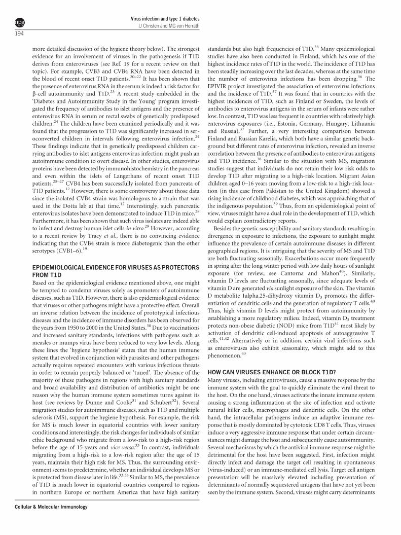
Figure 1 Sequential challenge by environmental factors. (a) Several events occurring throughout an individual’s lifetime might influence the balance of the immune
system towards autoimmune disease or protection. The genetic predisposition for disease might on the one hand determine a certain starting point and on the other
hand influence the magnitude of the different immune reactions against environmental challenges. Several events including detrimental and protective virus
infections, exposure to xenobiotica and molecular mimicry might be involved in the modulation of the immune repertoire and its balance. The accumulation of
detrimental events combined with a sufficient genetic susceptibility might push the immune balance over a certain threshold for the development of clinical
autoimmune disease. (b) Several scenarios might be feasible. First, a low genetic predisposition might be a reason for which the disease threshold is never reached.
In contrast, a high predisposition might accelerate the autoimmune process. Further, an impaired immune regulation might enhance all events that involve an
activation of the immune system, but might have no influence on the consequences of a b-cell toxin. Last, enhanced immune regulation by viral activation of regulatory
T cells or by enhanced exposure to sunlight and elevated vitamin D levels might cause a general dampening of the immune response and the disease threshold might
not be reached. T1D, type 1 diabetes.
Virus infection and type 1 diabetes
U Christen and MG von Herrath
196
Cellular & Molecular Immunology

autoimmune destruction of the pancreatic b-cells and the subsequent
clinical T1D. In Figure 1, we propose possible scenarios for the etiol-
ogy of T1D integrating the genetic predisposition and protective and
detrimental environmental factors. We postulate that the odds for
T1D might be the highest when genetic predisposition is paired with
several detrimental environmental triggering factors including viruses
and direct b-cell toxins. In addition, an impaired immune regulation
might exacerbate the disease. The Environmental Determinants of
Diabetes in the Young (TEDDY) study66 is designed to collect as much
information about such multiple triggering and/or protective factors.
The TEDDY study keeps track of the diet, infections, allergies and
other life experiences of neonates for up to an age of 15 years. After
an initial screening for genetic risk factors more than 7000 children
will be periodically screened for islet cell-specific autoantibodies. First
data from the TEDDY study are expected to be released in a few years
and will hopefully bring further insight in the etiology of T1D and/or
help to identify protective factors. Unfortunately, the TEDDY study
does not foresee sampling of stool and blood during infectious events,
which might be a huge drawback when trying to precisely identify the
infection agents.
An interesting, simple and unifying concept for how to distinguish
T1D enhancing from protective enteroviral infections was put forward
based on investigations by Tracy et al. (as discussed here previously),
who observed that more severe infections enhanced diabetes in the
NOD, whereas lower-replicating strains were protective.19 This
implies that antiviral vaccines should transform deleterious more
severe infections into protective ones and there is indeed an initiative
in Finland to develop such a vaccine and test this concept. It should
also be possible to test this concept in prospective studies such as
TEDDY, but to achieve this it might have been necessary to also
recover samples from children precisely at the time of febrile infec-
tions, which would likely encompass the more deleterious enterovirus
encounters.
ACKNOWLEDGEMENTS
UC is supported by grants of the German Research Foundation. MGvH is
supported by a scholar award of the Juvenile Diabetes Research Foundation and
a Program Project Grant of the National Institute of Health to the La Jolla
Institute for Allergy and Immunology.
1 Todd JA. Etiology of type 1 diabetes. Immunity 2010; 32: 457–467.
2 Ziegler AG, Nepom GT. Prediction and pathogenesis in type 1 diabetes. Immunity2010; 32: 468–478.
3 Redondo MJ, Rewers M, Yu L, Garg S, Pilcher CC, Elliott RB et al. Geneticdetermination of islet cell autoimmunity in monozygotic twin, dizygotic twin, andnon-twin siblings of patients with type 1 diabetes: prospective twin study. BMJ1999; 318: 698–702.
4 Bonifacio E, Hummel M, Walter M, Schmid S, Ziegler AG. IDDM1 and multiple familyhistory of type 1 diabetes combine to identify neonates at high risk for type 1 diabetes.Diabetes Care 2004; 27: 2695–700.
5 Christen U, Benke D, Wolfe T, Rodrigo E, Rhode A, Hughes AC et al. Cure ofprediabetic mice by viral infections involves lymphocyte recruitment along an IP-10gradient. J Clin Invest 2004; 113: 74–84.
6 Filippi CM, Estes EA, Oldham JE, von Herrath MG. Immunoregulatory mechanismstriggered by viral infections protect from type 1 diabetes in mice. J Clin Invest 2009;119: 1515–1523.
7 Ehlers S, Kaufmann SH. 99th Dahlem conference on infection, inflammation andchronic inflammatory disorders: lifestyle changes affecting the host-environmentinterface. Clin Exp Immunol 2010; 160: 10–14.
8 Filippi CM, von Herrath MG. 99th Dahlem conference on infection, inflammation andchronic inflammatory disorders: viruses, autoimmunity and immunoregulation. ClinExp Immunol 2010; 160: 113–119.
9 Chatenoud L, You S, Okada H, Kuhn C, Michaud B, Bach JF. 99th Dahlem conferenceon infection, inflammation and chronic inflammatory disorders: immune therapies oftype 1 diabetes: new opportunities based on the hygiene hypothesis. Clin ExpImmunol 2010; 160: 106–112.
10 Gamble DR, Kinsley ML, FitzGerald MG, Bolton R, Taylor KW. Viral antibodies indiabetes mellitus. Br Med J 1969; 3: 627–630.
11 Al-Hello H, Paananen A, Eskelinen M, Ylipaasto P, Hovi T, Salmela K et al. Anenterovirus strain isolated from diabetic child belongs to a genetic subcluster ofechovirus 11, but is also neutralised with monotypic antisera to coxsackievirus A9.J Gen Virol 2008; 89: 1949–1959.
12 Dotta F, Censini S, van Halteren AG, Marselli L, Masini M, Dionisi S et al. Coxsackie B4virus infection of beta cells and natural killer cell insulitis in recent-onset type 1diabetic patients. Proc Natl Acad Sci USA 2007; 104: 5115–5120.
13 Yoon JW, Austin M, Onodera T, Notkins AL. Virus-induced diabetes mellitus: isolationof a virus from the pancreas of a child with diabetic ketoacidosis. N Engl J Med 1979;300: 1173–1179.
14 Honeyman MC, Coulson BS, Stone NL, Gellert SA, Goldwater PN, Steele CE et al.Association between rotavirus infection and pancreatic islet autoimmunity in childrenat risk of developing type 1 diabetes. Diabetes 2000; 49: 1319–1324.
15 Hyoty H, Leinikki P, Reunanen A, Ilonen J, Surcel HM, Rilva A et al. Mumps infectionsin the etiology of type 1 (insulin-dependent) diabetes. Diabetes Res 1988; 9: 111–116.
16 Gale EA. Congenital rubella: citation virus or viral cause of type 1 diabetes?Diabetologia 2008; 51: 1559–1566.
17 Pak CY, Eun HM, McArthur RG, Yoon JW. Association of cytomegalovirus infectionwith autoimmune type 1 diabetes. Lancet 1988; 2: 1–4.
18 Jenson AB, Rosenberg HS, Notkins AL. Pancreatic islet-cell damage in children withfatal viral infections. Lancet 1980; 2: 354–358.
19 Tracy S, Drescher KM, Jackson JD, Kim K, Kono K. Enteroviruses, type 1 diabetes andhygiene: a complex relationship. Rev Med Virol 2010; 20: 106–116.
20 Andreoletti L, Hober D, Hober-Vandenberghe C, Belaich S, Vantyghem MC, Lefebvre Jet al. Detection of coxsackie B virus RNA sequences in whole blood samples from adultpatients at the onset of type I diabetes mellitus. J Med Virol 1997; 52: 121–127.
21 Schulte BM, Bakkers J, Lanke KH, Melchers WJ, Westerlaken C, Allebes W et al.Detection of enterovirus RNA in peripheral blood mononuclear cells of type 1diabetic patients beyond the stage of acute infection. Viral Immunol 2010; 23: 99–104.
22 Clements GB, Galbraith DN, Taylor KW. Coxsackie B virus infection and onset ofchildhood diabetes. Lancet 1995; 346: 221–223.
23 Lonnrot M, Salminen K, Knip M, Savola K, Kulmala P, Leinikki P et al. EnterovirusRNA in serum is a risk factor for beta-cell autoimmunity and clinical type 1 diabetes: aprospective study. Childhood Diabetes in Finland (DiMe) Study Group. J Med Virol2000; 61: 214–220.
24 Stene LC, Oikarinen S, Hyoty H, Barriga KJ, Norris JM, Klingensmith G et al.Enterovirus infection and progression from islet autoimmunity to type 1 diabetes:The Diabetes and Autoimmunity Study in the Young (DAISY). Diabetes 2010; 59:3174–3180.
25 Yoon JW, Austin M, Onodera T, Notkins AL. Isolation of a virus from the pancreas of achild with diabetic ketoacidosis. N Engl J Med 1979; 300: 1173–1179.
26 Richardson SJ, Willcox A, Bone AJ, Foulis AK, Morgan NG. The prevalence ofenteroviral capsid protein vp1 immunostaining in pancreatic islets in human type 1diabetes. Diabetologia 2009; 52: 1143–1151.
27 Richardson SJ, Willcox A, Hilton DA, Tauriainen S, Hyoty H, Bone AJ et al. Use ofantisera directed against dsRNA to detect viral infections in formalin-fixed paraffin-embedded tissue. J Clin Virol 2010; 49: 180–185.
28 Chatterjee NK, Nejman C, Gerling I. Purification and characterization of a strain ofcoxsackievirus B4 of human origin that induces diabetes in mice. J Med Virol 1988;26: 57–69.
29 Elshebani A, Olsson A, Westman J, Tuvemo T, Korsgren O, Frisk G. Effects on isolatedhuman pancreatic islet cells after infection with strains of enterovirus isolated atclinical presentation of type 1 diabetes. Virus Res 2007; 124: 193–203.
30 Bach JF. The effect of infections on susceptibility to autoimmune and allergicdiseases. N Engl J Med 2002; 347: 911–920.
31 Dunne DW, Cooke A. A worm’s eye view of the immune system: consequences forevolution of human autoimmune disease. Nat Rev Immunol 2005; 5: 420–426.
32 Schubert C. News feature: the worm has turned. Nat Med 2004; 10: 1271–1272.
33 Kurtzke JF. Epidemiologic evidence for multiple sclerosis as an infection. ClinMicrobiol Rev 1993; 6: 382–427.
34 Fujinami RS, von Herrath MG, Christen U, Whitton JL. Molecular mimicry, bystanderactivation, or viral persistence: infections and autoimmune disease. Clin MicrobiolRev 2006; 19: 80–94.
35 Zaccone P, Fehervari Z, Phillips JM, Dunne DW, Cooke A. Parasitic worms andinflammatory diseases. Parasite Immunol 2006; 28: 515–523.
36 Viskari H, Ludvigsson J, Uibo R, Salur L, Marciulionyte D, Hermann R et al.Relationship between the incidence of type 1 diabetes and maternal enterovirusantibodies: time trends and geographical variation. Diabetologia 2005; 48: 1280–1287.
37 Viskari H, Ludvigsson J, Uibo R, Salur L, Marciulionyte D, Hermann R et al.Relationship between the incidence of type 1 diabetes and enterovirus infections indifferent European populations: results from the EPIVIR project. J Med Virol 2004;72: 610–617.
38 Seiskari T, Kondrashova A, Viskari H, Kaila M, Haapala AM, Aittoniemi J et al. Allergicsensitization and microbial load—a comparison between Finland and Russian Karelia.Clin Exp Immunol 2007; 148: 47–52.
39 Bodansky HJ, Staines A, Stephenson C, Haigh D, Cartwright R. Evidence for anenvironmental effect in the aetiology of insulin dependent diabetes in atransmigratory population. BMJ 1992; 304: 1020–1022.
Virus infection and type 1 diabetesU Christen and MG von Herrath
197
Cellular & Molecular Immunology

40 Cantorna MT, Mahon BD. Mounting evidence for vitamin D as an environmental factoraffecting autoimmune disease prevalence. Exp Biol Med (Maywood) 2004; 229:1136–1142.
41 Gregori S, Giarratana N, Smiroldo S, Uskokovic M, Adorini L. A 1alpha,25-dihydroxyvitamin D3 analog enhances regulatory T-cells and arrests autoimmunediabetes in NOD mice. Diabetes 2002; 51: 1367–1374.
42 Decallonne B, van Etten E, Overbergh L, Valckx D, Bouillon R, Mathieu C. 1Alpha,25-dihydroxyvitamin D3 restores thymocyte apoptosis sensitivity in non-obese diabetic(NOD) mice through dendritic cells. J Autoimmun 2005; 24: 281–289.
43 Fisman DN. Seasonality of infectious diseases. Annu Rev Public Health 2007; 28:127–143.
44 Christen U, Hintermann E, Holdener M, von Herrath MG. Viral triggers for autoimmunity:is the ‘glass of molecular mimicry’ half full or half empty? J Autoimmun 2010; 34: 38–44.
45 Christen U, von Herrath MG. Induction, acceleration or prevention of autoimmunity bymolecular mimicry. Mol Immunol 2004; 40: 1113–1120.
46 Damian RT. Molecular mimicry: antigen sharing by parasite and host and itsconsequences. Am Nat 1964; 98: 129–149.
47 Oldstone MB. Molecular mimicry as a mechanism for the cause and as a probeuncovering etiologic agent(s) of autoimmune disease. Curr Top Microbiol Immunol1989; 145: 127–136.
48 von Herrath MG, Fujinami RS, Whitton JL. Microorganisms and autoimmunity: makingthe barren field fertile. Nat Rev Microbiol 2003; 1: 151–157.
49 Rhode A, Pauza ME, Barral AM, Rodrigo E, Oldstone MB, von Herrath MG et al. Islet-specific expression of CXCL10 causes spontaneous islet infiltration and acceleratesdiabetes development. J Immunol 2005; 175: 3516–3524.
50 Eizirik DL, Colli ML, Ortis F. The role of inflammation in insulitis and beta-cell loss intype 1 diabetes. Nat Rev Endocrinol 2009; 5: 219–226.
51 D’Alise AM, Auyeung V, Feuerer M, Nishio J, Fontenot J, Benoist C et al. The defect inT-cell regulation in NOD mice is an effect on the T-cell effectors. Proc Natl Acad SciUSA 2008; 105: 19857–19862.
52 Freimuth P, Philipson L, Carson SD. The coxsackievirus and adenovirus receptor. CurrTop Microbiol Immunol 2008; 323: 67–87.
53 Horwitz MS, Bradley LM, Harbertson J, Krahl T, Lee J, Sarvetnick N. Diabetes inducedby Coxsackie virus: initiation by bystander damage and not molecular mimicry. NatMed 1998; 4: 781–785.
54 Horwitz MS, Ilic A, Fine C, Balasa B, Sarvetnick N. Coxsackieviral-mediated diabetes:induction requires antigen-presenting cells and is accompanied by phagocytosis ofbeta cells. Clin Immunol 2004; 110: 134–144.
55 Serreze DV, Ottendorfer EW, Ellis TM, Gauntt CJ, Atkinson MA. Acceleration of type 1diabetes by a coxsackievirus infection requires a preexisting critical mass ofautoreactive T-cells in pancreatic islets. Diabetes 2000; 49: 708–711.
56 Coleman TJ, Gamble DR, Taylor KW. Diabetes in mice after Coxsackie B4 virusinfection. Br Med J 1973; 3: 25–27.
57 Tracy S, Drescher KM, Chapman NM, Kim KS, Carson SD, Pirruccello S et al. Towardtesting the hypothesis that group B coxsackieviruses (CVB) trigger insulin-dependentdiabetes: inoculating nonobese diabetic mice with CVB markedly lowers diabetesincidence. J Virol 2002; 76: 12097–12111.
58 Kanno T, Kim K, Kono K, Drescher KM, Chapman NM, Tracy S. Group B coxsackievirusdiabetogenic phenotype correlates with replication efficiency. J Virol 2006; 80:5637–5643.
59 Schulte BM, Kramer M, Ansems M, Lanke KH, van Doremalen N, Piganelli JD et al.Phagocytosis of enterovirus-infected pancreatic beta-cells triggers innate immuneresponses in human dendritic cells. Diabetes 2010; 59: 1182–1191.
60 Oldstone MB, Nerenberg M, Southern P, Price J, Lewicki H. Virus infection triggersinsulin-dependent diabetes mellitus in a transgenic model: Role of anti-self (virus)immune response. Cell 1991; 65: 319–331.
61 von Herrath MG, Dockter J, Oldstone MB. How virus induces a rapid or slow onsetinsulin-dependent diabetes mellitus in a transgenic model. Immunity 1994; 1: 231–242.
62 Oldstone MB. Prevention of type I diabetes in nonobese diabetic mice by virusinfection. Science 1988; 239: 500–502.
63 Christen U, von Herrath MG. Infections and autoimmunity—good or bad? J Immunol2005; 174: 7481–7486.
64 Selin LK, Welsh RM. Plasticity of T cell memory responses to viruses. Immunity 2004;20: 5–16.
65 Christen U, Edelmann KH, McGavern DB, Wolfe T, Coon B, Teague MK et al. A viralepitope that mimics a self antigen can accelerate but not initiate autoimmunediabetes. J Clin Invest 2004; 114: 1290–1298.
66 Homepage TEDDY Study. Available from: http://teddy.epi.usf.edu/
Virus infection and type 1 diabetes
U Christen and MG von Herrath
198
Cellular & Molecular Immunology