Embed Size (px)
Citation preview
DOBI MEDICAL INTERNATIONAL
DOBI COMFORTSCAN™ SYSTEMCLINICAL EFFECTIVENESS
EVALUATION REPORT
27-MARCH-2013
G. John Zhang, Ph.D.
DOBI ComfortScan™ SystemClinical Effectiveness Evaluation Report Page 2 of 147
CONTENT
PURPOSE ......................................................................................................................... 7SCOPE ............................................................................................................................. 7OBJECTIVE .................................................................................................................... 13CLINICAL EFFECTIVENESS REPORT............................................................................. 14
1. BACKGROUND ............................................................................................ 182. THE NEED FOR NEW DIAGNOSTIC TECHNOLOGIES ......................... 19
2.1 Mammography.......................................................................................... 202.2 Ultrasound, MRI and PET ........................................................................ 212.3 Biopsy ....................................................................................................... 22
3. BACKGROUND A NEW TECHNIQUE TO DETECT BREAST CANCER AT EARLY STAGE – DYANAMIC OPTICAL BREAST IMAGING........ 22
3.1 The Role of Angiogenesis in New Breast Cancer Diagnostic Technologies................................................................................................................... 23
3.2 Dynamic Opitical Breast Imaging Technology in Early Breast Cancer Diagnostic ................................................................................................. 29
3.3 Dynamic Optical Breast Imaging Principle .............................................. 344. MATERIAL and METHODS......................................................................... 36
4.1 ComfortScan Components and Description.............................................. 364.2 Use of the DOBI ComfortScan with a Patient .......................................... 394.3 ComfortScan Sanning Acquisition ........................................................... 404.4 ComfortScan Image Processing................................................................ 414.5 ComfortScan Image Interpretation ........................................................... 424.6 Comparison with Mammography ............................................................. 434.7 Clinical Testing......................................................................................... 44
5. CLINICAL EFFECTIVENESS...................................................................... 465.1 Report : Research Article, “The Dynamic Optical Breast Imaging in the
Preoperative Workflow of Women with Suspicious or Malignant Breast Lesions: Development of a New Comprehensive Score” by Massimiliano D'Aiuto, Giuseppe Frasci, Maria Luisa Barretta, Adolfo Gallipoli, Giovanni Maria Ciuffo, Flavia Musco, Sergio Orefice, Viviana Frattini, Ilves Guidi, Claudio Siani, Emanuela Esposito, Anna Crispo, Maurizio Montella, Andrea Chirico, Giuseppe D'Aiuto, and Aldo Vecchione atDepartment of Breast Disease, National Cancer Institute “G. Pascale” Foundation, Via Mariano Semmola, 80136 Naples, Italy, Department of Radiology, National Cancer Institute “G. Pascale” Foundation, Via Mariano Semmola, 80136 Naples, Italy, Breast Unit, Clinical Institute Zucchi, Via Zucchi 24, 20052 Monza, Italy, Breast Unit, Clinical Institute Pio X, Via Francesco Nava 31, 20159 Milano, Italy, Breast Surgery Divison, Medical Institute Monterosa, Via Monterosa 3, 20149 Milano, Italy, Breast Prevention Area, Physios Clinic, Via Chiesa Nord 52, 41016 Modena, Italy, Epidemiology Division, National Cancer Institute “G. Pascale” Foundation, Via Mariano Semmola, 80136 Naples, Italy, National Cancer Institute “G. Pascale” Foundation, Via Mariano
DOBI ComfortScan™ SystemClinical Effectiveness Evaluation Report Page 3 of 147
Semmola, 80136 Naples, Italy, ISRN Oncology, Volume 2012, Aritcle ID 631917, Accepted 29 July 2012................................................................ 47
5.2 Report : “Clinical Approach with Optical Imaging Instrument : Perspective analysis on 617 young females” by V. Frattini, L. Ghisoni, A. Teodoro, PL Vaj and S. Orefice at Habilita Group-Bergamo, Centro Medico MonteRosa of Milan, Humanitas-Rozzano of Milan and Habilita Group-Bergamo, Italy, Italian Journal of Gynaecology and Obstetrics, Volume 23, Issue 2-3, October 3, 2011 .................................................... 49
5.3 Report : DOBI Score Ongoing Project by DOBI Sough European Distributor, Socrate Medical, Milano, Italy, 2011 .................................... 50
5.4 Report : Dynamic Optical Imaging. Thesis Dissertation : “The Use of Dynamic Optical Imaging in Breast Cancer Detection” by Wilson, Kyle, Ph.D., MCMASTER UNIVERSITY and Publication submitted to Radiology “Dynamic Optical Imaging in Breast Cancer Detection” byKyle J. Wilson, Kavita Dhamanaskar, Terry Minuk, and Gerald R. Moran, Canada, 2011............................................................................................. 51
5.5 Report : Dynamic Optical Breast Imaging (DOBI): Prospective Study of ComfortScan Accuracy in Diagnosis of Breast Cancer. PRELIMINARY RESULTS, Thesis Dissertation by Rossella Dandolo, Ph.D., University of Rome Tor Vergata, Italy, 2010 ................................................................. 53
5.6 Report : Dynamic Optical Breast Imaging (DOBI), associated Ultrasound, allows to avoid unnecessary Biopsies - “Seno, luce rossa preventiva : Il Dynamic optical breast imaging (Dobi), associato all’ecografia, permette di evitare biopsie non necessarie” by di Piercarlo Salari, Oncologia, Tecnologie, October 12, 2009................................................................... 57
5.7 Report : “Dynamic optical breast imaging: A novel technique to detect and characterize tumor vessels” Published by Laure S. Fournier, Daniel Vanel, Alexandra Athanasiou, Wolfgang Gatzemeier, I.V. Masuykov, Anwar R. Padhani, Clarisse Dromain, Ken Galetti, Robert Sigal, Alberto Costa, Cornne Balleyguier on Europea Journal of Radiology 69 (2009) ............ 58
5.8 Report : “Optical mammography: a new technique for visualizing breast lesions in women presenting non palpable BIRADS 4–5 imaging findings:preliminary results with radiologic–pathologic correlation” Published by Alexandra Athanasiou, Daniel Vanel, Laure Fournier, and Corinne Balleyguier on International Cancer Imaging Society 7 (2007) ............... 60
5.9 Report : “Dynamic Optical Breast Imaging: A new technique to visualise breast vessels: Comparison with breast MRI and preliminary results”Published by Alexandra Athanasiou, Daniel Vanel, Cornne Balleyguier, Laure Fournier, Marie Christine Mathieu, Suzette Delaloge, Clarisse Dromain on Europea Journal of Radiology 54 (2005).............................. 64
5.10 Report : “Correlation of Dynamic Optical Breast Imaging Curve and Microvessel Density Count” Presented by Abraham A. Ghiatas, K Pavlaki, I Messini, N Karaglani, D Keramopoullos, V Gaki, D Baltas, and N Bredakis from Iaso Hospital in Greece in Cancer Imaging 6 (2006) ....... 65
DOBI ComfortScan™ SystemClinical Effectiveness Evaluation Report Page 4 of 147
5.11 Report : “Digital Optical Breast Imaging” Presented by Abraham A. Ghiatas from Iaso Hospital in Greece (2005) ........................................... 65
5.12 Report : The purpose of our study was to report: “New Perspective of Mammary Screening : Application of Non-Invasive DOBI”, THERESULTS OF DOBI EXAMINATIONS IN MASARYK MEMORIAL CANCER INSTITUTE in Czech Republic, by Irena Komorousova, Bartonkova H., Standara M., Schneiderova M., from 2004 to 2005 ........ 66
5.13 Report : “Dynamic Optical Breast Imaging: A non-invasive, adjunctive method to detect breast cancer” Presented by Gatzemeier W, Scelsi M, Galetti K, Villani L, Tinterri C, Secci A, and Costa A in San Antonio Breast Cancer Symposium (December 2004)........................................... 68
5.14 Report : “Dynamic Functional Mammoscopy : A Non-Ionizing Imaging Technique Ehahcing Early Detection of Breast Cancer” Published by Suzanne J. Smith, Scientific and Technical Aerospace Reports 42 (May, 2004) ......................................................................................................... 69
5.15 Report : “White Paper – Dynamic Optical Breast Imaging : Preliminary Results from Two Data Interpretation Methods” by DOBI Medical International (September, 2004) ............................................................... 70
5.16 Report : “The Value of Dynamic Optical Breast Imaging and Ultrasound in Early Breast Cancer Detection” Reported by Guojian Tan, Jie Wang, Cui Liu, Chunmian Li, Weiping Wang, John Zhang (2010) .................... 72
5.17 Report : “Preliminary Research on Dynamic Optical Breast Imaging in Breast Cancer” Published by Guojian Tan, Jie Wang, Cui Liu, Chunmian Li, John Zhang, Weiping Wang on Chinese Journal of Medical Imaging 18 (2010)................................................................................................... 73
5.18 Report : “The Diagnostic Value of Small Lesions within Breast by Ultrasound combined with DOBI” published by Mei Xu, Junlai Li, MEDICAL JOURNAL OF CHINESE PEOPLE'S LIBERATION ARMY,Vol 34, No. 8 on August 1, 2009 .............................................................. 74
5.19 Report : “The Value Analysis of ComfortScan System in Differentiating Benign Breast Lesions from Malignant” published by Mei Xu, Junlai Li, Yongfeng Zhang, Xuejuan Shi, Chunmian Li, Jie Tiang, and “The Application of Dynamic Optical Breast Imaging in Differentiating Benign Breast Lesions from Malignant” Thesis submitted by Mei Xu at Chinese PLA General Hospital & Postcarduate Medical School on June 1, 2009. 74
5.20 Report : “ComfortScan System and Ultrasound Imaging: The Value of Combined Application to Differentiate Benign Breast Lesions fromMalignant” Reported by Yongfeng Zhang, Junlai Li, Xuejuan Shi, Mei Xu, China, 2008 ........................................................................................ 75
5.21 Report : “ComfortScanTM System Clinical Evaluation Report” for SFDA Review by G. Zhang and W. Lin based on data collected from 2008 to 2009, China, Febuary, 2011...................................................................... 76
5.22 Report : “ComfortScanTM System Performance Report” for SFDA Review by G. Zhang and W. Lin based on data collected from 2008 to 2009, China, Febuary, 2011................................................................................ 78
DOBI ComfortScan™ SystemClinical Effectiveness Evaluation Report Page 5 of 147
5.23 Report : “RESULTS OF INVESTIGATIONAL USE OF DOBI COMFORTSCAN IN CHINA” by G. Zhang, W. Wang, D. Yang and H. Jiang from 2005 to 2006 ........................................................................... 79
6. CLINICAL ACCEPTANCE........................................................................... 936.1 The Italian League for the Fight against Cencer (LILT) has recommeded
to DOBI ComfortScan for younger women in detecting breast cancer at early stage on August 2, 2012................................................................... 94
6.2 The Italian League for the Fight against Cancer (LILT) – Underforty Women Breast Care, 2010 ........................................................................ 96
6.3 2nd Meeting DOBI Group in Italy, 2011................................................ 1006.4 1st Meeting DOBI Group in Italy, 2010 ................................................. 1016.5 ANT: from now on mammograms for women under 40: 22/10/2012 - As
of today young women may be subjected to investigation mammography. With the resort Cancer Prevention Foundation ANT, and DOBI mammography optical non-invasive and without radiation.................... 102
6.6 Breast cancer diagnosis today is born with "DOBI"............................... 1036.7 Breast cancer remains the second leading cause of death among women
................................................................................................................. 1046.8 UNIT 'ANT CANCER PREVENTION PROJECTS FOR POLICE AND
UNICREDIT........................................................................................... 1056.9 THE INNOVATIVE METHODS OF STUDY MEDICAL MANARA 31
................................................................................................................. 1066.10 INSTITUTE mammograms ANT EVEN UNDER 40 YEARS ............. 1076.11 Know, prevent, cure: the fundamental objectives of the Centre............. 1086.12 From tomorrow morning a new entry with the camper made available by
the project underforty LILT Bologna, with breast ultrasound and optical technology mammary (ComfortScan Dobi) for this occasion for the first time in southern Italy. ............................................................................. 110
6.13 As breast, ComfortScan presents as a solution for the prevention ......... 1116.14 Prevention of breast cancer: the solution Italian for a global problem... 1136.15 A New Weapon Without radiation for the diagnosis of breast cancer in
young women – Women’s Health in Italy .............................................. 1156.16 US-China Technical Fight against Cancer with no more X-rays, August
2011......................................................................................................... 1166.17 PREVENTING BREAST: NEW DIAGNOSTIC .................................. 1176.18 DOBI, the New Technology against Breast Cancer on Oct. 17, 2011.... 1186.19 Ready for a life-saving machine, March 14, 2011.................................. 1196.20 Monza, care of the breast. Institutes Zucchi intervene in Rome, Feburary
2011......................................................................................................... 1196.21 DOBI New Screening for Breast Cancer that does not use X-rays ........ 1206.22 The New Frontier Optical Survey, 2011................................................. 1206.23 Screening mammography: what to do?, April 19, 2011 ......................... 1216.24 Professor Rocca’s Trial........................................................................... 1226.25 READY FOR A LIFE SUPPORT MACHINE, 2011 ............................ 1236.26 A Step Forward to Prevent Breast Cancer - Health in Italy.................... 124
DOBI ComfortScan™ SystemClinical Effectiveness Evaluation Report Page 6 of 147
6.27 New Light in the diagnosis – LILT Magazine in Italy ........................... 1246.28 The City of Rome in the front line against Breast Cancer - The I.P.A. in
Italy ......................................................................................................... 1256.29 Breast Safe with the Examination at Red Lights – July Fitness in Italy. 1266.30 The Functional Optical Systems : Vanguard for the Fight against Breast
Cancer ..................................................................................................... 1276.31 The Technology to Fight Breast Cancer - Introduction of DOBI.......... 1276.32 Breast Cancer, Zucchi Hospital Leader in Italy - Use the Wavelength of
Right Light : DOBI Unit Acitve April 2009........................................... 1286.33 Private Clinics in Italy, A New Light In the Diagnosis of Brest Cancer ,
Breast Optical Mammography................................................................ 1296.34 Innovative Companies of the Year 2002 ................................................ 1306.35 Wall Street Reporter Magazine on February 25, 2004 ........................... 1316.36 Wall Street Transcript on July 11, 2005 ................................................. 1326.37 Rising Star Stocks – A Revolutionary Medical Imaging Device on August,
2004......................................................................................................... 1336.38 Rising Star Stocks – A Revolutionary Medical Imaging Device on August,
2004......................................................................................................... 1346.39 MD Buyline Leading Edge Report : 10 New Technologies that Could
Change Healthcare – January 2005......................................................... 1356.40 Enhancing Mammography: New Tactics for Early Brest Cancer Detection
– Oncology Time 25 on March 10, 2003................................................ 1366.41 Body Mind : The Future of Breast Cancer, More Magazine on October
2005......................................................................................................... 1376.42 Leading International Physicians Discuss DOBI ComfortScan in Prague,
the Czech Republic from September 9-11, 2005 .................................... 1387. CONCLUSION............................................................................................. 139
REFERENCES ......................................................................................................... 145
DOBI ComfortScan™ SystemClinical Effectiveness Evaluation Report Page 7 of 147
PURPOSE
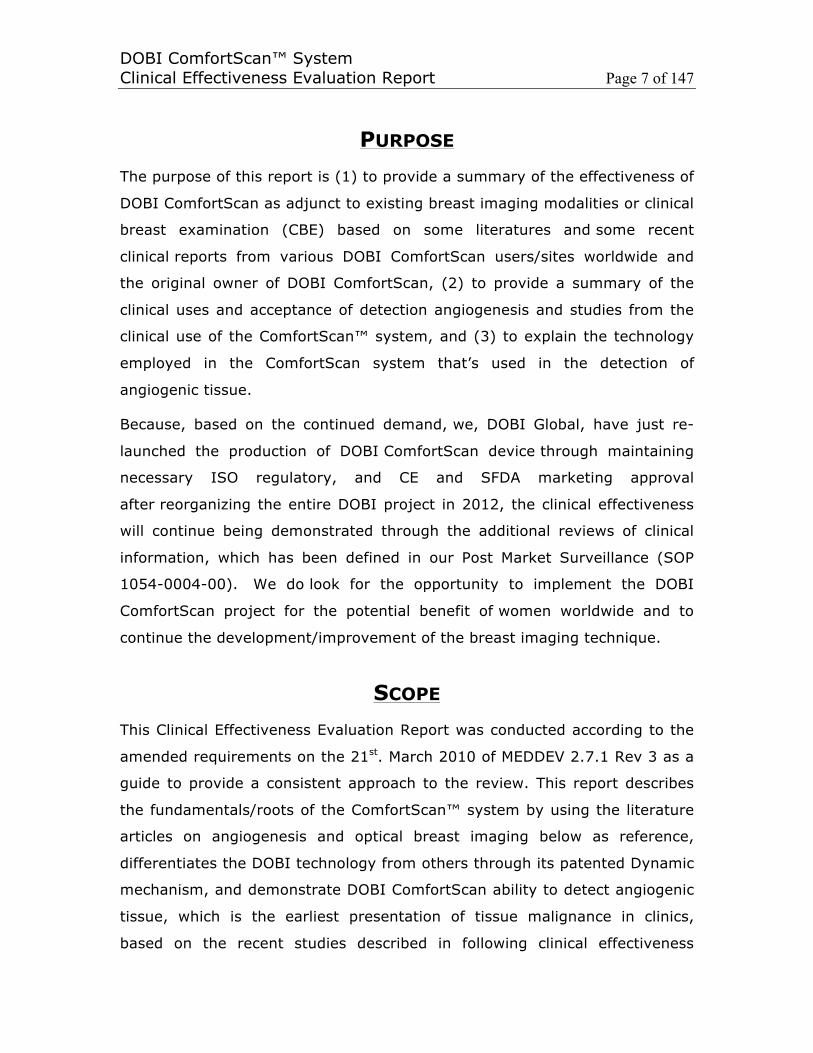
The purpose of this report is (1) to provide a summary of the effectiveness of
DOBI ComfortScan as adjunct to existing breast imaging modalities or clinical
breast examination (CBE) based on some literatures and some recent
clinical reports from various DOBI ComfortScan users/sites worldwide and
the original owner of DOBI ComfortScan, (2) to provide a summary of the
clinical uses and acceptance of detection angiogenesis and studies from the
clinical use of the ComfortScan™ system, and (3) to explain the technology
employed in the ComfortScan system that’s used in the detection of
angiogenic tissue.
Because, based on the continued demand, we, DOBI Global, have just re-
launched the production of DOBI ComfortScan device through maintaining
necessary ISO regulatory, and CE and SFDA marketing approval
after reorganizing the entire DOBI project in 2012, the clinical effectiveness
will continue being demonstrated through the additional reviews of clinical
information, which has been defined in our Post Market Surveillance (SOP
1054-0004-00). We do look for the opportunity to implement the DOBI
ComfortScan project for the potential benefit of women worldwide and to
continue the development/improvement of the breast imaging technique.
SCOPE
This Clinical Effectiveness Evaluation Report was conducted according to the
amended requirements on the 21st. March 2010 of MEDDEV 2.7.1 Rev 3 as a
guide to provide a consistent approach to the review. This report describes
the fundamentals/roots of the ComfortScan™ system by using the literature
articles on angiogenesis and optical breast imaging below as reference,
differentiates the DOBI technology from others through its patented Dynamic
mechanism, and demonstrate DOBI ComfortScan ability to detect angiogenic
tissue, which is the earliest presentation of tissue malignance in clinics,
based on the recent studies described in following clinical effectiveness
DOBI ComfortScan™ SystemClinical Effectiveness Evaluation Report Page 8 of 147
section five.
Since DOBI ComfortScan technique is based on the Angiogenesis Theory
published by Judah Folkman 1971 and the Optical Imaging of the Breast
introduced by Max Cutler in 1929, the clinical effectiveness and efficacy of
those two fundamentals will not addressed in this report, but their clinical
significances could be partially referred through following literature lists.
• “Clinical applications of research on angiogenesis”, Judah Folkman.
• “Chemotherapy targeted to tumor vasculature”, Wadih Arp, Renata
Pasqualini and Erlli Ruoslahti.
• “Inhibition of tumor Angiogenesis as a Strategy to Circumvent
Acquired Resistance to Anti-Cancer Therapeutic Agents”, Robert S.
Kerbel.
• “Functional imaging of the human body”, Eduard E. Godik and Yuri V.
Gulyaev.
• “Dynamic Optical Imaging” Edward Godik, Tamas Gergely, Vladimir
Liger, Vladimir Zlatov, Alex Taratorin.
• “Simulation of effect of mechanical loading on dynamics of breast
tissue optical properties” Alexander Dyachenko, John Gardner, Ivan
Masyukov, Alan Rego, Vladimir Zlatov.
• “The development of human gene therapy”, Theodore Friedmann.
• “Tumor development under Angiogenic Signaling: A dynamical theory
of tumor growth, treatment response and postvascular dormancy”,
Philip Hahnfeild, Dipak Panigrahy, Judah Folkman and Lynn Hlatky.
• “Vascular attack as a therapeutic strategy for cancer”, Juliana
Denekamp
• “The relationship between elevated interstitial fluid pressure and blood
flow in tumors: A bioengineering analysis”, Michael F. Milosevic,
DOBI ComfortScan™ SystemClinical Effectiveness Evaluation Report Page 9 of 147
Anthony W. Fyles and Richard P. Hill.
• “Optical tomography, photon migration and spectroscopy of tissue and
model media: Theory, human studies and instrumentation”, Britton
Chance, Abraham Katzir.
• “An Evaluation of transmission spectroscopy (Lightscanning) in the
diagnosis of symptomatic breast Lesions”, C. S. Dowle, Jennifer
Caseldine, Jennifer Tew, A. R. Manhire, E. J. Roeblck and R. W.
Blamey.
• “Diaphanography in the diagnosis of breast cancer”, Brad Drexler, J.
Leonard Davis and Gail Schofield.
• “Diagnostic Accuracy of Lightscanning and Mammography in Women
with Dense Breasts”, O. Jarlman, I. Andersson, G. Balldin and S. A.
Larsson.
• “Light scanning of nonpalpable breast lesions: Reevaluation”, Barbsrn
Monsees, Judy M. Destouet and Deborah Gersell.
• “LightScanning versus Mammography for the detection of breast
cancer in screening and clinical practice”, A Swedish Multicenter Study.
• “Relationship between lightscanning and the histologic and
mammographic appearance of malignant breast tumors”, O. Jarlman,
G. Balldin, I. Andersson, M. Löfgren, A. S. Larsson, and F. Linell.
• “Transillumination in breast cancer detection: screening failures and
potential”, G. Eric Geslien, J. Ronald Fisher and Colleen Delaney.
• “Recent advances in diffuse optical imaging”, A P Gibson, J C Hebden
and S R Arridge.
• “Optical imaging of the breast”, S.M.W.Y. van de Ven, S.G. Elias,
M.A.A.J. van den Bosch, P. Luijten and W.P.Th.M. Mali.
• “Assessing the future of diffuse optical imaging technologies”, Bruce J
Tromberg, Brian W Pogue, Keith D. Paulsen, Arjun G. Yodh, David A.
DOBI ComfortScan™ SystemClinical Effectiveness Evaluation Report Page 10 of 147
Boas, Albert E. Cerussi.
The improvements of the Optical Imaging Technology will be discussed within
this report as well as can be found from some literatures listed above.
Because of (1) the breakthrough of Angiogenesis Theory in clinics, (2) the
development of Semiconductor Technology in Optical Detector, and (3) the
tremendous efforts worldwide in past twenty years, optical breast imaging
technique has been developed significantly by comparing the study carried
out by A Swedish Multicenter Study with the publications of “LightScanning
versus Mammography for the detection of breast cancer in screening and
clinical practice” and “Relationship between lightscanning and the histologic
and mammographic appearance of malignant breast tumors”.
About ten (10) years ago, on March 8, 2001, in a press release announcing
the release of its comprehensive new study, Mammography and Beyond, the
National Academy of Sciences’ Institute of Medicine issued a call to action for
improvements in breast-imaging techniques. The Chapter 2, Breast Imaging
and Related Technologies, addressed as following:
• Optical imaging or tomography, which is relatively inexpensive and
simple in comparison with many other imaging modalities, is also
actively under investigation for a variety of cancers, including breast
cancer. The technique uses light in the near-infrared range
(wavelengths from 700 to 1,200 nm), which is nonionizing, to produce
an image of the breast. Potential advantages of the technology include
speed, comfort, and non-invasiveness. An optical scan can be taken in
less than 30 seconds by simply placing an image pad over the breast
without compression (Chance, 1998). Optical imaging methods offer
the potential to differentiate between soft tissues that are
indistinguishable by other modalities, and specific absorption by
natural chromophores (such as hemoglobin) can also provide biological
or functional information.
DOBI ComfortScan™ SystemClinical Effectiveness Evaluation Report Page 11 of 147
• Optical imaging systems are being commercially developed by Imaging
Diagnostic Systems Inc. (IMDS; Plantation, Florida), DOBI Medical
Systems (Mahwah, New Jersey), and Advanced Research and
Technology, Inc (ART; Montreal, Canada). The DOBI technology is
based on optical detection of angiogenesis in malignant lesions,
whereas the IMDS and ART technologies use laser-based technologies
to assess various optical properties of breast abnormalities. This
difference between DOBI and others is created by DOBI patented
Dynamic Technique. All three companies are conducting clinical trials
for FDA approval for diagnostic use of their devices, but they also plan
to pursue a screening approach in the future.
• In summary, optical imaging has long been thought to have potential
as a means of breast cancer detection, but to date that potential has
not yet been realized. Significant technological improvements in recent
years may eventually propel this technology into the clinic, but a
conclusion cannot yet be reached about its future utility.
The studies from the pioneer of Optical Breast Imaging, Professor Britton
Chance, who died in November 2010, led to the development of near infrared
(NIR) spectroscopy and imaging for real time metabolic studies of brain
(hematoma detection, prefrontal cortex monitoring, fetal brain oxygenation
in utero), breast (cancer detection using signals of angiogenesis and
hypermetabolism), skeletal muscle (metabolic monitoring) and cardiac
muscle (trans-thoracic detection of hypoxia of myocardium). His scholarly
articles, such as “Optical tomography, photon migration, and Spectroscop of
Tissue and Model Media: Theory, Human Studies, and Instrumentation”,
“Concurrent MRI and diffuse optical tomography of breast after indocyanine
green enhancement”, “Breast imaging technology: Probing physiology and
molecular function using optical imaging - applications to breast cancer”,
“MRI-Guided Diffuse Optical Spectroscopy of Malignant and Benign Breast
Lesions”, etc., published about ten (10) years ago have demonstrated the
DOBI ComfortScan™ SystemClinical Effectiveness Evaluation Report Page 12 of 147
capability of NIR Optical Imaging technique in detecting the malignancy of
the breast tumors.
The three publications, “Recent advances in diffuse optical imaging” by A P
Gibson, J C Hebden and S R Arridge; “Optical imaging of the breast” by
S.M.W.Y. van de Ven, S.G. Elias, M.A.A.J. van den Bosch, P. Luijten and
W.P.Th.M. Mali; “Assessing the future of diffuse optical imaging technologies”
by Bruce J Tromberg, Brian W Pogue, Keith D. Paulsen, Arjun G. Yodh, David
A. Boas, Albert E. Cerussi, in 2005, 2008 and 2008 respectively, have
described the development of optical imaging technology, which is an
emerging technique for functional imaging of biological tissue but still need
more clinical studies. A further role which optical imaging could fill is as a
low-cost, portable imaging system for use in primary care situations or at the
bedside. The transfer of new techniques and ideas for diffuse optical imaging
into clinical tools require close collaboration between engineers, clinicians,
scientists and mathematicians.
The paper of “Dynamical Optical Imaging” published on SPIE Vol. 2389/859
by E. Godik, T. Gergely, V. Liger, V. Zlatov, A. Taratorin, has addressed the
possibility for revealing and identifying pathology through the spatially
distributed low amplitude dynamic optical contrasts, which reflect the
physiological dynamics of the living tissue, described the a simple CCD-based
system and software for optical image sequence processing, and
demonstrated examples of the application of this approach for breast imaging
diagnostics. A USA “Dynamic-functional imaging of biological objects using a
non-rigid object holder” patent filed on July 1, 2003 and renewed on January
3, 2011 has described the technical improvement conventional Optical Breast
Imaging. The DOBI ComfortScan technique detects the differences of the
transilluminations between benign tissues and malignant tissues by
evaluating the light attenuation when an external pressure stimulus is
applied over time described by Dyachenko A “Dynamic imaging of breast
lesions; one dimensional optical model” (Asian Journal of Physics
DOBI ComfortScan™ SystemClinical Effectiveness Evaluation Report Page 13 of 147
2001;10;4:1-18). This patented Dynamic Technique differentiates DOBI
method from other optical imaging approach by enabling DOBI ComfortScan
ONLY to detect the angiogenesis in malignant tissue. For example, the
model described by Dowle et al is the Spectrascan lite scan model 10, which
is sometimes referred to as the Spectrascan or the Lite Scan. The
Spectrascan Lite scan passes infrared light through the breast to detect early
cancers. The machine produces a beam of light that alternates between the
red and/or infrared. The computer converts the image recorded by the video
camera into digital information, which is displayed on a color monitor. The
image shows area of different kinds of tissue in different colors with respect
to the different absorption of transmitted light. The Sepctrascan Model 10
made by Sepctrascan, Inc. has very high False Positive Rate. The DOBI
ComfortScan also detects the NIR light transmission through various
absorption tissues, and the recorded digital images also shows area of
different kinds of tissue in different colors with respect to the different
absorption of transmitted light. But the difference is that the uniform
pressure pulses applied on the breast decreases the False Positive Rate
through distinguishing the abnormal tissues, which contains the Angiogenesis,
from normal tissues. The detailed description of DOBI ComfortScan
Technique will be described in more details in following DOBI Technology
section.
OBJECTIVE
The purpose of this report is to demonstrate the EFFECTIVENESS of DOBI
ComfortScan as adjunct to existing breast imaging modalities, such as
Mammography or Ultrasound, in detecting breast cancer through recent
clinical studies worldwide and the improvement of the physician’s ability in
providing more accurate breast cancer diagnosis in earlier stage than current
imaging approaches through the market Acceptance and public Awareness of
the DOBI ComfortScan system in clinics.
DOBI ComfortScan™ SystemClinical Effectiveness Evaluation Report Page 14 of 147
CLINICAL EFFECTIVENESS REPORT
The DOBI ComfortScan System is designed to detect areas of abnormal vascularization
in breast tissue. DOBI technology is based on the now generally recognized
phenomenon of angiogenesis or the growth of new blood vessels around and in support
of malignant lesions. The increased vascularity associated with the growth of malignant
lesions is different from the vascularity supporting benign and normal tissue and
interstitial fluid pressure is elevated around malignant tumors. This vascularity in a
breast behaves differently in response to the stimulus of pressure modulation resulting in
different light absorption in the area of abnormal vascularization. The ComfortScan
system measures transmission of red light through the breast, recording the transient
response to a pressure stimulus that initiates changes in blood volume. As a result of
this pressure stimulus, the dynamic behavior of tissue optical properties creates different
contrast for areas of abnormal vascularization from neighboring normal breast tissue.
The potential for the ComfortScan system as a diagnostic tool is to non-invasively
differentiate between specific optical patterns of vascularized and non-vascularized
areas and therefore to provide the physician with additional information as to the
angiogenic status of the suspicious area. This information is intended to assist the
physician in the diagnostic process and possibly in treatment recommendations.
The effectiveness of DOBI ComfortScan as adjunct to existing breast imaging modalities
in detecting breast cancer are demonstrated through the recent study reports from
various DOBI ComfortScan users/sites worldwide and the original owner of DOBI
ComfortScan described in clinical effectiveness and clinical acceptance sections.
Because, based on the continued demand, the manufacturing of DOBI ComfortScan
device and application of a new CE Certification and ISO 13485 Certificate have just re-
launched after the intellectual property of the DOBI Technology and ComfortScan device
was reorganized on the May of 2010, the clinical effectiveness will continue being
demonstrated through the additional reviews of clinical information, which has been
defined in our Post Market Surveillance (SOP 1054-0004-00).
According to the amended requirements on the 21st March 2010 of the Medical Devices
Directive (93/42/EEC), we are going to demonstrate the Clinical Effectiveness of our
DOBI ComfortScan™ SystemClinical Effectiveness Evaluation Report Page 15 of 147
DOBI ComfortScan through following Clinical Effectiveness section which is the outcome
we have implemented our Post Market Surveillance of Dynamic Optical Breast Imaging
Systems (DOBI) until the December of 2012. Therefore, based on the MEDDEV 2.7.1
Rev 3 for the clinical effectiveness, we, DOBI Medical International, have followed our
Post-Marketing Surveillance in Section 5:
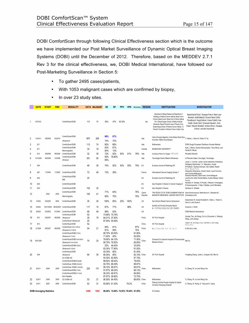
• To gather 2495 cases/patients,
• With 1053 malignant cases which are confirmed by biopsy,
• In over 23 study sites.
DATE START END MODALITY DATA MALIGNANT SE SP PPV VPN Accuracy REGION INSTITUATION INVESTIGATOR
1 9/27/2012 ComfortScan/DOBI 113 74 80% 87% 92.20% Italy
Department of Breast Disease and Department of Radiology of National Cancer Institute, Breast Unit of
Clinical Institute Zucchi, Breast Unit of Clinical Institute Pio X, Breast Surgery Divison of Medical Institute
Monterosa, Breast Prevention Area of Physios Clinic, Epidemiology Division of National Cancer Institute, “G. Pascale” Foundation of National Cancer Institute, Italy
MassimilianoD’Aiuto1, Giuseppe Frasci1, Maria Luisa Barretta2, AdolfoGallipoli2, Giovanni Maria Ciuffo3,
FlaviaMusco3, SergioOrefice4, Viviana Frattini5, Ilves Guidi6, Claudio Siani1, Emanuela Esposito1, Anna
Crispo7, Maurizio Montella7, Andrea Chirico1, Giuseppe D’Aiuto1, and Aldo Vecchione8
ComfortScan/DOBI 98% 87%Ultrasound 74% 70%
3 2011 ComfortScan/DOBI 113 74 80% 88% Italy Multicenters DOBI Sough European Distributor Socrate MedicalComfortScan/DOBI 83% 67%Mammography 94% 13%
5 2010 06/2009 09/2009 ComfortScan/DOBI 32 30 72% 92% 93% 61% 79% Italy University of Rome Tor Vergata Rossella DandoloComfortScan/DOBI 95% 78.80%Ultrasoun 66%
7 2009 ComfortScan/DOBI 46 35 74% 92% 93% 55% 79% EU Europea Journal of Radiology 69
Laure S. Fournier, Daniel Vanel,Alexandra Athanasiou, Wolfgang Gatzemeier, I.V. Masuykov, Anwar R.Padhani, Clarisse Dromain, Ken Galetti, Robert Sigal Alberto Costa
8 2007 11/2004 11/2005 ComfortScan/DOBI 72 49 73% 38% France International Cancer Imaging Society 7 Alexandra Athanasiou, Daniel Vanel, Laure Fournier, and Corinne Balleyguier
ComfortScan/DOBIMRI
10 2006 ComfortScan/DOBI EU Iaso Hospital in Greece in Cancer Imaging 6 Abraham A. Ghiatas, K Pavlaki, I Messini, N Karaglani, D Keramopoullos, V Gaki, D Baltas, and N Bredakis
11 2005 ComfortScan/DOBI 102 32 EU Iaso Hospital in Greece Abraham A. Ghiatas
ComfortScan/DOBI 71% 64% 76%
Mammography 66% 75% 76%
13 12/2004 03/2003 2004 ComfortScan/DOBI 35 30 100% 80% 20% 100% US San Antonio Breast Cancer Symposium Gatzemeier W, Scelsi M,Galetti K, Villani L, Tinterri C, Secci A, and Costa A
14 05/2004 10/01/2000 09/30/2001 ComfortScan/DOBI 117 15 87% 77% 98% US Scientific and Technical Aerospace Reports 42 42
Suzanne J. Smith
15 09/2004 03/2003 01/2004 ComfortScan/DOBI 68 49 98% 63% US DOBI Medical InternationalComfortScan/DOBI 52 73.68% 75.76%Ultrasound 35 84.21% 37.50%ComfortScan+Ultrasound 25 87.50% 55.56%
17 2010 ComfortScan/DOBI 52 China 301 PLA Hospital Guojian Tan, Jie Wang, Cui Liu, Chunmian Li, John Zhang, Weiping Wang
ComfortScan+UL(<=2cm) 95% 81% 87%Ultrasound (<=2cm) 81% 59% 70%ComfortScan/DOBI(<1cm) 85.70% 80.00% 81.80%Ultrasound (<1cm) 71.40% 40% 50.00%ComfortScan/DOBI(1cm-2cm) 78.60% 64.10% 71.00%Ultrasound (1cm-2cm) 85.70% 76.50% 80.60%ComfortScan/DOBI(>2cm) 25% 44.40% 33.00%Ultrasound (>2cm) 83.30% 77.80% 81.00%ComfortScan/DOBI 83.30% 80% 81.50%Ultrasound 80.00% 85% 83.10%CS DOBI+US 93.30% 82.90% 87.70%ComfortScan/DOBI(Overall) 89.62% 68.42% 78.63%ComfortScan DOBI(<2cm2) 93.75% 86.49% 88.67%ComfortScan DOBI(>=2cm2) 94.12% 66.67% 84.61%ComfortScan/DOBI(<1cm) 91.67% 88.24% 89.13%ComfortScan/DOBI/(>=1cm) 95.24% 66.67% 84.84%Non-Palpable 87.67% 58.82% 73.75%
22 02/2011 2008 2009 CS DOBI+US 53 21 90.50% 84.40% 86.60% China Multicenters G. Zhang, W. Lin and Wang Yan
23 6/2011 2005 2006 ComfortScan/DOBI 62 31 83.90% 61.30% 79.2% China Peking Univisity People Hospital & Capital Univisity Chaoyang Hospital G. Zhang, W. Wang, D. Yang and H. Jiang
DOBI Averaging Statistics 2495 1053 86.46% 74.60% 74.55% 78.64% 81.95% 23 Centers
220 131 China Multicenters
30 China 301 PLA Hospital Yongfeng Zhang, Junlai Li, Xuejuan Shi, Mei Xu
19 06/01/2009 74 33 ChinaChinese PLA General Hospital & Postcarduate Medical School
Mei Xu
di Piercarlo Salari, Oncologia, Tecnologie
9 2005 25 EU Europea Journal of Radiology 54Alexandra Athanasiou, Daniel Vanel, Cornne Balleyguier, LaureFournier, Marie Christine Mathieu, Suzette Delaloge, Clarisse Dromain
V. Frattinia, L. Ghisoni S. Orefice, PL Vaj
4 2011 33 19 Canada MCMASTER UNIVERSITY Kyle J. Wilson, Kavita Dhamanaskar, Terry Minuk, and Gerald R. Moran
2 10/3/2011 09/2008 03/2010 617 269 Italy Centro Chirurgico Magentino, Centro Medico Monte Rosa Humanitas, Habilita Group-Bergamo
China
6 10/12/2009 06/2008 12/2008 391 50 Italy Tecnologie Centro Medico Monterosa
12 2004 2005 100 41 Czech Republic
THE RESULTS OF DOBI EXAMINATIONS IN MASARYK MEMORIAL CANCER INSTITUTE
Irena Komorousova, Bartonkova H., Standara M., Schneiderova M.
16 2010 05/2007 06/2008 19 China 301 PLA Hospital Guojian Tan, Jie Wang, Cui Liu,Chunmian Li, Weiping Wang, John Zhang
18 8/1/2009 08/2007 06/2008 54 21 Xu Mei and Li Junlai
20 2008 64
21 02/2011 2008 2009 G. Zhang, W. Lin and Wang Yan
DOBI ComfortScan™ SystemClinical Effectiveness Evaluation Report Page 16 of 147
Those data are collected from the DOBI ComfortScan installed before 2012 to the fullest
extent practicable in accordance with the requirements of MEDDEV 2.12-2
GUIDELINES ON POST MARKET CLINICAL FOLLOW-UP (PMCF) and 1054-0004-00
Post-Marketing Surveillance of our company’s internal procedure through out
distributors, research partners and customers. To evaluate the clinical effectiveness of
DOBI ComfortScan comprehensively, some of the ComfortScan data are combined with
Mammography, Ultrasound and MRI in compliance with the requirements in MEDDEV
2.12-2 GUIDELINES on Post-Market Clinical Follow-up (PMCF) “in the form of follow up
specific sub-groups and/or prospective study”.
As an example of the collected data for this clinical evaluation from the a recent
publication at Italian Journal of Gynaecology and Obstetrics Volume 23 on October 3,
2011, with the supports of our Italy distributor, Dr. V. Frattini, L. Ghisoni, A. Teodoro, PL
Vaj and S. Orefice from Habilita Group-Bergamo, Centro Medico Monte Rosa, Istituto
Clinico Humanitas and Habilita Group-Bergamo in Italy conducted a multicenter study to
determine the Sensitivity and Specificity of the ComfortScan System to detect
malignancy as an adjunct to Ultrasound in patients between 25 to 39 years of age. A
total of 617 young females aged 25-39 with clinical risk to develop a cancer or dense
breast to live suspect breast cancer from standard imaging. There are 269 malignant
cases confirmed by biopsy. All subjects have been submitted to clinical investigation by
means of both DOBI ComfortScan and Ultrasound. When both ComfortScan and
Ultrasound clinical results were positive for neoplasm, the second step consisted surgery
biopsy. If only ComfortScan or Ultrasound are positive for neoplasm all patent submitted
to core biopsy. The dynamic optical imaging from DOBI ComfortScan showed a
statistical difference (p<0.005) in patient analyses compared with Ultrasound. In this
study, the dynamical optical breast imaging, DOBI, had a sensitivity equal to 98% and a
specificity equal to 87% while the sensitivity and specificity of the Ultrasound are equal
to 74% and 70% respectively. Because of the new recommendation from US Preventive
Services Task Force, we have sought Ultrasound instead of Mammography as the
adjunct of DOBI ComfortScan in detecting breast malignancy in patients at all age.
As summary of those studies based on the total 2495 data and 1053 malignant cases
collected from over 23 multicenter trials and the statistical analysis from a variety of
readers in compliance with MEDDEV.2.7.1 Rev.3, the averaging Sensitivity and
DOBI ComfortScan™ SystemClinical Effectiveness Evaluation Report Page 17 of 147
Specificity of DOBI ComfortScan in detecting breast cancer are 87% and 75%
separately.
Due to the breakthroughs of Clinical Tumor Angiogenesis and Industrial Semi-conductor
in past decades, the Optical Imaging technology has been developed significantly and
the Optical Imaging devices in detecting breast cancer at early stage have become
emerging. Some optical devices, such as CTLM, SoftScan, Optimus, etc., based on the
Diffuse Optical Tomography (DOT) technology in breast cancer detection, have been
approved by the Certification Authority of CE Notified Bodies. In order to improve the
specificity of breast cancer diagnosis, based on the same DOT theory, a patented
Dynamic Technique of DOBI ComfortScan has been developed through localizing tumor
angiogenesis and collecting the changes in both blood volume and metabolic rates
associated with the angiogenesis. By comparing with other optical imaging methods, the
marketing acceptance and clinical effectiveness of DOBI ComfortScan have been
increased significantly.
Overall, according to the DOBI ComfortScan Clinical Effectiveness Evaluation Report
attached with this letter, based on the MEDDEV 2.7.1 Rev 3 and 1054-0004-00 Post-
Marketing Surveillance of DOBI Medical International, we believe that we have
accomplished the Post-Marketing Surveillance on our DOBI ComfortScan Systems in
2012 and the Clinical Effectiveness of out DOBI ComfortScan have been demonstrated.
Based on the Angiogenesis Theory published by Judah Folkman 1971 and the Optical
Imaging of the Breast introduced by Max Cutler in 1929, DOBI ComfortScan system is
sensing near infrared light penetration through the breast tissue, recording of the
reaction of the tissue to a compression stimulus that induces changes in blood volume
and metabolic rates associated with tumor angiogenesis in the breast, and analyzing
spatial and temporal information that represents the appearance of tumor angiogenesis,
which is associated with the tissue malignancy of the breast. The ComfortScan is a
personal computer and microcontroller based data acquisition system intended to
acquire transillumination images of intact, implant free, in situ human tissue. The images
assist the physician to characterize normal and abnormal tissue under both static and
dynamic conditions. Applying a small external air pressure via a soft silicon membrane,
to the breast, creates the dynamic aspect. Preliminary results have showed that the

DOBI ComfortScan™ SystemClinical Effectiveness Evaluation Report Page 18 of 147
ComfortScan can help the performance and accuracy of clinical doctors through an In-
Vivo, Non-Invasive, Non-Ionizing and Non-painful molecular vesicular Dynamical Optical
Breast Imaging technology.
1. BACKGROUND
Methods contributing to the diagnostics of malignant tumors have been at the forefront of
interest of physicians and researchers for many years. According to the American
Cancer Society,1 breast cancer is the most common cancer in women and is a leading
cause of death among women worldwide. In 2008 it was estimated that worldwide, 1.38
million women were diagnosed with breast cancer, accounting for around a tenth
(10.9%) of all new cancers and nearly a quarter (23%) of all female cancer cases.
Female breast cancer incidence rates vary considerably, with the highest rates in
Europe and the lowest rates in Africa and Asia. An estimated 332,000 new cases of
breast cancer occurred in the countries of the European Union (EU-27) in 2008, and an
estimated 182,460 occur in the USA each year (http://info.cancerresearchuk.org/). It is
estimated that 207,090 women will be diagnosed with and 39,840 women will die of
cancer of the breast in 2010 in US (National Cancer Institute of U.S. NIH). The latest
statistics from the Health Ministry shows that breast cancer in China has a high
incidence among women aged 30 to 54 years, which are earlier about 15 years than
western countries. Currently, due to lack of preventive knowledge and ineffective early
diagnosis, tens of thousands of Chinese women are still at risk of unknowingly
developing the disease, and cancers are in advanced stage when they are found. The
recent data from the Chinese Anti-Cancer Association (CACA) shows the incidence and
death rates of breast cancer in China's major cities rose by 37 percent and 38.9 percent,
respectively, over the last 10 years, while the death rate in rural areas rose by 39.7
percent. The death rate from breast cancer has been increasing by three percent
annually in recent years.2
According to the Institute of Medicine,3 early detection of breast cancer, or screening,
has reduced breast cancer mortality by allowing intervention at an earlier stage of cancer
progression. In clinics, more than 90 percent of early breast cancer patients can live 10
more years and their breast can be kept as much as possible. So undeniably, an
DOBI ComfortScan™ SystemClinical Effectiveness Evaluation Report Page 19 of 147
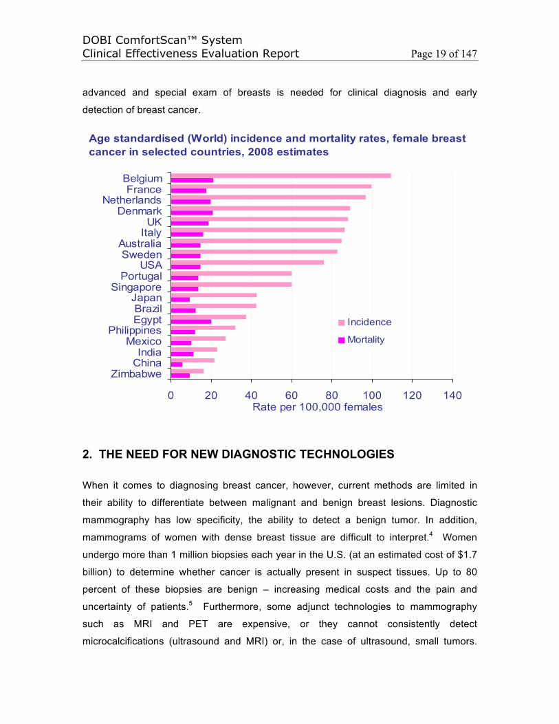
advanced and special exam of breasts is needed for clinical diagnosis and early
detection of breast cancer.
0 20 40 60 80 100 120 140
ZimbabweChinaIndia
MexicoPhilippines
EgyptBrazil
JapanSingapore
PortugalUSA
SwedenAustralia
ItalyUK
DenmarkNetherlands
FranceBelgium
Rate per 100,000 females
Incidence
Mortality
Age standardised (World) incidence and mortality rates, female breastcancer in selected countries, 2008 estimates
2. THE NEED FOR NEW DIAGNOSTIC TECHNOLOGIES
When it comes to diagnosing breast cancer, however, current methods are limited in
their ability to differentiate between malignant and benign breast lesions. Diagnostic
mammography has low specificity, the ability to detect a benign tumor. In addition,
mammograms of women with dense breast tissue are difficult to interpret.4 Women
undergo more than 1 million biopsies each year in the U.S. (at an estimated cost of $1.7
billion) to determine whether cancer is actually present in suspect tissues. Up to 80
percent of these biopsies are benign – increasing medical costs and the pain and
uncertainty of patients.5 Furthermore, some adjunct technologies to mammography
such as MRI and PET are expensive, or they cannot consistently detect
microcalcifications (ultrasound and MRI) or, in the case of ultrasound, small tumors.
DOBI ComfortScan™ SystemClinical Effectiveness Evaluation Report Page 20 of 147
What is needed in clincal breast cancer diagnosis is a noninvasive, cost-effective adjunct
to mammography that can discriminate between malignant and benign lesions – thus
preventing unnecessary biopsies.
In response to this need, on March 8, 2001, in a press release announcing the release of
its comprehensive new study, Mammography and Beyond, the National Academy of
Sciences’ Institute of Medicine issued a call to action for improvements in breast-imaging
techniques. In addition to characterizing film mammography as the “gold standard”
against which new imaging technologies will be measured, the press release states that
“no single imaging technology is capable of accurately detecting all breast abnormalities”
and that “ultimately, the best detection may come from using several different tools.”6
Currently, breast cancer detection encompasses three stages. First, a physical
examination or screening mammography identifies an abnormality in the breast tissue.
Second, additional imaging modalities may be used to help decide if a biopsy is
required. Third, if required, a biopsy is performed to diagnose the abnormality as either
benign or malignant. Malignant abnormalities are further characterized biochemically
and are staged according to the size of the tumor as well as the extent of invasion and
metastasis in order to determine a prognosis and treatment.7 The following sections
review the utilization of mammography, ultrasound, MRI, PET and biopsy as diagnostic
tools.
2.1 Mammography
Mammograms, x-rays of the breast, are generally categorized either as screening or as
diagnostic. Screening mammography is used to check for breast disease in women who
are asymptomatic. Diagnostic mammography is used to check for breast disease in
women who experience new symptoms, or it is used to further explore a suspicious
finding identified by a screening mammogram. Mammograms, however, do not detect
cancer per se. Instead, they help to identify tissue abnormalities, which in turn are
subject to interpretation or additional testing such as ultrasound or, definitively,
diagnostic biopsy.8 Although most studies demonstrate a substantial reduction in death
rates from breast cancer among women screened by mammography, women over age
50 benefit the most from screening mammography. Below age 50, the value of
DOBI ComfortScan™ SystemClinical Effectiveness Evaluation Report Page 21 of 147
screening is less clear.9 Since women in their 40s are generally premenopausal and
therefore more likely to have greater breast density than postmenopausal women, it is
more difficult to interpret mammograms – leading to some that are indeterminate.
Mammograms for postmenopausal women on estrogen-replacement therapy are
similarly difficult to interpret.10 Because the specificity of mammogram testing is quite
low, false-positive findings can have a detrimental effect on the screened population. As
many as 80 percent of all breast lesions that are biopsied as a result of suspicious
findings on a mammogram turn out to be benign.11 Studies show that abnormal
mammograms negatively affect a woman’s psychological and emotional state and may
impair daily functioning for 3 to 18 months.12 Because the greater density of breast
tissue in younger, premenopausal women renders mammography results more difficult
to interpret, improved specificity and sensitivity in diagnostic methods would benefit
younger women in particular.
2.2 Ultrasound, MRI and PET
Once an abnormality is identified through examination or mammography, the next stage
in cancer detection can be utilization of an adjunct technology (such as breast
ultrasound, MRI and PET) or, definitively, a biopsy. Ultrasound can help to determine if
an abnormality that appeared on a mammogram is a cyst or a solid mass.13 According
to the National Cancer Institute, however, about half of cancers detected by
mammography appear as a cluster of microcalcifications and ultrasound does not
consistently detect microcalcifications nor detect very small tumors.14
As an alternative to ultrasound, MRI (Nuclear Magnetic Resonance Imaging) may
eventually prove useful in a small number of cases for diagnosing breast lesions
identified through screening mammography or clinical breast examination. MRI,
however, remains an unproven technology for widespread use in breast cancer
detection. Furthermore, it is an expensive diagnostic alternative and it cannot detect
microcalcifications.15
Based upon the understanding that malignant tissue tends to metabolize glucose
differently from tissue with benign abnormalities, positron emission tomography (PET)
uses radioactive tracers such as labeled glucose to identify regions in the body with high
DOBI ComfortScan™ SystemClinical Effectiveness Evaluation Report Page 22 of 147
metabolic activity. PET scans, however, are an expensive alternative and are invasive in
that they require the injection of a radioactive substance into the body.
2.3 Biopsy
According to the American Cancer Society, a biopsy is the only way to detect whether or
not cancer is actually present. All biopsies remove a tissue sample for examination
under a microscope. Biopsies include fine-needle aspiration (FNA) biopsy, core-needle
biopsy (CNB) and surgical or excisional biopsy.16 Surgical or excisional biopsies are the
most traditional method of removing tissue for further study – they are also the most
expensive and invasive. FNA biopsy requires insertion of a very thin needle on a syringe
to remove either fluid from a cyst or clusters of cells from a palpable mass. CNB is more
traumatic than FNA biopsy because it uses a larger needle with a special cutting edge to
remove small cores of tissue. The tissue cores are usually large enough to enable
pathologists to distinguish between invasive and noninvasive types of breast cancer.
Because 80 percent of breast biopsies are conducted on benign tissue – raising
healthcare costs and causing pain, possible scarring and anxiety in patients – an adjunct
technology that supplements mammography and reduces the large number of
unnecessary biopsies would be of significant benefit to both patients and healthcare
providers. Because the non-invasive DOBI technology is designed to identify the minute
vascular changes associated with growing cancer in its earliest stages, it has the
potential to provide a new screening tool, as well as DOBI ComfortScan has the
potential to play a key role in improving current methods of breast cancer detection and
treatment monitoring.
3. BACKGROUND A NEW TECHNIQUE TO DETECT BREAST CANCER AT EARLY STAGE – DYANAMIC OPTICAL BREAST IMAGING
Physicians worldwide are looking for an innovative and inexpensive technology that
provides further diagnostic information to complement current diagnostic data, thus
allowing them to make a more complete and accurate diagnosis. Undeniably, a new

DOBI ComfortScan™ SystemClinical Effectiveness Evaluation Report Page 23 of 147
screening, following up and monitering tool for breast carcer early detection and breast
non-invasive treatment evaulation is increasingly paied great attention in the
examination of the breast. The Dynamic Optical Breast Imaging (DOBI®) ComfortScan,
while not intended to replace mammography, is a noninvasive, nonionizing medical
imaging system designed to assist physicians in the diagnosis of breast cancer by
providing new, image-based physiological information which mammography, ultrasound
and physical exams cannot provide. The medical and scientific foundation of the DOBI
technology is developed to image the body’s creation of new blood vessels
(neovascularization) associated with the support of tumor development. This process,
known as angiogenesis, has been scientifically linked to the development and growth of
most cancers and over 70 other human diseases. The ability for medical and scientific
professionals to image angiogenesis in the human body in this manner is virtually non-
existent. Thus, the ComfortScan system is designed to provide important physiology-
based information not readily available to physicians today for determining the presence
of abnormally vascularized lesions in the body.
3.1 The Role of Angiogenesis in New Breast Cancer Diagnostic Technologies
Since Judah Folkman’s seminal hypothesis was published in 1971, the formation and
growth of new blood vessels from preexisting blood vessels, called angiogenesis, has
become widely recognized as a key biological process that occurs in both healthy and
diseased tissues.17 When properly regulated, angiogenesis is necessary for
reproduction, embryonic development and wound repair. In such cases, the complex
angiogenic process is maintained in careful balance by a variety of angiogenesis-
stimulating growth factors, angiogenesis inhibitors, cell-bound molecules, the
surrounding extracellular matrix (ECM) and other mediators. When this balance is tipped
in favor of too much or too little angiogenesis, a variety of pathological conditions – such
as cancer, rheumatoid arthritis and coronary artery disease – can be the result.18,19 In
particular, the role of angiogenesis in breast cancer has been documented.20
As in all cells, the cells of an incipient tumor require constant nourishment and oxygen
as well as a way to remove waste products. As long as a tumor remains small –
approximately one millimeter in diameter – the process of diffusion (through a cell
membrane) can adequately provide nourishment and dispose of wastes. To grow
DOBI ComfortScan™ SystemClinical Effectiveness Evaluation Report Page 24 of 147
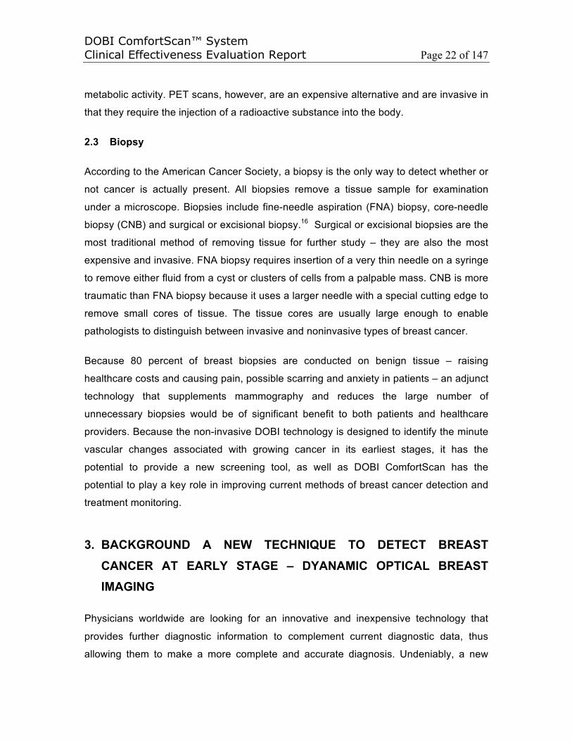
beyond the “one-millimeter limit,” the tumor cells must develop their own blood
circulation system – mimicking the circulatory system of healthy tissue nearby, as shown
in Figure below Normal cell tissue is interlaced with a dense network of capillaries.
Constructed from endothelial cells, these capillaries supply nourishment and carry off
wastes. When starved of oxygen, the cells of normal tissues are able to induce
endothelial cell proliferation and the formation of new capillaries by releasing angiogenic
growth factors such as vascular endothelial growth factor (VEGF) and basic fibroblast
growth factor (bFGF). In imitation of normal cells, some of the cells in the tumor acquire
the ability to secrete angiogenic growth factors – thereby attracting endothelial cells from
nearby tissues and inducing these endothelial cells to multiply. By encouraging
capillaries to grow into the tumor, tumor cells acquire direct access to oxygen-and-
nutrient-rich blood as well as a way of removing waste products. This enables the tumor
cells to grow explosively and spread widely. Some physicians use the presence or
absence of a dense capillary network in tumor samples to determine the stage of tumor
development and predict its future course.21
The Stage of Tumor Angiogenesis
Angiogenesis in tumors follows an orderly sequence of events:22,23,24
1. Initiation. The diseased tissue (tumor) produces and releases angiogenic growth
factors that diffuse into the surrounding tissue.
2. Proliferation and invasion by endothelial cells. The angiogenic growth factors
bind to receptors located on the endothelial cells of nearby blood vessels. Within
an endothelial cell, signals are sent from the cell’s surface to the nucleus and the
cell begins to produce new molecules including enzymes. The enzymes dissolve
DOBI ComfortScan™ SystemClinical Effectiveness Evaluation Report Page 25 of 147
tiny holes in the sheath-like basement membrane that surrounds the blood
vessel. The endothelial cells begin to divide (proliferate) and they migrate out
through the dissolved holes and toward the tumor. Specialized molecules, called
adhesion molecules or integrins (αvβ3 and αvβ5), help to pull the sprouting tip of
the blood vessel forward. Additional enzymes, called matrix metalloproteinases,
or MMPs, dissolve the tissue in front of the sprouting blood vessel tip and, as the
vessel extends, the tissue is remolded around the vessel. The sprouting
endothelial cells roll up to form a blood vessel tube and the individual blood
vessel tubes connect to form complete blood-vessel loops that can circulate
blood.
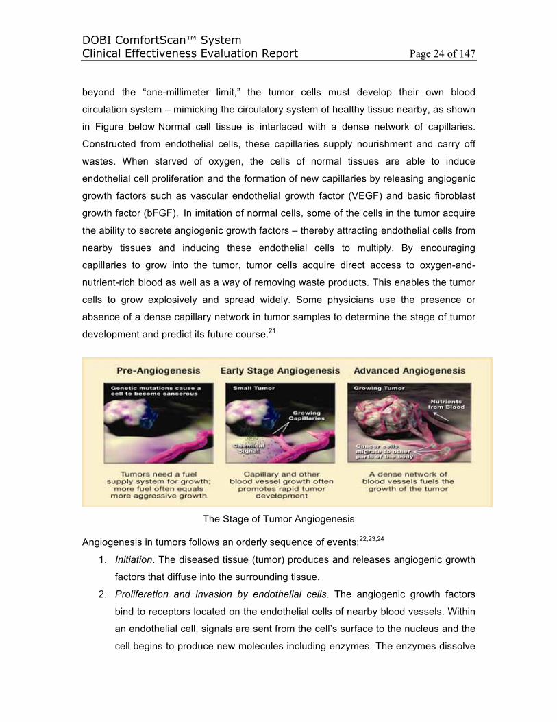
3. Maturation of blood vessels. The newly formed blood vessel tubes are stabilized
by the growth of specialized muscle cells, which provide structural support. Blood
then begins to flow through the new blood vessels. These new blood vessels are
the mechanism by which the tumor creates an oxygen-and-nutrient-rich
environment in which to grow explosively and spread throughout the body.
Without this environment, the tumor would remain confined to the “one-millimeter
limit” described earlier – starved for nutrients and choking on its own waste
products.
DOBI ComfortScan™ SystemClinical Effectiveness Evaluation Report Page 26 of 147
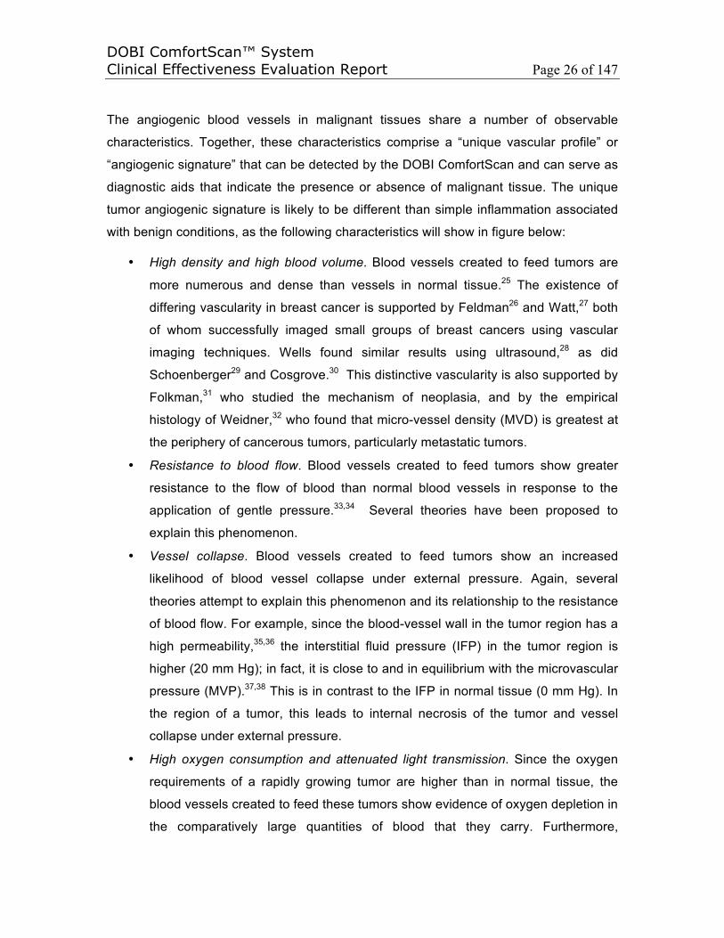
The angiogenic blood vessels in malignant tissues share a number of observable
characteristics. Together, these characteristics comprise a “unique vascular profile” or
“angiogenic signature” that can be detected by the DOBI ComfortScan and can serve as
diagnostic aids that indicate the presence or absence of malignant tissue. The unique
tumor angiogenic signature is likely to be different than simple inflammation associated
with benign conditions, as the following characteristics will show in figure below:
• High density and high blood volume. Blood vessels created to feed tumors are
more numerous and dense than vessels in normal tissue.25 The existence of
differing vascularity in breast cancer is supported by Feldman26 and Watt,27 both
of whom successfully imaged small groups of breast cancers using vascular
imaging techniques. Wells found similar results using ultrasound,28 as did
Schoenberger29 and Cosgrove.30 This distinctive vascularity is also supported by
Folkman,31 who studied the mechanism of neoplasia, and by the empirical
histology of Weidner,32 who found that micro-vessel density (MVD) is greatest at
the periphery of cancerous tumors, particularly metastatic tumors.
• Resistance to blood flow. Blood vessels created to feed tumors show greater
resistance to the flow of blood than normal blood vessels in response to the
application of gentle pressure.33,34 Several theories have been proposed to
explain this phenomenon.
• Vessel collapse. Blood vessels created to feed tumors show an increased
likelihood of blood vessel collapse under external pressure. Again, several
theories attempt to explain this phenomenon and its relationship to the resistance
of blood flow. For example, since the blood-vessel wall in the tumor region has a
high permeability,35,36 the interstitial fluid pressure (IFP) in the tumor region is
higher (20 mm Hg); in fact, it is close to and in equilibrium with the microvascular
pressure (MVP).37,38 This is in contrast to the IFP in normal tissue (0 mm Hg). In
the region of a tumor, this leads to internal necrosis of the tumor and vessel
collapse under external pressure.
• High oxygen consumption and attenuated light transmission. Since the oxygen
requirements of a rapidly growing tumor are higher than in normal tissue, the
blood vessels created to feed these tumors show evidence of oxygen depletion in
the comparatively large quantities of blood that they carry. Furthermore,
DOBI ComfortScan™ SystemClinical Effectiveness Evaluation Report Page 27 of 147
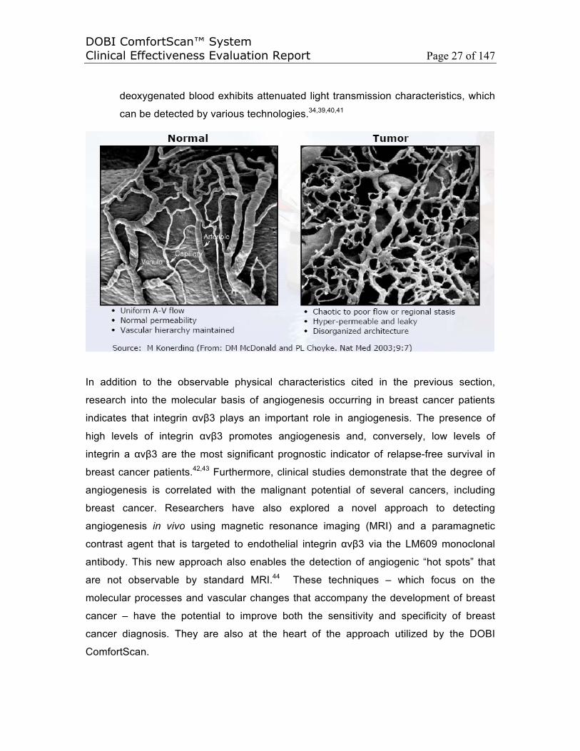
deoxygenated blood exhibits attenuated light transmission characteristics, which
can be detected by various technologies.34,39,40,41
In addition to the observable physical characteristics cited in the previous section,
research into the molecular basis of angiogenesis occurring in breast cancer patients
indicates that integrin αvβ3 plays an important role in angiogenesis. The presence of
high levels of integrin αvβ3 promotes angiogenesis and, conversely, low levels of
integrin a αvβ3 are the most significant prognostic indicator of relapse-free survival in
breast cancer patients.42,43 Furthermore, clinical studies demonstrate that the degree of
angiogenesis is correlated with the malignant potential of several cancers, including
breast cancer. Researchers have also explored a novel approach to detecting
angiogenesis in vivo using magnetic resonance imaging (MRI) and a paramagnetic
contrast agent that is targeted to endothelial integrin αvβ3 via the LM609 monoclonal
antibody. This new approach also enables the detection of angiogenic “hot spots” that
are not observable by standard MRI.44 These techniques – which focus on the
molecular processes and vascular changes that accompany the development of breast
cancer – have the potential to improve both the sensitivity and specificity of breast
cancer diagnosis. They are also at the heart of the approach utilized by the DOBI
ComfortScan.
DOBI ComfortScan™ SystemClinical Effectiveness Evaluation Report Page 28 of 147
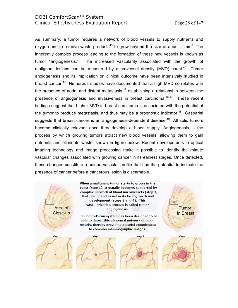
As summary, a tumor requires a network of blood vessels to supply nutrients and
oxygen and to remove waste products45 to grow beyond the size of about 2 mm3. The
inherently complex process leading to the formation of these new vessels is known as
tumor “angiogenesis.” The increased vascularity associated with the growth of
malignant lesions can be measured by microvessel density (MVD) count.46 Tumor
angiogenesis and its implication on clinical outcome have been intensively studied in
breast cancer.47 Numerous studies have documented that a high MVD correlates with
the presence of nodal and distant metastasis,18 establishing a relationship between the
presence of angiogenesis and invasiveness in breast carcinoma.48,49 These recent
findings suggest that higher MVD in breast carcinoma is associated with the potential of
the tumor to produce metastasis, and thus may be a prognostic indicator.48 Gasparini
suggests that breast cancer is an angiogenesis-dependent disease.49 All solid tumors
become clinically relevant once they develop a blood supply. Angiogenesis is the
process by which growing tumors attract new blood vessels, allowing them to gain
nutrients and eliminate waste, shown in figure below. Recent developments in optical
imaging technology and image processing make it possible to identify the minute
vascular changes associated with growing cancer in its earliest stages. Once detected,
these changes constitute a unique vascular profile that has the potential to indicate the
presence of cancer before a cancerous lesion is discernable.
DOBI ComfortScan™ SystemClinical Effectiveness Evaluation Report Page 29 of 147
3.2 Dynamic Opitical Breast Imaging Technology in Early Breast Cancer
Diagnostic
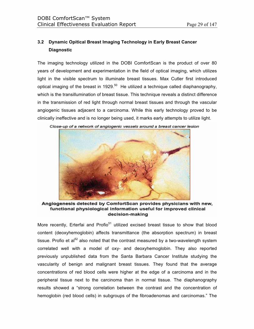
The imaging technology utilized in the DOBI ComfortScan is the product of over 80
years of development and experimentation in the field of optical imaging, which utilizes
light in the visible spectrum to illuminate breast tissues. Max Cutler first introduced
optical imaging of the breast in 1929.50 He utilized a technique called diaphanography,
which is the transillumination of breast tissue. This technique reveals a distinct difference
in the transmission of red light through normal breast tissues and through the vascular
angiogenic tissues adjacent to a carcinoma. While this early technology proved to be
clinically ineffective and is no longer being used, it marks early attempts to utilize light.
More recently, Erterfai and Profio51 utilized excised breast tissue to show that blood
content (deoxyhemoglobin) affects transmittance (the absorption spectrum) in breast
tissue. Profio et al52 also noted that the contrast measured by a two-wavelength system
correlated well with a model of oxy- and deoxyhemoglobin. They also reported
previously unpublished data from the Santa Barbara Cancer Institute studying the
vascularity of benign and malignant breast tissues. They found that the average
concentrations of red blood cells were higher at the edge of a carcinoma and in the
peripheral tissue next to the carcinoma than in normal tissue. The diaphanography
results showed a “strong correlation between the contrast and the concentration of
hemoglobin (red blood cells) in subgroups of the fibroadenomas and carcinomas.” The
DOBI ComfortScan™ SystemClinical Effectiveness Evaluation Report Page 30 of 147
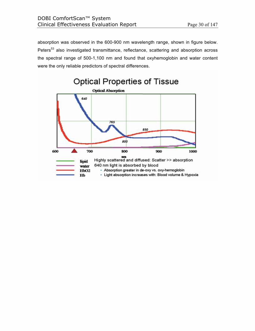
absorption was observed in the 600-900 nm wavelength range, shown in figure below.
Peters53 also investigated transmittance, reflectance, scattering and absorption across
the spectral range of 500-1,100 nm and found that oxyhemoglobin and water content
were the only reliable predictors of spectral differences.
DOBI ComfortScan™ SystemClinical Effectiveness Evaluation Report Page 31 of 147
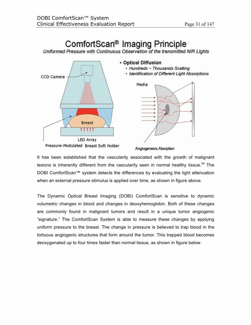
It has been established that the vascularity associated with the growth of malignant
lesions is inherently different from the vascularity seen in normal healthy tissue.54 The
DOBI ComfortScan™ system detects the differences by evaluating the light attenuation
when an external pressure stimulus is applied over time, as shown in figure above.
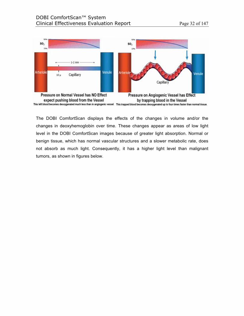
The Dynamic Optical Breast Imaging (DOBI) ComfortScan is sensitive to dynamic
volumetric changes in blood and changes in deoxyhemoglobin. Both of these changes
are commonly found in malignant tumors and result in a unique tumor angiogenic
“signature.” The ComfortScan System is able to measure these changes by applying
uniform pressure to the breast. The change in pressure is believed to trap blood in the
tortuous angiogenic structures that form around the tumor. This trapped blood becomes
deoxygenated up to four times faster than normal tissue, as shown in figure below.
DOBI ComfortScan™ SystemClinical Effectiveness Evaluation Report Page 32 of 147
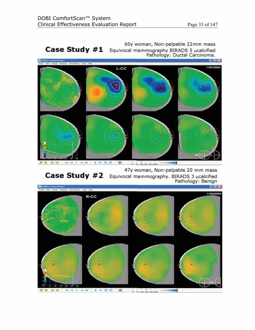
The DOBI ComfortScan displays the effects of the changes in volume and/or the
changes in deoxyhemoglobin over time. These changes appear as areas of low light
level in the DOBI ComfortScan images because of greater light absorption. Normal or
benign tissue, which has normal vascular structures and a slower metabolic rate, does
not absorb as much light. Consequently, it has a higher light level than malignant
tumors, as shown in figures below.
DOBI ComfortScan™ SystemClinical Effectiveness Evaluation Report Page 33 of 147
DOBI ComfortScan™ SystemClinical Effectiveness Evaluation Report Page 34 of 147
In contrast to early unsuccessful transillumination techniques, the DOBI ComfortScan
uses mechanical perturbation to create a dynamic signature. The DOBI ComfortScan’s
array of light-emitting diodes (LEDs) illuminates areas of vascular development in the
breast that possess characteristics unique to malignant tumors. As described earlier,
during the process of angiogenesis, a cancerous growth surrounds itself with a dense
network of tiny blood-filled capillaries. These capillaries provide oxygen and nutrients to
active tumors and they exhibit the unique physiological “markers” that the DOBI
ComfortScan can detect. These physiological markers include dense vascularity, high
blood-flow resistance, blood deoxygenation, attenuated light transmission and a greater
likelihood of the blood vessels to collapse under external pressure.34
3.3 Dynamic Optical Breast Imaging Principle
While other diagnostic imaging devices primarily detect static morphological (structural)
changes, the DOBI ComfortScan is designed to detect dynamic (physiologic) changes,
namely the dynamic flow, increased blood volume levels and depleted oxygen levels
(deoxygenated hemoglobin) that are characteristic of malignancies. The currment
ComfortScan system utilizes light from 127 light emitting diodes (LED), mounted on an
illuminator plate inclined 30° from horizontal plane. The LEDs emit red light with a
wavelength of 640nm for greater absorption and higher sensitivity of optical absorption
of de-oxy-hemoglobin. Transmitted light is recorded by a Charge-Coupled Device (CCD)
camera for approximately 45 seconds. As part of this process, the machine applies a
slight amount of uniform pressure via a patented silicone membrane system – a
pressure jump from 5mm Hg (setup pressure) to 10 mm Hg (analysis pressure) for about
30 seconds – to the breast, which already has been compressed lightly by the soft
breast holder. When an external pressure stimulus is applied uniformly around the
breast, the dynamics of blood redistribution, the capillary and vein collapse, and the
oxygenated state of the blood as a function of time after the initial pressure stimulus in
the area of abnormal vascularization will be different from those properties in normal
areas of the breast tissue. The system collects the images of the breast before, during
and after applying the pressure jump to record the changes as they occur.
Since a single static image alone does not reveal much information about abnormal
vascularization because the light beams are heavily scattered and diffused by tissues
DOBI ComfortScan™ SystemClinical Effectiveness Evaluation Report Page 35 of 147
resulting in very low spatial resolution, and only changes caused by re-distribution of
blood volume and oxygenation level are detected, the dynamic response of the breast to
pressure modulation is carried in the intensity variations among different images in the
whole sequence. The dynamic image sequence may be represented by I(x, y, t). The
DOBI ComfortScan uses the first image after the pressure jump as a reference image,
Iref, after the first illumination cycle following the onset of the pressure step (breast shape
has stabilized). This image is subtracted from the rest of the image sequence to
represent the dynamic signature or response to the pressure at each spatial point (x, y):
DS(x, y, t) = ( I(x, y, t) – Iref ) / Iref .
The vascular changes associated with cancerous lesions absorb more light than normal
tissue and this creates areas of low light level on the image. As the light from the LEDs
encounters the angiogenic tissue surrounding the tumor, the hemoglobin, which is
trapped in the blood-filled capillaries near the malignancy, absorbs the red light more
completely than in normal or benign tissue. Following each pressure jump, the system
records the changes in light transmission at the red wavelength.55 When compared with
the abnormal regions, blood volume and oxygen saturation levels decrease at a different
rate in the region with normal vascularity, normal interstitial fluid pressure, and normal
oxygen consumption rate. The difference in dynamic signatures in normal and abnormal
areas can be summarized as:
DSa (xa, ya, t) ≠ DSn(xn yn t).
To process the images recorded by the camera, the DOBI ComfortScan utilizes
proprietary computer algorithms that generate a graph of the changing light-transmission
values for each location over time. Consequently, it visually displays the unique vascular
profile of the angiogenic region of the breast that stands out in marked contrast to
normal or benign portions of the breast. On the image, bright areas indicate normal or
benign tissues and dark blue areas indicate a potential for malignancy. By displaying a
contrasting appearance, the DOBI ComfortScan has the potential to confirm the
presence of cancer and differentiate cancer from both benign lesions and normal tissue
within the breast. The potential for the ComfortScan system as a diagnostic tool is to
non-invasively differentiate between specific optical patterns of normal and abnormal
vascularization areas and therefore to provide the physician with additional information
as to the angiogenic status of the suspicious area. This information is intended to assist
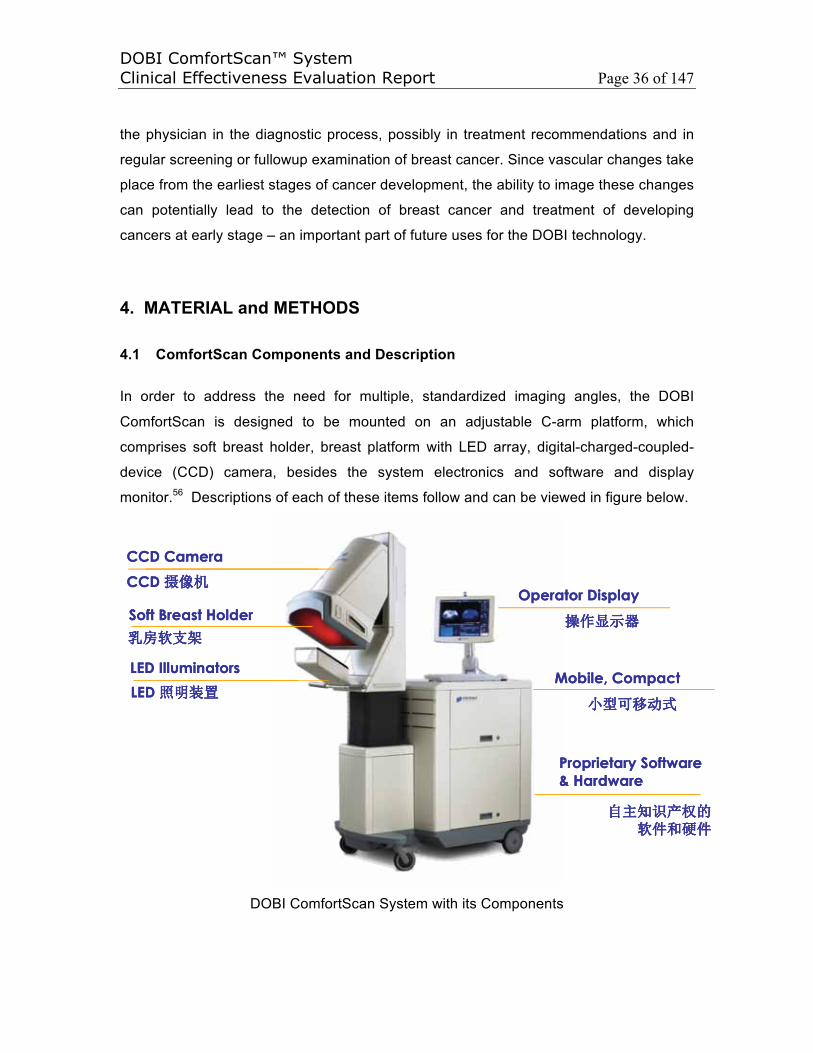
DOBI ComfortScan™ SystemClinical Effectiveness Evaluation Report Page 36 of 147
the physician in the diagnostic process, possibly in treatment recommendations and in
regular screening or fullowup examination of breast cancer. Since vascular changes take
place from the earliest stages of cancer development, the ability to image these changes
can potentially lead to the detection of breast cancer and treatment of developing
cancers at early stage – an important part of future uses for the DOBI technology.
4. MATERIAL and METHODS
4.1 ComfortScan Components and Description
In order to address the need for multiple, standardized imaging angles, the DOBI
ComfortScan is designed to be mounted on an adjustable C-arm platform, which
comprises soft breast holder, breast platform with LED array, digital-charged-coupled-
device (CCD) camera, besides the system electronics and software and display
monitor.56 Descriptions of each of these items follow and can be viewed in figure below.
CCD Camera
Soft Breast Holder
LED Illuminators
CCD
LED
CCD Camera
Soft Breast Holder
LED Illuminators
CCD
LED Mobile, Compact
Operator Display
Proprietary Software & Hardware
Mobile, Compact
Operator Display
Proprietary Software & Hardware
DOBI ComfortScan System with its Components
DOBI ComfortScan™ SystemClinical Effectiveness Evaluation Report Page 37 of 147
Soft breast holder. It is necessary to compress the breast to achieve acceptable image
contrast in the area of pathologic influence (API). The breast holder consists of a silicon
membrane that provides a soft contact surface and an airtight seal over a pressure
chamber. The pressure chamber can be inflated with low-pressure air. The silicon
membrane gently compresses the breast between the silicon balloon on the camera side
(top) and the flat breast platform (below) with its array of LEDs. The DOBI ComfortScan
controls and monitors the rise, fall and maintenance of pressure in the pneumatic system
with a custom-programmed microcontroller.
Breast platform with LED array. The DOBI ComfortScan utilizes a flat-plane array of
127 light emitting diodes (LEDs) that emit light in the single visible-red band (640 nm).
The custom-programmed microprocessor precisely controls the optical exposure time
and the intensity profile of the LED array, which enables versatile operation with breasts
of different sizes and densities. The system supports independent control of the light
intensity for each LED.
Digital CCD camera. The DOBI ComfortScan uses a 12-bit digital-charged-coupled-
device (CCD) camera. The sensitivity of this camera is necessary because the changes
in light intensity within the illuminated breast are small in amplitude in comparison to the
amount of light emitted by the LEDs. Consequently, it is necessary to utilize a high-gain,
low-noise device such as the CCD camera. The CCD camera employs an internal
thermal-electric cooler and operates at a controlled low temperature of -20° C. This low
temperature produces an extremely low dark-current output. The intrinsic spatial
resolution of the camera is 768 x 512 pixels. The 5 x 5 pixel-binning mechanism
increases the efficiency of photo collection. The final spatial resolution of the camera
output is effectively 102 x 128 pixels. Gray-scale resolution of the signal from each pixel
is up to 4,096 levels, which corresponds to 12 bits.
System electronics and software. The Controller is an electronic assembly that
interfaces with the LED illuminator, the computer, the soft holder assembly and two
pressure reservoirs located in the bottom of the base unit. Housed in the controller are
circuit boards for lighting the LED, operating the pneumatic pump, sensing the pressure
in the air chamber and interfacing with the computer. It houses also a microcomputer
and a programmable read-only memory. The Computer System provides the main user
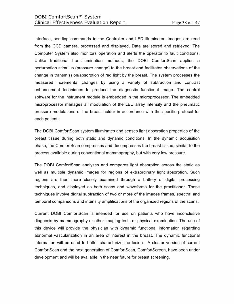
DOBI ComfortScan™ SystemClinical Effectiveness Evaluation Report Page 38 of 147
interface, sending commands to the Controller and LED illuminator. Images are read
from the CCD camera, processed and displayed. Data are stored and retrieved. The
Computer System also monitors operation and alerts the operator to fault conditions.
Unlike traditional transillumination methods, the DOBI ComfortScan applies a
perturbation stimulus (pressure change) to the breast and facilitates observations of the
change in transmission/absorption of red light by the breast. The system processes the
measured incremental changes by using a variety of subtraction and contrast
enhancement techniques to produce the diagnostic functional image. The control
software for the instrument module is embedded in the microprocessor. The embedded
microprocessor manages all modulation of the LED array intensity and the pneumatic
pressure modulations of the breast holder in accordance with the specific protocol for
each patient.
The DOBI ComfortScan system illuminates and senses light absorption properties of the
breast tissue during both static and dynamic conditions. In the dynamic acquisition
phase, the ComfortScan compresses and decompresses the breast tissue, similar to the
process available during conventional mammography, but with very low pressure.
The DOBI ComfortScan analyzes and compares light absorption across the static as
well as multiple dynamic images for regions of extraordinary light absorption. Such
regions are then more closely examined through a battery of digital processing
techniques, and displayed as both scans and waveforms for the practitioner. These
techniques involve digital subtraction of two or more of the images frames, spectral and
temporal comparisons and intensity amplifications of the organized regions of the scans.
Current DOBI ComfortScan is intended for use on patients who have inconclusive
diagnosis by mammography or other imaging tests or physical examination. The use of
this device will provide the physician with dynamic functional information regarding
abnormal vascularization in an area of interest in the breast. The dynamic functional
information will be used to better characterize the lesion. A cluster version of current
ComfortScan and the next generation of ComfortScan, ComfortScreen, have been under
development and will be available in the near future for breast screening.
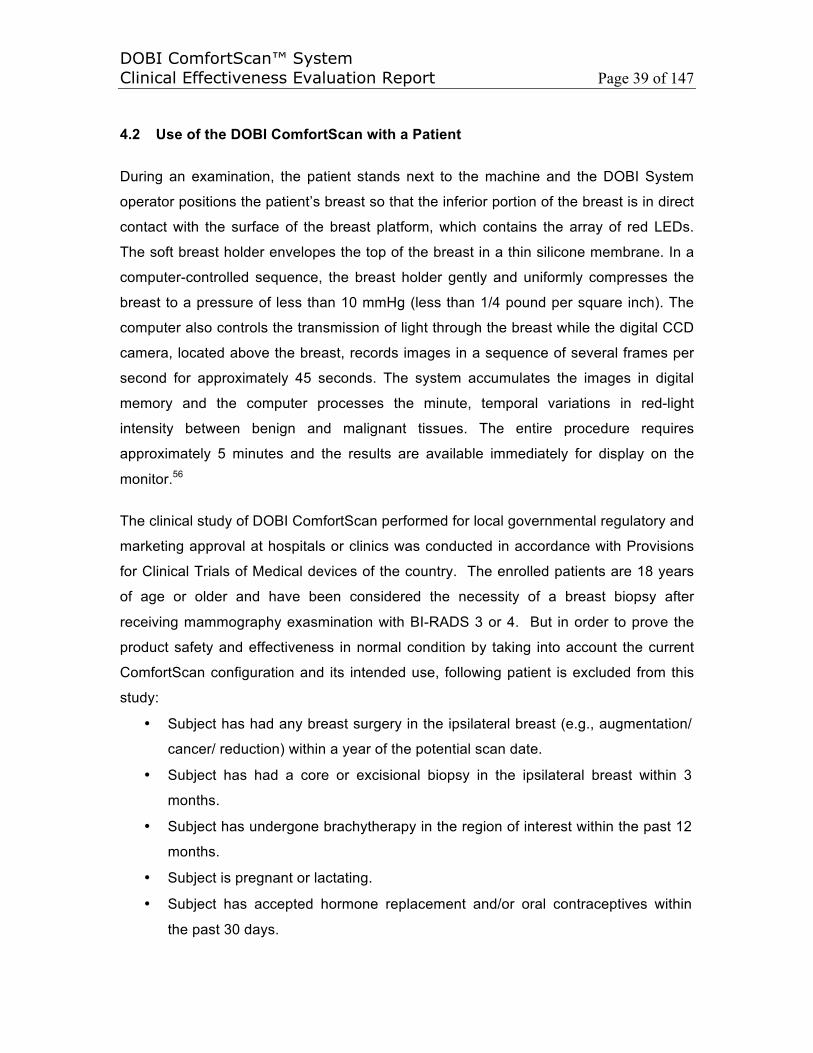
DOBI ComfortScan™ SystemClinical Effectiveness Evaluation Report Page 39 of 147
4.2 Use of the DOBI ComfortScan with a Patient
During an examination, the patient stands next to the machine and the DOBI System
operator positions the patient’s breast so that the inferior portion of the breast is in direct
contact with the surface of the breast platform, which contains the array of red LEDs.
The soft breast holder envelopes the top of the breast in a thin silicone membrane. In a
computer-controlled sequence, the breast holder gently and uniformly compresses the
breast to a pressure of less than 10 mmHg (less than 1/4 pound per square inch). The
computer also controls the transmission of light through the breast while the digital CCD
camera, located above the breast, records images in a sequence of several frames per
second for approximately 45 seconds. The system accumulates the images in digital
memory and the computer processes the minute, temporal variations in red-light
intensity between benign and malignant tissues. The entire procedure requires
approximately 5 minutes and the results are available immediately for display on the
monitor.56
The clinical study of DOBI ComfortScan performed for local governmental regulatory and
marketing approval at hospitals or clinics was conducted in accordance with Provisions
for Clinical Trials of Medical devices of the country. The enrolled patients are 18 years
of age or older and have been considered the necessity of a breast biopsy after
receiving mammography exasmination with BI-RADS 3 or 4. But in order to prove the
product safety and effectiveness in normal condition by taking into account the current
ComfortScan configuration and its intended use, following patient is excluded from this
study:
• Subject has had any breast surgery in the ipsilateral breast (e.g., augmentation/
cancer/ reduction) within a year of the potential scan date.
• Subject has had a core or excisional biopsy in the ipsilateral breast within 3
months.
• Subject has undergone brachytherapy in the region of interest within the past 12
months.
• Subject is pregnant or lactating.
• Subject has accepted hormone replacement and/or oral contraceptives within
the past 30 days.
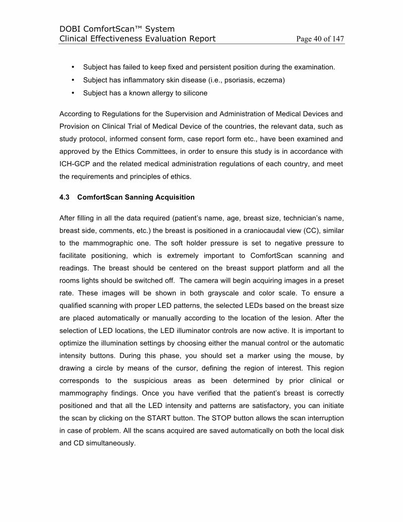
DOBI ComfortScan™ SystemClinical Effectiveness Evaluation Report Page 40 of 147
• Subject has failed to keep fixed and persistent position during the examination.
• Subject has inflammatory skin disease (i.e., psoriasis, eczema)
• Subject has a known allergy to silicone
According to Regulations for the Supervision and Administration of Medical Devices and
Provision on Clinical Trial of Medical Device of the countries, the relevant data, such as
study protocol, informed consent form, case report form etc., have been examined and
approved by the Ethics Committees, in order to ensure this study is in accordance with
ICH-GCP and the related medical administration regulations of each country, and meet
the requirements and principles of ethics.
4.3 ComfortScan Sanning Acquisition
After filling in all the data required (patient’s name, age, breast size, technician’s name,
breast side, comments, etc.) the breast is positioned in a craniocaudal view (CC), similar
to the mammographic one. The soft holder pressure is set to negative pressure to
facilitate positioning, which is extremely important to ComfortScan scanning and
readings. The breast should be centered on the breast support platform and all the
rooms lights should be switched off. The camera will begin acquiring images in a preset
rate. These images will be shown in both grayscale and color scale. To ensure a
qualified scanning with proper LED patterns, the selected LEDs based on the breast size
are placed automatically or manually according to the location of the lesion. After the
selection of LED locations, the LED illuminator controls are now active. It is important to
optimize the illumination settings by choosing either the manual control or the automatic
intensity buttons. During this phase, you should set a marker using the mouse, by
drawing a circle by means of the cursor, defining the region of interest. This region
corresponds to the suspicious areas as been determined by prior clinical or
mammography findings. Once you have verified that the patient’s breast is correctly
positioned and that all the LED intensity and patterns are satisfactory, you can initiate
the scan by clicking on the START button. The STOP button allows the scan interruption
in case of problem. All the scans acquired are saved automatically on both the local disk
and CD simultaneously.
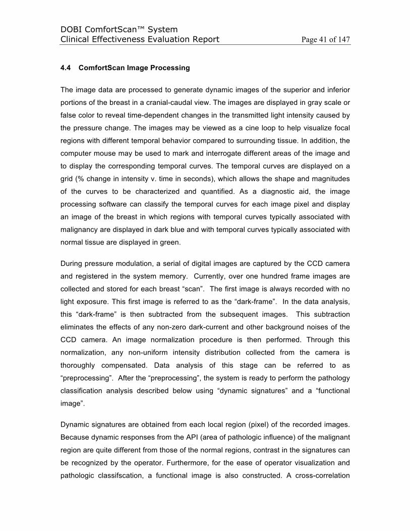
DOBI ComfortScan™ SystemClinical Effectiveness Evaluation Report Page 41 of 147
4.4 ComfortScan Image Processing
The image data are processed to generate dynamic images of the superior and inferior
portions of the breast in a cranial-caudal view. The images are displayed in gray scale or
false color to reveal time-dependent changes in the transmitted light intensity caused by
the pressure change. The images may be viewed as a cine loop to help visualize focal
regions with different temporal behavior compared to surrounding tissue. In addition, the
computer mouse may be used to mark and interrogate different areas of the image and
to display the corresponding temporal curves. The temporal curves are displayed on a
grid (% change in intensity v. time in seconds), which allows the shape and magnitudes
of the curves to be characterized and quantified. As a diagnostic aid, the image
processing software can classify the temporal curves for each image pixel and display
an image of the breast in which regions with temporal curves typically associated with
malignancy are displayed in dark blue and with temporal curves typically associated with
normal tissue are displayed in green.
During pressure modulation, a serial of digital images are captured by the CCD camera
and registered in the system memory. Currently, over one hundred frame images are
collected and stored for each breast “scan”. The first image is always recorded with no
light exposure. This first image is referred to as the “dark-frame”. In the data analysis,
this “dark-frame” is then subtracted from the subsequent images. This subtraction
eliminates the effects of any non-zero dark-current and other background noises of the
CCD camera. An image normalization procedure is then performed. Through this
normalization, any non-uniform intensity distribution collected from the camera is
thoroughly compensated. Data analysis of this stage can be referred to as
“preprocessing”. After the “preprocessing”, the system is ready to perform the pathology
classification analysis described below using “dynamic signatures” and a “functional
image”.
Dynamic signatures are obtained from each local region (pixel) of the recorded images.
Because dynamic responses from the API (area of pathologic influence) of the malignant
region are quite different from those of the normal regions, contrast in the signatures can
be recognized by the operator. Furthermore, for the ease of operator visualization and
pathologic classifscation, a functional image is also constructed. A cross-correlation
DOBI ComfortScan™ SystemClinical Effectiveness Evaluation Report Page 42 of 147
algorithm is used for the generation of this functional image. In the cross-correlation
process, a “reference signature” is selected from a normal breast region. Then a cross-
correlation process is applied between the dynamic signatures from all regions of the
breast with the reference signature. Because dynamic signatures from all normal
regions are similar to the reference signature, a high level of correlation is obtained from
these regions. On the other hand, dynamic signatures from the API are quite different
from the reference signature; a low level of correlation is obtained from these regions.
Consequently, the output from the correlation process produces a high-contrast
functional image. On the functional image, bright intensities present normal areas,
whereas dark intensities present in the API.
4.5 ComfortScan Image Interpretation
The interpretation of images is based in two separate physiological parameters: (1) the
color range in color window and (2) the dynamic curves corresponding to each area
selected. The blue or dark blue color is corresponding to areas of increased abnormal
vascularisation, as this is estimated by analyzing the signal of LED emission by means
of imaging functional programs incorporated into the computer system. The dynamic
curves offer a plus sophisticated means of calculating the patterns of increased
vascularisation, optimally permitting to predict the areas of neovascularisation.
There are two types of curves registered: (1) the progressive one that is correlated so far
with the areas of increased abnormal vascularisation and (2) the fluctuating one that
correlates with areas of 'normally' increased vascularisation, e.g. fibrokystic disease
areas or florid fibroadenomas. The physiological explanation for these curves is based in
the fluctuating rate of normal vascularisation, which is influenced by cardiac and
respiratory rate, thus presenting smoothly curved up and down lines, in accordance with
inspiration-expiration and systolic-diastolic rates. It is also worth mentioning the
significant role of capillary vessels, which permit an 'elasticity' of hematogenous flow. On
the contrary, areas of neovascularisation are typically characterised by the absence of
capillary trichoid vessels, and blood is 'pooling' into abnormal arteriovenous shunts, not
following the fluctuating cardiac and respiratory rate. This is represented by a
progressive curve, without 'elasticity'. From DOBI ComfortScan preliminary study
chosen to compare ComfortScan results with the findings of the considered-to-be the
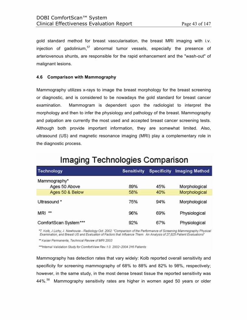
DOBI ComfortScan™ SystemClinical Effectiveness Evaluation Report Page 43 of 147
gold standard method for breast vascularisation, the breast MRI imaging with i.v.
injection of gadolinium,57 abnormal tumor vessels, especially the presence of
arteriovenous shunts, are responsible for the rapid enhancement and the "wash-out" of
malignant lesions.
4.6 Comparison with Mammography
Mammography utilizes x-rays to image the breast morphology for the breast screening
or diagnostic, and is considered to be nowadays the gold standard for breast cancer
examination. Mammogram is dependent upon the radiologist to interpret the
morphology and then to infer the physiology and pathology of the breast. Mammography
and palpation are currently the most used and accepted breast cancer screening tests.
Although both provide important information, they are somewhat limited. Also,
ultrasound (US) and magnetic resonance imaging (MRI) play a complementary role in
the diagnostic process.
Mammography has detection rates that vary widely: Kolb reported overall sensitivity and
specificity for screening mammography of 68% to 88% and 82% to 98%, respectively;
however, in the same study, in the most dense breast tissue the reported sensitivity was
44%.58 Mammography sensitivity rates are higher in women aged 50 years or older
DOBI ComfortScan™ SystemClinical Effectiveness Evaluation Report Page 44 of 147
compared with those less than 50 years old. This can be explained with the decrease of
breast density as a function of increasing age. Dense breasts (associated with a BI-
RADS™ breast density scale of 3 or 4) are often seen in younger women and fatty
breasts (e.g., BI-RADS density value of 1 and 2) are more common in the older
women59. Kerlikowske also reported that age had a strong effect on mammography
sensitivity, which was highest among women 60 to 69 years of age (87.0%) and lowest
among women 30 to 39 years of age (67.9%).60 A comparison based on the same trial
criteria is listed in the above.
These findings suggest that there is a need to improve on the tools currently used to
detect breast disease in younger women or those with more dense breasts. To
demonstrate the DOBI ComfortScan is an improved tool for young woman breast
examination and an useful tool for breast cancer diagnostic with addtional new
physiological information, all the enrolled patients during the clinical study of DOBI
ComfortScan in Beijing were performed mammography examinaitons but have
inconclusive findings, which are catogrized as Breast Imaging Reporting and Data
System (BI-RADS) 3 or 4. The results are shown in Table above.
4.7 Clinical Testing
During the entire developmental period of the DOBI System beginning in 1996,
approximately 1,300 patients have been scanned. Based on the analysis of scans
completed on typical patients Q3/4 2000 through Q1 2001, the DOBI System is
performing with a sensitivity (“true positive”) of 92% and specificity (“true negative”) of
76%, based on 123 patients with a malignancy rate of 20%. Negative predictive value is
97%.
Some of these scans were performed for the purpose of testing potential hardware and
software configurations of the System while others assessed its accuracy in
differentiating benign from malignant lesions. During the period of mid-1999 to mid-2001,
several scan protocols were used to test the device and the results were used to refine
both the hardware and the software and to develop additional analytical tools. This test-
refine-retest process was repeated until the upgrades resulted in the current System
DOBI ComfortScan™ SystemClinical Effectiveness Evaluation Report Page 45 of 147
configuration being tested at six investigational clinical sites under a well-defined set of
rules. These investigational clinical sites are:
• Asheville Women’s Medical Center, Asheville, NC
• Columbia University College of Physicians and Surgeons, New York, NY
• Hackensack University Medical Center, Hackensack, NJ
• Massachusetts General Hospital, Boston, MA
• University of Massachusetts Medical School, Worcester, MA
• Weill Medical College of Cornell University, New York, NY
The current trial protocol being followed is designed to incorporate DOBI System
scanning of patients scheduled for biopsy on the basis of equivocal mammogram and
other clinical findings. Approximately 450 patients were scanned under this protocol from
June 2000 through June 2001. The DOBI System scans are interpreted by consulting
radiologists and results of interpretation of 200 of these scans by an independent
radiologist show the following: 26/28 malignant lesions were recommended for biopsy,
for a sensitivity of 93 percent and 116/172 benign lesions would have been correctly
followed clinically without biopsy, for a specificity of 67 percent. Another way to assess
the value of these results is to look at the negative predictive value, that is, the
percentage of cases that were correctly recommended for clinical follow-up without
biopsy. The negative predictive value is 98 percent. Specifically, 116 of 118 cases that
DOBI System scan interpretation recommended for clinical follow-up without biopsy
were correctly assessed.
Following Clinical Effectiveness section lists some recent clinical studies and
demonstrates the clinical effectiveness of DOBI ComfortScan device. A total 2328 data
and 958 malignant cases are collected from over 15 multicenters and the averaging
Sensitivity and Specificity of DOBI ComfortScan in detecting breast cancer are 83.87%
and 73.9 separately by the statistical analysis from a variety of readers in compliance
with MEDDEV.2.7.1 Rev.3.
DOBI ComfortScan™ SystemClinical Effectiveness Evaluation Report Page 46 of 147
5. CLINICAL EFFECTIVENESS
Because DOBI ComfortScan could be used for common sizes and densities in detecting
breast cancer by comparing with Mammography, the major differences of breasts
between different races, such as Chinese women, North American women, European
women, are the size and density. Chinese women tend to have small and dense breasts.
For example, the “RESULTS OF INVESTIGATIONAL USE OF DOBI COMFORTSCAN
IN CHINA” Reported by G. Zhang, W. Wang, D. Yang and H. Jiang in Beijing shows
among 62 patients 90.3% patients have dense breasts (10 very dense and 46 dense
breasts). Because the sizes and densities of breasts have no influence on the scans of
DOBI ComfortScan, thus, the performances using DOBI ComfortScan among different
races, Chinese, North American and European women have no difference.
Based on the published studies from Cornell University, Saint Louis University, Chinese
Anti-Cancer Association, etc., race is not considered a factor that might increase a
woman’s chance of getting breast cancer. However, the rates of developing and dying
from the disease differ among ethnic groups. This may be due to differences in specific
risk factors, the biology of the breast cancer or in breast cancer screening rates and
treatment (Ref: ww5.komen.org/uploadedFiles/Content_Binaries/806-373a.pdf and
envirocancer.cornell.edu/Factsheet/general/fs47.ethnicity.cfm). For example, the latest
statistics from the Health Ministry shows that breast cancer in China has a high
incidence among women aged 30 to 54 years, which are earlier about 15 years than
western countries. Due to lack of preventive knowledge and ineffective early diagnosis,
tens of thousands of Chinese women are still at risk of unknowingly developing the
disease, and cancers are in advanced stage when they are found. Because DOBI
ComfortScan is Currently used as adjunct to the mammography in detecting/diagnosing
breast cancer (Not Screening), the Sensitivity (1-False negative rate) and Specificity (1-
False positive rate) of a quantitative test are dependent on the cut-off value above or
below which the test is positive. In general, the higher the sensitivity, the lower the
specificity, and vice versa. Unlike Positive predicative value and Negative predicative
value, the Sensitivity and Specificity are independent of the population of interest
subjected to the test and are not influenced by the prevalence of the disease. Therefore,
DOBI ComfortScan™ SystemClinical Effectiveness Evaluation Report Page 47 of 147
the performances using DOBI ComfortScan among different races, such as Chinese,
North American and European populations are equivalent physiologically.
Following sections will summarize some preliminary clinical reports from various DOBI
ComfortScan users/sites worldwide and the original owner of DOBI ComfortScan to
demonstrate the effectiveness of DOBI ComfortScan as adjunct to existing breast
imaging modalities, such as Mammography or Ultrasound, in diagnosing breast cancer
through detecting tumor angiogenesis in clinical use of the ComfortScan system.
5.1 Report : Research Article, “The Dynamic Optical Breast Imaging in the
Preoperative Workflow of Women with Suspicious or Malignant Breast
Lesions: Development of a New Comprehensive Score” by Massimiliano
D'Aiuto, Giuseppe Frasci, Maria Luisa Barretta, Adolfo Gallipoli, Giovanni
Maria Ciuffo, Flavia Musco, Sergio Orefice, Viviana Frattini, Ilves Guidi,
Claudio Siani, Emanuela Esposito, Anna Crispo, Maurizio Montella, Andrea
Chirico, Giuseppe D'Aiuto, and Aldo Vecchione at Department of Breast
Disease, National Cancer Institute “G. Pascale” Foundation, Via Mariano
Semmola, 80136 Naples, Italy, Department of Radiology, National Cancer
Institute “G. Pascale” Foundation, Via Mariano Semmola, 80136 Naples, Italy,
Breast Unit, Clinical Institute Zucchi, Via Zucchi 24, 20052 Monza, Italy,
Breast Unit, Clinical Institute Pio X, Via Francesco Nava 31, 20159 Milano,
Italy, Breast Surgery Divison, Medical Institute Monterosa, Via Monterosa 3,
20149 Milano, Italy, Breast Prevention Area, Physios Clinic, Via Chiesa Nord
52, 41016 Modena, Italy, Epidemiology Division, National Cancer Institute “G.
Pascale” Foundation, Via Mariano Semmola, 80136 Naples, Italy, National
Cancer Institute “G. Pascale” Foundation, Via Mariano Semmola, 80136
Naples, Italy, ISRN Oncology, Volume 2012, Aritcle ID 631917, Accepted 29
July 2012
To determine the diagnostic accuracy of DOBIComfortScan in patients with Breast
Imaging Reporting suspect breast lesions (BI-RADS) 4-5 breast lesions. Materials
and Methods. One-hundred and thirteen patients underwent DOBI ComfortScan
examination before surgery. Twelve parameters were taken into consideration to

DOBI ComfortScan™ SystemClinical Effectiveness Evaluation Report Page 48 of 147
define DOBI findings. Results. Twenty-seven radical mastectomies, 47
quadrantectomies and 39 wide excisions, were performed. Overall, 65 invasive
cancer, 9 in situ carcinoma and 39 nonmalignant lesions, were observed. Ten out
of 12 considered parameters resulted significantly in association with histology at
discriminant analysis. A summation score of 30.5 resulted to be the best cut off at
ROC analysis, giving a sensitivity and specificity of 80% and 87%, respectively,
and a positive predictive value of 92.2%. Finally the following DOBI-BI-RADS
model was developed: malignant (B5 >=38 score); possibly malignant (B4 = 25~37
score); benign but the possibility of malignancy can not be excluded (B3 = 20~24
score); benign (B2<20).
The DOBI-level
The summation score derived from the discriminant analysis was evaluated by
using the receiver-operating characteristic (ROC) curves to assess the best cutoff.
The area under ROC curve (AUC) illustrates the performance of a classifier over
all sensitivity and specificity levels. The AUC can range from 0.5 (chance
performance) to 1.0 (perfect performance). In order to minimize the rate of false
negative, we decided to take into account a performance at very high sensitivity
level.
The Conclusion is that the DOBI ComfortScan is a low-cost, noninvasive technique
with a good potential for discriminating benign from malignant lesions. According to
the results, the definition of other parameters besides pixel intensity permits to
improve the accuracy of this diagnostic procedure. Further studies are warranted to
define the potential role of DOBI ComfortScan in breast cancer imaging.
DOBI ComfortScan™ SystemClinical Effectiveness Evaluation Report Page 49 of 147
5.2 Report : “Clinical Approach with Optical Imaging Instrument : Perspective
analysis on 617 young females” by V. Frattini, L. Ghisoni, A. Teodoro, PL Vaj
and S. Orefice at Habilita Group-Bergamo, Centro Medico MonteRosa of
Milan, Humanitas-Rozzano of Milan and Habilita Group-Bergamo, Italy, Italian
Journal of Gynaecology and Obstetrics, Volume 23, Issue 2-3, October 3,
2011
This perspective study determines the diagnostic accuracy of using Dynamic
Optical Breast Imaging in addition to Ultrasound systems for prevention, diagnosis
and monitoring of breast cancer in young females by means of a non-invasive
examination methodology. This examination methodology can represent a
methodical support in uncertain and particular neoplasms or an alternative
approach especially for young women. The study aims to evaluating the usage
flexibility of the DOBI methodology (differentiated diagnosis between benign and
malignant neoplasm, breast physiopathology variation and evaluation) on female
patients respect and with US methodology that is the first step for diagnosis in
young woman. This study has been carried out at the “Centro Medico Monterosa
(CMM)”, Milan Italy. On total of 617 consecutive woman (from September 2008 to
march 2010), age range 20-39, average 35±1.1 years All patients have been
submitted to clinical investigation by means of both US and DOBI. When both US
and DOBI clinical results were positive for neoplasm, the second step consisted
surgery biopsy. If only US or DOBI are positive for neoplasm all patent submitted to
core biopsy. The dynamic optical imaging showed a statistical difference
(p<0.005) in patient analyses compared with Ultrasound; in this study, the
dynamical optical breast imaging had a sensitivity equal to 98% and a specificity
equal to 87%. This non-invasive imaging-based methodology has a high potential
for breast cancer prevention independently of breast size, density, hormonal status
and it is particularly suited for young females, being non-radiant.
In all study cases the anamnesis data have been evaluated by their collection in a
purposely devoted Data Base and elaboration according to a specific protocol and
method. After anamnesis and clinical visit, all patients have been submitted to both
breast US by a Kretz-Voluson 730 and additional DOBI examination, independently
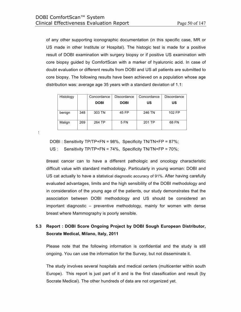
DOBI ComfortScan™ SystemClinical Effectiveness Evaluation Report Page 50 of 147
of any other supporting iconographic documentation (in this specific case, MR or
US made in other Institute or Hospital). The histogic test is made for a positive
result of DOBI examination with surgery biopsy or if positive US examination with
core biopsy guided by ComfortScan with a marker of hyaluronic acid. In case of
doubt evaluation or different results from DOBI and US all patients are submitted to
core biopsy. The following results have been achieved on a population whose age
distribution was: average age 35 years with a standard deviation of 1.1:
Histology Concordance
DOBIDiscordance
DOBIConcordance
USDiscordance
US
benign 348 303 TN 45 FP 246 TN 102 FP
Malign 269 264 TP 5 FN 201 TP 68 FN
!
DOBI : Sensitivity TP/TP+FN = 98%, Specificity TN/TN+FP = 87%;
US : Sensitivity TP/TP+FN = 74%, Specificity TN/TN+FP = 70%;
Breast cancer can to have a different pathologic and oncology characteristic
difficult value with standard methodology. Particularly in young woman: DOBI and
US cat actually to have a statistical diagnostic accuracy of 91%. After having carefully
evaluated advantages, limits and the high sensibility of the DOBI methodology and
in consideration of the young age of the patients, our study demonstrates that the
association between DOBI methodology and US should be considered an
important diagnostic – preventive methodology, mainly for women with dense
breast where Mammography is poorly sensible.
5.3 Report : DOBI Score Ongoing Project by DOBI Sough European Distributor,
Socrate Medical, Milano, Italy, 2011
Please note that the following information is confidential and the study is still
ongoing. You can use the information for the Survey, but not disseminate it.
The study involves several hospitals and medical centers (multicenter within south
Europe). This report is just part of it and is the first classification and result (by
Socrate Medical). The other hundreds of data are not organized yet.

DOBI ComfortScan™ SystemClinical Effectiveness Evaluation Report Page 51 of 147
The study involves symptomatic women of different age. All women diagnostic
path includes visit, US, X-Ray , Biopsy and obviously DOBI.
The study objective is the DOBI level evaluation:
Patient Number: 113 women
Biopsy: – 39 (34.5%) nonmalignant
– 74 (65.4%) malignant
Result table:
Nonmalignant Malignant Total
Nonmalignant 31 (83.8) 6 (16.2) 37
Malignant 15 (20.3) 59 (79.7) 74
46 65
59/74 malignant correct classification (79,7%)
31/37 nonmalignant correct classification (83,7%)
Cut-off DOBI-Score = 30.5,
• Sensitivity : 80%
• Specificity : 88% .
When the cut-off DOBI Score is adjusted based on the design of the study or the
need of the clinics, different outcomes, Sensitivity and Specificity, will be obtained.
At the same time, since the DOBI Score is easy to be adapted or computerized, a
standard diagnostics will be implemented and a clinical significance will be
achieved.
5.4 Report : Dynamic Optical Imaging. Thesis Dissertation : “The Use of
Dynamic Optical Imaging in Breast Cancer Detection” by Wilson, Kyle, Ph.D.,
MCMASTER UNIVERSITY and Publication submitted to Radiology “Dynamic
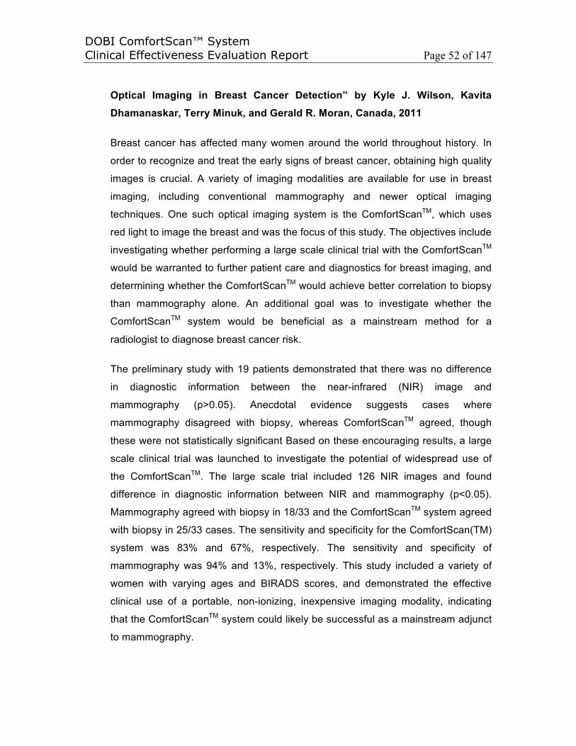
DOBI ComfortScan™ SystemClinical Effectiveness Evaluation Report Page 52 of 147
Optical Imaging in Breast Cancer Detection” by Kyle J. Wilson, Kavita
Dhamanaskar, Terry Minuk, and Gerald R. Moran, Canada, 2011
Breast cancer has affected many women around the world throughout history. In
order to recognize and treat the early signs of breast cancer, obtaining high quality
images is crucial. A variety of imaging modalities are available for use in breast
imaging, including conventional mammography and newer optical imaging
techniques. One such optical imaging system is the ComfortScanTM, which uses
red light to image the breast and was the focus of this study. The objectives include
investigating whether performing a large scale clinical trial with the ComfortScanTM
would be warranted to further patient care and diagnostics for breast imaging, and
determining whether the ComfortScanTM would achieve better correlation to biopsy
than mammography alone. An additional goal was to investigate whether the
ComfortScanTM system would be beneficial as a mainstream method for a
radiologist to diagnose breast cancer risk.
The preliminary study with 19 patients demonstrated that there was no difference
in diagnostic information between the near-infrared (NIR) image and
mammography (p>0.05). Anecdotal evidence suggests cases where
mammography disagreed with biopsy, whereas ComfortScanTM agreed, though
these were not statistically significant Based on these encouraging results, a large
scale clinical trial was launched to investigate the potential of widespread use of
the ComfortScanTM. The large scale trial included 126 NIR images and found
difference in diagnostic information between NIR and mammography (p<0.05).
Mammography agreed with biopsy in 18/33 and the ComfortScanTM system agreed
with biopsy in 25/33 cases. The sensitivity and specificity for the ComfortScan(TM)
system was 83% and 67%, respectively. The sensitivity and specificity of
mammography was 94% and 13%, respectively. This study included a variety of
women with varying ages and BIRADS scores, and demonstrated the effective
clinical use of a portable, non-ionizing, inexpensive imaging modality, indicating
that the ComfortScanTM system could likely be successful as a mainstream adjunct
to mammography.
DOBI ComfortScan™ SystemClinical Effectiveness Evaluation Report Page 53 of 147
The potential of using polyvinyl alcohol cryogel (PVA-C) as a breast tissue mimic
was investigated and PVA-C was then used to validate the mode of action of the
ComfortScanTM system. Two experimental methods reported the absorption
coefficients and reduced scattering coefficients of PVA-C. Using a double
integrating sphere, the values were μa = 0.012 ± 0.002 mm-1 and μs' = 1.5 ± 0.2
mm-1 and using steady-state spatially resolved diffuse reflectance, the values were
μa = 0.017 ± 0.005 mm-1 and μs' = 1.3 ± 0.2 mm-1 at 640 nm. These values are
comparable to typical absorption coefficients for tissue reported by others.
The mode of action suggested by DOBI (Dynamic Optical Breast Imaging) Medical
for the ComfortScanTM system is that under compression a malignant tumour will
highly attenuate light, due to a partial collapse in the tumourous vasculature,
resulting in an increased deoxygenation of blood over time. Using a PVA-C breast
mimicking phantom, it was shown that by deoxygenating horse blood in a cavity,
there was an increase in the attenuation of 640 nm light as compared with the
surrounding phantom material; which suggests that the colour representative of
malignancies on the ComfortScanTM is caused by deoxygenating blood. Further
evidence suggests that the ComfortScanTM system is not recognizing a total
collapse of the vasculature and subsequent void of blood from the tumour as the
trigger for malignant detection. The mode of action suggested by DOBI Medical is
supported by our findings.
5.5 Report : Dynamic Optical Breast Imaging (DOBI): Prospective Study of
ComfortScan Accuracy in Diagnosis of Breast Cancer. PRELIMINARY
RESULTS, Thesis Dissertation by Rossella Dandolo, Ph.D., University of
Rome Tor Vergata, Italy, 2010
Current methods of breast imaging (mammography, ultrasonography and MRI) are
burdened with some considerable limitations that affect diagnostic accuracy, since
all characterized by a satisfactory sensitivity but variable specificity. In fact,
mammography is strongly influenced by the degree of radiopacity of the breast and
exposes patients to ionizing radiation; Ultrasonography provides an in-depth
clinical and mammographic findings diagnostic doubts, but has a sensitivity and
DOBI ComfortScan™ SystemClinical Effectiveness Evaluation Report Page 54 of 147
specificity of operator-dependent imaging methods such as additional MRI
(Magnetic Resonance Imaging) can be expensive and inconclusive, for example in
the detection of microcalcifications, and finally the cyto-histology, a diagnosis that
in nature highly suspect in the case of images, is not without limits, identified in the
most cost greater injury and greater susceptibility to sampling errors.
The DOBI (Dynamic Optical Breast Imaging) is an innovative tool for optical
imaging, non-invasive, which exploits the properties of light absorption by breast
tissue in order to measure in static and dynamic conditions, the ability to reduce
the light signal dependent neoplastic processes in the mammary gland. DOBI
physiological data related to functional tumor-induced neoangiogenesis. The
process of neovascularization associated with tumor growth is characterized by
progressive replacement of normal tissue vasculature with numerous arterio-
venous shunts that increase the blood volume in the context of the neoplastic
process in response to increased metabolic demand. The angiogenic blood
vessels in malignant tissues share several characteristics observed. Together,
these features create a "profile vascular only" or "angiogenic footprint" that can be
identified by DOBI in the diagnosis of breast lesions. The aim of our study is to
assess the diagnostic accuracy of DOBI, determining the indications and
limitations of the method and, ultimately, identifying the benefits, in order to
propose it as a useful tool for integration in the study of breast diseases.
Between June and September 2009, 32 patients, aged between 23 and 75 years,
were selected for our study and subjected to DOBI and MRI. Object of our study
were patients whose mammographic findings had shown the presence of opacity,
parenchymal distortion or microcalcifications suggestive of malignancy. To
complete diagnosis, all patients selected were under investigation or cytological
microistologica. The average size of tumors detected was 15 mm (range, 0.6-25
mm).
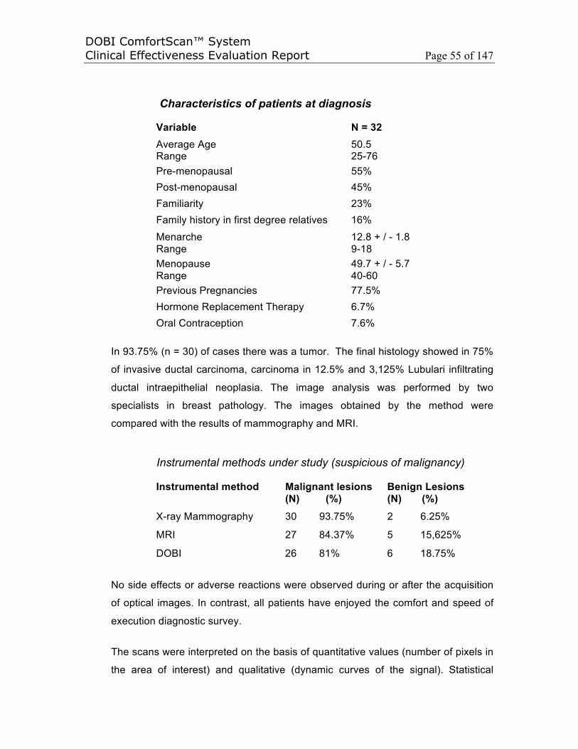
DOBI ComfortScan™ SystemClinical Effectiveness Evaluation Report Page 55 of 147
Characteristics of patients at diagnosis
Variable N = 32Average Age Range
50.5 25-76
Pre-menopausal 55% Post-menopausal 45% Familiarity 23% Family history in first degree relatives 16%
Menarche Range
12.8 + / - 1.8 9-18
Menopause Range
49.7 + / - 5.7 40-60
Previous Pregnancies 77.5% Hormone Replacement Therapy 6.7% Oral Contraception 7.6%
In 93.75% (n = 30) of cases there was a tumor. The final histology showed in 75%
of invasive ductal carcinoma, carcinoma in 12.5% and 3,125% Lubulari infiltrating
ductal intraepithelial neoplasia. The image analysis was performed by two
specialists in breast pathology. The images obtained by the method were
compared with the results of mammography and MRI.
Instrumental methods under study (suspicious of malignancy)
Instrumental method Malignant lesions(N) (%)
Benign Lesions(N) (%)
X-ray Mammography 30 93.75% 2 6.25%
MRI 27 84.37% 5 15,625%
DOBI 26 81% 6 18.75%
No side effects or adverse reactions were observed during or after the acquisition
of optical images. In contrast, all patients have enjoyed the comfort and speed of
execution diagnostic survey.
The scans were interpreted on the basis of quantitative values (number of pixels in
the area of interest) and qualitative (dynamic curves of the signal). Statistical
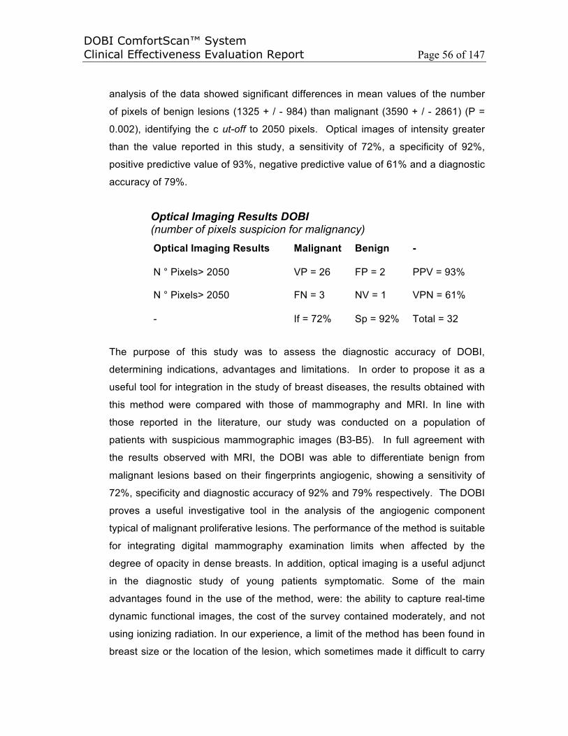
DOBI ComfortScan™ SystemClinical Effectiveness Evaluation Report Page 56 of 147
analysis of the data showed significant differences in mean values of the number
of pixels of benign lesions (1325 + / - 984) than malignant (3590 + / - 2861) (P =
0.002), identifying the c ut-off to 2050 pixels. Optical images of intensity greater
than the value reported in this study, a sensitivity of 72%, a specificity of 92%,
positive predictive value of 93%, negative predictive value of 61% and a diagnostic
accuracy of 79%.
Optical Imaging Results DOBI (number of pixels suspicion for malignancy)
Optical Imaging Results Malignant Benign -
N ° Pixels> 2050 VP = 26 FP = 2 PPV = 93%
N ° Pixels> 2050 FN = 3 NV = 1 VPN = 61%
- If = 72% Sp = 92% Total = 32
The purpose of this study was to assess the diagnostic accuracy of DOBI,
determining indications, advantages and limitations. In order to propose it as a
useful tool for integration in the study of breast diseases, the results obtained with
this method were compared with those of mammography and MRI. In line with
those reported in the literature, our study was conducted on a population of
patients with suspicious mammographic images (B3-B5). In full agreement with
the results observed with MRI, the DOBI was able to differentiate benign from
malignant lesions based on their fingerprints angiogenic, showing a sensitivity of
72%, specificity and diagnostic accuracy of 92% and 79% respectively. The DOBI
proves a useful investigative tool in the analysis of the angiogenic component
typical of malignant proliferative lesions. The performance of the method is suitable
for integrating digital mammography examination limits when affected by the
degree of opacity in dense breasts. In addition, optical imaging is a useful adjunct
in the diagnostic study of young patients symptomatic. Some of the main
advantages found in the use of the method, were: the ability to capture real-time
dynamic functional images, the cost of the survey contained moderately, and not
using ionizing radiation. In our experience, a limit of the method has been found in
breast size or the location of the lesion, which sometimes made it difficult to carry
DOBI ComfortScan™ SystemClinical Effectiveness Evaluation Report Page 57 of 147
out the survey. The study of small breasts has been problematic for the conduct of
the examination in relation to the structural characteristics of the machine. The
same difficulty was found in the analysis of large breasts. In the study, the
diagnostic accuracy in the localization of the disease was limited when lesions at
the chest wall or nipple-areola complex (NAC). Further application methods in the
study of cancer in situ will be useful in the diagnostic definition of this disease with
important clinical and therapeutic implications if the method proves able to
establish accurately the nature. Subsequent studies will be needed in order to
optimize the technique, defining the optical aspects of the breasts are not
pathological; evaluating sensitivity and specificity of the method on a larger
population of patients, and, ultimately, improving the knowledge about its potential
applications.
5.6 Report : Dynamic Optical Breast Imaging (DOBI), associated Ultrasound,
allows to avoid unnecessary Biopsies - “Seno, luce rossa preventiva : Il
Dynamic optical breast imaging (Dobi), associato all’ecografia, permette di
evitare biopsie non necessarie” by di Piercarlo Salari, Oncologia, Tecnologie,
October 12, 2009
“Breast, red light prior : Dynamic optical breast imaging (Dobi), associated
ultrasound, thus avoiding unnecessary biopsies” describes a study has shown that
Dobi has obtained a value of 95% sensitivity and specificity of 78.8%.
The aim of the present study is to determine the diagnostic accuracy of using
Dynamic Optical Breast Imaging (DOBI) in addition to Ultrasound systems (US) for
prevention, diagnosis and monitoring of breast cancer in young females by means
of a non invasive examination methodology. This study included 391 patients with
clinical risk to develop a cancer. All patients have been submitted to clinical
investigation by means of both US and DOBI. The dynamic optical imaging
showed a statistical difference (p<0.005) in patient analyses compared with
standard examinations. In this study, the dynamical optical breast imaging had a
sensitivity equal to 95% and a specificity equal to 78.8%. Under the same trial
criteria, the mammographic sensitivity is 67% as reported by Kerlikowske et al
DOBI ComfortScan™ SystemClinical Effectiveness Evaluation Report Page 58 of 147
(Evaluation of abnormal mammography results and palpable breast abnormalities.
Ann Intern Med. 2004 May 4; 140(9):764), and the average Ultrasound sensibility
is only 58–64%.
At present, ComfortScan® DOBI in this study proves to be a powerful system to
be used for diagnostic purposes in synergy with traditional methodologies such as
mammography and ultrasound, especially in case of women with dense breast and
high risk from a clinical and anamnesis viewpoint. In particular, in young women
the clinical and ultrasound examinations associated with DOBI represent a valid,
non invasive, accurate and quick method able to discriminate between truly benign
lesions and like-benign lesions such as the marrow and lobular or medullar
carcinomas. In case of persisting doubts, MR or core biopsy are mandatory. DOBI
can be used for non invasive monitoring, too. This non invasive imaging-based
methodology has a very high potential for breast cancer prevention independently
of breast size, density, hormonal status.
5.7 Report : “Dynamic optical breast imaging: A novel technique to detect and
characterize tumor vessels” Published by Laure S. Fournier, Daniel Vanel,
Alexandra Athanasiou, Wolfgang Gatzemeier, I.V. Masuykov, Anwar R.
Padhani, Clarisse Dromain, Ken Galetti, Robert Sigal, Alberto Costa, Cornne
Balleyguier on Europea Journal of Radiology 69 (2009)
Optical Imaging: In a prospective cohort trial, Fournier et al. (2008) prospectively
determined the diagnostic accuracy of dynamic optical breast imaging (DOBI)
compared to pathology in 46 women with BI-RADS 3–5 classification lesions.
DOBI was conducted immediately prior to biopsy. Twelve lesions (26%) were
diagnosed as benign and 35 (74%) as malignant. Results demonstrated a
sensitivity of 74%, a specificity of 92%, a positive predictive value of 93%, a
negative predictive value of 55% and a diagnostic accuracy of 79%. The authors
noted that further evaluation will be required to optimize the technique, evaluate its
sensitivity and specificity in a wider range of patients, and explore its potential role
in patient management.
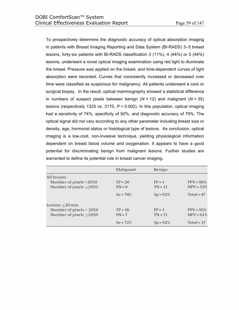
DOBI ComfortScan™ SystemClinical Effectiveness Evaluation Report Page 59 of 147
To prospectively determine the diagnostic accuracy of optical absorption imaging
in patients with Breast Imaging Reporting and Data System (BI-RADS) 3–5 breast
lesions, forty-six patients with BI-RADS classification 3 (11%), 4 (44%) or 5 (44%)
lesions, underwent a novel optical imaging examination using red light to illuminate
the breast. Pressure was applied on the breast, and time-dependent curves of light
absorption were recorded. Curves that consistently increased or decreased over
time were classified as suspicious for malignancy. All patients underwent a core or
surgical biopsy. In the result, optical mammography showed a statistical difference
in numbers of suspect pixels between benign (N = 12) and malignant (N = 35)
lesions (respectively 1325 vs. 3170, P = 0.002). In this population, optical imaging
had a sensitivity of 74%, specificity of 92%, and diagnostic accuracy of 79%. The
optical signal did not vary according to any other parameter including breast size or
density, age, hormonal status or histological type of lesions. As conclusion, optical
imaging is a low-cost, non-invasive technique, yielding physiological information
dependent on breast blood volume and oxygenation. It appears to have a good
potential for discriminating benign from malignant lesions. Further studies are
warranted to define its potential role in breast cancer imaging.
DOBI ComfortScan™ SystemClinical Effectiveness Evaluation Report Page 60 of 147
5.8 Report : “Optical mammography: a new technique for visualizing breast
lesions in women presenting non palpable BIRADS 4–5 imaging findings:
preliminary results with radiologic–pathologic correlation” Published by
Alexandra Athanasiou, Daniel Vanel, Laure Fournier, and Corinne Balleyguier
on International Cancer Imaging Society 7 (2007)
The purpose of this prospective study is to determine the diagnostic accuracy of
near-infrared breast optical absorption imaging in patients with Breast Imaging
Reporting and Data System (BIRADS) 4–5 non-palpable lesions scheduled for
biopsy, using pathology after core or excisional biopsy as a reference. The
patient's breast was positioned onto a panel of red light-emitting diodes (640 nm).
A soft membrane was inflated to exert a uniform pressure on the breast.
Transmitted light was detected using a CCD camera. The entire acquisition
sequence took 1 minute. Image processing generated dynamic images displayed
in colour scale, to reveal time-dependent changes in the transmitted light intensity
caused by the pressure change. Dynamic curves were classified in two categories:
consistently decreasing intensity suspicious for malignancy, and sinusoidal
increasing intensity considered as benign.
Between November 2004 and November 2005, a total of 78 women (age range
41–72 years) participated in this study. All of them presented non-palpable
BIRADS 4–5 mammographic and/or ultrasonographic findings and were referred to
our institution for further investigation in this study. In six out of 78 cases, scan
acquisition was considered invalid, mainly due to ambient light problems. We finally
included 72 patients: 40 of them presenting BIRADS 4 lesions and 32 presenting
BIRADS 5 lesions. Breast parenchyma density in mammographic images was
evaluated according to the BIRADS classification system (density ranging from 0
(totally fatty) to 4 (totally fibroglandular) breast parenchyma). Evaluation of optical
scans was based on three parameters:
• The presence of early, focal, blue “blush” in the area of interest, suggesting
an underlying lesion with strong deoxyhaemoglobin concentration.
• The pixel intensity of focal blue blush areas calculated by means of
dedicated software: a high number, usually more than 90, indicated a high
DOBI ComfortScan™ SystemClinical Effectiveness Evaluation Report Page 61 of 147
light absorption. This threshold was in accord with previously published
studies in which total haemoglobin concentration and tumour hypoxia had
been calculated from oxyhaemoglobin and deoxyhaemoglobin distributions.
These distributions were proven to be highly correlated with lesion
malignancy[9,10]. A mean haemoglobin concentration of 95 µmol/l was used
as a threshold to separate malignant lesions from benign lesions.
• The type of temporal signature of dynamic curves that was further classified
into consistently decreasing negative-spectral and sinusoidal increasing
positive-spectral.
A numeric level of suspicion (LOS) score was calculated based on all these
elements and taking into consideration the intensity and colour polarity of blush
areas as well as the shape of dynamic curves, as follows:
where P is the colour polarity (red, orange, blue), S is the dynamic curve shape, I
is the maximum intensity and A is the area of interest in cm2. A score >5 was
considered suspicious. Pathologic correlation was obtained for all cases.
At histological analysis, 49 out of 72 lesions were found to correspond to
malignancies. This represented a total of 31 BIRADS 5 and 18 BIRADS 4 lesions.
Among these 49 carcinomas, 17 corresponded to ductal carcinoma in situ (DCIS)
and 32 to infiltrating ductal or lobular carcinoma (IDC or ILC). Twenty-three cases
were found to correspond to benign or high-risk lesions at histology, such as
fibrocystic changes, sclerosing adenosis, atypical hyperplasia or radial scar. They
corresponded to one case of BIRADS 5 and 22 cases of BIRADS 4 classification.
Dynamic optical breast imaging was positive in 41 cases. Among them 30
corresponded to malignant lesions and 11 to benign proliferative lesions. Optical
findings in benign proliferative lesions were different, as in these cases the
hypervascularized areas were not as hypoxic as the malignant hypermetabolic
tumours that consume a greater amount of oxygen. Early “blush” in benign cases
was displayed as red-coloured areas and the calculated dynamic curves were in
the positive scale. The optical signal was positive in the mean of high absorption,
but the corresponding area of interest was represented as a red-coloured zone.
DOBI ComfortScan™ SystemClinical Effectiveness Evaluation Report Page 62 of 147
Dynamic curves were in the positive scale, in accordance with an underlying
hypermetabolic but not strongly hypoxic area. In the remaining 31 cases, optical
acquisition was negative; no signal was detected in the areas of interest. However,
19 cases of negative optical imaging were finally diagnosed as malignant at
histology. The majority (11 out of 19) corresponded to DCIS of small size
(calcifications did not exceed 10 mm) and of low or intermediate grade. An
imaging–histology correlation was obtained for seventy-two patients, the remaining
six patients were excluded for technical optical scan reasons. We experienced an
overall sensitivity of 73% and specificity of 38%, the false negative results being
mainly small size (<10 mm) infiltrating malignant lesions and ductal carcinoma in
situ (DCIS). False positive results were seen in benign proliferative lesions.
Recently optical mammography has emerged as a potential and revolutionary
imaging method targeting the detection and, if possible, the characterization of
vascular stroma in normal and abnormal tissues. In vitro and ex vivo, many
experiments have already been performed in order to validate the feasibility and
evaluate the sensibility of this method. In vivo, optical imaging has almost
exclusively been used in cases of breast tissue lesions. The main reason for this
is the relatively small volume of breast and the superficial lesion location compared
to other deep intra-abdominal organs, where light could possibly never reach the
target with a sufficient intensity. It is noteworthy that optical mammography uses
almost exclusively infrared light emission (spectrum varying between 640 and 800
nm depending on the various studies already published in the literature). Blood
vessels and highly vascularized areas feature a high optical contrast due to
increased infrared light absorption, thus providing indispensable spatial resolution
information. Various algorithms permit the quantitative analysis of the images
obtained (whether static or dynamic), mainly by estimating the haemoglobin
concentration and the oxygenation, providing the so-called spectral information.
To date, publications have demonstrated the promising role of optical
mammography, used either alone or combined with other non-invasive and non-
ionizing imaging modalities such as ultrasonography or magnetic resonance
imaging. However, there are not many studies dealing with optical, infrared breast
imaging in patients presenting non-palpable BIRADS 4–5 lesions. Our results
DOBI ComfortScan™ SystemClinical Effectiveness Evaluation Report Page 63 of 147
reflected the performance of optical imaging according to the different stages of
tumour angiogenesis: intraductal carcinomas were difficult to depict due to several
physiologic factors; a malignant lesion confined to the basement membrane may
not substantially influence the physical milieu, whereas a more invasive lesion
would. Another factor may be that angiogenesis is less advanced during the earlier
stages of ductal carcinoma in situ when the tumour is still confined to the duct.
However, we should mention that a suspicious signal in DCIS lesions was easily
detected, as the micro-calcification surface was larger, mainly because larger
DCIS were more often associated with micro-invasion. Certainly a number of
malignancies have been missed during our pilot study, however initial results are
rather promising, especially if this technique is used as complementary to the
traditional ones, where it could possibly increase the degree of suspicion of non-
palpable breast lesions. With breast cancer incidence showing no signs of
abatement, every imaging modality used as complement to the traditional ones
could be of interest. This is still a work-in-progress. The system software and the
evaluation parameters are subject to modifications and improvement. Potential
clinical applications include additional information on non-palpable breast lesion
diagnosis, as well as monitoring of tumour response to neo-adjuvant
chemotherapy. Optical mammography has already been used for this purpose with
promising results. Further studies and continuous system improvement are
necessary for a better evaluation and clinical application of this innovative method.
The initial results of this study indicate that dynamic optical mammography is an
innovative, simple, well-tolerated, non-ionizing imaging method that could be of
interest in the detection of hypermetabolic, hypoxic breast areas, suspicious for
malignancies in women presenting non-palpable BIRADS 4–5 lesions. However,
further technical improvement and larger studies are needed to define any possible
clinical applications. Dynamic optical breast imaging is a novel, low-cost, non-
invasive technique yielding a new type of information about the physiology of
breast lesions. Absorption is due to haemoglobin and its products, therefore
reflecting the angiogenic status of breast tumours.
DOBI ComfortScan™ SystemClinical Effectiveness Evaluation Report Page 64 of 147
5.9 Report : “Dynamic Optical Breast Imaging: A new technique to visualise
breast vessels: Comparison with breast MRI and preliminary results”
Published by Alexandra Athanasiou, Daniel Vanel, Cornne Balleyguier, Laure
Fournier, Marie Christine Mathieu, Suzette Delaloge, Clarisse Dromain on
Europea Journal of Radiology 54 (2005)
The purpose of our study was to explore and evaluate an innovative imaging
approach, which consists on imaging the breast parenchyma by means of
photoluminescence detectors (LED) and analysis of dynamic data. Breast
magnetic resonance imaging (MRI) was chosen as the reference imaging method,
as this is considered to be nowadays the gold standard for breast vascularisation
evaluation. Preliminary results reveal a good correlation between breast MRI
findings and light images.
In the 25-patient study, patients underwent both breast MRI and optical scans in
addition to standard mammography and ultrasonography. Patients' ages ranged
between 28 and 78 years of age and all patients scanned did not have recent
trauma, breast surgery or biopsy. ComfortScan system images were positive in
four of the five cases where MRI was positive. In the only case where the
ComfortScan system disagreed with the MRI finding, a three-month follow-up with
MRI showed up as negative when scanned a second time, aligning the MRI with
the ComfortScan system findings. Investigators noted that they found the potential
advantages of ComfortScan system imaging included the ease of patient
positioning, quick exam time, and the absence of ionizing (X-ray) radiation.
Investigators also concluded that ComfortScan system imaging may also be
valuable in cases of claustrophobic patients or in other cases where MRI may be
contraindicated.
Dynamic optical breast imaging can be a promising complementary imaging
modality in women with inconclusive mammography and/or physical examination.
Our study includes a small number of patients, but the preliminary results are
encouraging enough. However, further evaluation with a larger number of patients
should be carried out. "The promise of the ComfortScan system, as demonstrated
in this initial study, warrants additional investigation, but the preliminary results are
DOBI ComfortScan™ SystemClinical Effectiveness Evaluation Report Page 65 of 147
encouraging," stated Dr. Daniel Vanel. "The sensitivity of the ComfortScan system
technology was extremely strong and the specificity rates also were positive."
5.10 Report : “Correlation of Dynamic Optical Breast Imaging Curve and
Microvessel Density Count” Presented by Abraham A. Ghiatas, K Pavlaki, I
Messini, N Karaglani, D Keramopoullos, V Gaki, D Baltas, and N Bredakis
from Iaso Hospital in Greece in Cancer Imaging 6 (2006)
The aim of this stud is to investigate the behaviour of the dynamic optical breast
imaging (DOBI) curve in relation to the microvessel density (MVD) count of surgical
specimens from breast biopsies. Forty-six patients underwent DOBI evaluation for
mammographic findings suggesting biopsy. The DOBI evaluation was performed
the day before or on the day of the scheduled biopsy. The MVD count was
performed from the site of the specimen where the pathology was located. The
characteristics of the DOBI curve were correlated to the MVD count and to the
pathology results of the biopsy. The results show All malignant lesions had a high
MVD count and a DOBI curve with a downslope direction, rather straight and
without any initiation delays. The benign cases with a high MVD count had a
downslope DOBI curve but not always straight and with some initiation delays. The
rest of the cases had a low MVD count and most of them an elevated DOBI curve.
Conclusion Our preliminary results indicate a relationship between malignant
breast lesions with a characteristic DOBI curve and high MVD count.
5.11 Report : “Digital Optical Breast Imaging” Presented by Abraham A. Ghiatas
from Iaso Hospital in Greece (2005)
The PURPOSE of this study is to Assess the utility of digital breast optical imaging
in the evaluation of breast malignancies. The METHOD AND MATERIALS are One
hundred two patients with mammographic findings suggesting biopsy underwent
digital optical breast imaging performed by DOBI ComfortScan, which utilizes high-
intensity light-emitting diodes and gentle pressure in order to identify areas of
neoangiogenesis within the breast. The results of this evaluation were compared to
the pathology results. The RESULTS from the 102 cases are that 32 had proven
malignancies which were categorized by our breast surgeons into three categories
DOBI ComfortScan™ SystemClinical Effectiveness Evaluation Report Page 66 of 147
according to their aggressive behavior, taking into consideration the size of the
malignancy, lymph node involvement, hormone receptors, and grade. The digital
optical breast imaging curve from the region of malignancy in all malignant cases
had a downslope configuration indicating neoangiogenesis, and for each of the cat-
egories of aggressiveness, the curve had a rather characteristic configuration
indicating the level of aggressiveness (high aggressive, aggressive, low
aggressive). As CONCLUSIONS, in our study, the initial results indicate there is
an association of the morphology of the digital optical breast imaging curve and the
aggressiveness of the evaluated breast malignancies.
5.12 Report : The purpose of our study was to report: “New Perspective of
Mammary Screening : Application of Non-Invasive DOBI”, THE RESULTS OF
DOBI EXAMINATIONS IN MASARYK MEMORIAL CANCER INSTITUTE in
Czech Republic, by Irena Komorousova, Bartonkova H., Standara M.,
Schneiderova M., from 2004 to 2005
The purpose of our study was to evaluate an upcoming breast cancer diagnostic
modality based on measurements of the red light absorption in the breast and
response on pressure stimulus inducing circulating blood volume changes.
Patients and Method were 100 Patients (n=100) for age above 45 with suspicious
mammography finding underwent Dynamic Optical Breast Imaging (DOBI) followed
by core-cut biopsy in Masaryk Memorial Cancer Institute. Sensitivity and specificity
were calculated to compare DOBI with mammography. The Results of both
sensitivity and specificity (71% and 64% respectively) indicates that DOBI can be a
promising minimum-stress method in malignant breast lesion detection.
By comparing Table 1 and 2 below, the difference between both methods in
sensitivity and specificity is small. It is necessary to be aware of the fact that the
DOBI method is an entirely new approach, it is not possible to compare these early
experiences with the many years of experience with mammography. Based on
other preliminary evaluation, sensitivity 87-92% and specificity 75-82% of DOBI
ComfortScan would be hopeful in clinics.
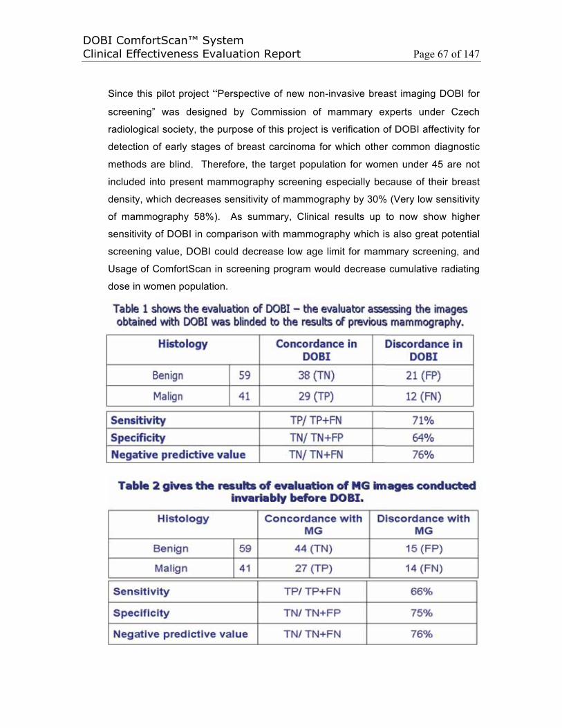
DOBI ComfortScan™ SystemClinical Effectiveness Evaluation Report Page 67 of 147
Since this pilot project “Perspective of new non-invasive breast imaging DOBI for
screening” was designed by Commission of mammary experts under Czech
radiological society, the purpose of this project is verification of DOBI affectivity for
detection of early stages of breast carcinoma for which other common diagnostic
methods are blind. Therefore, the target population for women under 45 are not
included into present mammography screening especially because of their breast
density, which decreases sensitivity of mammography by 30% (Very low sensitivity
of mammography 58%). As summary, Clinical results up to now show higher
sensitivity of DOBI in comparison with mammography which is also great potential
screening value, DOBI could decrease low age limit for mammary screening, and
Usage of ComfortScan in screening program would decrease cumulative radiating
dose in women population.
DOBI ComfortScan™ SystemClinical Effectiveness Evaluation Report Page 68 of 147
5.13 Report : “Dynamic Optical Breast Imaging: A non-invasive, adjunctive
method to detect breast cancer” Presented by Gatzemeier W, Scelsi M,
Galetti K, Villani L, Tinterri C, Secci A, and Costa A in San Antonio Breast
Cancer Symposium (December 2004)
From March 2003 to 2004, as part of a larger prospective international polite study
aimed at developing data acquisition methods and dynamic signature
interpretation/reading rules, a total 105 patients, aged 23-79, scheduled for open
biopsies (palpable and non-palpable lesions) were entered into this study from
Division of Senology and Division of Pathology at the Maugeri Foundation in Italy.
All patients were scanned with DOBI ComfortScan preoperatively and findings
were compared with those of the definite histology report and previous imaging.
Approximate half of the patients participated in the training section and only 35
qualified scans from the rest of the half patients are used for this study.
The calculated sensitivity and specificity to discriminate between malignant and
benign lesions were 100% and 80% respectively, i.e. 20% False Positive Rate.
The results from this dataset indicated that the ComfortScan System could be a
valuable tool in breast cancer diagnosis when paired with other imaging methods.
In addition, the system provides the physician with information obtained through a
dynamic physiological method to distinguish non-invasively between begin and
malignant lesions, which may avoid unnecessary interventions. Further studies
are needed to support these data.
Since this study aimed at developing the data acquisition methods and dynamic
signature interpretation/reading rules as same as all the studies/trials since 2000
after the product was designed and manufactured, the software for data acquisition
methods and dynamic signature interpretation/reading rules are the ongoing
development/improvement of the DOBI technique. As a result of the studies/trials
the DOBI ComfortScan software of ComfortScan and ComfortView have been
upgraded to Version 2.0 released on April 27, 2005 and January 15, 2006
respectively.
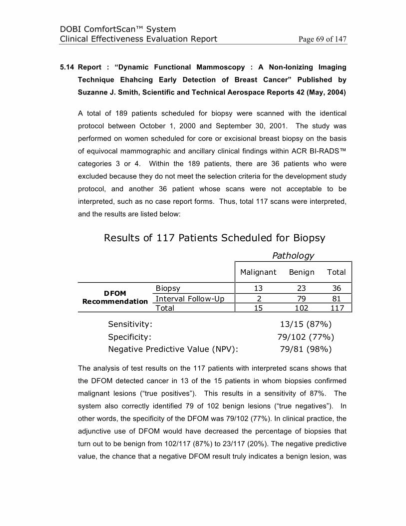
DOBI ComfortScan™ SystemClinical Effectiveness Evaluation Report Page 69 of 147
5.14 Report : “Dynamic Functional Mammoscopy : A Non-Ionizing Imaging
Technique Ehahcing Early Detection of Breast Cancer” Published by
Suzanne J. Smith, Scientific and Technical Aerospace Reports 42 (May, 2004)
A total of 189 patients scheduled for biopsy were scanned with the identical
protocol between October 1, 2000 and September 30, 2001. The study was
performed on women scheduled for core or excisional breast biopsy on the basis
of equivocal mammographic and ancillary clinical findings within ACR BI-RADS™
categories 3 or 4. Within the 189 patients, there are 36 patients who were
excluded because they do not meet the selection criteria for the development study
protocol, and another 36 patient whose scans were not acceptable to be
interpreted, such as no case report forms. Thus, total 117 scans were interpreted,
and the results are listed below:
Results of 117 Patients Scheduled for Biopsy
Pathology
Malignant Benign Total
Biopsy 13 23 36Interval Follow-Up 2 79 81
DFOMRecommendation
Total 15 102 117
Sensitivity: 13/15 (87%)
Specificity: 79/102 (77%)Negative Predictive Value (NPV): 79/81 (98%)
The analysis of test results on the 117 patients with interpreted scans shows that
the DFOM detected cancer in 13 of the 15 patients in whom biopsies confirmed
malignant lesions (“true positives”). This results in a sensitivity of 87%. The
system also correctly identified 79 of 102 benign lesions (“true negatives”). In
other words, the specificity of the DFOM was 79/102 (77%). In clinical practice, the
adjunctive use of DFOM would have decreased the percentage of biopsies that
turn out to be benign from 102/117 (87%) to 23/117 (20%). The negative predictive
value, the chance that a negative DFOM result truly indicates a benign lesion, was
DOBI ComfortScan™ SystemClinical Effectiveness Evaluation Report Page 70 of 147
79/81(98%) for the cases included thus far. While encouraging, these results
suggest the need for further patient studies on specificity.
5.15 Report : “White Paper – Dynamic Optical Breast Imaging : Preliminary
Results from Two Data Interpretation Methods” by DOBI Medical
International (September, 2004)
A total of 245 data sets were acquired with the ComfortScan system from March
2003 to January 2004. These data were collected in 4 clinical sites from 4
countries (Italy, France, Spain, and United States). The data were collected with a
new acquisition modality and utilized to develop data acquisition methods and
dynamic signature interpretation rules. The interpretation rules were developed
and verified using different and somewhat overlapping subsets of the initial 245
data sets. These rules were used for the interpretations described below and were
applied to the filtered subset selected as described below. Of these 245 data sets,
15 had no usable information (either test scans or images of the tool used to
calibrate the ComfortScan system; i.e., the calibration phantom) and were
excluded. From each data set, information regarding pixel saturation, light intensity
acquisition range, and ratio of focal area of illumination to breast area were entered
in a database. A set of standards, compatible with the most recent acquisition
hardware and proprietary software, and with the most recent reading application
and interpretation rules, was used to filter the database. The data sets with values
falling outside these rules were not included in the analysis. The following standard
filter rules applied: Number of saturated pixels >20; light intensity (detected by the
CCD camera) <400 counts; illuminated area to breast area ratio ≤25%. After
application of the filter, the database returned 111 data sets. Of these data sets, 43
had incomplete patient documentation (biopsy pathology report, mammography
report or sonography report) or had the focal illumination not matching the area of
interest indicated by the mammography or sonography reports and were excluded
from the analysis. A total of 68 data sets were available for interpretation. Of these
scans, 49 were from lesions determined to be malignant by biopsy and 19 were
from lesions determined to be benign.
DOBI ComfortScan™ SystemClinical Effectiveness Evaluation Report Page 71 of 147
Each set was interpreted by a physician (A.S.) blinded to the results of the biopsy.
The blinded reader was given information regarding the location of the lesion as
indicated in the mammography or sonography reports (quadrant and depth) and
the age of the patient. The reader was requested to interpret each scan as
“Malignant” or “Benign.” No undecided or dubious scores were allowed. Reader
interpretations were compared with the biopsy results reported in the pathology
laboratory reports. The reader used two different methods to interpret the
ComfortScan sets. The first method was manual, relying on the expertise of the
reader to locate suspicious areas and interpret the corresponding dynamic
signatures. The second method (computeraided) relies on a map of suspicious
curves generated by the computer and on a standard area curve counter (a circle
with a 113 pixels area). The curve counter was positioned manually by the reader,
under guidance of the computer-generated map, to include the highest possible
number of suspicious curves. A cut-off of 40 curves was selected. This second
method is much less dependent on reader interpretation and requires less training
and expertise than the manual method.
The results shows that a total of 68 data sets were interpreted by a blinded reader;
49 of these data sets were from malignant lesions and 19 from benign lesions. The
average age of the patients was 57 (28 to 79). Following list reports a summary of
the lesions as indicated by the pathology reports. Lesion Type as Indicated by
Pathology Report:
Lesion Type Number of Cases Reported
Ductal carcinoma 31
Lobular carcinoma 12
Fibrocystic disease 9
Fibroadenoma 6
Non-specified invasive carcinoma 4
DCIS 2
Fatty tissue 2
Cyst 1
Micro-calcifications 1
DOBI ComfortScan™ SystemClinical Effectiveness Evaluation Report Page 72 of 147
The sensitivity obtained by the blinded reader, using the first method (manual) was
98%, and the average specificity was 63%. Only one malignant case (ductal
carcinoma, in a 58 year old patient) was interpreted by the blinded reader as a
benign case Table 3 below reports the blinded reader results. The sensitivity
obtained by the blinded reader, using the second method (computer generated
map of suspicious curves and standard size curve counter) was 92%, and the
average specificity was 63%. These results suggest that the ComfortScan system
can be a useful tool, in combination with other diagnostic methods, in the
assessment of suspicious breast lesions.
5.16 Report : “The Value of Dynamic Optical Breast Imaging and Ultrasound in
Early Breast Cancer Detection” Reported by Guojian Tan, Jie Wang, Cui Liu,
Chunmian Li, Weiping Wang, John Zhang (2010)
The purpose of this study for the Dynamic Optical Breast Imaging and Ultrasound
were evaluated in diagnosing the early breast cancer. There were 62 patients
enrolled in this study, who had biopsies during the period of May, 2007 to June,
2008. All of them are female, the average age is 47 According to the
examinations, they can be divided into 3 groups: DOBI Examination 52 cases;
Ultrasound Examination of 35 cases; Both DOBI and Ultrasound combined
Examination of 25 cases, excluding patients in pregnant/lactation period and the
ones that accept galactophore operation within half a year. Sensitivity(SE),
specificity(SP), the rate of misdiagnosis(α) and the rate of missed diagnosis(β) of
DOBI technology, Ultrasound and their combination’s diagnosis to breast
carcinoma are calculated and compared respectively.
For the DOBI ComfortScan 52 tests alone, the sensitivity (SE) is 14/19 = 73.68%;
specificity (SP) is 25/33 = 75.76%; false positive rate (FPR) is 8/33 = 24.24%; false
negative rate (FNR) is 5/19 = 26.32%. For the Ultrasound 35 tests alone, the
sensitivity (SE) is 16/19 = 84.21%; specificity (SP) is 6/16 = 37.50%; false positive
rate (FPR) is 10/16 = 62.50%; false negative rate (FNR) is 3/19 = 15.79%. During
the 25 combination test by DOBI ComfortScan first and then Ultrasound second,
the patients were taken biopsy if both of them indicated malignant lesions. The
DOBI ComfortScan™ SystemClinical Effectiveness Evaluation Report Page 73 of 147
sensitivity (SE) is 14/16 = 87.50%; specificity (SP) is 5/9 = 55.56%; false positive
rate (FPR) is 4/9 = 44.44%; false negative rate (FNR) is 2/16 = 12.50%.
As a conclusion, the Dynamic Optical Breast Imaging is a new technology. It is
non-invasive and non-radiation. It is an appropriate approach for the early
diagnosing breast cancer by combining the Dynamic Optical Breast Imaging and
Ultrasound. While it needs a large number of case studies to distinguish malignant
and benign tumors, including in different stages of cancer development, with
different physical signs in patients, quantify the metabolic rate of malignant and
benign tumors by statistics and so on.
5.17 Report : “Preliminary Research on Dynamic Optical Breast Imaging in Breast
Cancer” Published by Guojian Tan, Jie Wang, Cui Liu, Chunmian Li, John
Zhang, Weiping Wang on Chinese Journal of Medical Imaging 18 (2010)
A new way in breast cancer detection, Dynamic Optical Breast Imaging (DOBI)
was studied. Fifty two (52) patients receiving breast biopsy and DOBI were
enrolled in this study. 94.8% of “blue area” in non-breast cancer lesions was
found as wandering or diffusive pattern, while 68.42% breast cancer showed focal
pattern. 86.46% of the curve signature of “blue area” in non-breast cancer lesions
was wavy or flat, while 57.37% of the breast cancer showed a steep decline. In
64.58% non-breast cancer, the curve of “blue area” was similar to that of non-blue
area. The absolute value of amplitude (-5.77±12.14) of “blue area” in cancer was
higher than in non-cancer (-3.34±0.87). The differences were all statistically
significant (P<0.05). As conclusion, the spatial and temporal characteristics of
DOBI were of diagnostic and differential value for breast cancer. The abolute
value of amplitude, over |-5|, also helped the diagnosis of breast cancer.
DOBI ComfortScan™ SystemClinical Effectiveness Evaluation Report Page 74 of 147
5.18 Report : “The Diagnostic Value of Small Lesions within Breast by Ultrasound
combined with DOBI” published by Mei Xu, Junlai Li, MEDICAL JOURNAL OF
CHINESE PEOPLE'S LIBERATION ARMY, Vol 34, No. 8 on August 1, 2009
Female breast screening use of ultrasound technology, ultrasound detection rate
for microcalcifications lower limits of small breast lesions benign and malignant
correct judgment. Dynamic optical breast imaging (DOBI) system is an analysis of
the lesion of new blood vessels to generate blood and oxygen content of new
technologies for the diagnosis of breast disease. This article aims to observe the
clinical diagnostic value of ultrasound combined with DOBI of small breast lesions.
From August of 2007 to June of 2008, there are 53 women at age 43.7, who were
subjected the breast examination by using Ultrasound and DOBI at 301 Hospital.
The sensitivity, specificity and accuracy of Ultrasound is about 81%, 59% and 70%
respectively. But by combining with DOBI, the sensitivity, specificity and accuracy
of Ultrasound increase to 95%, 81% and 87%. Overall, ultrasound combined with
DOBI can improve the sensitivity, specificity, and accuracy of diagnosis of breast
lesions, the better to avoid the misdiagnosis of small breast lesions. However, due
to the DOBI technical application time is short, its accuracy and experience of the
operator have a certain relationship, combined with a relatively small number of
cases, the clinical value needs further study.
5.19 Report : “The Value Analysis of ComfortScan System in Differentiating
Benign Breast Lesions from Malignant” published by Mei Xu, Junlai Li,
Yongfeng Zhang, Xuejuan Shi, Chunmian Li, Jie Tiang, and “The Application
of Dynamic Optical Breast Imaging in Differentiating Benign Breast Lesions
from Malignant” Thesis submitted by Mei Xu at Chinese PLA General
Hospital & Postcarduate Medical School on June 1, 2009
The study is to analyze the value of Dynamic Optical Breast Imaging in
Differentiating Benign Breast Lesions from Malignant. A total of 74 patients at
averaged 44 years‘ old were examined with the Duplex Ultrasonography and
ComfortScan System. The patients were grouped based on the lesion sizes into
three groups: <1 cm, 1~2 cm and >2 cm through the Ultrasound measures.
DOBI ComfortScan™ SystemClinical Effectiveness Evaluation Report Page 75 of 147
After comparing the pathological results, DOBI ComfortScan has higher sensitivity
(85.7%), specificity (80.0%) and accuracy (81.8) than Ultrasound’s sensitivity
(71.4%), specificity (40.0%) and accuracy (50.0%) in <1 cm group. In 1~2 cm
group, the ComfortScan and Ultrasound are similar : (78.6% 64.7% 71.0%) and
(85.7% 76.5% 80.6%) respectively. But in >2 cm group, the ComfortScan is
much lower than Ultrasound : (25.0% 44.4% 33.3%) and (83.3% 77.8%
81.0%).
As a conclusion, DOBI ComfortScan has much higher sensitivity, specificity and
accuracy for smaller breast lesion sizes, and could be very useful in diagnosing
breast cancer at early stage. Because the cases in this study is relatively small,
further study with more data is recommended.
5.20 Report : “ComfortScan System and Ultrasound Imaging: The Value of
Combined Application to Differentiate Benign Breast Lesions from
Malignant” Reported by Yongfeng Zhang, Junlai Li, Xuejuan Shi, Mei Xu,
China, 2008
The purpose of this study is to explore the value of ComfortScan System in
differentiating benign breast lesions from malignant ones. A total of 65 breast
tumors in 64 patients were examined with the Duplex Ultrasonography and
ComfortScan System, and the sonograms and ComfortScan-grams were analyzed
respectively. The biopsy confirms 30 malignances.
The results were compared with histopathology. The results show there was no
significant difference between the two groups for differentiating benign from
malignant breast lesions, which means the similar diagnosing value. By
combination of these two techniques, the sensitivity was significantly improved to
93.3% (P≤0.01), which is much higher than that of the Duplex Ultrasonography.
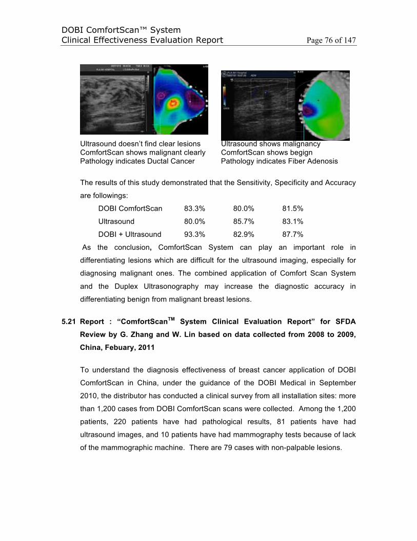
DOBI ComfortScan™ SystemClinical Effectiveness Evaluation Report Page 76 of 147
Ultrasound doesn’t find clear lesions Ultrasound shows malignancyComfortScan shows malignant clearly ComfortScan shows begignPathology indicates Ductal Cancer Pathology indicates Fiber Adenosis
The results of this study demonstrated that the Sensitivity, Specificity and Accuracy
are followings:
DOBI ComfortScan 83.3% 80.0% 81.5%
Ultrasound 80.0% 85.7% 83.1%
DOBI + Ultrasound 93.3% 82.9% 87.7%
As the conclusion, ComfortScan System can play an important role in
differentiating lesions which are difficult for the ultrasound imaging, especially for
diagnosing malignant ones. The combined application of Comfort Scan System
and the Duplex Ultrasonography may increase the diagnostic accuracy in
differentiating benign from malignant breast lesions.
5.21 Report : “ComfortScanTM System Clinical Evaluation Report” for SFDA
Review by G. Zhang and W. Lin based on data collected from 2008 to 2009,
China, Febuary, 2011
To understand the diagnosis effectiveness of breast cancer application of DOBI
ComfortScan in China, under the guidance of the DOBI Medical in September
2010, the distributor has conducted a clinical survey from all installation sites: more
than 1,200 cases from DOBI ComfortScan scans were collected. Among the 1,200
patients, 220 patients have had pathological results, 81 patients have had
ultrasound images, and 10 patients have had mammography tests because of lack
of the mammographic machine. There are 79 cases with non-palpable lesions.
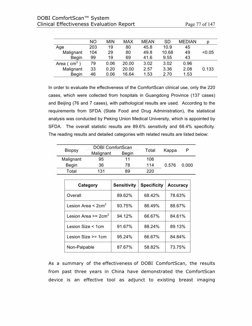
DOBI ComfortScan™ SystemClinical Effectiveness Evaluation Report Page 77 of 147
NO MIN MAX MEAN SD MEDIAN pAge 203 19 80 45.8 10.9 45
Malignant 104 29 80 49.8 10.68 49 <0.05Begin 99 19 69 41.6 9.55 43
Area cm2 79 0.06 20.00 3.02 3.02 0.96Malignant 33 0.20 20.00 2.57 3.36 2.08 0.133
Begin 46 0.06 16.64 1.53 2.70 1.53
In order to evaluate the effectiveness of the ComfortScan clinical use, only the 220
cases, which were collected from hospitals in Guangdong Province (137 cases)
and Beijing (76 and 7 cases), with pathological results are used. According to the
requirements from SFDA (State Food and Drug Administration), the statistical
analysis was conducted by Peking Union Medical University, which is appointed by
SFDA. The overall statistic results are 89.6% sensitivity and 68.4% specificity.
The reading results and detailed categories with related results are listed below:
Biopsy DOBI ComfortScan Total Kappa PMalignant Begin
Malignant 95 11 1060.576 0.000Begin 36 78 114
Total 131 89 220
Category Sensitivity Specificity Accuracy
Overall 89.62% 68.42% 78.63%
Lesion Area < 2cm2 93.75% 86.49% 88.67%
Lesion Area >= 2cm2 94.12% 66.67% 84.61%
Lesion Size < 1cm 91.67% 88.24% 89.13%
Lesion Size >= 1cm 95.24% 66.67% 84.84%
Non-Palpable 87.67% 58.82% 73.75%
As a summary of the effectiveness of DOBI ComfortScan, the results
from past three years in China have demonstrated the ComfortScan
device is an effective tool as adjunct to existing breast imaging
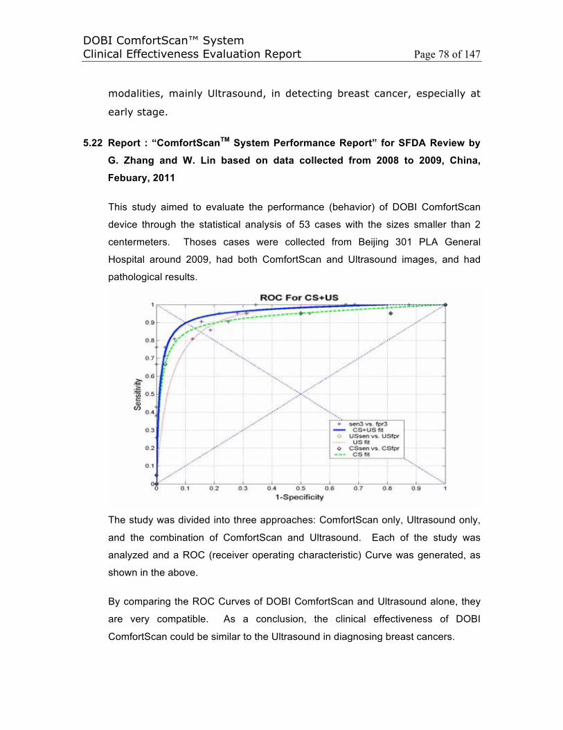
DOBI ComfortScan™ SystemClinical Effectiveness Evaluation Report Page 78 of 147
modalities, mainly Ultrasound, in detecting breast cancer, especially at
early stage.
5.22 Report : “ComfortScanTM System Performance Report” for SFDA Review by
G. Zhang and W. Lin based on data collected from 2008 to 2009, China,
Febuary, 2011
This study aimed to evaluate the performance (behavior) of DOBI ComfortScan
device through the statistical analysis of 53 cases with the sizes smaller than 2
centermeters. Thoses cases were collected from Beijing 301 PLA General
Hospital around 2009, had both ComfortScan and Ultrasound images, and had
pathological results.
The study was divided into three approaches: ComfortScan only, Ultrasound only,
and the combination of ComfortScan and Ultrasound. Each of the study was
analyzed and a ROC (receiver operating characteristic) Curve was generated, as
shown in the above.
By comparing the ROC Curves of DOBI ComfortScan and Ultrasound alone, they
are very compatible. As a conclusion, the clinical effectiveness of DOBI
ComfortScan could be similar to the Ultrasound in diagnosing breast cancers.
DOBI ComfortScan™ SystemClinical Effectiveness Evaluation Report Page 79 of 147
In order to clearly show the difference between individual use and the combination
of the two breast imaging techniques, a portional of the ROC Curve, which is
generally used for both sensitivity and specificity greater than 60%, is shown
below:
In the case, which needs the sensitivity greater than 90%, the specificity of the
combination of ComfortScan and Ultrasound is 15.6% and 34.4% higher than
ComfortScan and Ultrasound individually. If a 80% or highter specificity is needed,
the sensitivity of the combination of ComfortScan and Ultrasound can reach 95%
while ComfortScna sensitivity is about 66.7% and Ultrasound sensitivity is only
38.1%. As another conclusion, the clinical effectiveness of DOBI ComfortScan as
adjucnt to Ultrasound in detecting breast cancers is significant.
5.23 Report : “RESULTS OF INVESTIGATIONAL USE OF DOBI COMFORTSCAN IN
CHINA” by G. Zhang, W. Wang, D. Yang and H. Jiang from 2005 to 2006
This clinical study of DOBI ComfortScan performed at the two hospitals in Beijing
was conducted in accordance with The Provisions for Clinical Trials of Medical
devices in SFDA Order No. 5, as well as MEDDEV2.11/1 (2005/50/CEE) to
DOBI ComfortScan™ SystemClinical Effectiveness Evaluation Report Page 80 of 147
demonstrate the clinical EFFECTIVENESS of DOBI ComfortScan device. The
enrolled patients are 18 years of age or older and have been considered the
necessity of a breast biopsy after receiving mammography exasmination with BI-
RADS 3 or 4. But in order to prove the product safety and effectiveness in normal
condition by taking into account the current ComfortScan configuration and its
intended use, following patient is excluded from this study:
• Subject has had any breast surgery in the ipsilateral breast (e.g.,
augmentation/ cancer/ reduction) within a year of the potential scan date.
• Subject has had a core or excisional biopsy in the ipsilateral breast within 3
months.
• Subject has undergone brachytherapy in the region of interest within the
past 12 months.
• Subject is pregnant or lactating.
• Subject has accepted hormone replacement and/or oral contraceptives
within the past 30 days.
• Subject has failed to keep fixed and persistent position during the
examination.
• Subject has inflammatory skin disease (i.e., psoriasis, eczema)
• Subject has a known allergy to silicone
According to "Regulations for the Supervision and Administration of Medical
Devices" and "Provision on Clinical Trial of Medical Device" of SFDA of China, the
relevant data, such as study protocol, informed consent form, case report form etc.,
have been examined and approved by the Ethics Committees, in order to ensure
this study is in accordance with ICH-GCP and the related medical administration
regulations of China, and meet the requirements and principles of ethics.
A multi-center, Blinded Read Study to determine the sensitivity, specificity and
safety of the ComfortScan System in detecting malignancy in lesions of the breast
was conducted from year 2005 to 2006. All valid 62 women were examined with
ComfortScan at Peking University People’s Hospital and Capital Medical School
Chaoyang Hospital in Beijing as part of the present clinical study over a period of 6
months. The study included only age 18 or older women who had pathological
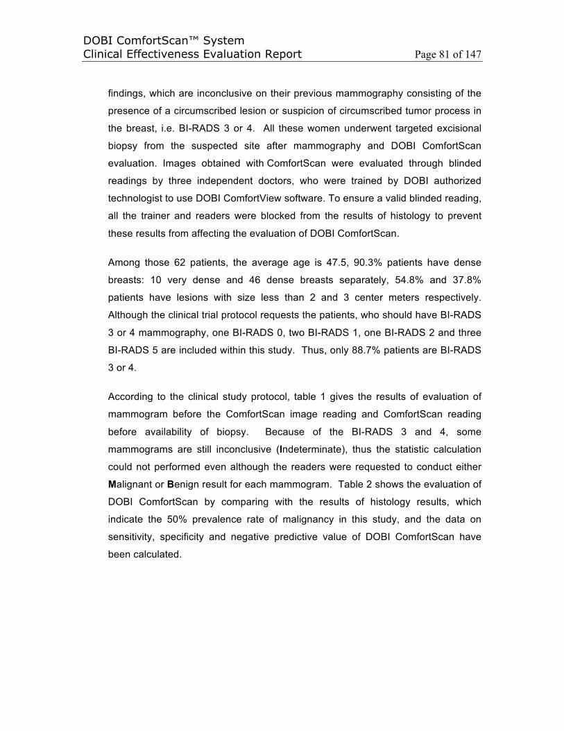
DOBI ComfortScan™ SystemClinical Effectiveness Evaluation Report Page 81 of 147
findings, which are inconclusive on their previous mammography consisting of the
presence of a circumscribed lesion or suspicion of circumscribed tumor process in
the breast, i.e. BI-RADS 3 or 4. All these women underwent targeted excisional
biopsy from the suspected site after mammography and DOBI ComfortScan
evaluation. Images obtained with ComfortScan were evaluated through blinded
readings by three independent doctors, who were trained by DOBI authorized
technologist to use DOBI ComfortView software. To ensure a valid blinded reading,
all the trainer and readers were blocked from the results of histology to prevent
these results from affecting the evaluation of DOBI ComfortScan.
Among those 62 patients, the average age is 47.5, 90.3% patients have dense
breasts: 10 very dense and 46 dense breasts separately, 54.8% and 37.8%
patients have lesions with size less than 2 and 3 center meters respectively.
Although the clinical trial protocol requests the patients, who should have BI-RADS
3 or 4 mammography, one BI-RADS 0, two BI-RADS 1, one BI-RADS 2 and three
BI-RADS 5 are included within this study. Thus, only 88.7% patients are BI-RADS
3 or 4.
According to the clinical study protocol, table 1 gives the results of evaluation of
mammogram before the ComfortScan image reading and ComfortScan reading
before availability of biopsy. Because of the BI-RADS 3 and 4, some
mammograms are still inconclusive (Indeterminate), thus the statistic calculation
could not performed even although the readers were requested to conduct either
Malignant or Benign result for each mammogram. Table 2 shows the evaluation of
DOBI ComfortScan by comparing with the results of histology results, which
indicate the 50% prevalence rate of malignancy in this study, and the data on
sensitivity, specificity and negative predictive value of DOBI ComfortScan have
been calculated.
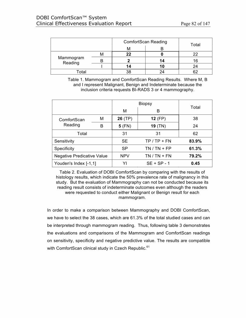
DOBI ComfortScan™ SystemClinical Effectiveness Evaluation Report Page 82 of 147
ComfortScan ReadingTotal
M B
Mammogram Reading
M 22 0 22B 2 14 16I 14 10 24
Total 38 24 62
Table 1. Mammogram and ComfortScan Reading Results. Where M, B and I represent Malignant, Benign and Indeterminate because the
inclusion criteria requests BI-RADS 3 or 4 mammography.
BiopsyTotal
M B
ComfortScanReading
M 26 (TP) 12 (FP) 38
B 5 (FN) 19 (TN) 24
Total 31 31 62
Sensitivity SE TP / TP + FN 83.9%Specificity SP TN / TN + FP 61.3%Negative Predicative Value NPV TN / TN + FN 79.2%Youden's Index [-1,1] YI SE + SP - 1 0.45
Table 2. Evaluation of DOBI ComfortScan by comparing with the results of histology results, which indicate the 50% prevalence rate of malignancy in this study. But the evaluation of Mammography can not be conducted because its reading result consists of indeterminate outcomes even although the readers
were requested to conduct either Malignant or Benign result for each mammogram.
In order to make a comparison between Mammography and DOBI ComfortScan,
we have to select the 38 cases, which are 61.3% of the total studied cases and can
be interpreted through mammogram reading. Thus, following table 3 demonstrates
the evaluations and comparisons of the Mammogram and ComfortScan readings
on sensitivity, specificity and negative predictive value. The results are compatible
with ComfortScan clinical study in Czech Republic.61
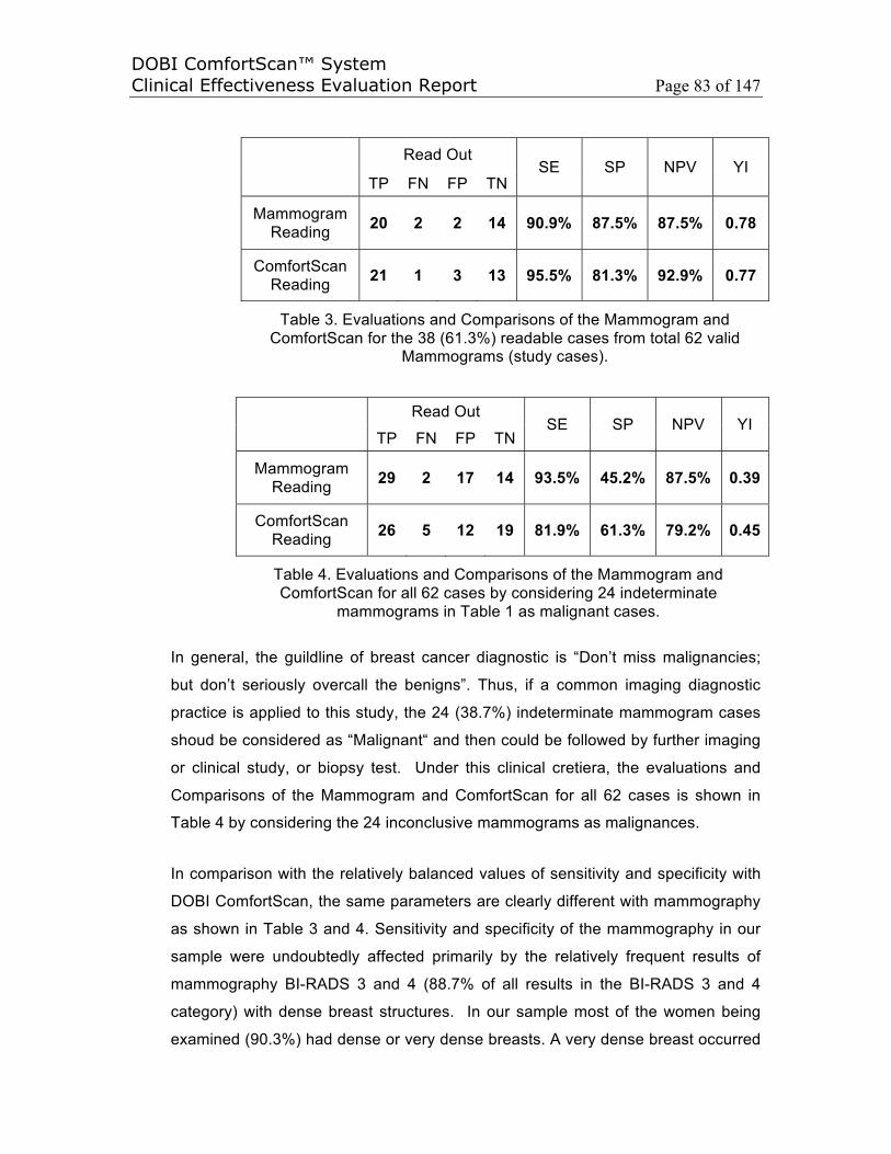
DOBI ComfortScan™ SystemClinical Effectiveness Evaluation Report Page 83 of 147
Read OutSE SP NPV YI
TP FN FP TN
MammogramReading 20 2 2 14 90.9% 87.5% 87.5% 0.78
ComfortScanReading 21 1 3 13 95.5% 81.3% 92.9% 0.77
Table 3. Evaluations and Comparisons of the Mammogram and ComfortScan for the 38 (61.3%) readable cases from total 62 valid
Mammograms (study cases).
Read OutSE SP NPV YI
TP FN FP TN
MammogramReading 29 2 17 14 93.5% 45.2% 87.5% 0.39
ComfortScanReading 26 5 12 19 81.9% 61.3% 79.2% 0.45
Table 4. Evaluations and Comparisons of the Mammogram and ComfortScan for all 62 cases by considering 24 indeterminate
mammograms in Table 1 as malignant cases.
In general, the guildline of breast cancer diagnostic is “Don’t miss malignancies;
but don’t seriously overcall the benigns”. Thus, if a common imaging diagnostic
practice is applied to this study, the 24 (38.7%) indeterminate mammogram cases
shoud be considered as “Malignant“ and then could be followed by further imaging
or clinical study, or biopsy test. Under this clinical cretiera, the evaluations and
Comparisons of the Mammogram and ComfortScan for all 62 cases is shown in
Table 4 by considering the 24 inconclusive mammograms as malignances.
In comparison with the relatively balanced values of sensitivity and specificity with
DOBI ComfortScan, the same parameters are clearly different with mammography
as shown in Table 3 and 4. Sensitivity and specificity of the mammography in our
sample were undoubtedly affected primarily by the relatively frequent results of
mammography BI-RADS 3 and 4 (88.7% of all results in the BI-RADS 3 and 4
category) with dense breast structures. In our sample most of the women being
examined (90.3%) had dense or very dense breasts. A very dense breast occurred
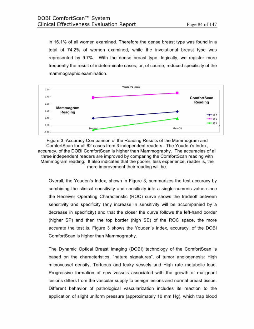
DOBI ComfortScan™ SystemClinical Effectiveness Evaluation Report Page 84 of 147
in 16.1% of all women examined. Therefore the dense breast type was found in a
total of 74.2% of women examined, while the involutional breast type was
represented by 9.7%. With the dense breast type, logically, we register more
frequently the result of indeterminate cases, or, of course, reduced specificity of the
mammographic examination.
Figure 3. Accuracy Comparison of the Reading Results of the Mammogram and ComfortScan for all 62 cases from 3 independent readers. The Youden’s Index,
accuracy, of the DOBI ComfortScan is higher than Mammography. The accuracies of all three independent readers are improved by comparing the ComfortScan reading with Mammogram reading. It also indicates that the poorer, less experience, reader is, the
more improvement their reading will be.
Overall, the Youden’s Index, shown in Figure 3, summarizes the test accuracy by
combining the clinical sensitivity and specificity into a single numeric value since
the Receiver Operating Characteristic (ROC) curve shows the tradeoff between
sensitivity and specificity (any increase in sensitivity will be accompanied by a
decrease in specificity) and that the closer the curve follows the left-hand border
(higher SP) and then the top border (high SE) of the ROC space, the more
accurate the test is. Figure 3 shows the Youden’s Index, accuracy, of the DOBI
ComfortScan is higher than Mammography.
The Dynamic Optical Breast Imaging (DOBI) technology of the ComfortScan is
based on the characteristics, “nature signatures”, of tumor angiogenesis: High
microvessel density, Tortuous and leaky vessels and High rate metabolic load.
Progressive formation of new vessels associated with the growth of malignant
lesions differs from the vascular supply to benign lesions and normal breast tissue.
Different behavior of pathological vascularization includes its reaction to the
application of slight uniform pressure (approximately 10 mm Hg), which trap blood
Youden's Index
-0.10
0.00
0.10
0.20
0.30
0.40
0.50
MamI=M Mam+CS
Dr 1
Dr 2
Dr 3
ComfortScanReading
MammogramReading
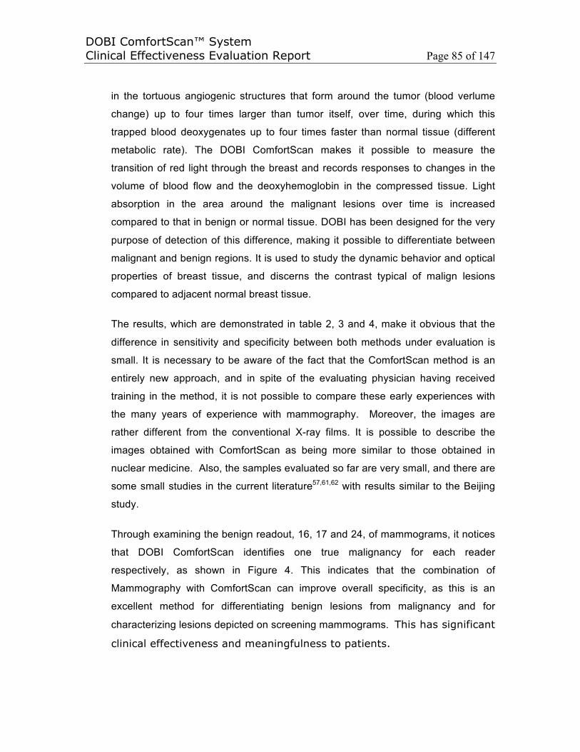
DOBI ComfortScan™ SystemClinical Effectiveness Evaluation Report Page 85 of 147
in the tortuous angiogenic structures that form around the tumor (blood verlume
change) up to four times larger than tumor itself, over time, during which this
trapped blood deoxygenates up to four times faster than normal tissue (different
metabolic rate). The DOBI ComfortScan makes it possible to measure the
transition of red light through the breast and records responses to changes in the
volume of blood flow and the deoxyhemoglobin in the compressed tissue. Light
absorption in the area around the malignant lesions over time is increased
compared to that in benign or normal tissue. DOBI has been designed for the very
purpose of detection of this difference, making it possible to differentiate between
malignant and benign regions. It is used to study the dynamic behavior and optical
properties of breast tissue, and discerns the contrast typical of malign lesions
compared to adjacent normal breast tissue.
The results, which are demonstrated in table 2, 3 and 4, make it obvious that the
difference in sensitivity and specificity between both methods under evaluation is
small. It is necessary to be aware of the fact that the ComfortScan method is an
entirely new approach, and in spite of the evaluating physician having received
training in the method, it is not possible to compare these early experiences with
the many years of experience with mammography. Moreover, the images are
rather different from the conventional X-ray films. It is possible to describe the
images obtained with ComfortScan as being more similar to those obtained in
nuclear medicine. Also, the samples evaluated so far are very small, and there are
some small studies in the current literature57,61,62 with results similar to the Beijing
study.
Through examining the benign readout, 16, 17 and 24, of mammograms, it notices
that DOBI ComfortScan identifies one true malignancy for each reader
respectively, as shown in Figure 4. This indicates that the combination of
Mammography with ComfortScan can improve overall specificity, as this is an
excellent method for differentiating benign lesions from malignancy and for
characterizing lesions depicted on screening mammograms. This has significant
clinical effectiveness and meaningfulness to patients.
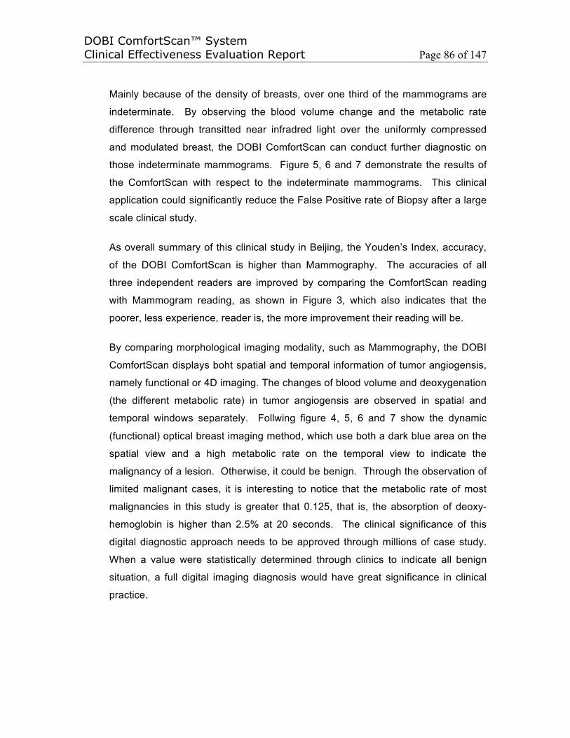
DOBI ComfortScan™ SystemClinical Effectiveness Evaluation Report Page 86 of 147
Mainly because of the density of breasts, over one third of the mammograms are
indeterminate. By observing the blood volume change and the metabolic rate
difference through transitted near infradred light over the uniformly compressed
and modulated breast, the DOBI ComfortScan can conduct further diagnostic on
those indeterminate mammograms. Figure 5, 6 and 7 demonstrate the results of
the ComfortScan with respect to the indeterminate mammograms. This clinical
application could significantly reduce the False Positive rate of Biopsy after a large
scale clinical study.
As overall summary of this clinical study in Beijing, the Youden’s Index, accuracy,
of the DOBI ComfortScan is higher than Mammography. The accuracies of all
three independent readers are improved by comparing the ComfortScan reading
with Mammogram reading, as shown in Figure 3, which also indicates that the
poorer, less experience, reader is, the more improvement their reading will be.
By comparing morphological imaging modality, such as Mammography, the DOBI
ComfortScan displays boht spatial and temporal information of tumor angiogensis,
namely functional or 4D imaging. The changes of blood volume and deoxygenation
(the different metabolic rate) in tumor angiogensis are observed in spatial and
temporal windows separately. Follwing figure 4, 5, 6 and 7 show the dynamic
(functional) optical breast imaging method, which use both a dark blue area on the
spatial view and a high metabolic rate on the temporal view to indicate the
malignancy of a lesion. Otherwise, it could be benign. Through the observation of
limited malignant cases, it is interesting to notice that the metabolic rate of most
malignancies in this study is greater that 0.125, that is, the absorption of deoxy-
hemoglobin is higher than 2.5% at 20 seconds. The clinical significance of this
digital diagnostic approach needs to be approved through millions of case study.
When a value were statistically determined through clinics to indicate all benign
situation, a full digital imaging diagnosis would have great significance in clinical
practice.
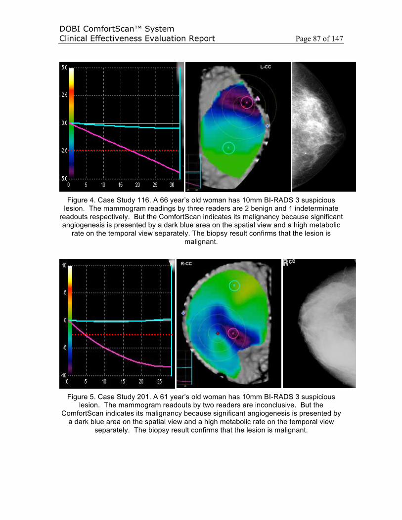
DOBI ComfortScan™ SystemClinical Effectiveness Evaluation Report Page 87 of 147
Figure 4. Case Study 116. A 66 year’s old woman has 10mm BI-RADS 3 suspicious lesion. The mammogram readings by three readers are 2 benign and 1 indeterminate
readouts respectively. But the ComfortScan indicates its malignancy because significant angiogenesis is presented by a dark blue area on the spatial view and a high metabolic
rate on the temporal view separately. The biopsy result confirms that the lesion is malignant.
Figure 5. Case Study 201. A 61 year’s old woman has 10mm BI-RADS 3 suspicious lesion. The mammogram readouts by two readers are inconclusive. But the
ComfortScan indicates its malignancy because significant angiogenesis is presented by a dark blue area on the spatial view and a high metabolic rate on the temporal view
separately. The biopsy result confirms that the lesion is malignant.
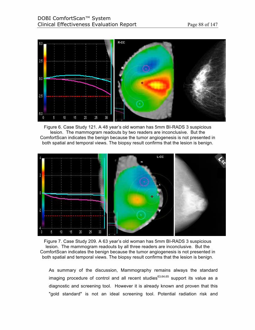
DOBI ComfortScan™ SystemClinical Effectiveness Evaluation Report Page 88 of 147
Figure 6. Case Study 121. A 48 year’s old woman has 5mm BI-RADS 3 suspicious lesion. The mammogram readouts by two readers are inconclusive. But the
ComfortScan indicates the benign because the tumor angiogenesis is not presented in both spatial and temporal views. The biopsy result confirms that the lesion is benign.
Figure 7. Case Study 209. A 63 year’s old woman has 5mm BI-RADS 3 suspicious lesion. The mammogram readouts by all three readers are inconclusive. But the
ComfortScan indicates the benign because the tumor angiogenesis is not presented in both spatial and temporal views. The biopsy result confirms that the lesion is benign.
As summary of the discussion, Mammography remains always the standard
imaging procedure of control and all recent studies63,64,65 support its value as a
diagnostic and screening tool. However it is already known and proven that this
"gold standard" is not an ideal screening tool. Potential radiation risk and
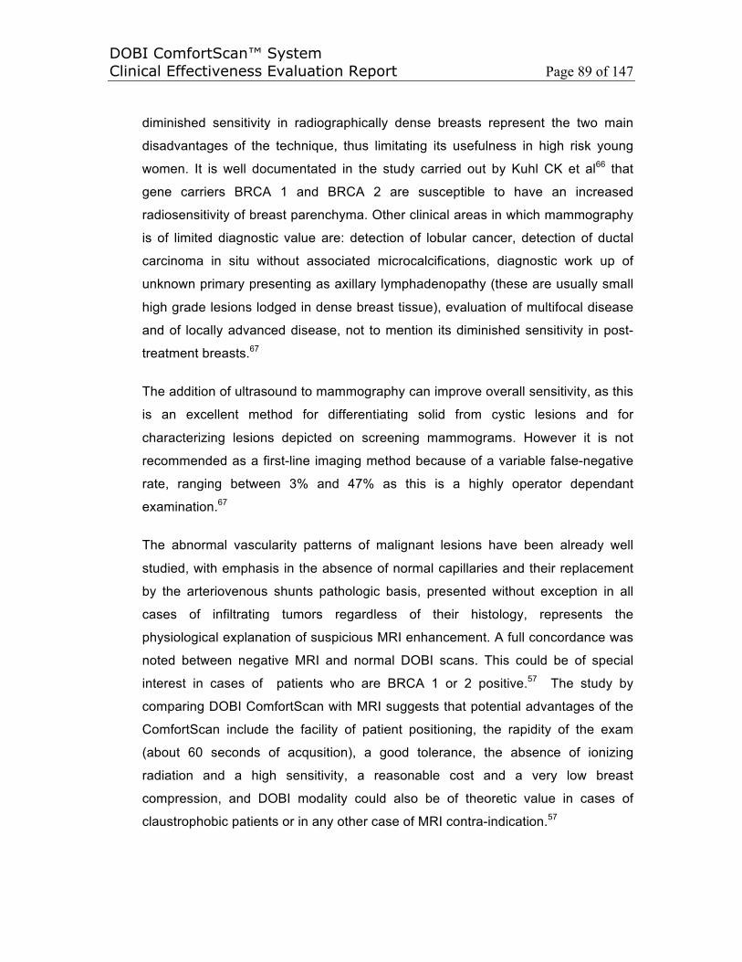
DOBI ComfortScan™ SystemClinical Effectiveness Evaluation Report Page 89 of 147
diminished sensitivity in radiographically dense breasts represent the two main
disadvantages of the technique, thus limitating its usefulness in high risk young
women. It is well documentated in the study carried out by Kuhl CK et al66 that
gene carriers BRCA 1 and BRCA 2 are susceptible to have an increased
radiosensitivity of breast parenchyma. Other clinical areas in which mammography
is of limited diagnostic value are: detection of lobular cancer, detection of ductal
carcinoma in situ without associated microcalcifications, diagnostic work up of
unknown primary presenting as axillary lymphadenopathy (these are usually small
high grade lesions lodged in dense breast tissue), evaluation of multifocal disease
and of locally advanced disease, not to mention its diminished sensitivity in post-
treatment breasts.67
The addition of ultrasound to mammography can improve overall sensitivity, as this
is an excellent method for differentiating solid from cystic lesions and for
characterizing lesions depicted on screening mammograms. However it is not
recommended as a first-line imaging method because of a variable false-negative
rate, ranging between 3% and 47% as this is a highly operator dependant
examination.67
The abnormal vascularity patterns of malignant lesions have been already well
studied, with emphasis in the absence of normal capillaries and their replacement
by the arteriovenous shunts pathologic basis, presented without exception in all
cases of infiltrating tumors regardless of their histology, represents the
physiological explanation of suspicious MRI enhancement. A full concordance was
noted between negative MRI and normal DOBI scans. This could be of special
interest in cases of patients who are BRCA 1 or 2 positive.57 The study by
comparing DOBI ComfortScan with MRI suggests that potential advantages of the
ComfortScan include the facility of patient positioning, the rapidity of the exam
(about 60 seconds of acqusition), a good tolerance, the absence of ionizing
radiation and a high sensitivity, a reasonable cost and a very low breast
compression, and DOBI modality could also be of theoretic value in cases of
claustrophobic patients or in any other case of MRI contra-indication.57
DOBI ComfortScan™ SystemClinical Effectiveness Evaluation Report Page 90 of 147
Larger samples, evaluated over longer time periods, will undoubtedly tell more
distinctly whether one can predict possible future use of DOBI ComfortScan as part
of standard diagnostic investigation for malign breast gland lesions. This would
bring distinct benefits, not only in terms of broader diagnostic possibilities, but also
and more importantly in terms of radiation load, which is obviously zero in dynamic
optical imaging, paving the way for future use as a method that is more suitable for
mammographic screening for all ages.
The total of 62 scans have been acquired and interpreted by three independent
blinded readers with encouraging results. This device could provide the physician
with dynamic functional information regarding abnormal vascularization in an area
of interest in the breast and this information could be used to better characterize
the lesion. ComfortScan can help the performance and accuracy of averaging,
under averaging or less experienced doctors in their clinics most significantly.
Dynamic optical breast imaging can be a promising complementary imaging
modality for further investigation in cases of women with inconclusive
mammography and/or physical examination. The study in Beijing comprised a
small number of patients, but the preliminary results were encouraging enough,
especially in cases of indeterminate mammographic cases. However further
evaluation with a larger number of patients should be carried out.
As the conclusion, based upon its performance in clinical studies worldwide, the
DOBI ComfortScan is a novel imaging technology with clinical effectiveness that is
appropriate as an imaging modality in diagnosing breast cancer at early stage. As
a diagnostic tool of breast cancer, a large scale number of cases should be studied
to charaterize different malignant and benign tumors at different stages
respectively, different statues of patients, such as menopause stages with related
nipple blue, and to statistically quantatize the metabolic rates of both malignant
and benign tumors.
As a result, the ComfortScan system focuses on physiology-based dynamic
functional imaging (i.e., what is occurring within the tissue in near real time) rather
than a singular morphological image (i.e., a static anatomical snapshot showing
physical details at a single point in time), such as those created by mammography.
DOBI ComfortScan™ SystemClinical Effectiveness Evaluation Report Page 91 of 147
When combined with mammography or ultrasound, both of which provide simple
morphologic images, the ComfortScan system’s images of physiological changes
in the breast is intended to provide physicians with a more complete data set to
improve the physician’s ability to provide an accurate breast cancer diagnosis.
With its negative predictive value of 98 percent and sepcificity of 87 percent,68 the
DOBI ComfortScan represents an opportunity to reduce the incidence and severity
of invasive diagnostic intervention and, thus, to potentially reduce the number of
unnecessary, painful and costly biopsies that are conducted on patients with
healthy tissue. Furthermore, the safety profile, convenience, comfort and low
comparative cost of the DOBI ComfortScan correspond closely to the call to action
delivered by the National Academy of Sciences’ Institute of Medicine.
Because it is an aid in detecting the minute vascular changes that accompany the
process of angiogenesis during the earliest stages of malignant tumor growth, the
DOBI ComfortScan could potentionally become a useful breast cancer screening
tool if a full FOV (filed of view) cluster ComfortScan or DOBI ComfortScreen, next
generation of the ComfortScan, could maintain a high negative predictive value,
above 95%.
In addition to disease diagnosis, therapeutic monitoring of both pro- and anti-
angiogenic drugs may also be a longer-term application of this technology and,
since angiogenesis is found in many significant disease states (such as rheumatoid
arthritis and adult blindness), the DOBI technology may have future applications in
addition to cancer.
DOBI’s dynamic analysis is a significant improvement over current static imaging.
Breast density does not affect DOBI images, making DOBI especially important in
the evaluation of dense breasts, as often seen in young women or those on
Hormone Replacement Therapy (HRT). The initial results obtained with this rather
new method, which is associated with no radiation load and well tolerated by
women, hold promise for further development, particularly in the area of software
development and standardization of evaluation parameters. Another important
point to stress is the need for high-quality training of evaluating physicians which is,
DOBI ComfortScan™ SystemClinical Effectiveness Evaluation Report Page 92 of 147
in our view, extremely important and affects the results of the investigation rather
significantly.
DOBI ComfortScan is an Office, In-Vivo, Non-Invasive, Non-Ionizing and Non-
painful molecular vesicular Dynamical Optical Breast Imaging modality. DOBI
technology/modality will continue to improve as new features are added, much the
same as other imaging modalities such as MRI, PET, CT, and digital
mammography have evolved over time.
DOBI ComfortScan™ SystemClinical Effectiveness Evaluation Report Page 93 of 147
6. CLINICAL ACCEPTANCE
In the past 6 years over 50 diagnostic centers worldwide have used the
ComfortScan, including nearly 30 US sites participating in the FDA PMA
clinical trials, and over 30 sites in Italy and China have bought the devices,
resulting in the use of the device on thousands patients. The cumulative
device experiences to date, as well as the above clinical effectiveness, have
demonstrated that this device, DOBI ComfortScan, could provide the
physician with dynamic functional information regarding abnormal
vascularization in an area of interest in the breast and this information could
be used to better characterize the lesion. DOBI ComfortScan can help the
performance and accuracy of averaging, under averaging or less experienced
doctors in their clinics most significantly. Dynamic optical breast imaging can
be a promising complementary imaging modality for further investigation in
cases of women with inconclusive mammography and/or physical
examination.
As a result, the clinical acceptance of DOBI ComfortScan could be shown in
following examples.
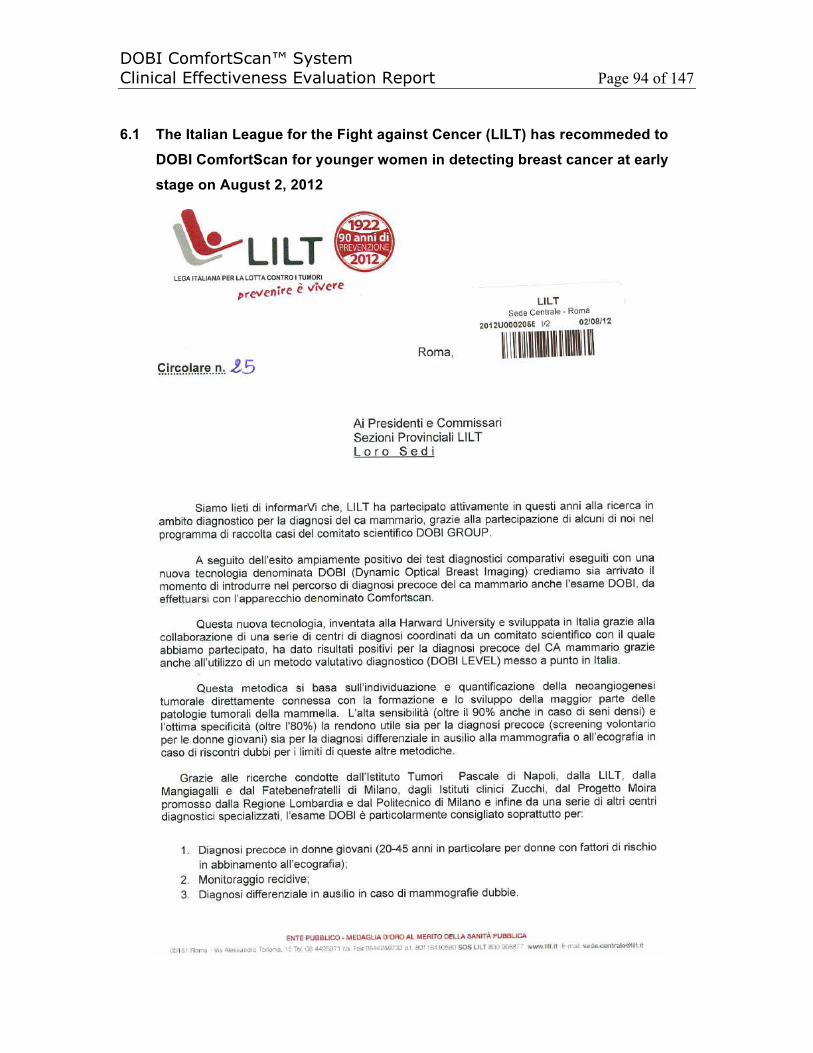
DOBI ComfortScan™ SystemClinical Effectiveness Evaluation Report Page 94 of 147
6.1 The Italian League for the Fight against Cencer (LILT) has recommeded to
DOBI ComfortScan for younger women in detecting breast cancer at early
stage on August 2, 2012
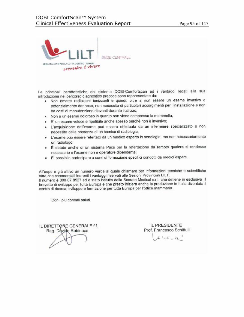
DOBI ComfortScan™ SystemClinical Effectiveness Evaluation Report Page 95 of 147
DOBI ComfortScan™ SystemClinical Effectiveness Evaluation Report Page 96 of 147
6.2 The Italian League for the Fight against Cancer (LILT) – Underforty Women
Breast Care, 2010
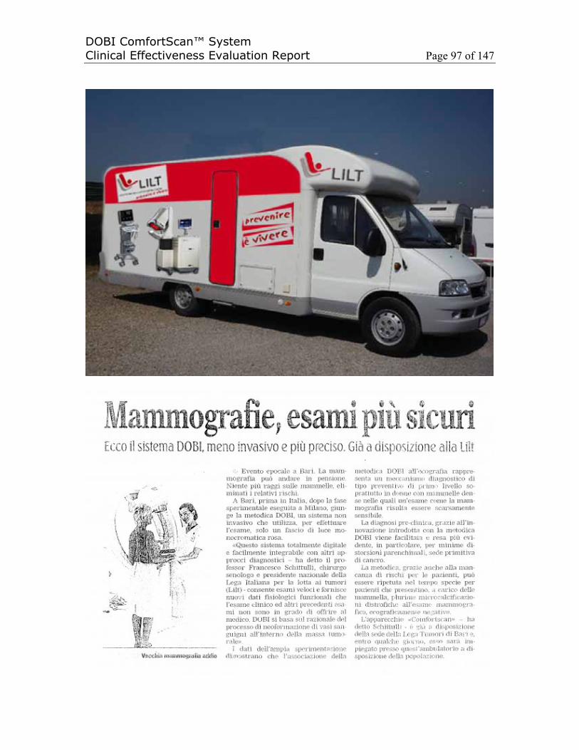
DOBI ComfortScan™ SystemClinical Effectiveness Evaluation Report Page 97 of 147
DOBI ComfortScan™ SystemClinical Effectiveness Evaluation Report Page 98 of 147
DOBI ComfortScan™ SystemClinical Effectiveness Evaluation Report Page 99 of 147
DOBI ComfortScan™ SystemClinical Effectiveness Evaluation Report Page 100 of 147
6.3 2nd Meeting DOBI Group in Italy, 2011
DOBI ComfortScan™ SystemClinical Effectiveness Evaluation Report Page 101 of 147
6.4 1st Meeting DOBI Group in Italy, 2010
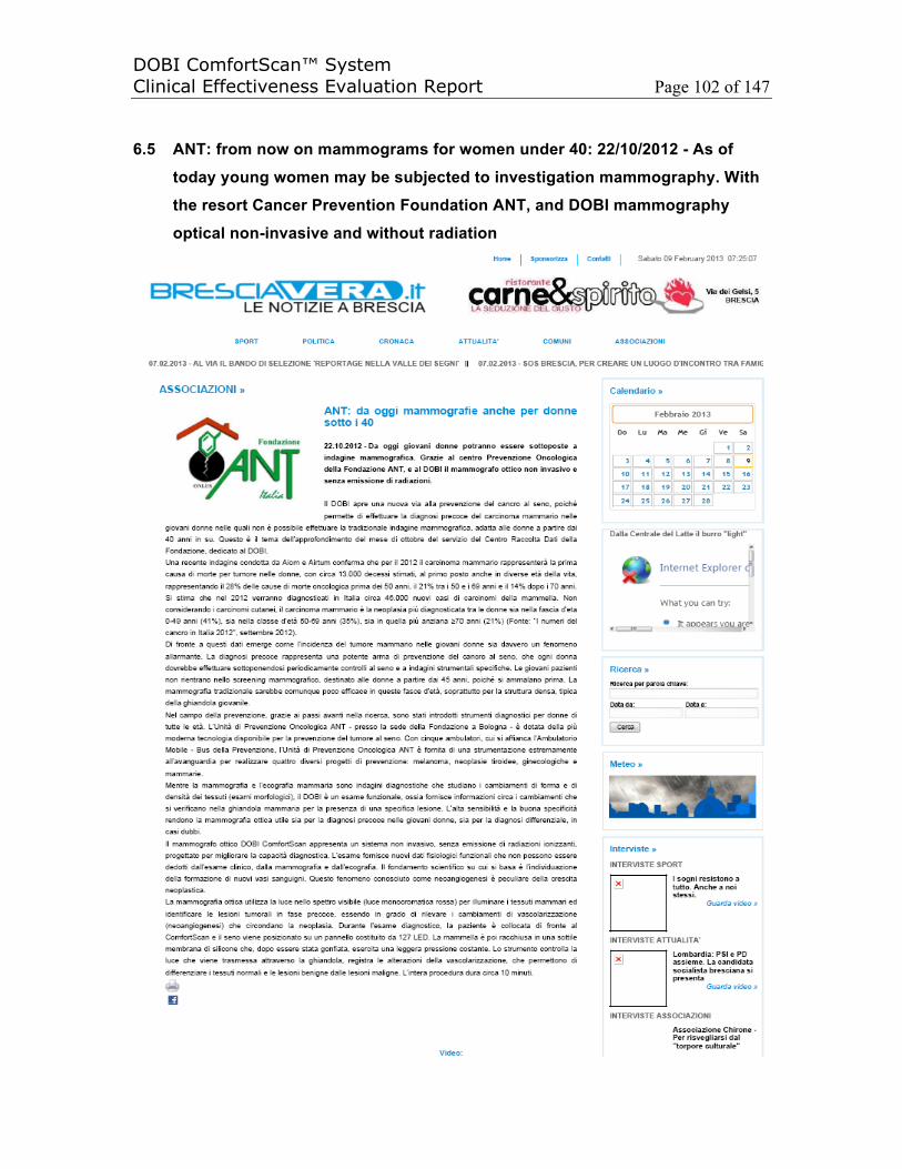
DOBI ComfortScan™ SystemClinical Effectiveness Evaluation Report Page 102 of 147
6.5 ANT: from now on mammograms for women under 40: 22/10/2012 - As of
today young women may be subjected to investigation mammography. With
the resort Cancer Prevention Foundation ANT, and DOBI mammography
optical non-invasive and without radiation
DOBI ComfortScan™ SystemClinical Effectiveness Evaluation Report Page 103 of 147
6.6 Breast cancer diagnosis today is born with "DOBI"
DOBI ComfortScan™ SystemClinical Effectiveness Evaluation Report Page 104 of 147
6.7 Breast cancer remains the second leading cause of death among women
DOBI ComfortScan™ SystemClinical Effectiveness Evaluation Report Page 105 of 147
6.8 UNIT 'ANT CANCER PREVENTION PROJECTS FOR POLICE AND UNICREDIT
DOBI ComfortScan™ SystemClinical Effectiveness Evaluation Report Page 106 of 147
6.9 THE INNOVATIVE METHODS OF STUDY MEDICAL MANARA 31
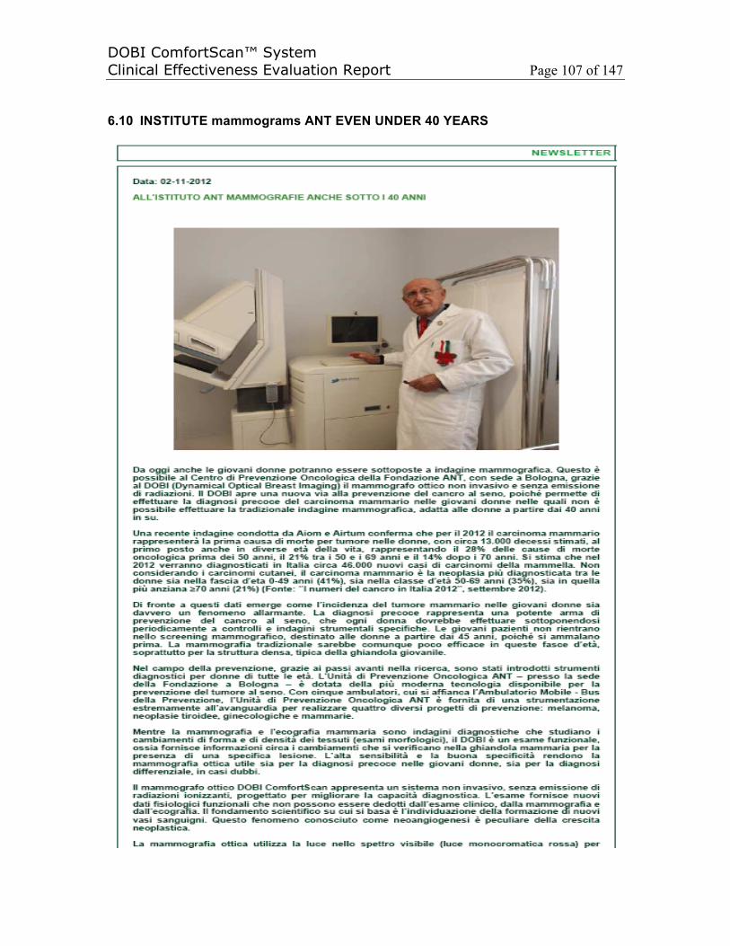
DOBI ComfortScan™ SystemClinical Effectiveness Evaluation Report Page 107 of 147
6.10 INSTITUTE mammograms ANT EVEN UNDER 40 YEARS
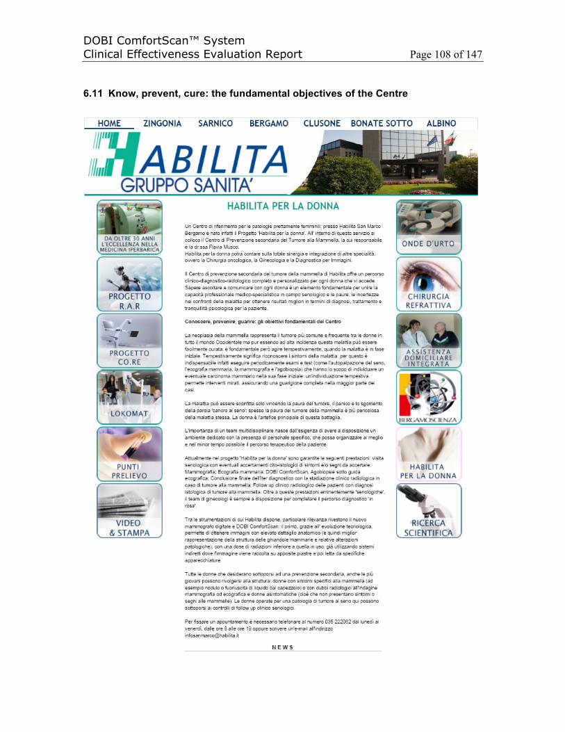
DOBI ComfortScan™ SystemClinical Effectiveness Evaluation Report Page 108 of 147
6.11 Know, prevent, cure: the fundamental objectives of the Centre
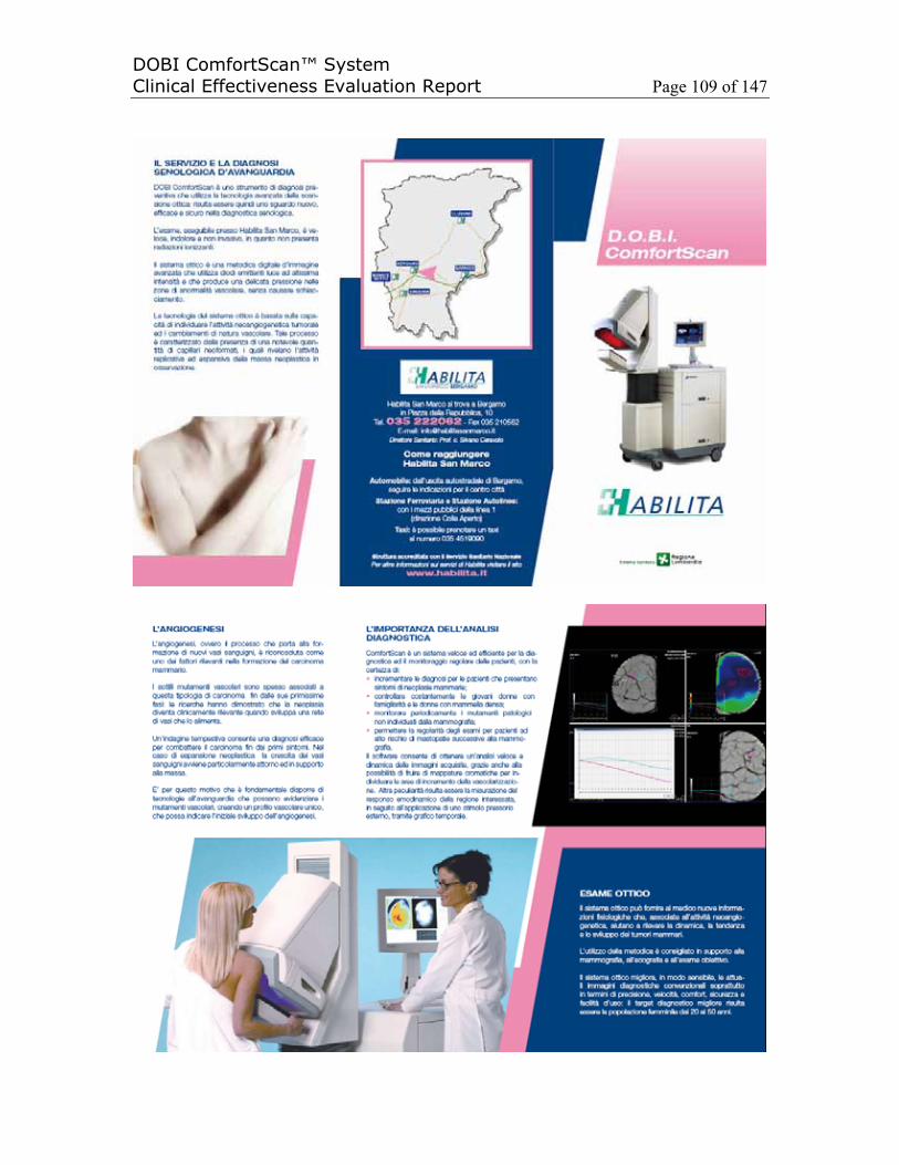
DOBI ComfortScan™ SystemClinical Effectiveness Evaluation Report Page 109 of 147
DOBI ComfortScan™ SystemClinical Effectiveness Evaluation Report Page 110 of 147
6.12 From tomorrow morning a new entry with the camper made available by the project underforty LILT Bologna, with breast ultrasound and optical technology mammary (ComfortScan Dobi) for this occasion for the first time in southern Italy.
DOBI ComfortScan™ SystemClinical Effectiveness Evaluation Report Page 111 of 147
6.13 As breast, ComfortScan presents as a solution for the prevention
DOBI ComfortScan™ SystemClinical Effectiveness Evaluation Report Page 112 of 147
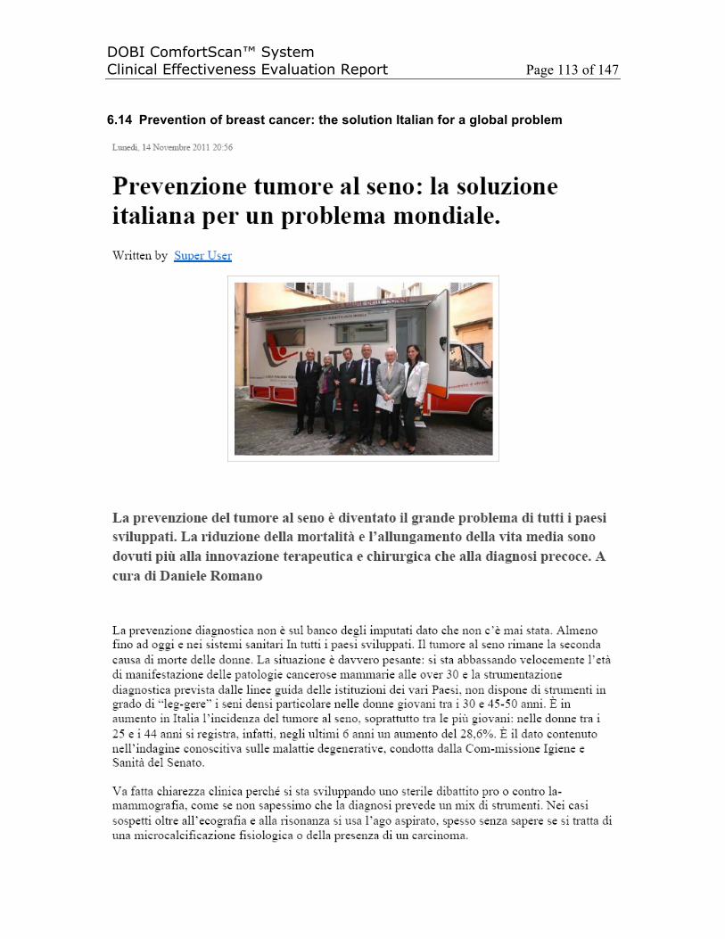
DOBI ComfortScan™ SystemClinical Effectiveness Evaluation Report Page 113 of 147
6.14 Prevention of breast cancer: the solution Italian for a global problem
DOBI ComfortScan™ SystemClinical Effectiveness Evaluation Report Page 114 of 147
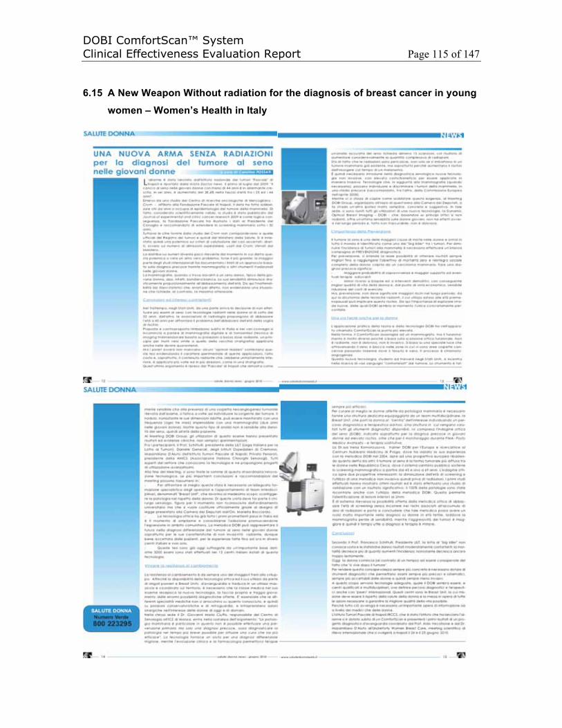
DOBI ComfortScan™ SystemClinical Effectiveness Evaluation Report Page 115 of 147
6.15 A New Weapon Without radiation for the diagnosis of breast cancer in young
women – Women’s Health in Italy
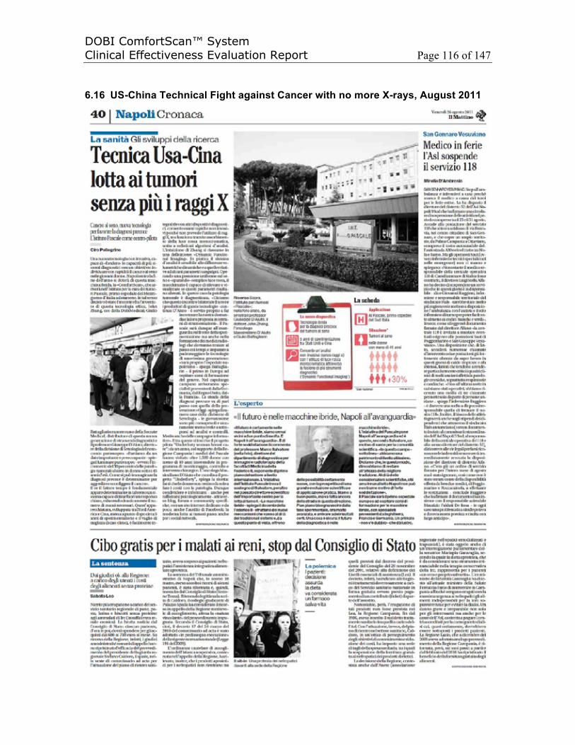
DOBI ComfortScan™ SystemClinical Effectiveness Evaluation Report Page 116 of 147
6.16 US-China Technical Fight against Cancer with no more X-rays, August 2011
DOBI ComfortScan™ SystemClinical Effectiveness Evaluation Report Page 117 of 147
6.17 PREVENTING BREAST: NEW DIAGNOSTIC
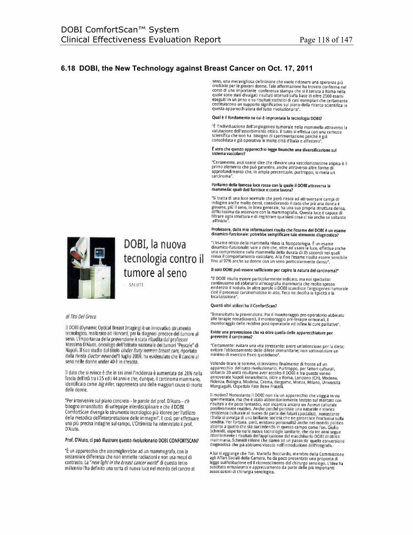
DOBI ComfortScan™ SystemClinical Effectiveness Evaluation Report Page 118 of 147
6.18 DOBI, the New Technology against Breast Cancer on Oct. 17, 2011
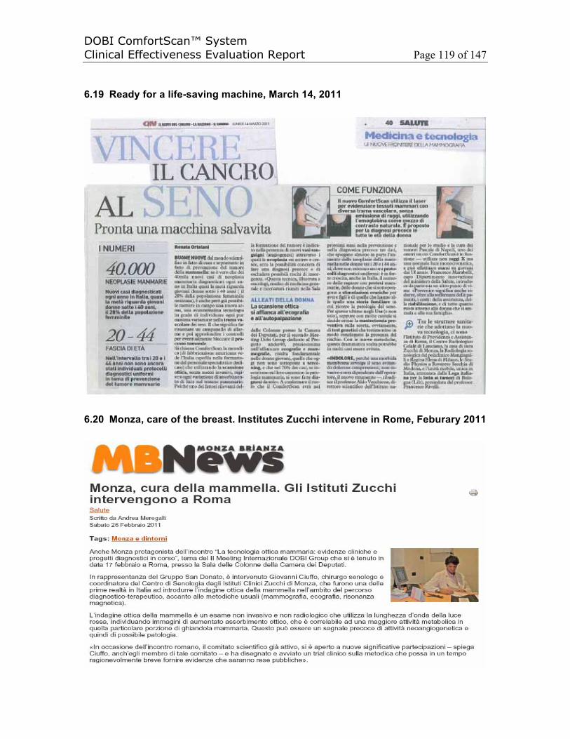
DOBI ComfortScan™ SystemClinical Effectiveness Evaluation Report Page 119 of 147
6.19 Ready for a life-saving machine, March 14, 2011
6.20 Monza, care of the breast. Institutes Zucchi intervene in Rome, Feburary 2011
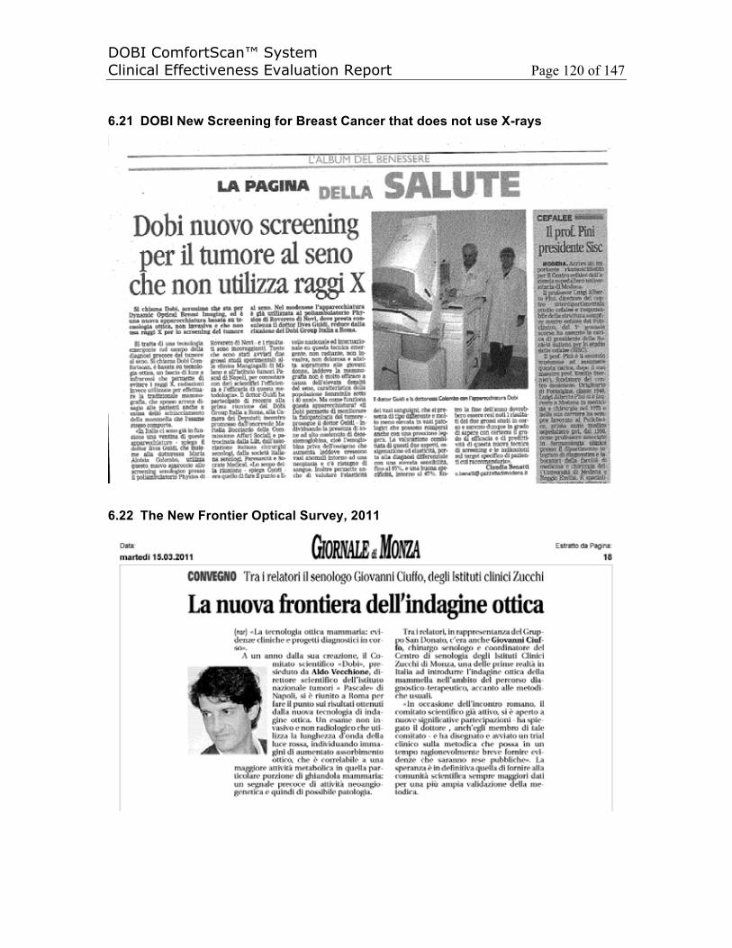
DOBI ComfortScan™ SystemClinical Effectiveness Evaluation Report Page 120 of 147
6.21 DOBI New Screening for Breast Cancer that does not use X-rays
6.22 The New Frontier Optical Survey, 2011
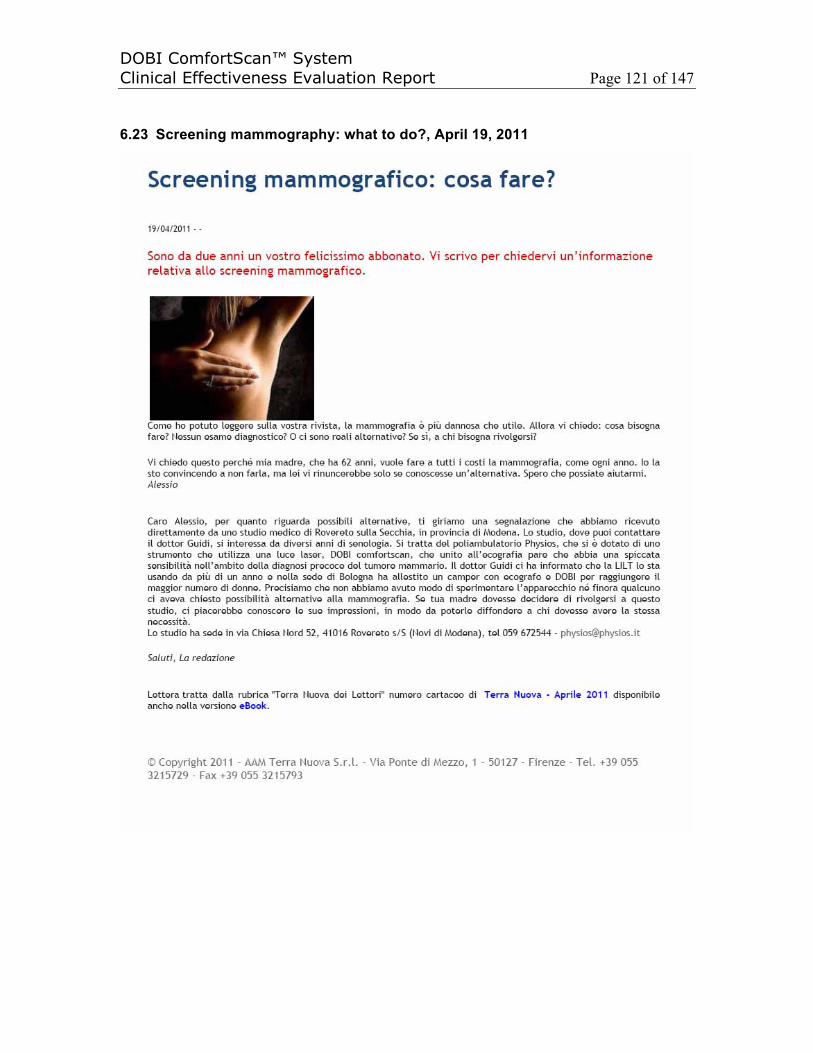
DOBI ComfortScan™ SystemClinical Effectiveness Evaluation Report Page 121 of 147
6.23 Screening mammography: what to do?, April 19, 2011
DOBI ComfortScan™ SystemClinical Effectiveness Evaluation Report Page 122 of 147
6.24 Professor Rocca’s Trial
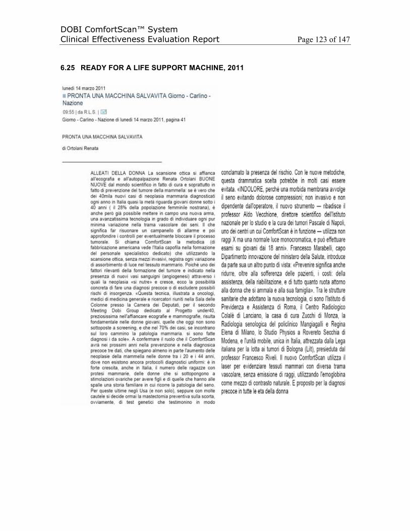
DOBI ComfortScan™ SystemClinical Effectiveness Evaluation Report Page 123 of 147
6.25 READY FOR A LIFE SUPPORT MACHINE, 2011
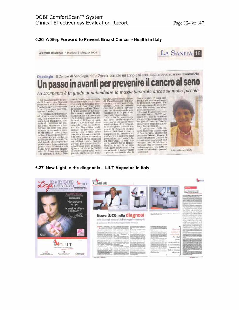
DOBI ComfortScan™ SystemClinical Effectiveness Evaluation Report Page 124 of 147
6.26 A Step Forward to Prevent Breast Cancer - Health in Italy
6.27 New Light in the diagnosis – LILT Magazine in Italy
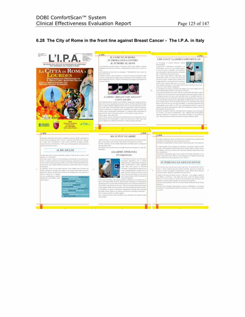
DOBI ComfortScan™ SystemClinical Effectiveness Evaluation Report Page 125 of 147
6.28 The City of Rome in the front line against Breast Cancer - The I.P.A. in Italy
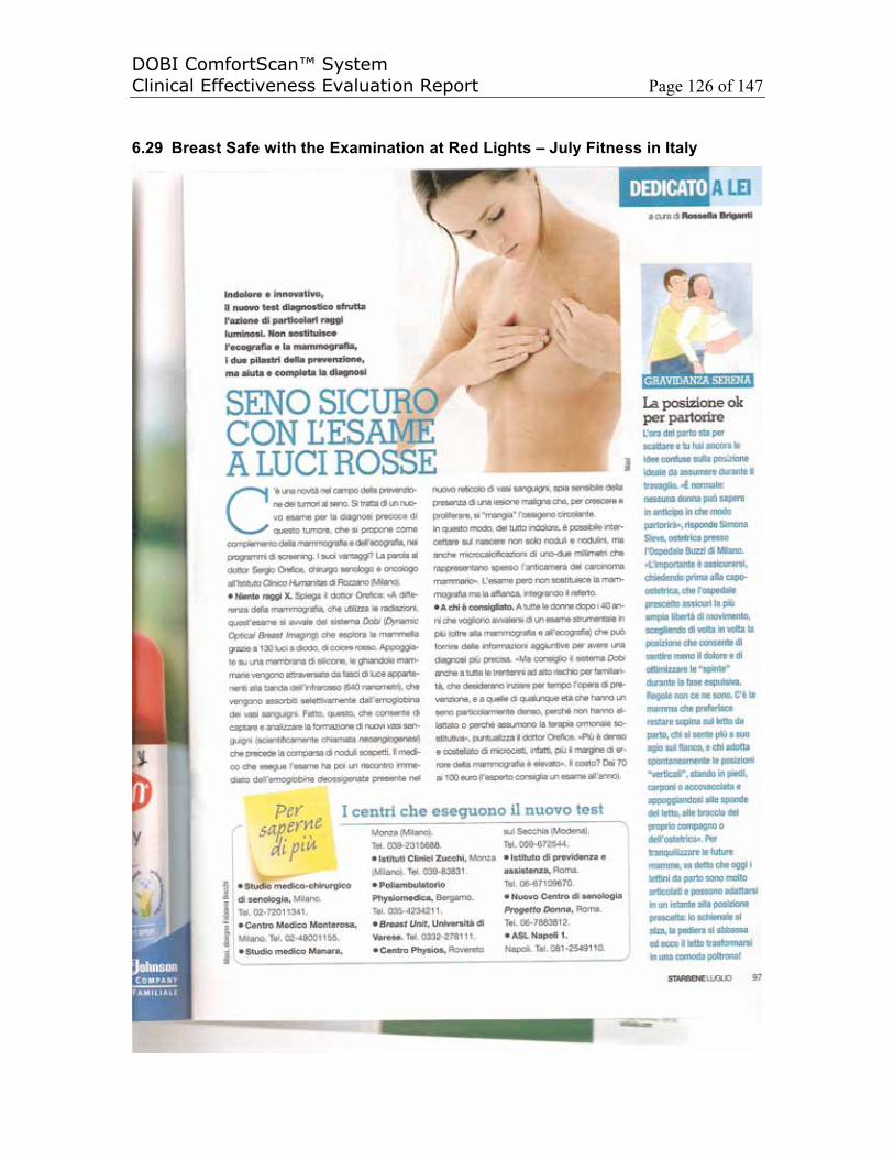
DOBI ComfortScan™ SystemClinical Effectiveness Evaluation Report Page 126 of 147
6.29 Breast Safe with the Examination at Red Lights – July Fitness in Italy
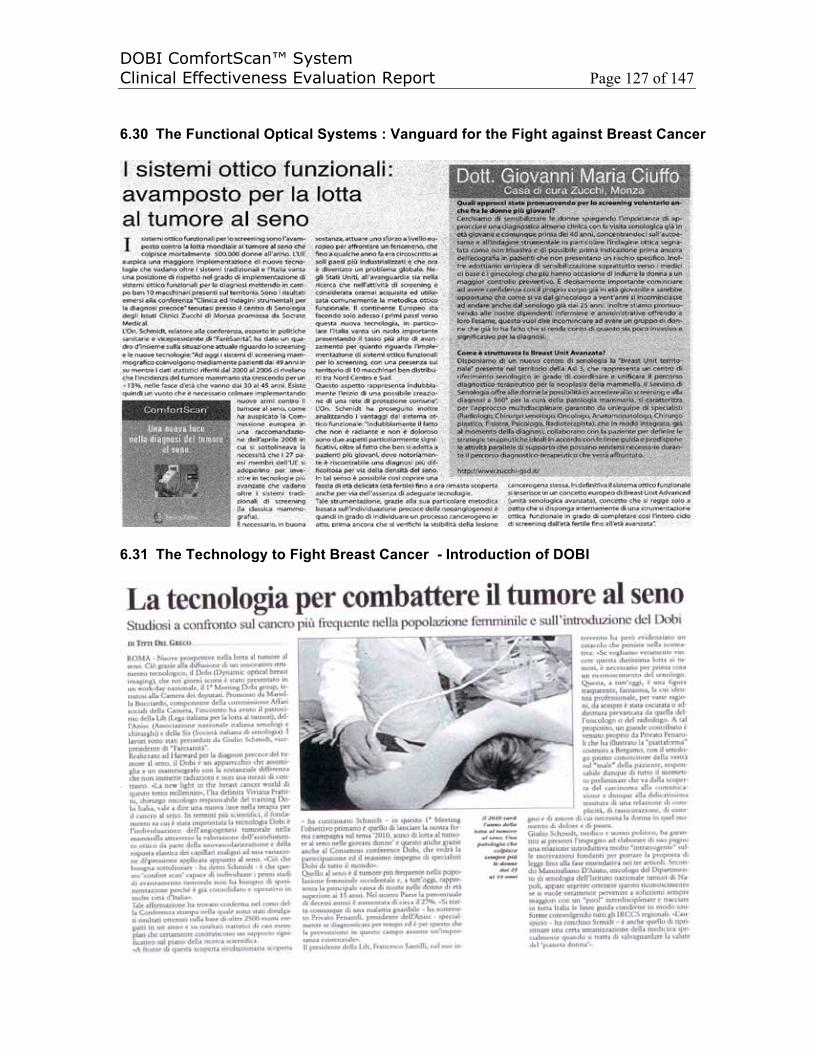
DOBI ComfortScan™ SystemClinical Effectiveness Evaluation Report Page 127 of 147
6.30 The Functional Optical Systems : Vanguard for the Fight against Breast Cancer
6.31 The Technology to Fight Breast Cancer - Introduction of DOBI

DOBI ComfortScan™ SystemClinical Effectiveness Evaluation Report Page 128 of 147
6.32 Breast Cancer, Zucchi Hospital Leader in Italy - Use the Wavelength of Right Light : DOBI Unit Acitve April 2009
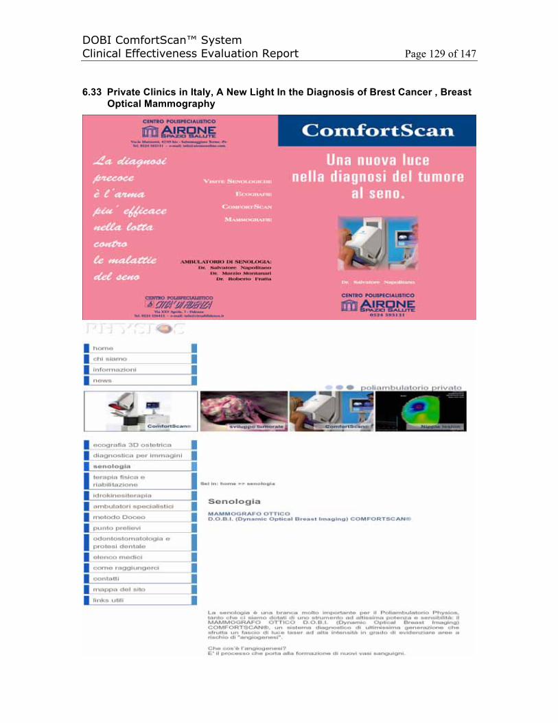
DOBI ComfortScan™ SystemClinical Effectiveness Evaluation Report Page 129 of 147
6.33 Private Clinics in Italy, A New Light In the Diagnosis of Brest Cancer , Breast Optical Mammography
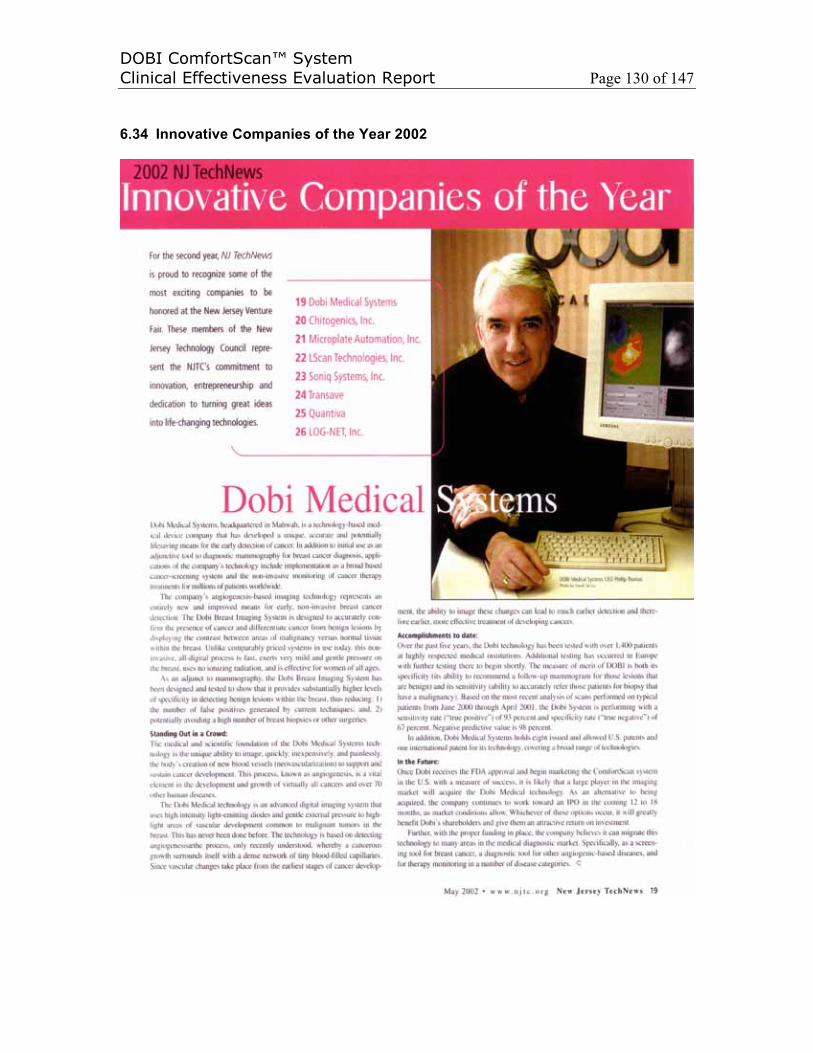
DOBI ComfortScan™ SystemClinical Effectiveness Evaluation Report Page 130 of 147
6.34 Innovative Companies of the Year 2002
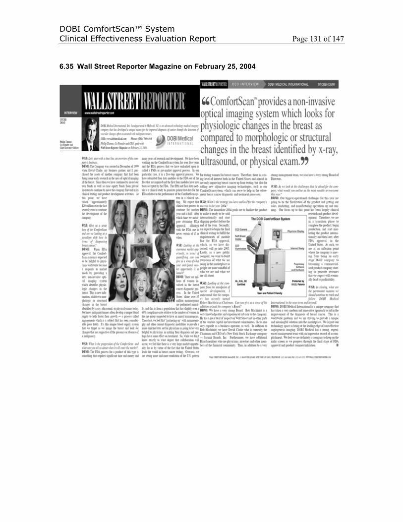
DOBI ComfortScan™ SystemClinical Effectiveness Evaluation Report Page 131 of 147
6.35 Wall Street Reporter Magazine on February 25, 2004
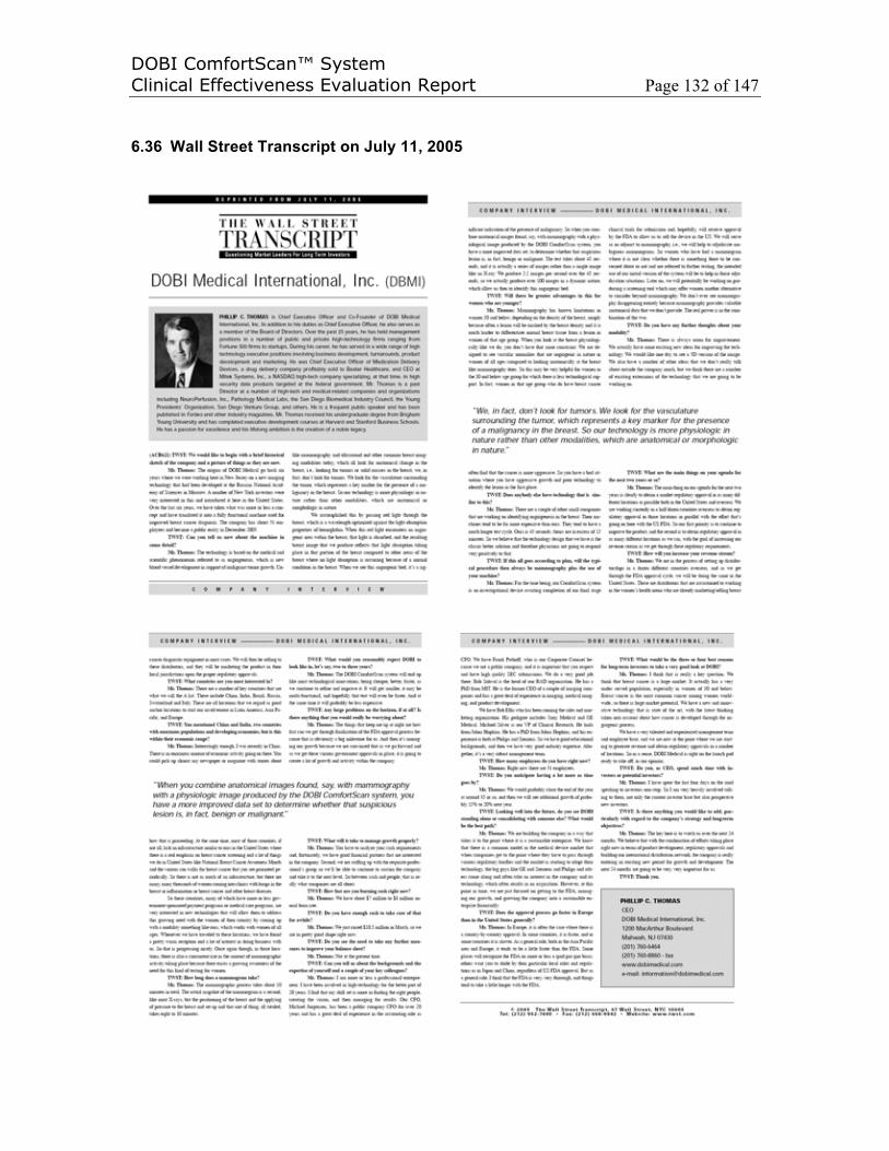
DOBI ComfortScan™ SystemClinical Effectiveness Evaluation Report Page 132 of 147
6.36 Wall Street Transcript on July 11, 2005
DOBI ComfortScan™ SystemClinical Effectiveness Evaluation Report Page 133 of 147
6.37 Rising Star Stocks – A Revolutionary Medical Imaging Device on August, 2004
DOBI ComfortScan™ SystemClinical Effectiveness Evaluation Report Page 134 of 147
6.38 Rising Star Stocks – A Revolutionary Medical Imaging Device on August, 2004
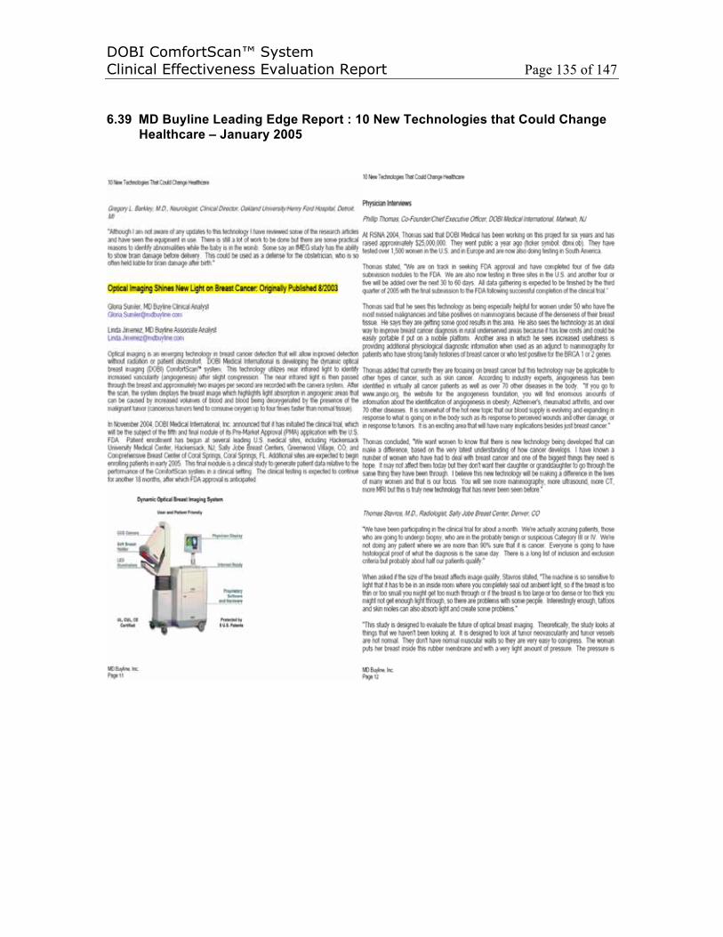
DOBI ComfortScan™ SystemClinical Effectiveness Evaluation Report Page 135 of 147
6.39 MD Buyline Leading Edge Report : 10 New Technologies that Could ChangeHealthcare – January 2005
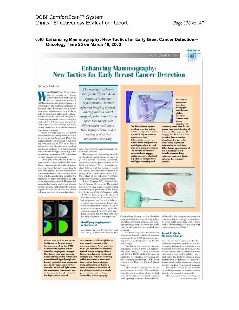
DOBI ComfortScan™ SystemClinical Effectiveness Evaluation Report Page 136 of 147
6.40 Enhancing Mammography: New Tactics for Early Brest Cancer Detection –Oncology Time 25 on March 10, 2003
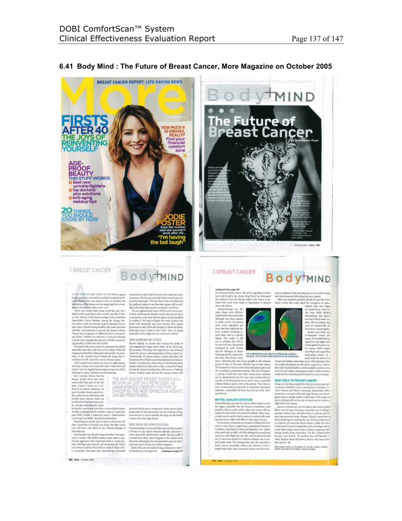
DOBI ComfortScan™ SystemClinical Effectiveness Evaluation Report Page 137 of 147
6.41 Body Mind : The Future of Breast Cancer, More Magazine on October 2005
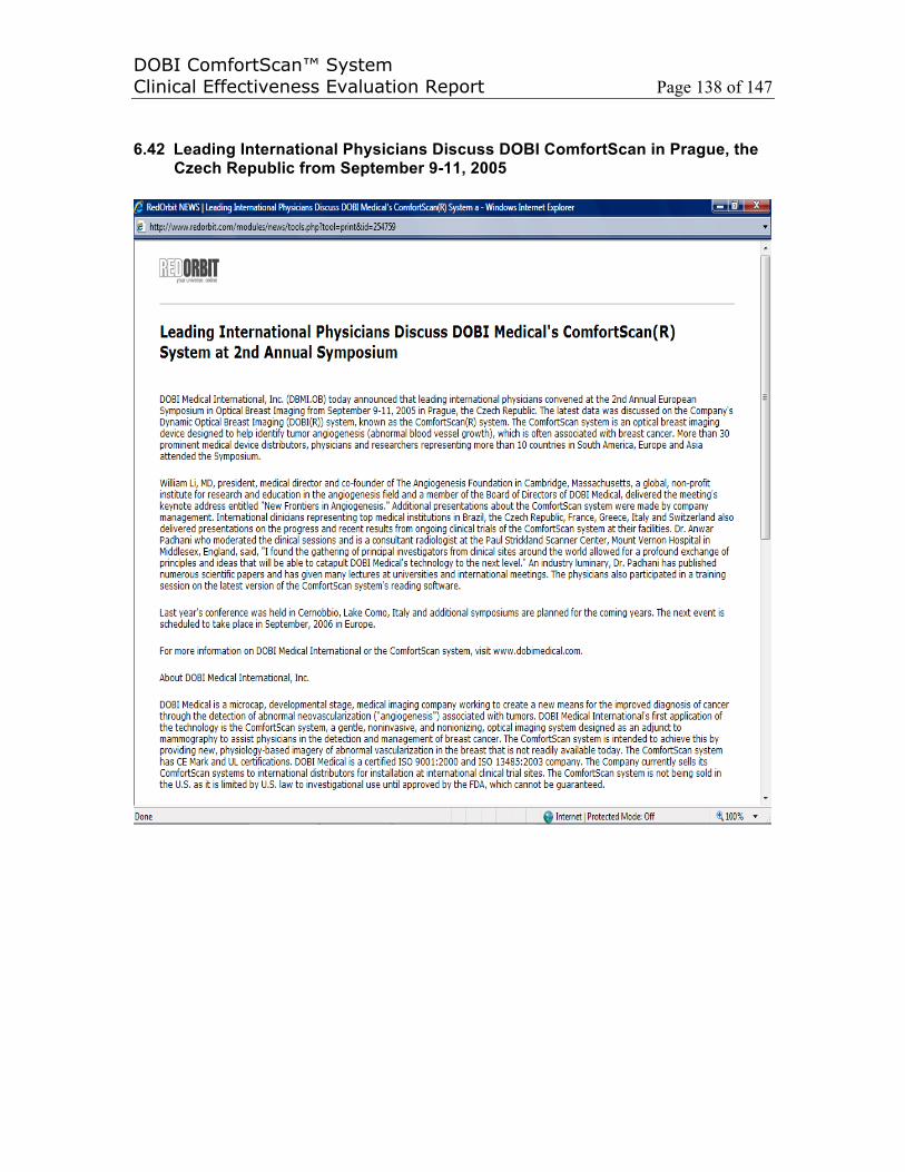
DOBI ComfortScan™ SystemClinical Effectiveness Evaluation Report Page 138 of 147
6.42 Leading International Physicians Discuss DOBI ComfortScan in Prague, the Czech Republic from September 9-11, 2005
DOBI ComfortScan™ SystemClinical Effectiveness Evaluation Report Page 139 of 147
7. CONCLUSION
Based on the purpose of this clinical effective evaluation report, through (1) the
introduction of DOBI ComfortScan, which uses the Tumor Angiogenesis Theory, Optical
Imaging Technique and the Significant Improvements of them in past decades in clinics
as the fundamentals, (2) some clinical study/trial publications/reports from recent clinical
cases, and (3) some collections of clinical acceptances, the clinical EFFECTIVENESS of
the DOBI ComfortScan as adjunct to existing breast imaging modalities in diagnosing
breast cancer has been demonstrated through detecting the characteristics of tumor
angiogenesis of malignant breast lesions and employing the appearance of the tumor
angiogenesis in detection of breast cancers in clinics. Because, based on the continued
demand, DOBI Global has just re-manufactured the ComfortScan device with necessary
CE and ISO regulatories after acquiring the intellectual property on May of 2010, the
significant clinical effectiveness will continue being demonstrated through the additional
reviews of clinical information, which has been defined in our Post Market Surveillance
(SOP 1054-0004-00). An opportunity for the potential benefit of women worldwide and
to continue the development/improvement of the breast imaging techniques, such as
DOBI ComfortScan, would be expected by scientists/developers of the
techniques/devices, medical society and general publics.
As the summary, in the past 7 years about 70 diagnostic centers worldwide have tested
the ComfortScan, including nearly 30 US sites participating in the FDA PMA clinical
trials, and over 30 sites in Italy and China have installed the devices, resulting in
thoughts of individuals whom were scanned by this device. The cumulative device
experiences to date, as well as the above literatures, have demonstrated that this
device, DOBI ComfortScan, could provide the physician with dynamic functional
information regarding abnormal vascularization in an area of interest in the breast and
this information could be used to better characterize the malignant lesions at early stage.
DOBI ComfortScan can help the performance and accuracy of averaging, under
averaging or less experienced doctors in their clinics most significantly. Dynamic optical
breast imaging, DOBI, technique, which is different from others by identifying
angiogenesis of malignant lesions, can be a promising complementary imaging modality
for further investigation in cases of women with inconclusive mammography and/or
DOBI ComfortScan™ SystemClinical Effectiveness Evaluation Report Page 140 of 147
physical examination. Based upon its performance in clinical studies worldwide, the
DOBI ComfortScan is a novel imaging technology that is appropriate as an imaging
modality in diagnosing breast cancer at early stage. As a diagnostic tool of breast
cancer, a large scale number of cases should be studied to characterize different
malignant and benign tumors at different stages respectively, different statues of
patients, such as menopause stages with related nipple blue, and to statistically
quantitatize the metabolic rates of both malignant and benign tumors. As a result, the
ComfortScan system focuses on physiology-based dynamic functional imaging (i.e.,
what is occurring within the tissue in near real time) rather than a singular morphological
image (i.e., a static anatomical snapshot showing physical details at a single point in
time), such as those created by mammography. When combined with mammography or
ultrasound, both of which provide simple morphologic images, the ComfortScan
system’s images of physiological changes in the breast is intended to provide physicians
with a more complete data set to improve the physician’s ability to provide an accurate
breast cancer diagnosis. With its negative predictive value of 98 percent and specificity
of 87 percent, the DOBI ComfortScan represents an opportunity to reduce the incidence
and severity of invasive diagnostic intervention and, thus, to potentially reduce the
number of unnecessary, painful and costly biopsies that are conducted on patients with
healthy tissue. Furthermore, the safety profile, convenience, comfort and low
comparative cost of the DOBI ComfortScan correspond closely to the call to action
delivered by the National Academy of Sciences’ Institute of Medicine. Because it is an
aid in detecting the minute vascular changes that accompany the process of
angiogenesis during the earliest stages of malignant tumor growth, the DOBI
ComfortScan could potentially become a useful breast cancer screening tool if a full FOV
(filed of view) cluster ComfortScan or DOBI ComfortScreen, next generation of the
ComfortScan, could maintain a high negative predictive value, above 95%. In addition to
disease diagnosis, therapeutic monitoring of both pro- and anti-angiogenic drugs may
also be a longer-term application of this technology and, since angiogenesis is found in
many significant disease states (such as rheumatoid arthritis and adult blindness), the
DOBI technology may have future applications in addition to cancer. DOBI’s dynamic
analysis is a significant improvement over current static imaging. Breast density does
not affect DOBI images, making DOBI especially important in the evaluation of dense
breasts, as often seen in young women or those on Hormone Replacement Therapy
DOBI ComfortScan™ SystemClinical Effectiveness Evaluation Report Page 141 of 147
(HRT). The initial results obtained with this rather new method, which is associated with
no radiation load and well tolerated by women, hold promise for further development,
particularly in the area of software development and standardization of evaluation
parameters. Another important point to stress is the need for high-quality training of
evaluating physicians which is, in our view, extremely important and affects the results of
the investigation rather significantly.
As the statement of Mammography and Beyond published by the National Academy of
Sciences’ Institute of Medicine, Mammography remains always the standard imaging
procedure of control and all recent studies63,64,65 support its value as a diagnostic and
screening tool. However it is already known and proven that this "gold standard" is not
an ideal tool. Potential radiation risk and diminished sensitivity in radiographically dense
breasts represent the two main disadvantages of the technique, thus limitating its
usefulness in high risk young women. It is well documentated in the study carried out by
Kuhl CK et al66 that gene carriers BRCA 1 and BRCA 2 are susceptible to have an
increased radiosensitivity of breast parenchyma. Other clinical areas in which
mammography is of limited diagnostic value are: detection of lobular cancer, detection of
ductal carcinoma in situ without associated microcalcifications, diagnostic work up of
unknown primary presenting as axillary lymphadenopathy (these are usually small high
grade lesions lodged in dense breast tissue), evaluation of multifocal disease and of
locally advanced disease, not to mention its diminished sensitivity in post-treatment
breasts.67 Since women in their 40s are generally premenopausal and therefore more
likely to have greater breast density than postmenopausal women, it is more difficult to
interpret mammograms – leading to some that are indeterminate. Mammograms for
postmenopausal women on estrogen-replacement therapy are similarly difficult to
interpret.10 Because the specificity of mammogram testing is quite low, false-positive
findings can have a detrimental effect on the screened population. As many as 80
percent of all breast lesions that are biopsied as a result of suspicious findings on a
mammogram turn out to be benign.11 Because the greater density of breast tissue in
younger, premenopausal women renders mammography results more difficult to
interpret, improved specificity and sensitivity in diagnostic methods would benefit
younger women in particular. In the study report of “Results of Investigational Use of
DOBI ComfortScan in China by G Zhang et al, the table below shows there are 24
DOBI ComfortScan™ SystemClinical Effectiveness Evaluation Report Page 142 of 147
indeterminate (I) case out of total 62 cases because of the 88.7% BI-RADS 3 or 4 in
Mammography from dense breasts, and the Youden’s Index indicates that the Accuracy
of DOBI ComfortScan is higher than Mammography with respect to three independent
blinding readers. The above finding suggests that there is a need to improve on the
tools currently used to detect breast disease in younger women or those with more
dense breasts. To demonstrate the DOBI ComfortScan is an improved tool for young
woman breast examination and an useful tool for breast cancer diagnostic with addtional
new physiological information, all the enrolled patients during the clinical study of DOBI
ComfortScan in Beijing were performed mammography examinaitons but have
inconclusive findings, which are catogrized as Breast Imaging Reporting and Data
System (BI-RADS) 3 or 4. According to the a recent publication at Italian Journal of
Gynaecology and Obstetrics Volume 23 on October 3, 2011, Dr. V. Frattini, L. Ghisoni, A.
Teodoro, PL Vaj and S. Orefice conducted a multicenter study to determine the
Sensitivity and Specificity of the ComfortScan System to detect malignancy as an
adjunct to Ultrasound in 617 young females between 25 to 39 years of age. After DOBI
ComfortScan and Ultrasound, 269 malignant cases were confirmed by biopsy. In this
study, the dynamical optical breast imaging, DOBI, had a sensitivity equal to 98% and a
specificity equal to 87% while the sensitivity and specificity of the Ultrasound are equal
to 74% and 70% respectively. Dynamic optical breast imaging in European Journal of
Radiology 2008 by a group of scientists from France, Italy UK and US) has concluded as
following: “We have used a novel imaging instrument that combines infrared imaging
with light breast compression in women with equivocal or suspect mammographic
abnormalities and have shown that it has potential in distinguishing benign
frommalignant lesions. This is an early evaluation of this technique which relies on
physiological properties of breast tissue to impart optical contrast on images. Further
evaluation will be required to optimize the technique, evaluate its sensitivity and
specificity in a wider range of patients, and explore its potential role in patient
management.” Based upon its performance in clinical studies worldwide, the DOBI
ComfortScan is a novel imaging technology that is appropriate as an imaging modality in
diagnosing breast cancer at early stage. As a diagnostic tool of breast cancer, a large
scale number of cases should be studied to charaterize different malignant and benign
tumors at different stages respectively, different statues of patients, such as menopause
stages with related nipple blue, and to statistically quantatize the metabolic rates of both
DOBI ComfortScan™ SystemClinical Effectiveness Evaluation Report Page 143 of 147
malignant and benign tumors. DOBI’s dynamic analysis is a significant improvement
over current static imaging. Breast density does not affect DOBI images, making DOBI
especially important in the evaluation of dense breasts, as often seen in young women
or those on Hormone Replacement Therapy (HRT). The initial results obtained with this
rather new method, which is associated with no radiation load and well tolerated by
women, hold promise for further development, particularly in the area of software
development and standardization of evaluation parameters. Another important point to
stress is the need for high-quality training of evaluating physicians which is, in our view,
extremely important and affects the results of the investigation rather significantly.
As a result of a continued demand for this device, DOBI Global has acquired all the
intellectual property including the trade mark and DOBI Medical International name in
the May of 2010, and has decided to re-launch the device and obtain a new CE
Certification and ISO 13485 Certification. The clinical efficacy will continue being
demonstrated through the collection of more clinical data and the publications based on
the clinical data if the CE Mark could be approved. The SOP 1054-0004-00 has defined
Post Market Surveillance activities that additional reviews of clinical information will be
conducted as part of the on going Post Market Surveillance activity, which complies and
will continue to comply with the requirements of the Medical Device Directive. Because
DOBI Medical had stopped its clinical trial of FDA Premarket Approval (PMA) due to the
financial issues in 2007, there are about hundreds of controlled patient data from USA
and hundreds of controlled patient data, which are locked. Because of the changes
within FDA ((On Monday, November 22, 2010 who has accepted a Premarket
Notification 510(k) of Imaging Diagnostic Systems), those data and more data from a
large scale number of trials might be used for the evaluation of clinical efficacy below
after FDA Approval/Notification:
• Adjunct to existing breast imaging modalities in diagnosing breast cancer at early
stage: As a diagnostic tool of breast cancer, a large scale number of cases
should be studied to charaterize different malignant and benign tumors at
different stages respectively, different statues of patients, such as menopause
stages with related nipple blue, and to statistically quantatize the metabolic rates
of both malignant and benign tumors.
DOBI ComfortScan™ SystemClinical Effectiveness Evaluation Report Page 144 of 147
• Physiology-based dynamic functional imaging (i.e., what is occurring within the
tissue in near real time) rather than a singular morphological image (i.e., a static
anatomical snapshot showing physical details at a single point in time): When
combined with mammography or ultrasound, both of which provide simple
morphologic images, the ComfortScan system’s images of physiological changes
in the breast is intended to provide physicians with a more complete data set to
improve the physician’s ability to provide an accurate breast cancer diagnosis.
• Screening Tool: Because it is an aid in detecting the minute vascular changes
that accompany the process of angiogenesis during the earliest stages of
malignant tumor growth, the DOBI ComfortScan could potentionally become a
useful breast cancer screening tool if the Negative Predicative Value could be
maintained at the level or above 95%.
• Therapeutic Monitoring: Both pro- and anti-angiogenic drug monitoring may also
be a longer-term application of this technology, and since angiogenesis is found
in many significant disease states (such as rheumatoid arthritis and adult
blindness), the DOBI technique may have future applications in addition to
cancer.
• High-quality Training: the need for high-quality training of physicians are
necessary because (1) it is not taught at school and (2) it affects the results
significantly.
As conclusion, the Clinical Effectiveness of DOBI ComfortScan device has been
demonstrated through recent clinical studies, which include total 2495 patients and 1053
malignant cases in over 23 multicenters, and achieve averaging Sensitivity and
Specificity in detecting malignant breast lesions are 87% and 75% separately by the
statistical analysis from a variety of readers in compliance with MEDDEV.2.7.1 Rev.3.
DOBI ComfortScan is an Office, In-Vivo, Non-Invasive, Non-Ionizing and Non-painful
molecular vesicular Dynamical Optical Breast Imaging modality. DOBI
technology/modality will continue to improve as new features are added, much the same
as other imaging modalities such as MRI, PET, CT, and digital mammography have
evolved over time.
DOBI ComfortScan™ SystemClinical Effectiveness Evaluation Report Page 145 of 147
REFERENCES1. American Cancer Society, Breast Cancer Resource Center, www.cancer.org, April 2001.2. China Medical Devices Net, www.zgylqxw.cn/Html/2007-03-10/320313.shtml.3. Nass SJ, Henderson C, Lashof JC, eds. Mammography and Beyond: Developing
Technologies for the Early Detection of Breast Cancer. Washington, DC: National Academy Press. 2001. Prepublication copy:13.
4. Ibid:1.5. Medical Data International, Inc. U.S. Markets for Diagnostic Oncology Products, 1999-2005.
Santa Ana, CA:Medical Data International, Inc. 2000. #RP-481430:2-14.6. National Academy of Sciences, Institute of Medicine (Recent Reports), www.iom.edu, April
2001.7. Nass SJ, Henderson C, Lashof JC, eds. Mammography and Beyond: Developing
Technologies for the Early Detection of Breast Cancer. Washington, DC: National Academy Press. 2001. Prepublication copy:16.
8. Ibid:19.9. Ibid:18.10. Ibid:23.11. Medical Data International, Inc. U.S. Markets for Diagnostic Oncology Products, 1999-2005.
Santa Ana, CA: Medical Data International, Inc. 2000. #RP-481430:2-14.12. Nass SJ, Henderson C, Lashof JC, eds. Mammography and Beyond: Developing
Technologies for the Early Detection of Breast Cancer. Washington, DC: National Academy Press. 2001. Prepublication copy:21.
13. Ibid:54.14. National Cancer Institute, Cancer Information, CancerNet, Types of Cancer, Breast Cancer
www.nci.nih.gov, April 2001.15. National Cancer Institute, Cancer Information, CancerNet, Types of Cancer, Breast Cancer
www.nci.nih.gov, April 2001.16. American Cancer Society, Breast Cancer Resource Center, www.cancer.org, April 2001.17. Folkman J. Tumor angiogenesis: therapeutic implications, New England Journal of Medicine
1971; 285:1182-1186.18. Angiogenesis Foundation, Understanding Angiogenesis, www.angio.org, April 2001.19. Li WW, Li VW, Tsakayannis D, Casey R, Jaffe M, Atwater LA, eds. Market Study and
Analysis of Angiogenesis- Dependent Diseases. Cambridge, MA: Angiogenesis Foundation, 2001:17.
20. Ibid:13.21. Weinberg RA, One Renegade Cell: How Cancer Begins. New York, NY: Basic Books.
1998:143-146.22. Eliceiri BP, Cheresh DA. The role of αv integrins during angiogenesis. Molecular Medicine
1998;4:741.23. Angiogenesis Foundation, Understanding Angiogenesis, www.angio.org, April 2001.24. Li WW, Tumor angiogenesis: molecular pathology, therapeutic targeting and imaging.
Academic Radiology 2000; 7:800-811.25. Gasparini G, Brooks PC, Biganzoli E, et al. Vascular integrin avb3: a new prognostic indicator
in breast cancer. Clinical Cancer Research 1998;4:2625.26. Feldman F, Habif DV, Fleming RJ, Kanter IE, Seaman WB. Arteriography of the breast.
Radiology 1967;89:1053-1061.27. Watt AC, Ackerman LV, Shetty PC, et al. Differentiation between benign and malignant
disease of the breast using digital subtraction angiography of the breast. Cancer 1985;56:1287-1292.
28. Wells PNT, Halliwell M, Skidmore R, Webb AJ, Woodcock JP. Tumor detection by ultrasound doppler bloodflow signals. Ultrasound. 1977;15:231-232.
DOBI ComfortScan™ SystemClinical Effectiveness Evaluation Report Page 146 of 147
29. Schoenberger SG, Sutherland CM, Robinson AE. Breast neoplasms: duplex sonographic imaging as an adjunct in diagnosis. Radiology 1988;168:665-668.
30. Cosgrove DO, Bamber JC, Davey JB, McKinna JA, Sinnett HD. Color doppler signals from breast tumors. Work in progress. Radiology 1990;176:175.
31. Folkman J, Watson K, Ingber D, Hanahan D. Induction of angiogenesis during the transition from hyperplasia to neoplasia. Nature 1989:339:58-61.
32. Weidner N, Semple JP, Welch WR, Folkman J. Tumor angiogenesis and metastasis –correlation in invasive breast carcinoma. New England Journal of Medicine 1991;324:1-8.
33. Baish JW, Netti PA, Jain RK. Transmural coupling of fluid flow in microcirculatory network and interstitium in tumors. Microvascular Research 1997;53:128.
34. Dyachenko A. Dynamic imaging of breast lesions; one dimensional optical model. Asian Journal of Physics 2001;10;4:1-18.
35. Boucher Y, Leunig M, Jain RK. Tumor angiogenesis and interstitial hypertension. Cancer Research 1996;56:4264.
36. Netti PA, Roberage S, Boucher Y, Baxter LT, Jain RK. Effect of transvascular fluid exchange on pressure—flow relationship in tumors: a proposed mechanism for tumor blood flow heterogeneity. Microvascular Research 1996;52:27.
37. Boucher Y, Baxter LT, Jain RK. Interstitial pressure gradients in tissue-isolated and subcutaneous tumors: implications for therapy. Cancer Research 1990;50:4478.
38. Boucher Y, Jain RK. Microvascular pressure is the principle driving force for interstitial hypertension in solid tumors. Cancer Research 1992;52:5110.
39. Dewhirst MW, Secomb TW, Ong ET, Hsu R, Gross JF. Determination of local oxygen consumption rates in tumors. Cancer Research 1994;54:3333.
40. Ertefai S, Profio AE. Spectral transmittance and contrast in breast diaphanography. Medical Physics 1985;12:393-400.
41. Profio AE, Navarro GA, Sartorius OW. Scientific basis of breast diaphanography. Medical Physics 1989;16:60-65.
42. Eliceiri BP, Cheresh DA. The role of αv integrins during angiogenesis. Molecular Medicine 1998;4:743.
43. Gasparini G, Brooks PC, Biganzoli E, et al. Vascular integrin αvβ3: a new prognostic indicator in breast cancer.Clinical Cancer Research 1998;4:2625.
44. Sipkins DA, Cheresh DA, Kazemi MR, Nevin LM, Bednarski MD, Li KCP. Detection of tumor45. Cristofanilli,C, et al, Angiogenesis Modulation in Cancer Research: Novel Clinical
Approaches. Nature Reviews Drug Discovery 2002, June 1(6): 414-26.46. Sauer G, Deissler H., Angiogenesis: Prognostic and Therapeutic Implications in Gynecologic
and Breast Malignancies. Current Opinion in Obstetrics and Gynecology; 2003 February, 15(45)-9.
47. Weidner N, Folkman J, Pozza F, Bevilacqua P, Allred EN, Moore DH, Meli S, Gasparini G. Tumor Angiogenesis: A New Significant and Independent Prognostic Indicator in Early-Stage Breast Carcinoma. Journal of the Natl. Cancer Institute December 1992.
48. Weidner N, Semple JP, Welch WR, Folkman J., Tumor Angiogenesis and Metastasis—Correlation in Invasive Breast Carcinoma. New England Journal of Medicine 1991, Jan. 3, 324(1): 1-8.
49. Gasparini, G., Clinical Significance of the Determination of Angiogenesis in Human Breast Cancer: Update of the Biological Background and Overview of the Vicenza Studies. Eur. J. Cancer 1996, 32A: 2485-93.
50. Cutler M. Transillumination as an aid to diagnosis of breast lesions. Surgery, Gynecology and Obstetrics 1929;48:721-727.
51. Ertefai S, Profio AE. Spectral transmittance and contrast in breast diaphanography. Medical Physics 1985;12:393-400.
52. Profio AE, Navarro GA, Sartorius OW. Scientific basis of breast diaphanography. Medical Physics 1989;16:60-65.
DOBI ComfortScan™ SystemClinical Effectiveness Evaluation Report Page 147 of 147
53. Peters VG, Wyman DR, Patterson MS, Frank GL. Optical properties of normal and diseased human breast tissues in the visible and near infrared. Physics in Medicine and Biology 1990;35:1317-1334.
54. McDonald DM, Choyke PL. Imaging of Angiogenesis: From Microscope to Clinic. Nature Medicine 2003, 9(6): 713-725.
55. Ibid.56. DOBI System data on file, DOBI Medical Systems, LLC, 2001.57. Alexandra Athanasiou, Daniel Vanel, Corinne Balleyguier, Laure Fournier, Marie Christine
Mathieu, Suzette Delaloge and Clasrisse Dromain, Dynamic Optical Breast Imaging: A New Technique to Visualise Breast Vessels: Comparison with Breath MRI and Preliminary Results. European Journal of Radiolog 54(2005) 72-79.
58. Kolb TM. Lichy J., Hewhouse J.H., Comparison of the Performance of Screening Mammography, Physical Examination, and Breast US and Evaluation of Factors that Influence Them: An Analysis or 27,825 Patient Evaluations. Radiology 2002, October: 225 (1): 165-75.
59. Kerlikowske K, Carney PA, Geller B, Mandelson MT, Taplin SH, Malvin K, Ernster V, Urban N, Cutter G, Rosenberg R, Ballard-Barbash R. Performance of Screening Mammography Among Women With and Without a First-Degree Relative with Breast Cancer. Annals of Internal Medicine 2000, Dec. 5; 133 (11): 855-63.
60. McDonald DM, Choyke PL. Imaging of Angiogenesis: From Microscope to Clinic. Nature Medicine 2003, 9(6): 713-725.
61. Bartoňková, H., Standara M. , Schneiderová M., THE RESULTS OF DOBI EXAMINATIONS IN MASARYK MEMORIAL CANCER INSTITUTE, Czech Ongological Society CLS JEP, KLINICKA ONKOLOGIE 18 4/2005: 149-151.
62. Gatzemeier W, Scelsi M, Galetti K, Villani L, Tinterri C, Secci A and Costa A, Dynamic Optical Breast Imaing: A non-invasive, adjunctive method to detect breast cancer, San Antonio Breast Cancer Symposium December 2004, Poster 6011.
63. Tabar L, Vitak B, Chen HH et al. Beyond randomized controlled trials : organized mammographic screening substantially reduces breast carcinoma mortality . Cancer 2001;91:1724-1731.
64. Olsen O,Gotzsche PC. Cochrane review on screening for breast cancer with mammography. Lancet 2001;358:1340-1342.
65. Preventive Services Task Force. Screening for breast cancer: recommendations and rationale. Ann Intern Med 2002;137:344-346
66. Kuhl KC, Schrading S, Leutner CC et al. Surveillance of 'high risk' women with proven or suspected familial (hereditary) breast cancer: first mid-term results of a multi-modality clinical screening trial J Clin Oncol 2003;21 (23suppl) :238
67. Smith JA, Andreopoulou E. An overview of the status of imaging screening technology for breast cancer. Ann of Oncol 2004; 15 (Suppl 1):18-26
68. DOBI Technical Paper, DOBI Medical Systems, LLC, August 2001.