Embed Size (px)
Citation preview
Docosahexaenoic acid-containing phosphatidylcholine a¡ects the bindingof monoclonal antibodies to puri¢ed Kb reconstituted into liposomes
Laura J. Jenski *, Paul K. Nanda, Philip Jiricko, William StillwellDepartment of Biology, Indiana University-Purdue University at Indianapolis, 723 W. Michigan Street, Indianapolis, IN 46202-5132, USA
Received 4 January 2000; received in revised form 18 April 2000; accepted 20 April 2000
Abstract
Class I major histocompatibility complex (MHC I) molecules are transmembrane proteins that bind and present peptidesto T-cell antigen receptors. The role of membrane lipids in controlling MHC I structure and function is not understood,although membrane lipid composition influences cell surface expression of MHC I. We reconstituted liposomes with purifiedMHC I (Kb) and probed the effect of lipid composition on MHC I structure (monoclonal anti-MHC I antibody binding).Four phospholipids were compared; each had a phosphocholine head group, stearic acid in the sn-1 position, and eitheroleic, K-linolenic, arachidonic, or docosahexaenoic acid (DHA) in the sn-2 position. The greatest binding of monoclonalantibody AF6-88.5, which detects a conformationally sensitive epitope in the extracellular region of the MHC I K-chain, wasachieved with DHA-containing proteoliposomes. Other epitopes (CTKb, 5041.16.1) showed some sensitivity to lipidcomposition. The addition of L2-microglobulin, which associates non-covalently with the K-chain and prevents K-chainaggregation, did not equalize antibody binding to proteoliposomes of different lipid composition, suggesting that freeK-chain aggregation was not responsible for disparate antibody binding. Thus, DHA-containing membrane lipids mayfacilitate conformational change in the extracellular domains of the K-chain, thereby modulating MHC I function througheffects on that protein's structure. ß 2000 Elsevier Science B.V. All rights reserved.
Keywords: Docosahexaenoic acid; Omega-3 fatty acid; Liposome; Major histocompatibility complex class I antigen
1. Introduction
Class I major histocompatibility complex (MHC I)molecules are membrane glycoproteins found on the
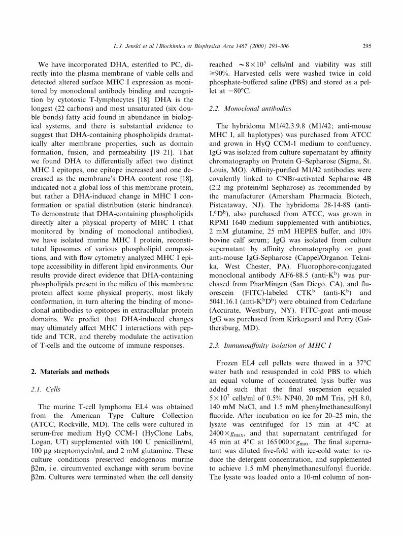
surface of all nucleated cells. Their principal functionis to present antigen peptides to T-lymphocytes, atask that is accomplished when a heterotrimer com-posed of the V45-kDa MHC I K-chain, a non-co-valently associated 12-kDa L2-microglobulin (L2m)protein, and peptide interacts with the antigen recep-tor (TCR) on T-cells (Fig. 1). If the interaction issu¤ciently avid, the antigen-speci¢c T-cell becomesactivated. Our current understanding of the molecu-lar interaction between the TCR and MHC I trimeris that the peptide provides one set of contacts withthe TCR, and that several critical amino acid resi-dues of the MHC I K-chain also contact the TCR [1],and hence the conformation of the K-chain is
0005-2736 / 00 / $ ^ see front matter ß 2000 Elsevier Science B.V. All rights reserved.PII: S 0 0 0 5 - 2 7 3 6 ( 0 0 ) 0 0 2 2 7 - 3
Abbreviations: ATCC, American Type Culture Collection;L2m, L2-microglobulin; DHA, docosahexaenoic acid; FITC,£uorescein isothiocyanate; FRET, £uorescence resonance energytransfer; IAEDANS, 5-((((2-iodoacetyl)amino)ethyl)amino)naph-thalene-1-sulfonic acid; MHC I, major histocompatibility com-plex class I; PBS, phosphate-bu¡ered saline; PC, phosphatidyl-choline; TCR, T-cell antigen receptor
* Corresponding author. Fax: +1-317-274-2846;E-mail : [email protected]
BBAMEM 77889 9-8-00
Biochimica et Biophysica Acta 1467 (2000) 293^306www.elsevier.com/locate/bba
thought to contribute substantially to TCR interac-tion [2].
MHC I functions naturally while spanning the bi-layer of the plasma membrane, and thus it is impor-tant to ask whether membrane lipids may controlMHC I structure and thereby ¢ne tune T-cell activa-tion. Recently, three-dimensional structure was de-termined for membrane-bound murine MHC I, i.e.soluble MHC I whose transmembrane and cytoplas-mic regions were replaced by a histidine tag to an-chor the molecule in a monolayer of nickel-chelatinglipids [3]. The extracellular domains of MHC I ap-pear positioned such that L2m contacts the lipidlayer and the peptide binding groove is directedaway from the lipid surface toward the TCR. How-ever, little else is known about the relationship be-tween membrane lipids and MHC I structure andfunction. We have directed our e¡orts to exploringhow the omega-3 fatty acid docosahexaenoic acid(DHA, 22:6g3), an important dietary componentthat becomes incorporated into membrane phospho-lipids, may control MHC I properties.
Lateral mobility, clustering, internalization, andprotein conformation are critical properties ofMHC I molecules to examine for lipid modulation.Lateral mobility of MHC I transmembrane proteinsis limited by the proteins' interactions with cytoskel-etal components, rather than by membrane lipids asis the case for a few proteins, such as visual rhodop-sin [4]. Similarly, clustering of MHC I may not bedriven directly by membrane lipid composition. Thatis, self aggregates of MHC I appear to result fromloss or lack of L2m [5^9] and do not correlate withlipid domains detectable in plasma membranes [6].Aggregation may serve to promote internalizationof free K-chains [8,10], thereby removing conforma-tionally altered MHC I from the cell surface. Mem-brane protein conformation, on the other hand, isin£uenced by membrane lipids. For example, mem-brane lipids alter the apparent pK for the conforma-tional equilibrium of metarhodopsin IHmetarho-dopsin II [11] ; DHA-containing phospholipidsfavor this conformational change purportedly by in-ducing negative curvature stress in membranes [12].In the case of calcium ATPase, conformationalchanges associated with catalytic activity are a¡ectedby alterations in membrane phospholipid composi-tion [13], and several indicators of K-fetoprotein con-formation (number of estradiol binding sites, estra-diol equilibrium dissociation constant, UVabsorption spectrum, and monoclonal antibody reac-tivity) varied with the concentration of free DHA[14]. Albeit limited, there do exist provocative dataimplicating the membrane and membrane lipids incontrolling MHC I conformation. Bene et al. [15]used intramolecular £uorescence resonance energytransfer (FRET) to detect a conformational changein MHC I induced by depolarization of JY (B-lym-phoblastoid) cells, and proposed that physicalchanges in membrane lipids may mediate, directlyor indirectly, MHC I's conformational change. Bind-ing of monoclonal antibodies to MHC I on normalmouse lymphocytes is decreased by treatment of thecells with cholesterol hemisuccinate (which increaseslipid packing), and increased by a PC preparation(which decreases lipid packing) [16]. Similarly, cho-lesterol enrichment augments the self-aggregationand reduces the expression of human MHC I onJY cells, whereas cholesterol depletion, which £uid-izes the membrane, enhances MHC I expression [17].
Fig. 1. Cartoons of MHCI in the native and hairpin con¢gura-tions. The K-chain consists of the K1-, K2-, and K3-domains,transmembrane region, and cytoplasmic tail. The intact hetero-trimeric MHCI molecule includes, in addition to the K-chain,the L2m-protein, and a peptide bound in the groove formed bythe K1- and K2-domains. In the hairpin con¢guration, thetransmembrane region fails to span the bilayer, i.e. remains inthe outer lea£et, causing the cytoplasmic tail to emerge on the`extracellular' side of the bilayer. The locations of the two freesulfhydryl groups (cysteines) available for IAEDANS labelingare indicated by arrows.
BBAMEM 77889 9-8-00
L.J. Jenski et al. / Biochimica et Biophysica Acta 1467 (2000) 293^306294
We have incorporated DHA, esteri¢ed to PC, di-rectly into the plasma membrane of viable cells anddetected altered surface MHC I expression as moni-tored by monoclonal antibody binding and recogni-tion by cytotoxic T-lymphocytes [18]. DHA is thelongest (22 carbons) and most unsaturated (six dou-ble bonds) fatty acid found in abundance in biolog-ical systems, and there is substantial evidence tosuggest that DHA-containing phospholipids dramat-ically alter membrane properties, such as domainformation, fusion, and permeability [19^21]. Thatwe found DHA to di¡erentially a¡ect two distinctMHC I epitopes, one epitope increased and one de-creased as the membrane's DHA content rose [18],indicated not a global loss of this membrane protein,but rather a DHA-induced change in MHC I con-formation or spatial distribution (steric hindrance).To demonstrate that DHA-containing phospholipidsdirectly alter a physical property of MHC I (thatmonitored by binding of monoclonal antibodies),we have isolated murine MHC I protein, reconsti-tuted liposomes of various phospholipid composi-tions, and with £ow cytometry analyzed MHC I epi-tope accessibility in di¡erent lipid environments. Ourresults provide direct evidence that DHA-containingphospholipids present in the milieu of this membraneprotein a¡ect some physical property, most likelyconformation, in turn altering the binding of mono-clonal antibodies to epitopes in extracellular proteindomains. We predict that DHA-induced changesmay ultimately a¡ect MHC I interactions with pep-tide and TCR, and thereby modulate the activationof T-cells and the outcome of immune responses.
2. Materials and methods
2.1. Cells
The murine T-cell lymphoma EL4 was obtainedfrom the American Type Culture Collection(ATCC, Rockville, MD). The cells were cultured inserum-free medium HyQ CCM-1 (HyClone Labs,Logan, UT) supplemented with 100 U penicillin/ml,100 Wg streptomycin/ml, and 2 mM glutamine. Theseculture conditions preserved endogenous murineL2m, i.e. circumvented exchange with serum bovineL2m. Cultures were terminated when the cell density
reached V8U105 cells/ml and viability was stillv90%. Harvested cells were washed twice in coldphosphate-bu¡ered saline (PBS) and stored as a pel-let at 380³C.
2.2. Monoclonal antibodies
The hybridoma M1/42.3.9.8 (M1/42; anti-mouseMHC I, all haplotypes) was purchased from ATCCand grown in HyQ CCM-1 medium to con£uency.IgG was isolated from culture supernatant by a¤nitychromatography on Protein G^Sepharose (Sigma, St.Louis, MO). A¤nity-puri¢ed M1/42 antibodies werecovalently linked to CNBr-activated Sepharose 4B(2.2 mg protein/ml Sepharose) as recommended bythe manufacturer (Amersham Pharmacia Biotech,Pistcataway, NJ). The hybridoma 28-14-8S (anti-LdDb), also purchased from ATCC, was grown inRPMI 1640 medium supplemented with antibiotics,2 mM glutamine, 25 mM HEPES bu¡er, and 10%bovine calf serum; IgG was isolated from culturesupernatant by a¤nity chromatography on goatanti-mouse IgG-Sepharose (Cappel/Organon Tekni-ka, West Chester, PA). Fluorophore-conjugatedmonoclonal antibody AF6-88.5 (anti-Kb) was pur-chased from PharMingen (San Diego, CA), and £u-orescein (FITC)-labeled CTKb (anti-Kb) and5041.16.1 (anti-KbDb) were obtained from Cedarlane(Accurate, Westbury, NY). FITC-goat anti-mouseIgG was purchased from Kirkegaard and Perry (Gai-thersburg, MD).
2.3. Immunoa¤nity isolation of MHC I
Frozen EL4 cell pellets were thawed in a 37³Cwater bath and resuspended in cold PBS to whichan equal volume of concentrated lysis bu¡er wasadded such that the ¢nal suspension equaled5U107 cells/ml of 0.5% NP40, 20 mM Tris, pH 8.0,140 mM NaCl, and 1.5 mM phenylmethanesulfonyl£uoride. After incubation on ice for 20^25 min, thelysate was centrifuged for 15 min at 4³C at2400Ugmax, and that supernatant centrifuged for45 min at 4³C at 165 000Ugmax. The ¢nal superna-tant was diluted ¢ve-fold with ice-cold water to re-duce the detergent concentration, and supplementedto achieve 1.5 mM phenylmethanesulfonyl £uoride.The lysate was loaded onto a 10-ml column of non-
BBAMEM 77889 9-8-00
L.J. Jenski et al. / Biochimica et Biophysica Acta 1467 (2000) 293^306 295
immune IgG2a (Sigma, St. Louis, MO) coupled toSepharose 4B, and the unbound £ow through waschanneled directly to the 1-ml M1/42 a¤nity columnat a £ow rate of 30 ml/h. The a¤nity column waswashed with 20 column volumes of pre-elution bu¡er(0.5% deoxycholate, 140 mM NaCl, 15 mM Tris, pH8.0) and bound MHC I was eluted with 20 columnvolumes of elution bu¡er (21 mM n-octyl-K-D-gluco-pyranoside, 20 mM NaHCO3, pH 11.5) at 4³C at0.5^1.0 ml/min. One-milliliter fractions of both thepre-elution wash and eluant were bu¡ered with100 Wl of 1 M Tris, pH 8.0, and saved for proteinand PAGE analyses. Protein analysis was conductedwith a BCA protein assay kit (Pierce, Rockford, IL)as described by the manufacturer. Protein in samplesfor SDS^PAGE was precipitated with cold 12.5%trichloroacetic acid and pelleted by centrifugationat 7800Ugmax for 15 min at room temperature. Thepellets were washed twice with ice-cold acetone toremove residual detergent, dissolved in sample bu¡er(1^4 Wg protein/20 Wl bu¡er), heated to 100³C for5 min, and electrophoresed by the method ofLaemmli [22]. The gels were stained either with0.3% w/v Coomassie brilliant blue R-250 or with asilver stain kit (Boehringer Mannheim Biochemicals/Roche, Indianapolis, IN).
2.4. Construction of MHC I proteoliposomes
The following phospholipids were purchased fromAvanti Polar Lipids (Alabaster, AL): 1-stearoyl, 2-oleoyl-phosphatidylcholine (18:0, 18:1 PC), 1-stearo-yl, 2-K-linolenoylphosphatidylcholine (18:0, 18:3PC), 1-stearoyl, 2-arachidonoylphosphatidylcholine(18:0, 20:4 PC), 1-stearoyl, 2-docosahexaenoylphos-phatidylcholine (18:0, 22:6 PC), and egg PC. Solventwas evaporated from the lipids under a stream ofnitrogen followed by vacuum pumping, and the lip-ids were rehydrated to 1 mg/ml of 20 mM NaHCO3,pH 9, for 10 min at room temperature with frequentvortexing. The lipids were then extruded v10 timesthrough a 2-Wm Nucleopore membrane (NucleoporeFiltration Products, Pleasanton, CA) using a syringe-type extruder (Avestin, Ottawa, Canada). Puri¢edMHC I (2 Wg) was added to 20 Wg of 2-Wm liposomesand 13 mM n-octyl-K-D-glucopyranoside (¢nal con-centration) in a total volume of 300 Wl. The n-octyl-K-D-glucopyranoside concentration corresponds to
that required to saturate, but not solubilize, the lipo-somes, and was determined empirically as describedby Rigaud et al. [23]. After 1 min at room temper-ature, complete and rapid detergent removal wasachieved with 80 mg of degassed SM-2 Bio-Beads/ml (Bio-Rad, Hercules, CA) for 3 h with agitation inthe dark at room temperature followed by a 1-h in-cubation with a fresh aliquot of Bio-Beads [23].
2.5. Flow cytometry
Proteoliposomes (2 Wg of protein in 300 Wl totalvolume) were incubated with 1 Wg of £uorescentlylabeled anti-MHC I monoclonal antibodies for30 min at room temperature in the dark. Cytometrywas accomplished with a Coulter Epics Elite ESP cellsorter equipped with an argon laser. Antibody bind-ing to liposomes, i.e. £uorescence, is described in twoways: the mean and median £uorescence intensitychannels (range, 0^1023), and the percent of lipo-somes having a given £uorescence intensity. In thelatter case, £uorescence intensities are divided intofour levels corresponding to the four decades (setsof 256 channels) of the log scale on a 1024-channelphotomultiplier tube. The gating of forward versusside scatter histograms, to select comparable proteo-liposomes for analysis, is shown in the ¢gures. Expo2.0 software (Beckman Coulter, Fullerton, CA) wasused for data analysis and exportation for the crea-tion of graphics.
2.6. Assessment of native vs. hairpin con¢guration
The £uorescent sulfhydryl probe 5-((((2-iodoace-tyl)amino)ethyl)amino)naphthalene-1-sulfonic acid(IAEDANS, Molecular Probes, Eugene, OR), wasused to label the two free sulfhydryl groups (Cys-142, Cys-358) present on the MHC I K-chain [24].IAEDANS (4.3 mg) was dissolved in 1 ml of dimeth-ylsulfoxide, added to 2 Wg of puri¢ed MHC I, andmixed gently for 1 h at room temperature in thedark. An excess (105 Wl) of 2-mercaptoethanol wasadded to the sample to consume unbound IAE-DANS and the sample was dialyzed for 24 h against1.38 mM n-octyl-K-D-glucopyranoside, 20 mMNaHCO3, pH 9 (three changes) in a 100^500 WlSlide-A-Lyzer dialysis cassette (Pierce, Rockford,IL). The labeled MHC I was then inserted into the
BBAMEM 77889 9-8-00
L.J. Jenski et al. / Biochimica et Biophysica Acta 1467 (2000) 293^306296
preformed 2-Wm liposomes as described above forthe construction of proteoliposomes. Proteolipo-somes were diluted and placed into an LS50B £uo-rimeter (Perkin Elmer, Norwalk, CT) and excited at350 nm; £uorescence emission was monitored from360 to 600 nm, and then CoCl2 (29^50 mM ¢nalconcentration) was added to quench [Cys-142]-AEDANS £uorescence from the external surface ofthe proteoliposomes. Finally, 0.3% Triton X-100(¢nal concentration) was added to solubilize all pro-teoliposomes and allow quenching of [Cys-358]-AEDANS in the liposomes' interior.
2.7. Human L2m addition
MHC I proteoliposomes (2 Wg protein in 300 Wltotal volume) were incubated with 30 Wl of 0.22-Wm¢ltered human serum (Sigma) or 10 Wl of a 0.1 mg/mlsolution of puri¢ed human L2m (Sigma) for 1 h atroom temperature. After incubation, 1 Wg of FITC-labeled monoclonal anti-Kb (clone AF6-88.5) wasadded, and the proteoliposomes were incubated for30 min at room temperature in the dark and thenanalyzed by £ow cytometry.
2.8. Indirect staining of cells and proteoliposomes for£ow cytometry
To assess whether Db was present in the MHC Iprotein used for proteoliposome reconstitution, 1 Wgof anti-Db (28-14-8S), puri¢ed from hybridomaculture supernatant as described previously in Sec-tion 2, was incubated for 30 min at room temper-ature with proteoliposomes containing 2 Wg of MHCI protein and with 1U106 live EL4 cells (the originalcell source of the MHC I). A secondary FITC-la-beled anti-mouse IgG antibody was added in 5-foldmolar excess, and after an additional 30-min incuba-tion at room temperature, the cells and proteolipo-somes were analyzed for £uorescence by £ow cyto-metry.
2.9. Statistical methods
The Wilcoxon paired sample test was used to as-sess signi¢cant di¡erences between antibody bindingto proteoliposomes of di¡erent phospholipid compo-sition. A non-parametric test was necessary because
proteoliposome £uorescence intensities were not nor-mally distributed. To accommodate day-to-day var-iations introduced with di¡erent MHC I puri¢cationlots, proteoliposome preparation, and instrumentperformance, the statistical method is a paired sam-ple test. The software package used was WinStar(Anderson Bell, Arvada, CO), and P9 0.05 was ac-cepted as statistically signi¢cant.
3. Results and discussion
3.1. Immunoa¤nity puri¢ed MHC I is incorporatedinto large unilamellar vesicles in a nativeconformation
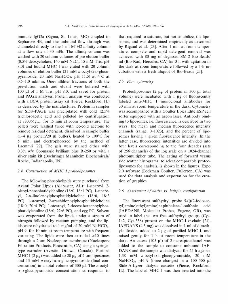
MHC I isolated from EL4 (H-2b) cells by a¤nitychromatography on M1/42-Sepharose consisted,under denaturing conditions, of a heavy chain (i.e.K-chain) with an apparent molecular weight V50kDa and a 12-kDa light chain (L2m) (Fig. 2). Densi-tometry of the Coomassie blue-stained gel indicatedthe relative densities of these two bands to be 1.3^2.5(K-chain/L2m), which varied with the puri¢cation lotand elution fraction. This relative recovery of the
Fig. 2. Electrophoretic properties of immunoa¤nity puri¢edMHC I. MHC I puri¢ed by a¤nity chromatography was elec-trophoresed under denaturing conditions in a 14% polyacryl-amide (resolving) gel, and the gel was stained with Coomassiebrilliant blue R-250. The left lane provides the molecular weightmarkers and the right lane shows the trichloroacetic acid-pre-cipitable protein eluted from the M1/42 a¤nity column.
BBAMEM 77889 9-8-00
L.J. Jenski et al. / Biochimica et Biophysica Acta 1467 (2000) 293^306 297
two chains is similar to that presented in the illustra-tions of Stallcup et al. [25], whose MHC I puri¢ca-tion procedure is the model upon which our immu-noa¤nity method is based. Contaminating bandsconstituted 4.5^6.6% of the total mass detected den-sitometrically, and thus were unlikely to interfere sig-ni¢cantly with proteoliposome reconstitution. Theincorporation of protein into liposomes was highlye¤cient (80% of added protein was incorporated)and equivalent for di¡erent liposome preparations
(Table 1). Equivalent protein incorporation is impor-tant (it is assumed in subsequent assays), but notsurprising; saturation of the bilayers with detergentprior to MHC I addition obviates the in£uence ofthe phospholipids on protein insertion.
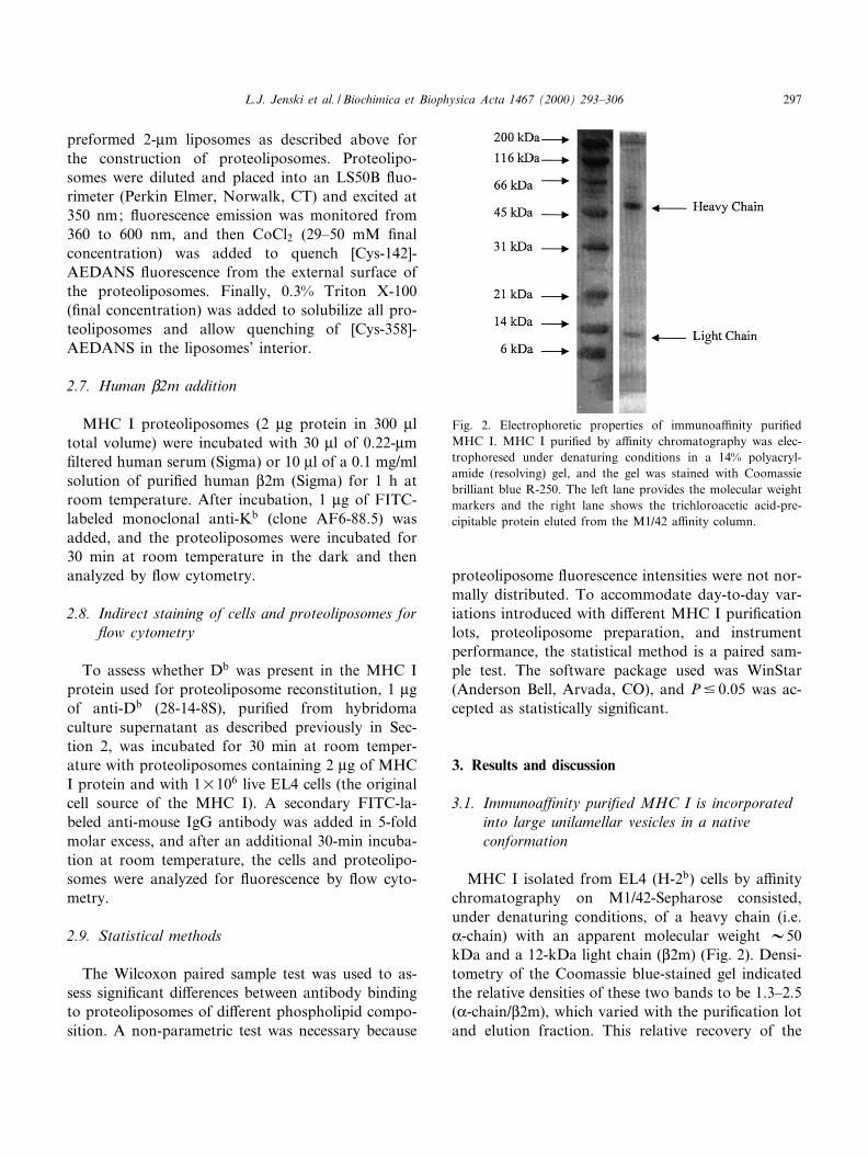
Fig. 3 demonstrates that £uorescent monoclonalantibodies directed against Kb will intensely stainliposomes reconstituted with a¤nity puri¢ed MHCI (Fig. 3B), whereas liposomes lacking MHC I arenot stained (Fig. 3D), nor are MHC I-reconstituted
Fig. 3. MHC I reconstituted into proteoliposomes displays speci¢c staining. Egg PC liposomes were reconstituted with MHC I andstained with FITC-labeled AF6-88.5 (anti-Kb) (A, B) or with FITC-labeled anti-Thy-1 (an irrelevant antibody) (E,F). In C and D,egg PC liposomes lacking MHC I were stained with FITC-labeled AF6-88.5. A, C, and E display the forward (y-axis) and side(x-axis) scatter characteristics of the liposomes and illustrate the gating (box) for the corresponding £uorescence histograms in B, D,and F (x-axis, £uorescence channel; y-axis, number).
BBAMEM 77889 9-8-00
L.J. Jenski et al. / Biochimica et Biophysica Acta 1467 (2000) 293^306298
proteoliposomes treated with FITC-anti-Thy-1, anirrelevant antibody (Fig. 3F). The correspondingplots of liposome size versus complexity (forwardscatter versus side scatter) are provided in Fig. 3A,C and E; variations in size (forward scatter) andtrapped internal membranes (side scatter) were likelyintroduced during initial liposome formation as wellas subsequent manipulations to insert protein. Thegates shown in Fig. 3A, C, and E are those assignedto the £uorescence histograms (Fig. 3B,D,F).Although di¡erent numbers of liposomes werecounted in the three samples (ranging from 2187 to9641 events), it is clear that intense staining is onlyachieved for MHC I-reconstituted proteoliposomestreated with £uorescent anti-MHC I antibodies.
The reconstitution method, insertion of proteininto pre-formed detergent-saturated liposomes fol-lowed by rapid detergent removal with Bio-Beads,was chosen to maximize MHC I rightside-out orien-tation and non-hairpin conformation. Earlier recon-stitution methods utilized extensive dialysis startingwith mixed micelles of protein and lipid, and resultedin proteoliposomes with heterogeneous protein^lipidratios and orientations [23] and a putative hairpinconformation for Kk [24]. In contrast, when pre-formed liposomes and rapid detergent removal wereused, the protein^lipid ratios were more homogene-ous and the protein orientation was highly asymmet-rical, with the protein hydrophobic domain inserted¢rst into the membrane (i.e. 85^95% of protein mol-ecules inserted rightside-out) [23]. Although we can-not completely exclude the possibility that someMHC I is oriented inside-out, this possibility seemsremote. Furthermore, antibodies, which are large(s 150 kDa) globular proteins, do not penetratethe liposomes to stain MHC I and therefore MHC
I with an inside-out orientation would not be ana-lyzed by £ow cytometry.
Our principal concern was that MHC I may as-sume a hairpin con¢guration (extracellular domainsand cytoplasmic tail all facing out, Fig. 1); this con-formation may obscure the e¡ects of membrane lip-ids and have little biological relevance. Hence, toassess the likelihood of a hairpin con¢guration forreconstituted MHC I we used the approach described
Table 1Protein is incorporated e¤ciently into preformed 2-Wm liposomesa
Liposomes Protein added (Wg) Protein recovered (Wg) Protein incorporatedb
18:0, 18:1 PC 2.0 1.7 (Expt. 1) 83%1.6 (Expt. 2) (80^85%)
18:0, 22:6 PC 2.0 1.7 (Expt. 1) 80%1.5 (Expt. 2) (75^85%)
aPreformed liposomes were incubated with immunoa¤nity isolated MHC I at a saturating n-octyl-K-D-glucopyranoside concentration.After detergent removal with Bio-Beads, the liposomes were washed in 15 vols. of PBS and harvested by centrifugation at100 000Ugmax for 30 min at 4³C, and the protein was quanti¢ed with a CBQCA protein assay kit.bThe values for protein incorporated are means of two experiments, with the range shown in parentheses.
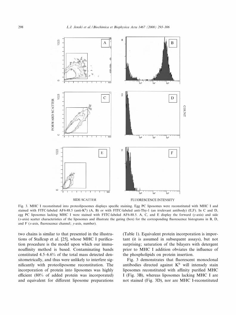
Fig. 4. Db, predicted to be lost during a¤nity chromatography,is not detected in proteoliposomes. Proteoliposomes reconsti-tuted with immunoa¤nity puri¢ed MHC I were treated withanti-Db monoclonal antibodies (clone 28-14-8S, black histo-gram) plus FITC-labeled anti-mouse IgG, or with the secondaryantibody alone (gray histogram). Positive staining of the cellsfrom which the MHC I was obtained is shown in the inset.
BBAMEM 77889 9-8-00
L.J. Jenski et al. / Biochimica et Biophysica Acta 1467 (2000) 293^306 299
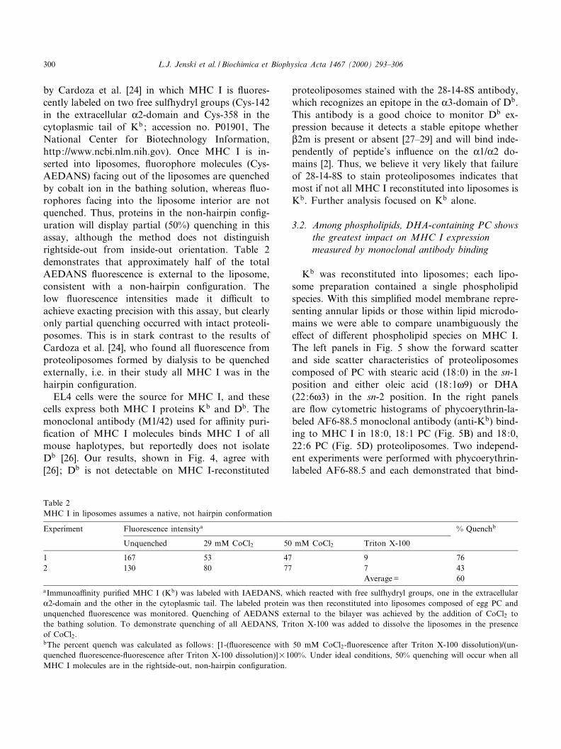
by Cardoza et al. [24] in which MHC I is £uores-cently labeled on two free sulfhydryl groups (Cys-142in the extracellular K2-domain and Cys-358 in thecytoplasmic tail of Kb ; accession no. P01901, TheNational Center for Biotechnology Information,http://www.ncbi.nlm.nih.gov). Once MHC I is in-serted into liposomes, £uorophore molecules (Cys-AEDANS) facing out of the liposomes are quenchedby cobalt ion in the bathing solution, whereas £uo-rophores facing into the liposome interior are notquenched. Thus, proteins in the non-hairpin con¢g-uration will display partial (50%) quenching in thisassay, although the method does not distinguishrightside-out from inside-out orientation. Table 2demonstrates that approximately half of the totalAEDANS £uorescence is external to the liposome,consistent with a non-hairpin con¢guration. Thelow £uorescence intensities made it di¤cult toachieve exacting precision with this assay, but clearlyonly partial quenching occurred with intact proteoli-posomes. This is in stark contrast to the results ofCardoza et al. [24], who found all £uorescence fromproteoliposomes formed by dialysis to be quenchedexternally, i.e. in their study all MHC I was in thehairpin con¢guration.
EL4 cells were the source for MHC I, and thesecells express both MHC I proteins Kb and Db. Themonoclonal antibody (M1/42) used for a¤nity puri-¢cation of MHC I molecules binds MHC I of allmouse haplotypes, but reportedly does not isolateDb [26]. Our results, shown in Fig. 4, agree with[26] ; Db is not detectable on MHC I-reconstituted
proteoliposomes stained with the 28-14-8S antibody,which recognizes an epitope in the K3-domain of Db.This antibody is a good choice to monitor Db ex-pression because it detects a stable epitope whetherL2m is present or absent [27^29] and will bind inde-pendently of peptide's in£uence on the K1/K2 do-mains [2]. Thus, we believe it very likely that failureof 28-14-8S to stain proteoliposomes indicates thatmost if not all MHC I reconstituted into liposomes isKb. Further analysis focused on Kb alone.
3.2. Among phospholipids, DHA-containing PC showsthe greatest impact on MHC I expressionmeasured by monoclonal antibody binding
Kb was reconstituted into liposomes; each lipo-some preparation contained a single phospholipidspecies. With this simpli¢ed model membrane repre-senting annular lipids or those within lipid microdo-mains we were able to compare unambiguously thee¡ect of di¡erent phospholipid species on MHC I.The left panels in Fig. 5 show the forward scatterand side scatter characteristics of proteoliposomescomposed of PC with stearic acid (18:0) in the sn-1position and either oleic acid (18:1g9) or DHA(22:6g3) in the sn-2 position. In the right panelsare £ow cytometric histograms of phycoerythrin-la-beled AF6-88.5 monoclonal antibody (anti-Kb) bind-ing to MHC I in 18:0, 18:1 PC (Fig. 5B) and 18:0,22:6 PC (Fig. 5D) proteoliposomes. Two independ-ent experiments were performed with phycoerythrin-labeled AF6-88.5 and each demonstrated that bind-
Table 2MHC I in liposomes assumes a native, not hairpin conformation
Experiment Fluorescence intensitya % Quenchb
Unquenched 29 mM CoCl2 50 mM CoCl2 Triton X-100
1 167 53 47 9 762 130 80 77 7 43
Average = 60aImmunoa¤nity puri¢ed MHC I (Kb) was labeled with IAEDANS, which reacted with free sulfhydryl groups, one in the extracellularK2-domain and the other in the cytoplasmic tail. The labeled protein was then reconstituted into liposomes composed of egg PC andunquenched £uorescence was monitored. Quenching of AEDANS external to the bilayer was achieved by the addition of CoCl2 tothe bathing solution. To demonstrate quenching of all AEDANS, Triton X-100 was added to dissolve the liposomes in the presenceof CoCl2.bThe percent quench was calculated as follows: [1-(£uorescence with 50 mM CoCl2-£uorescence after Triton X-100 dissolution)/(un-quenched £uorescence-£uorescence after Triton X-100 dissolution)]U100%. Under ideal conditions, 50% quenching will occur when allMHC I molecules are in the rightside-out, non-hairpin con¢guration.
BBAMEM 77889 9-8-00
L.J. Jenski et al. / Biochimica et Biophysica Acta 1467 (2000) 293^306300
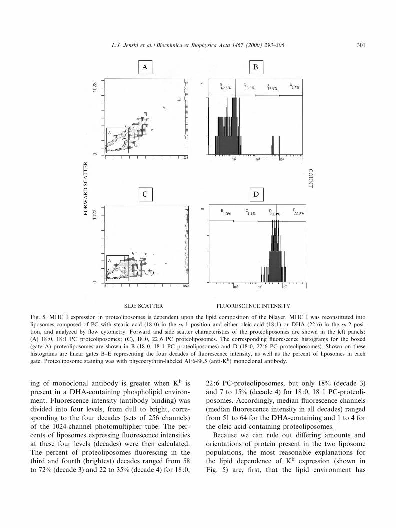
ing of monoclonal antibody is greater when Kb ispresent in a DHA-containing phospholipid environ-ment. Fluorescence intensity (antibody binding) wasdivided into four levels, from dull to bright, corre-sponding to the four decades (sets of 256 channels)of the 1024-channel photomultiplier tube. The per-cents of liposomes expressing £uorescence intensitiesat these four levels (decades) were then calculated.The percent of proteoliposomes £uorescing in thethird and fourth (brightest) decades ranged from 58to 72% (decade 3) and 22 to 35% (decade 4) for 18:0,
22:6 PC-proteoliposomes, but only 18% (decade 3)and 7 to 15% (decade 4) for 18:0, 18:1 PC-proteoli-posomes. Accordingly, median £uorescence channels(median £uorescence intensity in all decades) rangedfrom 51 to 64 for the DHA-containing and 1 to 4 forthe oleic acid-containing proteoliposomes.
Because we can rule out di¡ering amounts andorientations of protein present in the two liposomepopulations, the most reasonable explanations forthe lipid dependence of Kb expression (shown inFig. 5) are, ¢rst, that the lipid environment has
Fig. 5. MHC I expression in proteoliposomes is dependent upon the lipid composition of the bilayer. MHC I was reconstituted intoliposomes composed of PC with stearic acid (18:0) in the sn-1 position and either oleic acid (18:1) or DHA (22:6) in the sn-2 posi-tion, and analyzed by £ow cytometry. Forward and side scatter characteristics of the proteoliposomes are shown in the left panels:(A) 18:0, 18:1 PC proteoliposomes; (C), 18:0, 22:6 PC proteoliposomes. The corresponding £uorescence histograms for the boxed(gate A) proteoliposomes are shown in B (18:0, 18:1 PC proteoliposomes) and D (18:0, 22:6 PC proteoliposomes). Shown on thesehistograms are linear gates B^E representing the four decades of £uorescence intensity, as well as the percent of liposomes in eachgate. Proteoliposome staining was with phycoerythrin-labeled AF6-88.5 (anti-Kb) monoclonal antibody.
BBAMEM 77889 9-8-00
L.J. Jenski et al. / Biochimica et Biophysica Acta 1467 (2000) 293^306 301
changed the conformation of Kb thereby a¡ectingthe expression of the AF6-88.5 epitope and, second,that the MHC I proteins are self aggregating in the18:0, 18:1 PC bilayer, thereby obscuring the bindingof antibodies. Of course, these two processes are notmutually exclusive and may both be operative. Selfaggregation of free MHC I heavy chains (K-chainsdissociated from L2m) has been reported, and havingKb epitopes in close proximity may sterically hinderbinding of the monoclonal antibodies or bring to-gether £uorophore molecules, such that £uorophoreself quenching occurs. Phycoerythrin is relatively re-sistant to self quenching [30] and thus self quenchingis unlikely to be at play in the experiment shown inFig. 5. Fluorescein, on the other hand, is quite sen-sitive to self quenching, and £uorescein-conjugatedantibodies were used in various aspects of this study.We therefore addressed the participation of MHC Iself aggregation in our system. Chakrabarti et al. [9]reported that human MHC I reconstituted into di-myristoylphosphatidylcholine (14:0, 14:0 PC) dis-played self aggregation (measured by FRET), whichcould be blocked with exogenous L2m. Note, how-ever, that 14:0, 14:0 PC is not typically found inmammalian plasma membranes and does not pro-duce the usual surface bilayer thickness; this resultsin a hydrophobic mismatch between the bilayer andthe protein's transmembrane region and promotesprotein oligomerization to minimize exposed hydro-phobic area [31]. EL4 cells (our source for Kb) re-
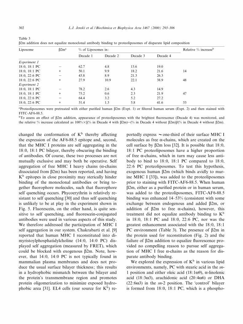
portedly express Vone-third of their surface MHC Imolecules as free K-chains, which are created on thecell surface by L2m loss [32]. It is possible that 18:0,18:1 PC proteoliposomes have a higher proportionof free K-chains, which in turn may cause less anti-body to bind to 18:0, 18:1 PC compared to 18:0,22:6 PC proteoliposomes. To test this hypothesis,exogenous human L2m (which binds avidly to mur-ine MHC I [33]), was added to the proteoliposomesprior to staining with FITC-AF6-88.5. When humanL2m, either as a puri¢ed protein or in human serum,was added to the proteoliposomes, FITC-AF6-88.5binding was enhanced 14^53% (consistent with someexchange between endogenous and added L2m, oraddition of L2m to free K-chains), however, thistreatment did not equalize antibody binding to Kb
in 18:0, 18:1 PC and 18:0, 22:6 PC, nor was thegreatest enhancement associated with the 18:0, 18:1PC environment (Table 3). The presence of L2m inthe protein used for reconstitution (Fig. 2) and thefailure of L2m addition to equalize £uorescence pro-vided no compelling reason to pursue self aggrega-tion of MHC I free K-chains as the reason for dis-parate antibody binding.
We explored the expression of Kb in various lipidenvironments, namely, PC with stearic acid in the sn-1 position and either oleic acid (18:1g9), K-linolenicacid (18:3g3), arachidonic acid (20:4g6) or DHA(22:6g3) in the sn-2 position. The `control' bilayeris formed from 18:0, 18:1 PC, which is a phospho-
Table 3L2m addition does not equalize monoclonal antibody binding to proteoliposomes of disparate lipid composition
Liposome L2ma % of Liposomes in: Relative % increaseb
Decade 1 Decade 2 Decade 3 Decade 4
Experiment 118:0, 18:1 PC 3 62.7 4.8 13.6 19.018:0, 18:1 PC + 50.1 9.9 18.2 21.6 1418:0, 22:6 PC 3 43.8 8.9 21.3 26.318:0, 22:6 PC + 27.9 10.9 22.1 38.9 48Experiment 218:0, 18:1 PC 3 78.2 2.6 4.3 14.918:0, 18:1 PC + 75.2 0.6 2.3 21.9 4718:0, 22:6 PC 3 64.4 3.2 5.2 27.218:0, 22:6 PC + 51.4 1.3 5.8 41.6 53aProteoliposomes were pretreated with either puri¢ed human L2m (Expt. 1) or ¢ltered human serum (Expt. 2) and then stained withFITC-AF6-88.5.bTo assess an e¡ect of L2m addition, appearance of proteoliposomes with the brightest £uorescence (Decade 4) was monitored, andthe relative % increase calculated as 100%U[(% in Decade 4 with L2m)3(% in Decade 4 without L2m)]/(% in Decade 4 without L2m).
BBAMEM 77889 9-8-00
L.J. Jenski et al. / Biochimica et Biophysica Acta 1467 (2000) 293^306302
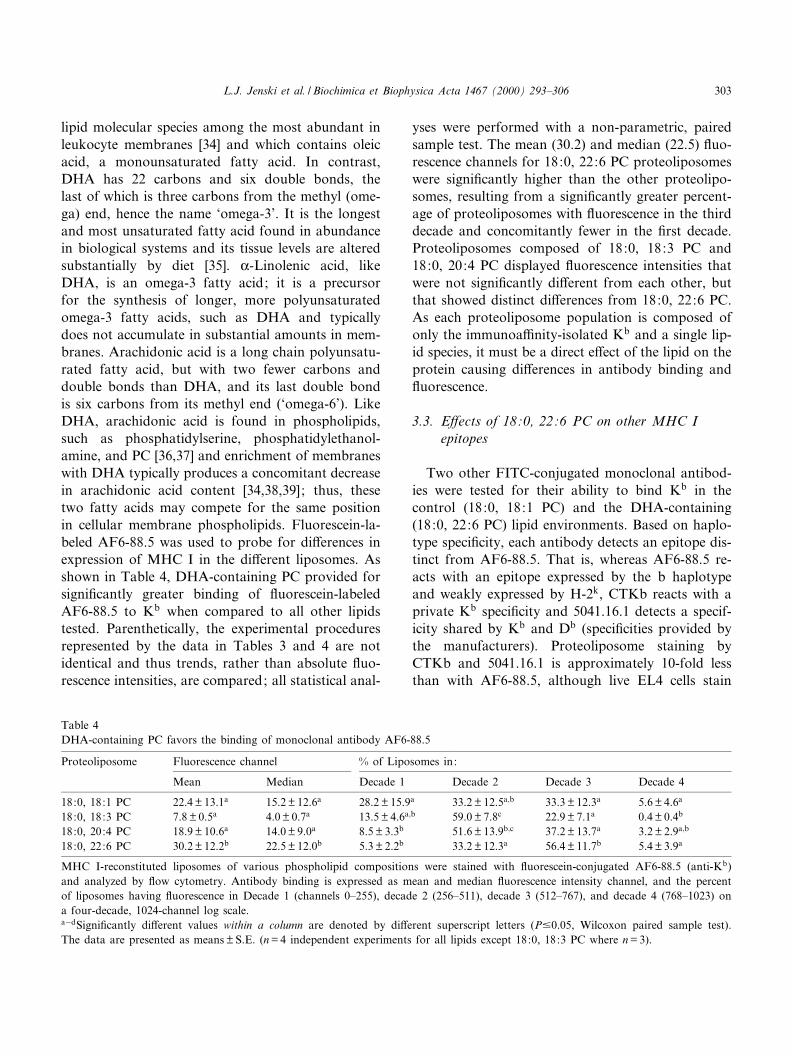
lipid molecular species among the most abundant inleukocyte membranes [34] and which contains oleicacid, a monounsaturated fatty acid. In contrast,DHA has 22 carbons and six double bonds, thelast of which is three carbons from the methyl (ome-ga) end, hence the name `omega-3'. It is the longestand most unsaturated fatty acid found in abundancein biological systems and its tissue levels are alteredsubstantially by diet [35]. K-Linolenic acid, likeDHA, is an omega-3 fatty acid; it is a precursorfor the synthesis of longer, more polyunsaturatedomega-3 fatty acids, such as DHA and typicallydoes not accumulate in substantial amounts in mem-branes. Arachidonic acid is a long chain polyunsatu-rated fatty acid, but with two fewer carbons anddouble bonds than DHA, and its last double bondis six carbons from its methyl end (`omega-6'). LikeDHA, arachidonic acid is found in phospholipids,such as phosphatidylserine, phosphatidylethanol-amine, and PC [36,37] and enrichment of membraneswith DHA typically produces a concomitant decreasein arachidonic acid content [34,38,39] ; thus, thesetwo fatty acids may compete for the same positionin cellular membrane phospholipids. Fluorescein-la-beled AF6-88.5 was used to probe for di¡erences inexpression of MHC I in the di¡erent liposomes. Asshown in Table 4, DHA-containing PC provided forsigni¢cantly greater binding of £uorescein-labeledAF6-88.5 to Kb when compared to all other lipidstested. Parenthetically, the experimental proceduresrepresented by the data in Tables 3 and 4 are notidentical and thus trends, rather than absolute £uo-rescence intensities, are compared; all statistical anal-
yses were performed with a non-parametric, pairedsample test. The mean (30.2) and median (22.5) £uo-rescence channels for 18:0, 22:6 PC proteoliposomeswere signi¢cantly higher than the other proteolipo-somes, resulting from a signi¢cantly greater percent-age of proteoliposomes with £uorescence in the thirddecade and concomitantly fewer in the ¢rst decade.Proteoliposomes composed of 18:0, 18:3 PC and18:0, 20:4 PC displayed £uorescence intensities thatwere not signi¢cantly di¡erent from each other, butthat showed distinct di¡erences from 18:0, 22:6 PC.As each proteoliposome population is composed ofonly the immunoa¤nity-isolated Kb and a single lip-id species, it must be a direct e¡ect of the lipid on theprotein causing di¡erences in antibody binding and£uorescence.
3.3. E¡ects of 18:0, 22:6 PC on other MHC Iepitopes
Two other FITC-conjugated monoclonal antibod-ies were tested for their ability to bind Kb in thecontrol (18:0, 18:1 PC) and the DHA-containing(18:0, 22:6 PC) lipid environments. Based on haplo-type speci¢city, each antibody detects an epitope dis-tinct from AF6-88.5. That is, whereas AF6-88.5 re-acts with an epitope expressed by the b haplotypeand weakly expressed by H-2k, CTKb reacts with aprivate Kb speci¢city and 5041.16.1 detects a specif-icity shared by Kb and Db (speci¢cities provided bythe manufacturers). Proteoliposome staining byCTKb and 5041.16.1 is approximately 10-fold lessthan with AF6-88.5, although live EL4 cells stain
Table 4DHA-containing PC favors the binding of monoclonal antibody AF6-88.5
Proteoliposome Fluorescence channel % of Liposomes in:
Mean Median Decade 1 Decade 2 Decade 3 Decade 4
18:0, 18:1 PC 22.4 þ 13.1a 15.2 þ 12.6a 28.2 þ 15.9a 33.2 þ 12.5a;b 33.3 þ 12.3a 5.6 þ 4.6a
18:0, 18:3 PC 7.8 þ 0.5a 4.0 þ 0.7a 13.5 þ 4.6a;b 59.0 þ 7.8c 22.9 þ 7.1a 0.4 þ 0.4b
18:0, 20:4 PC 18.9 þ 10.6a 14.0 þ 9.0a 8.5 þ 3.3b 51.6 þ 13.9b;c 37.2 þ 13.7a 3.2 þ 2.9a;b
18:0, 22:6 PC 30.2 þ 12.2b 22.5 þ 12.0b 5.3 þ 2.2b 33.2 þ 12.3a 56.4 þ 11.7b 5.4 þ 3.9a
MHC I-reconstituted liposomes of various phospholipid compositions were stained with £uorescein-conjugated AF6-88.5 (anti-Kb)and analyzed by £ow cytometry. Antibody binding is expressed as mean and median £uorescence intensity channel, and the percentof liposomes having £uorescence in Decade 1 (channels 0^255), decade 2 (256^511), decade 3 (512^767), and decade 4 (768^1023) ona four-decade, 1024-channel log scale.aÿdSigni¢cantly di¡erent values within a column are denoted by di¡erent superscript letters (P90.05, Wilcoxon paired sample test).The data are presented as means þ S.E. (n = 4 independent experiments for all lipids except 18:0, 18:3 PC where n = 3).
BBAMEM 77889 9-8-00
L.J. Jenski et al. / Biochimica et Biophysica Acta 1467 (2000) 293^306 303
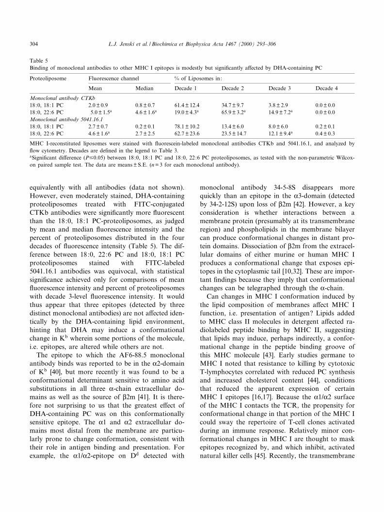
equivalently with all antibodies (data not shown).However, even moderately stained, DHA-containingproteoliposomes treated with FITC-conjugatedCTKb antibodies were signi¢cantly more £uorescentthan the 18:0, 18:1 PC-proteoliposomes, as judgedby mean and median £uorescence intensity and thepercent of proteoliposomes distributed in the fourdecades of £uorescence intensity (Table 5). The dif-ference between 18:0, 22:6 PC and 18:0, 18:1 PCproteoliposomes stained with FITC-labeled5041.16.1 antibodies was equivocal, with statisticalsigni¢cance achieved only for comparisons of mean£uorescence intensity and percent of proteoliposomeswith decade 3-level £uorescence intensity. It wouldthus appear that three epitopes (detected by threedistinct monoclonal antibodies) are not a¡ected iden-tically by the DHA-containing lipid environment,hinting that DHA may induce a conformationalchange in Kb wherein some portions of the molecule,i.e. epitopes, are altered while others are not.
The epitope to which the AF6-88.5 monoclonalantibody binds was reported to be in the K2-domainof Kb [40], but more recently it was found to be aconformational determinant sensitive to amino acidsubstitutions in all three K-chain extracellular do-mains as well as the source of L2m [41]. It is there-fore not surprising to us that the greatest e¡ect ofDHA-containing PC was on this conformationallysensitive epitope. The K1 and K2 extracellular do-mains most distal from the membrane are particu-larly prone to change conformation, consistent withtheir role in antigen binding and presentation. Forexample, the K1/K2-epitope on Dd detected with
monoclonal antibody 34-5-8S disappears morequickly than an epitope in the K3-domain (detectedby 34-2-12S) upon loss of L2m [42]. However, a keyconsideration is whether interactions between amembrane protein (presumably at its transmembraneregion) and phospholipids in the membrane bilayercan produce conformational changes in distant pro-tein domains. Dissociation of L2m from the extracel-lular domains of either murine or human MHC Iproduces a conformational change that exposes epi-topes in the cytoplasmic tail [10,32]. These are impor-tant ¢ndings because they imply that conformationalchanges can be telegraphed through the K-chain.
Can changes in MHC I conformation induced bythe lipid composition of membranes a¡ect MHC Ifunction, i.e. presentation of antigen? Lipids addedto MHC class II molecules in detergent a¡ected ra-diolabeled peptide binding by MHC II, suggestingthat lipids may induce, perhaps indirectly, a confor-mational change in the peptide binding groove ofthis MHC molecule [43]. Early studies germane toMHC I noted that resistance to killing by cytotoxicT-lymphocytes correlated with reduced PC synthesisand increased cholesterol content [44], conditionsthat reduced the apparent expression of certainMHC I epitopes [16,17]. Because the K1/K2 surfaceof the MHC I contacts the TCR, the propensity forconformational change in that portion of the MHC Icould sway the repertoire of T-cell clones activatedduring an immune response. Relatively minor con-formational changes in MHC I are thought to maskepitopes recognized by, and which inhibit, activatednatural killer cells [45]. Recently, the transmembrane
Table 5Binding of monoclonal antibodies to other MHC I epitopes is modestly but signi¢cantly a¡ected by DHA-containing PC
Proteoliposome Fluorescence channel % of Liposomes in:
Mean Median Decade 1 Decade 2 Decade 3 Decade 4
Monoclonal antibody CTKb18:0, 18:1 PC 2.0 þ 0.9 0.8 þ 0.7 61.4 þ 12.4 34.7 þ 9.7 3.8 þ 2.9 0.0 þ 0.018:0, 22:6 PC 5.0 þ 1.5a 4.6 þ 1.6a 19.0 þ 4.3a 65.9 þ 3.2a 14.9 þ 7.2a 0.0 þ 0.0Monoclonal antibody 5041.16.118:0, 18:1 PC 2.7 þ 0.7 0.2 þ 0.1 78.1 þ 10.2 13.4 þ 6.0 8.0 þ 6.0 0.2 þ 0.118:0, 22:6 PC 4.6 þ 1.6a 2.7 þ 2.5 62.7 þ 23.6 23.5 þ 14.7 12.1 þ 9.4a 0.4 þ 0.3
MHC I-reconstituted liposomes were stained with £uorescein-labeled monoclonal antibodies CTKb and 5041.16.1, and analyzed by£ow cytometry. Decades are de¢ned in the legend to Table 3.aSigni¢cant di¡erence (P90.05) between 18:0, 18:1 PC and 18:0, 22:6 PC proteoliposomes, as tested with the non-parametric Wilcox-on paired sample test. The data are means þ S.E. (n = 3 for each monoclonal antibody).
BBAMEM 77889 9-8-00
L.J. Jenski et al. / Biochimica et Biophysica Acta 1467 (2000) 293^306304
region of human MHC I was found to be responsiblefor inhibiting certain natural killer clones through anundetermined mechanism likely to involve control ofprotein conformation rather than MHC I oligomeri-zation, association with other proteins, or cellularlocalization [46]. We therefore consider it an essentialnext step to test the T-cell activating ability of MHCI proteoliposomes of various lipid compositions. Ad-ditionally, we are currently undertaking additionalassays of conformational change of MHC I reconsti-tuted into liposomes. By understanding the directe¡ect of DHA-containing phospholipids on MHC Iproperties, such as conformation and antigen presen-tation, we hope not only to better predict the e¡ectsof DHA in vivo (e.g. when provided in the diet), butalso to devise novel immunotherapies that utilizeDHA's abilities to in£uence antigen presentation.
Acknowledgements
This work was supported by Grant RO1CA57212from the National Institutes of Health and fundsfrom the Phi Beta Psi Sorority.
References
[1] D.N. Garboczi, W.E. Biddison, Shapes of MHC restriction,Immunity 10 (1999) 1^7.
[2] E.M. Rohren, D.J. McCormick, L.R. Pease, Peptide-inducedconformational changes in class I molecules. Direct detectionby £ow cytometry, J. Immunol. 152 (1994) 5337^5343.
[3] H. Celia, E. Wilson-Kubalek, R.A. Milligan, L. Teyton,Structure and function of a membrane-bound murineMHC class I molecule, Proc. Natl. Acad. Sci. USA 96(1999) 5634^5639.
[4] M. Edidin, S.C. Kuo, M.P. Sheetz, Lateral movements ofmembrane glycoproteins restricted by dynamic cytoplasmicbarriers, Science 254 (1991) 1379^1382.
[5] S. Damjanovich, G. Vereb, A. Schaper, A. Jenei, J. Matko,J.P.P. Starink, G.Q. Fox, D.J. Arndt-Jovin, T.M. Jovin,Structural hierarchy in the clustering of HLA class I mole-cules in the plasma membrane of human lymphoblastoidcells, Proc. Natl. Acad. Sci. USA 92 (1995) 1122^1126.
[6] J. Hwang, L.A. Gheber, L. Margolis, M. Edidin, Domainsin cell plasma membranes investigated by near-¢eld scanningoptical microscopy, Biophys. J. 74 (1998) 2184^2190.
[7] J. Matko, Y. Bushkin, T. Wei, M. Edidin, Clustering of classI HLA molecules on the surfaces of activated and trans-formed human cells, J. Immunol. 152 (1994) 3353^3360.
[8] G.G. Capps, B.E. Robinson, K.D. Lewis, M.C. Zuniga, Invivo dimeric association of class I MHC heavy chains,J. Immunol. 151 (1993) 159^169.
[9] A. Chakrabarti, J. Matko, N.A. Rahman, B.G. Barisas, M.Edidin, Self-association of class I major histocompatibilitycomplex molecules in liposome and cell surface membranes,Biochemistry 31 (1992) 7182^7189.
[10] A.M. Little, E. Nossner, P. Parham, Dissociation of L2-mfrom HLA class I heavy chains correlates with acquisition ofepitopes in the cytoplasmic tail, J. Immunol. 154 (1995)5205^5215.
[11] N.J. Gibson, M.F. Brown, Lipid headgroup and acyl chaincomposition modulate the MI^MII equilibrium of rhodopsinin recombinant membranes, Biochemistry 32 (1993) 2438^2454.
[12] M.F. Brown, Modulation of rhodopsin function by proper-ties of the membrane bilayer, Chem. Phys. Lipids 73 (1994)159^180.
[13] G.W. Hunter, D.J. Bigelow, T.C. Squier, Lysophosphatidyl-choline modulates catalytically important motions of the Ca-ATPase phosphorylation domain, Biochemistry 38 (1999)4604^4612.
[14] G. Vallette, R. Vranckx, M.E. Martin, C. Benassayag, E.A.Nunez, Conformational changes in rodent and human al-pha-fetoprotein: in£uence of fatty acids, Biochim. Biophys.Acta 997 (1989) 302^312.
[15] L. Bene, J. Szollosi, M. Balazs, L. Matyus, R. Gaspar, M.Ameloot, R.E. Dale, S. Damjanovich, Major histocompati-bility complex class I protein conformation altered by trans-membrane potential changes, Cytometry 27 (1997) 353^357.
[16] C.P. Muller, D.A. Stephany, M. Shinitzky, J.R. Wunderlich,Changes in cell-surface expression of MHC and Thy1.2 de-terminants following treatment with lipid modulating agents,J. Immunol. 131 (1983) 1356^1362.
[17] A. Bodnar, A. Jenei, L. Bene, S. Damjanovich, J. Matko,Modi¢cation of membrane cholesterol level a¡ects expres-sion and clustering of class I HLA molecules at the surfaceof JY human lymphoblasts, Immunol. Lett. 54 (1996) 221^226.
[18] A.W. Pascale, W.D. Ehringer, W. Stillwell, L.K. Sturdevant,L.J. Jenski, Omega-3 fatty acid modi¢cation of membranestructure and function. II. Alteration by docosahexaenoicacid of tumor cell sensitivity to immune cytolysis, Nutr.Cancer 19 (1993) 147^158.
[19] W. Ehringer, D. Belcher, S. Wassall, W. Stillwell, A compar-ison of the e¡ects of linolenic (1863) and docosahexaenoic(22:663) acids on phospholipid bilayers, Chem. Phys. Lipids54 (3) (1990) 79^88.
[20] W. Stillwell, W. Ehringer, L.J. Jenski, Docosahexaenoic acidincreases permeability of lipid vesicles and tumor cells, Lip-ids 28 (1993) 103^108.
[21] A.C. Dumaual, L.J. Jenski, W. Stillwell, Liquid crystalline/gel state phase separation in docosahexaenoic acid-contain-ing bilayers and monolayers, Biochim. Biophys. Acta 1463(2000) 395^406.
[22] U.K. Laemmli, Cleavage of structural proteins during the
BBAMEM 77889 9-8-00
L.J. Jenski et al. / Biochimica et Biophysica Acta 1467 (2000) 293^306 305
assembly of the head of bacteriophage T4, Nature 227 (1970)680^685.
[23] J.L. Rigaud, B. Pitard, D. Levy, Reconstitution of mem-brane protein insertion into liposomes: application to energytransducing membrane proteins, Biochim. Biophys. Acta1234 (1995) 223^246.
[24] J.D. Cardoza, A.M. Kleinfeld, K.C. Stallcup, M.F. Mescher,Hairpin con¢guration of H-2Kk in liposomes formed by de-tergent dialysis, Biochemistry 23 (1984) 4401^4409.
[25] K.C. Stallcup, T.A. Springer, M.F. Mescher, Characteriza-tion of an anti-H-2 monoclonal antibody and its use in large-scale antigen puri¢cation, J. Immunol. 127 (1981) 923^930.
[26] T.A. Springer, Cell-surface di¡erentiation in the mouse.Characterization of `jumping' and `lineage' antigens usingxenogeneic rat monoclonal antibodies, in: R.H. Kennett,T.J. McKearn, K.B. Bechtol (Eds.), Monoclonal Antibodies,Plenum Press, New York, 1980, pp. 185^217.
[27] K. Ozato, T.H. Hansen, D.H. Sachs, Monoclonal antibodiesto mouse MHC antigens. II. Antibodies to the H-2Ld anti-gen, the products of a third polymorphic locus of the mousemajor histocompatibility complex, J. Immunol. 125 (1980)2473^2477.
[28] K. Ozato, D.H. Sachs, Monoclonal antibodies to mouseMHC antigens. III. Hybridoma antibodies reacting to anti-gens of the H-2b haplotype reveal genetic control of isotypeexpression, J. Immunol. 126 (1981) 317^321.
[29] H. Allen, D. Wraith, P. Pala, B. Askonas, R.A. Flavell,Domain interactions of H-2 class I antigens alter cytotoxicT-cell recognition sites, Nature 309 (1984) 279^281.
[30] A.N. Glazer, Light guides. Directional energy transfer in aphotosynthetic antenna, J. Biol. Chem. 264 (1989) 1^4.
[31] J.A. Killian, Hydrophobic mismatch between proteins andlipids in membranes, Biochim. Biophys. Acta 1376 (1998)401^416.
[32] M.H. Smith, B.H. Barber, The conformational £exibility ofclass I H-2 molecules as revealed by anti-peptide antibodiesspeci¢c for intracytoplasmic determinants: di¡erential reac-tivity of L2-microglobulin `bound' and `free' H-2Kb heavychains, Mol. Immunol. 27 (1990) 169^180.
[33] M.J. Shields, L.E. Mo¡at, R.K. Ribaudo, Functional com-parison of bovine, murine, and human beta2-microglobulin:interactions with murine MHC I molecules, Mol. Immunol.35 (1998) 919^928.
[34] E.E. Williams, B.D. May, W. Stillwell, L.J. Jenski, Docosa-hexaenoic acid (DHA) alters the phospholipid molecularspecies composition of membranous vesicles exfoliatedfrom the surface of a murine leukemia cell line, Biochim.Biophys. Acta 1418 (1999) 185^196.
[35] N.J. Salem, H.-Y. Kim, J.A. Yergey, Docosahexaenoic acid:membrane function and metabolism, in: A.P. Simopoulos,R.R. Kifer, R.E. Martin (Eds.), Health E¡ects of Polyun-saturated Fatty Acids in Seafoods, Academic Press, NewYork, 1986, pp. 319^351.
[36] M. Goppelt-Strube, K. Resch, Polyunsaturated fatty acidsare enriched in the plasma membranes of mitogen-stimulatedT-lymphocytes, Biochim. Biophys. Acta 904 (1987) 22^28.
[37] M. Zerouga, W. Stillwell, J. Stone, A. Powner, L.J. Jenski,Phospholipid class as a determinant in docosahexaenoicacid's e¡ect on tumor cell viability, Anticancer Res. 16(1996) 2863^2868.
[38] C. Feng, D.H. Keisler, K.L. Fritsche, Dietary omega-3 poly-unsaturated fatty acids reduce IFN-Q receptor expression inmice, J. Interferon Cytokine Res. 19 (1999) 41^48.
[39] T. Sasaki, Y. Kanke, K. Kudoh, Y. Misawa, J. Shimizu, T.Takita, E¡ects of dietary docosahexaenoic acid on surfacemolecules involved in T cell proliferation, Biochim. Biophys.Acta 1436 (1999) 519^530.
[40] W. Chen, J. Fecondo, J. McCluskey, The structural in£uenceof individual residues located within peptide antigen dependsupon their sequence context, Mol. Immunol. 31 (1994) 1069^1075.
[41] S.T. Kuhns, L.R. Pease, A region of conformational varia-bility outside the peptide-binding site of a class I MHC mol-ecule, J. Immunol. 161 (1998) 6745^6750.
[42] G.R. Otten, E. Biko¡, R.K. Ribaudo, S. Kozlowski, D.H.Margulies, R.N. Germain, Peptide and L2-microglobulin reg-ulation of cell surface MHC class I conformation and ex-pression, J. Immunol. 148 (1992) 3723^3732.
[43] R.W. Roof, I.F. Luescher, E.R. Unanue, Phospholipids en-hance the binding of peptides to class II major histocompat-ibility molecules, Proc. Natl. Acad. Sci. USA 87 (1990)1735^1739.
[44] S.I. Schlager, S.H. Ohanian, Role of membrane lipids in theimmunological killing of tumor cells : I. Target cell lipids,Lipids 18 (1983) 475^482.
[45] M.H. Claesson, M.H. Nisse, Binding of human L2-micro-globulin to murine EL4 thymoma cells upregulates MHCclass I heavy-chain epitopes, inhibits IL-2 secretion and in-duces resistance to killing by natural killer cells, Immunol.Lett. 39 (1994) 195^202.
[46] D.M. Davis, O. Mandelboim, I. Luque, E. baba, J. Boyson,J.L. Strominger, The transmembrane sequence of humanhistocompatibility leukocyte antigen (HLA)-C as a determi-nant in inhibition of a subset of natural killer cells, J. Exp.Med. 189 (1999) 1265^1274.
BBAMEM 77889 9-8-00
L.J. Jenski et al. / Biochimica et Biophysica Acta 1467 (2000) 293^306306