Embed Size (px)
Citation preview

Doctoral Thesis
Molecular Physiological Study on the Adaptive Mechanisms
to Salinity Stress in Egyptian Rice Cultivars
Ahmad Mohammad Mohammad Mekawy
Graduate School of Biosphere Science
Hiroshima University
March 2016

Doctoral Thesis
Molecular Physiological Study on the Adaptive Mechanisms
to Salinity Stress in Egyptian Rice Cultivars
Ahmad Mohammad Mohammad Mekawy
Department of Environmental Dynamics and Management
Graduate School of Biosphere Science
Hiroshima University
March 2016

Abbreviations
DW Dry weight
RWC Relative water content
ELR Electrolyte leakage ratio
EC Electrical conductivity
HKT/HAK High affinity K transporter
KORC Potassium outwardly rectifying channel
KIRC Potassium inward rectifying channel
NHX Na+/H+ antiporter
SOS Salt overly sensitive
NSCCs Non selective cation channels
CNGCs Cyclic nucleotide gated channels
LCT Low affinity cation transporter
PMP3 Plasma membrane protein 3
Lti-6a/b Low temperature induced 6a/b
PCR Polymerase chain reaction
qRT-PCR Quantitative real-time polymerase chain reaction
cDNA Complementary deoxyribonucleic acid
CT Threshold cycle
ΔCT Change in threshold cycle
MTs Metallothioneins
ROS Reactive oxygen species

IPTG Isopropyl β-D-1-thiogalactopyranoside
OD Optical density
NBT Nitro-blue tetrazolium
O2− Oxygen superoxide
Contents
Page
Chapter 1 General introduction …………………………………………… 1
1.1 Soil salinity ………………………………………………….. 2
1.2 Salt stress, Na+ toxicity and its effects on plant growth ……… 2
1.3 Mechanisms of Na+-uptake in plants ………………………… 4
1.4 Transporters involved in Na+-uptake ………………………… 5
1.5 Salinity tolerance mechanisms in plants …………………….. 6
1.6 Plant biotechnology for developing salinity tolerant plants …. 7
1.7 Study rationale ……………………………………………….. 8
1.8 Study objectives ……………………………………………… 11
Chapter 2 Growth, physiological adaptation and gene expression
analysis of two Egyptian rice cultivars under salt stress ……..
12
2.1 Introduction…………………………………………………...
2.2 Materials and methods………………………………………..
2.3 Results………………………………………………………...
2.4 Discussion…………………………………………………….
2.5 Conclusion………………………………………………….....
13
15
19
36
41
Chapter 3 Identification of a type 3 metallothionein-like gene (OsMT-3a)
from rice through functional screening analysis in Escherichia
coli, confers tolerance against salinity and heavy-metal
stresses……………………………………………………………
42

3.1 Introduction…………………………………………………...
3.2 Materials and methods………………………………………..
3.3 Results………………………………………………………..
3.4 Discussion…………………………………………………….
3.5 Conclusion………………………………………………….....
43
45
52
69
74
Chapter 4 General discussion………………………………………………. 76
Summary ………………………………………………………………………………..
References ………………………………………………………………………………
Acknowledgments………………………………………………………………………
82
86
98

1
Chapter 1
General introduction

2
1.1 Soil salinity
Soil salinization is one of the factors causing serious soil degradations, which can arise from
natural causes and anthropogenic activity, such as irrigation in arid and semi-arid regions. Saline
soils contain a variety of salts, including Na2SO4, NaCl, MgSO4, CaSO4, MgCl2, KCl and Na2CO3,
but are normally dominated by NaCl, which causes most of the salt problems for higher plants in
nature (Flowers et al., 1977). Saline soils can be generally found in arid regions, estuaries, and
coastal fringes, which are dominated by Na+ ions with electrical conductivity (EC) of more than 4
dS/m that corresponds to approximately 40 mM NaCl (USDA-ARS 2008; Munns and Tester
2008). It has been estimated that more than 800 million hectares of land throughout the world are
salt affected, accounting for more than 6% of the world’s total land area (FAO, 2008).
1.2 Salt stress, Na+ toxicity, and its effects on plant growth
Soil salinity is one of the major constrains affecting plant growth and results in reduction of
crop yield throughout the world (Flowers, 2004; Cuartero et al., 2006). It is estimated that if the
current scenario of salinity stress would persist, up to 50 % of present cultivated land for
agriculture could be lost by 2050 (Wang et al., 2003). Generally, salt stress causes both osmotic
stress and ionic stress. Osmotic stress is triggered by an excess accumulation of salts in the soil,
and ionic stress is caused by the over-accumulation of salts in the cells. These stresses can
individually or simultaneously affect the physiological status of plants (Lefèvre et al., 2001; Ueda
et al., 2003). Na+ is very harmful in cells for most plants when it is present in the cytosol at a
concentration higher than the adequate level (1-10 mM) (Munns and Tester, 2008). Beyond this
range, Na+ interferes with many cellular functions. K+ is one of the essential and most abundant
monovalent cations in cells, and needs to be maintained within 100-200 mM range in cytosol for

3
efficient metabolic functioning (Cuin et al., 2003). As a co-factor in cytosol, K+ activates more
than 50 enzymes, which are very susceptible to high cytosolic Na+ and high Na+/K+ ratios (Munns
et al., 2006). Therefore, apart from low cytosolic Na+, maintenance of a low cytosolic Na+/K+ ratio
is also critical for the function of cells (Zhu et al., 1998). Under salt stress conditions, Na+ often
competitively inhibits the uptake of K+ leading to an imbalance in Na+/K+ ratios within plant cells
since Na+ and K+ are physico-chemically similar monovalent cations, thus competing for the
binding sites of many enzymes (Tester and Davenport, 2003). High uptake of Na+ and leakage of
K+ result in an imbalance in the Na+/K+ ratio in the cytosol, which, in turn, leads to many
imbalances in the cellular enzymatic reactions. An unfortunate consequence of salinity stress in
plants is the excessive generation of reactive oxygen species (ROS) such as superoxide, hydrogen
peroxide and hydroxyl radical (Foyer et al., 1994; Mittler, 2002). The excess production of ROS
during salinity stress results from impaired electron transport processes in chloroplast and
mitochondria as well as from pathways such as photorespiration. This is supported by reports,
which demonstrate that the oxygenase activity of Rubisco (which carries out the first step of the
photorespiratory pathway) gets significantly enhanced under conditions of salinity stress
(Sivakumar et al., 2000). Under normal growth conditions, the production of ROS in the cell is as
low as 240 µM s−1 O2• − and the steady state level of H2O2 in chloroplasts is 0.5 µM (Mittler, 2002;
Polle, 2001). However, under conditions of abiotic stress such as salinity, the disrupted cellular
homeostasis leads to the production of ROS like O2• − to a level as high as 720 µM s−1 and H2O2
to a level as high as 15 µM (Mittler, 2002; Polle, 2001). H2O2 at a concentration of 10 µM in
chloroplasts has been reported to cause a net reduction in photosynthesis in plants by over 50%
(Kaiser, 1979). The main toxicity of superoxide and hydrogen peroxide has been attributed to their
ability to initiate cascade reactions that result in the production of hydroxyl radicals and other

4
destructive species such as lipid peroxides (Noctor and Foyer, 1998) Hydroxyl radicals are very
reactive and can damage vital cellular macromolecules, e.g., via denaturation of proteins, mutation
of DNA and peroxidation of lipids.
1.3 Mechanisms of Na+-uptake in plants
Under salt stress, a plant root is the first organ to combat high Na+ in the soil solution. The root
tip in particular seems to be the major nutrient uptake region rather than the elongated mature
segments of the root (Golldack et al., 2003). This nutrient uptake occurs primarily in the plasma
membrane of root epidermal cells including root hairs. The uptake of Na+ proceeds through the
same path like any other mineral nutrient taken up by cells through the plasma membrane of either
the root epidermal cells or cortical cells. Structurally the plant plasma membrane is a lipid bilayer,
which surrounds cell cytoplasm and, in principle, is semi-permeable to solutes. There are,
however, many transport proteins, specific for one or a group of solutes, spanning the lipid bilayer
of the plasma membrane and facilitate the movement of solutes in and out of the cytosol. In plant
cells, the plasma membrane potential is negative inside (-120 to - 200 mV), but Na+ is a positive
ion (Munns and Tester, 2008). Therefore, Na+ mainly enters the cytosol through the passive way.
When it enters in epidermal cells or cortical cells, Na+ may follow a symplastic pathway or
apoplastic pathway before it encounters the endodermis layer. Since the endodermis with the
Casparian strip is an effective barrier for apoplastic Na+ movement (White, 2001; Karahara et al.,
2004), Na+ enters it symplastically. Thus, membrane transport at the level of root epidermal and
cortex cells is a critical point for the uptake or rejection of toxic ions like Na+ from the
environment. When Na+ is eventually taken up by roots, it is loaded into the xylem for long-
distance xylem transport following the transpiration stream to shoots. Finally, Na+ reaches to all
cells including metabolically active mesophyll cells in leaves. Here, the leaf mesophyll cells

5
conveys it to the phloem for another long-distance transport to other tissues (Sondergaard et al.,
2004). Thus, Na+ may also be recirculated in different cells and/or tissues.
1.4 Transporters involved in Na+-uptake
Different uptake mechanisms for Na+ into plant cells have been suggested. Nonselective cation
channels (NSCCs) are proposed to be the dominant pathways for Na+-influx in many plant species
(Davenport and Tester, 2000). However, the molecular identity of these NSCCs is still unknown.
High-affinity potassium transporters (HKTs) were also suggested to mediate a substantial Na+-
influx in some species (Horie et al., 2001; Golldack et al., 2002; Gárciadeblás et al., 2003). In rice,
nine HKT homologues (OsHKT1-9) have been identified of which only OsHKT5 is considered as
a non- functional gene, due to the existence of three stop codons in its mRNA (Gárciadeblás et al.,
2003). All of these functional genes encode proteins with distinct transport activities, which might
be expressed in various tissues and/or organs. Horie et al., (2001) suggested that OsHKT1 encodes
a Na+-transporter and OsHKT2 a Na+/K+-coupled transporter. Garciadeblás et al., (2003) also
showed that OsHKT1 (recently, OsHKT2;1) could be a high affinity Na+-transporter and OsHKT4
(recently, OsHKT1;1) a low affinity Na+-transporter. SKC1 (recently, OsHKT1;5) has recently
been shown to be a Na+-transporter, but contributing to the increased ability of salt tolerance by
maintaining shoot K+ homeostasis under salt stress (Ren et al., 2005). This is orthologous to the
function of AtHKT1 gene in Arabidopsis, which is a Na+-transporter, and interestingly, plays a
very important role in controlling the cytosolic Na+ detoxification (Sunarpi et al., 2005). AtHKT1
(recently, AtHKT1;1) functions in mediating tolerance to salt stress by unloading Na+ from xylem
vessels to xylem parenchyma cells and thus, protecting the plant leaves from salt stress (Sunarpi
et al., 2005). This transporter might also be responsible for unloading of Na+ from the phloem
(Berthomieu et al., 2003). Therefore, it is very likely that the HKT gene family in rice has an

6
important role for plant ion homeostasis even though some of the members evidently transport
Na+. Apart from these NSCCs and HKTs, other transport proteins that might be involved in
mediating Na+ influx under salinity stress are HAK/KT/KUP-type transporters, inward-rectifying
potassium channels, and low-affinity cation transporters of the LCT-1 type (Golldack et al., 2003).
1.5 Salinity tolerance mechanisms in plants
Many studies have revealed some of the mechanisms evolved by plants to adjust their
physiology and metabolisms to salt stress. First, Na+ entry to plants may be restricted by selective
ion uptake. Second, internalized Na+ can be stored in vacuoles. Third, Na+ in the cytosol may be
exported back to the growth medium or to apoplastic space. Fourth, the Na+ recirculation from
shoots into roots by the phloem sap, limiting Na+ accumulation in leaves, could be important for
salt tolerance. Finally, plants have also developed an antioxidant defense system to cope with
oxidative stress caused by high concentration salts in soil (Zhu, 2001; Gao et al., 2003).
Under saline conditions the protection of Na+-sensitive metabolic mechanisms in cell cytosol
partly depends on the ability to keep cytosolic Na+ levels low (Carden et al., 2003). For plant cells,
the most important way of keeping cytosolic Na+ concentration at a low level is to minimize Na+-
influx into the cytosol. Na+ entry into plant cells may be restricted either by down-regulating or
inactivating the Na+-influx channels and transporters in cells. Once Na+ enters the cytosol at a
toxic level, plant cells can deal with the internal Na+ by sequestering it either into the apoplast or
into the vacuole. The SOS pathway, has been extensively studied as it plays an important role for
maintaining Na+ homeostasis in the cytosol. The plasma membrane Na+/H+ antiporter, SOS1, is
the primary transport system responsible for cellular Na+-efflux (Zhu, 2002; Zhu 2003).
Overexpression of SOS1 increases the salt tolerance in Arabidopsis (Zhu, 2002; Shi et al., 2003).

7
In this species it controls Na+ loading into the xylem of the root and thus, restricts the accumulation
of Na+ in the shoot (Shi et al., 2002).
Na+ compartmentalization into vacuoles is an efficient strategy for plant cells to cope with
salinity stress (Fukuda et al., 1998, 2004; Apse et al., 1999; Blumwald, 2000; Tester and
Davenport, 2003). Once compartmentalized into the vacuole, Na+ is no more toxic for cells
(Subbarao et al., 2003) and has an advantage for growth via osmotic adjustment (Zhu, 2003;
Rodriguez-Navarro and Rubio, 2006). The tonoplast Na+/H+ antiporter, NHX1, has been
established to control the vacuolar sequestration of excess Na+ and/or K+ (Fukuda et al., 2004).
Oxidative stress is caused by accumulation of ROS that damage membrane lipids, proteins,
and nucleic acids. The protection of plants against salt-induced ROS is achieved by means of
different strategies including, in particular, partial suppression of ROS production, and scavenging
of ROS already produced. Scavenging of ROS is achieved by a complex interplay between
different enzymatic and non-enzymatic molecules. These include compounds, such as ascorbate,
glutathione, polyamines, carotenoids and metallothioneins, as well as antioxidant enzymes such as
superoxide dismutase (SOD), catalase (CAT), glutathione reductase (GR), and ascorbate
peroxidase (APX) (Zhu, 2001).
1.6 Plant biotechnology for developing salinity tolerant plants
Conventional approaches to breeding crop plants with improved stress tolerance have thus far
met with limited success because of the difficulty of breeding tolerance associated traits from
diverse plant backgrounds. Hence, an increasing emphasis has been directed to molecular
strategies aimed at enhancing the intrinsic ability of plants to survive under stress conditions.
Current approaches proposed to date focus attention on identification of genes associated with
salinity, drought and other abiotic stress resistance, followed by genetic modification of the plants
8
expressing genes enabling them to withstand restrictive growth imposed by unfavorable
environmental conditions (Sreenivasulu et al., 2007).
Agricultural biotechnology can provide powerful solutions to the problem of maintaining
agricultural productivity in saline soils. Application of crop biotechnology can expedite the
development of new crop varieties that would be more tolerant to physical stresses such as saline
soils, and drought, in an environmentally friendly way (Arzani, 2008). However, because of the
complexity of salt tolerance, it is hard to assume that transferring a single gene encoding a single
specific stress protein to a transgenic plant could dramatically improve salt tolerance in plants
(Wang et al., 2003).
In recent years, many transgenic crop varieties have been developed through both over-
production of compatible solutes, and overexpression of a vacuolar Na+/H+ antiporter gene in
transgenic plants. For instance, transgenic crops including rice (Mohanty et al., 2002), wheat
(Abebe et al., 2003) and tobacco (Hong et al., 2000) have been generated by overexpressing
compatible osmolytes synthesizing genes in these plants. The resultant transgenic plants have been
reported to show better germination, seedling growth and seed production under high salt and
osmotic stresses. Likewise, transgenic plants have been generated by over expressing the Na+/H+
antiporter gene in Arabidopsis (Apse et al., 1999), tomato (Zhang and Blumwald, 2001), Brassica
napus (Zhang et al., 2001), maize (Yan et al., 2004), wheat (Xue et al., 2004) and tobacco (Wang
et al., 2004). These transgenic plants exhibited improved salt tolerance compared to their wild type
progenitors.
1.7 Study rationale
With higher population densities in developing countries, there is an urgent need to increase
productivity of cultivated land. This makes also understanding of environmental stress phenomena

9
and related tolerant mechanisms more important in countries such as Egypt. Soil salinity is one of
the major problems for agriculture in semi-arid regions. In Egypt, plants are subjected to extreme
climatic factors such as high temperatures and drought. Under these conditions, dissolved salts
may accumulate in soils because of the insufficient leaching of ions. An accumulation of salts in
upper soil layers may be also due to an unsuitable irrigation management. Challenges faced by
crop plants cultivated in the presence of excess salts are disturbance of osmotic regulation, ion
imbalance, and oxidative stress, which impair plant metabolism and growth. Rice is one of the
most important crops in Egypt and its production plays a significant role in the strategy to
overcome food shortage and improvement of self-sufficiency for local consumption and export. In
2005 season, the total rice production in Egypt reached 6.6 million tons with a national average of
10.0 tons/ha (Badawi, 2005).
An experiment has been conducted under normal and drought conditions to examine the
magnitude of yield response of diverse genotypes to drought stress and to identify traits that may
confer drought resistance including 18 Egyptian, six Italian and nine Chinese rice varieties (Abd
Allah et al., 2010). Among the Egyptian rice cultivars, Giza 178 and Giza 182 were proved to be
drought-tolerant cultivars, while Sakha 104 was shown to be drought-sensitive in terms of
agronomic traits. Kandil et al., (2012), investigated the response of ten Egyptian rice cultivars
(Giza 178, Giza 181, Giza 182, Sakha 102, Sakha 104, Sakha 105, Sakha 106, Egyptian Yasmine,
Egyptian Hybrid 1, Egyptian Hybrid 2) to germination under salinity stress conditions to confirm
growth performance and to examine a range of genetic variability for salinity tolerance among the
rice cultivars under seven salinity levels. The results showed that rice cultivars significantly varied
in means of final germination percentage, germination rate, germination index, vigor index, root
and shoot lengths, root and shoot fresh weights, root dry weight, relative dry weight and seedling

10
height reduction. Moreover, the highest germination percentage and the enhance growth
parameters under salinity stress were in Sakha 102, Sakha 106, Sakha 104 and Egyptian Yasmine
cultivars, and that these cultivars were more tolerant to salinity. However, the authors did not
estimate the Na+ and K+ contents in the studied cultivars. Another study showed the effect of salt
stress on phospholipid signaling responses in the leaves of the Egyptian rice cultivars Sakha 102
and Sakha 104 (Darwish et al., 2009). Under both long- and short-term exposure to salinity stress
in hydroponics, Sakha 102 appeared to be salinity-sensitive, while Sakha 104 was shown to be
salinity-tolerant. Moreover, the results showed that salt stress rapidly activates several lipid
responses in rice leaves but that these responses do not explain the difference in salt tolerance
between sensitive and tolerant cultivars. A study conducted by Shehata et al., (2009) showed the
development of salt tolerant rice lines through mutation breeding. That investigation dealt with the
development of early maturity, non-lodging and high yielding with salinity tolerance, employing
a mutation approach of gamma rays irradiation on traditional Egyptian rice cultivars; Sakha 101,
Sakha 102, Egyptian Yasmine and the line, AC 1453. Four mutant lines, Sakha 101-M30, Sakha
102-M20, AC- M50 and Egyptian Yasmine M30 were developed. These mutants were tested for
yield performance in the salt-affected soils with a pH 8.00 to 8.20 and EC = 6.00 to 6.50 dS m-1.
Mutant lines were highly promising and far better than their respective control in the areas affected
by salinity conditions and produced higher paddy rice yield on salt-affected soils than the
commercial cultivar, Giza 178. In addition maintaining many desirable traits, such as early
maturity, non-lodging, quality traits and blast resistance, comparing with their respective control.
From all the previously mentioned reports, the response of the Egyptian rice cultivars to
salinity stress has not been yet clearly established. Up to date, there are no reports or information
published regarding the regulatory mechanisms of salinity stress tolerance in those cultivars.

11
Therefore, the objective of this study was to characterize the physiological responses and
adaptation mechanisms to salinity stress in some Egyptian rice cultivars by comparing the
physiological parameters, Na+ accumulation patterns and expression profiles of the genes that
encode Na+ and K+ transport proteins. Moreover, further elucidation of salinity-tolerance
mechanisms in the salinity-tolerant cultivar through the isolation and characterization of salinity-
inducible genes which might function in other tolerance pathways under salinity stress conditions,
thus could be another adaptation strategy to abiotic stress in this cultivar.
1.8 Study objectives
1. Investigating the physiological responses and mode of adaptation to salinity stress in the
Egyptian rice cultivars, Sakha 102 and Egyptian Yasmine by comparing the growth criteria,
Na+ and K+ accumulation patterns and other physiological parameters under salinity stress
conditions in a hydroponic culture system.
2. Elucidation of differences in the mechanisms of salinity tolerance (Na+ and /or K+
accumulation patterns), between the salinity-tolerant and salinity-sensitive rice cultivars by
analyzing the expression profiles of the genes that encode Na+ and/or K+ transport proteins.
3. Further elucidation of the molecular mechanisms of salinity tolerance in the salinity-
tolerant rice cultivar, through the isolation and characterization of salinity-inducible genes
and examine their functional roles which could be involved in other adaptation processes
under salinity stress conditions.
12
Chapter 2
Growth, physiological adaptation and gene
expression analysis of two Egyptian rice
cultivars under salt stress

13
2.1 Introduction
Soil salinity is a major environmental problem that has deleterious effects on world agriculture,
especially in irrigated lands (Flowers, 1999). Salinity stress causes serious damage to many cellular
and physiological processes including photosynthesis, nutrient uptake, water absorption, root
growth, and cellular metabolism, which all lead to yield reduction (Pardo, 2010). Furthermore,
excess Na+ causes an imbalance in cellular ion homeostasis, resulting in ion toxicity (Mandhania
et al., 2006; Assaha et al., 2013). Cellular Na+ and K+ homeostasis plays a fundamental role in the
growth and development of higher plants. As one of the most important macronutrients in plants,
K+ is necessary for the maintenance of membrane potential and turgor pressure, activation of
enzymes, regulation of osmotic pressure, stomatal movement, and tropisms (Golldack et al., 2003).
In contrast to animal cells, Na+ is not an essential element for plant cells; therefore, high K+/Na+
ratios maintain osmotic balance in plant cells.
Genes from many functional classes including those encoding transcription factors, signal
transduction, cell wall components, and membrane transporters were found to be differentially
regulated in response to salt stress (Walia et al., 2007; Ueda et al., 2002, 2004, 2006).
The transmembrane movement of Na+ and K+ in plants is mediated by several types of
transporters and/or channels (Yao et al., 2010), and a number of transporters have been implicated
in leaf Na+ exclusion. These include members of the high-affinity K+ transporters (HKTs),
including Arabidopsis thaliana HKT (AtHKT1;1) and its ortholog in rice (OsHKT1;5), which
retrieves Na+ from the xylem to the surrounding parenchyma cells (Ren et al., 2005; Horie et al.,
2009). Plasma membrane protein 3 (PMP3) is a small hydrophobic peptide that plays a role in
shoot Na+ exclusion by preventing excess Na+ entry into the plant roots (Nylander et al., 2001). In
addition to Na+ exclusion, plants may avoid toxic Na+ accumulation in the cytosol by sequestering
14
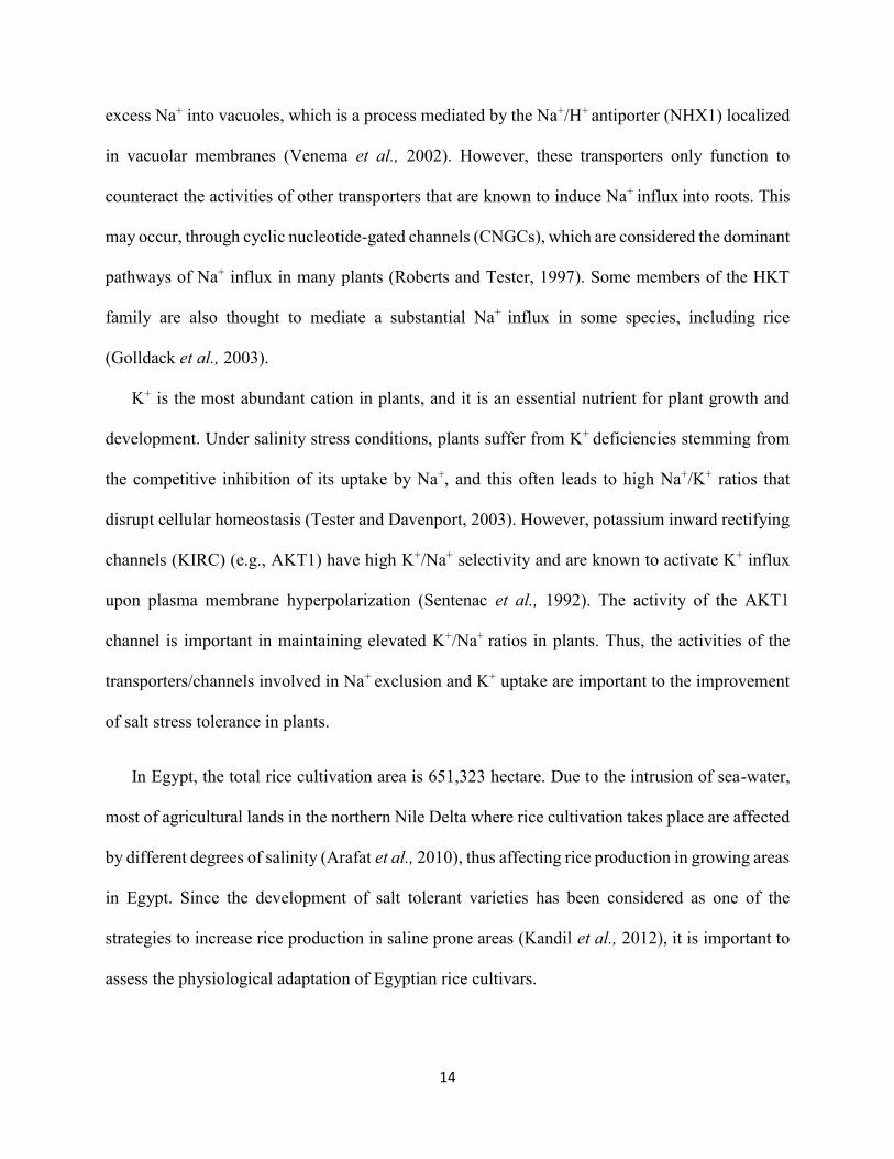
excess Na+ into vacuoles, which is a process mediated by the Na+/H+ antiporter (NHX1) localized
in vacuolar membranes (Venema et al., 2002). However, these transporters only function to
counteract the activities of other transporters that are known to induce Na+ influx into roots. This
may occur, through cyclic nucleotide-gated channels (CNGCs), which are considered the dominant
pathways of Na+ influx in many plants (Roberts and Tester, 1997). Some members of the HKT
family are also thought to mediate a substantial Na+ influx in some species, including rice
(Golldack et al., 2003).
K+ is the most abundant cation in plants, and it is an essential nutrient for plant growth and
development. Under salinity stress conditions, plants suffer from K+ deficiencies stemming from
the competitive inhibition of its uptake by Na+, and this often leads to high Na+/K+ ratios that
disrupt cellular homeostasis (Tester and Davenport, 2003). However, potassium inward rectifying
channels (KIRC) (e.g., AKT1) have high K+/Na+ selectivity and are known to activate K+ influx
upon plasma membrane hyperpolarization (Sentenac et al., 1992). The activity of the AKT1
channel is important in maintaining elevated K+/Na+ ratios in plants. Thus, the activities of the
transporters/channels involved in Na+ exclusion and K+ uptake are important to the improvement
of salt stress tolerance in plants.
In Egypt, the total rice cultivation area is 651,323 hectare. Due to the intrusion of sea-water,
most of agricultural lands in the northern Nile Delta where rice cultivation takes place are affected
by different degrees of salinity (Arafat et al., 2010), thus affecting rice production in growing areas
in Egypt. Since the development of salt tolerant varieties has been considered as one of the
strategies to increase rice production in saline prone areas (Kandil et al., 2012), it is important to
assess the physiological adaptation of Egyptian rice cultivars.

15
The traditional Egyptian rice cultivars; Sakha 102 and Egyptian Yasmine are important local
rice cultivars which have superiority over other local ones due to their exclusive quality; early
maturity, non-lodging and high yielding. Thus, as soil salinization is becoming increasingly
challenge for agriculture in Egypt, it is imperative to seek ways of improving these cultivars to
withstand salt stress. Whereas Sakha 102 has been established as a salt-sensitive cultivar (Darwish
et al., 2009), the response of Egyptian Yasmine has not been clearly established. Therefore, the
objective of this study was to investigate the physiological responses to salinity stress in the
cultivars Sakha 102 and Egyptian Yasmine and to elucidate differences in the mechanisms of
salinity tolerance between the two cultivars, by comparing the physiological parameters, Na+ and
K+ accumulation patterns and expression profiles of the genes that encode Na+ and K+ transport
proteins under salinity stress conditions.
2.2 Materials and Methods
2.2.1 Plant material, growth conditions, and salt treatment
The Egyptian rice cultivars Sakha 102 and Egyptian Yasmine used in this study were obtained
from the Field Crops Research Institute, Giza, Egypt. These genotypes were chosen from the
germplasm collections based on their reputation in terms of agronomic performance. The subset
of the germplasm samples included the subspecies O. sativa ssp. indica and O. sativa ssp. japonica.
Seeds were surface sterilized via immersion in a 5% NaClO solution for 30 min, and were then
thoroughly rinsed with distilled water. Seeds were subsequently soaked in tap water for 24 h at 28
ºC. After germination, the seeds were transferred to a nylon mesh floating on 20 L of tap water for
two days. Water was then replaced with half-strength Kimura B solution (0.18 mM (NH4)2SO4,
0.27 mM MgSO4.7H2O, 0.09 mM KNO3, 0.18 mM Ca(NO3)2
.4H2O, and 0.09 mM KH2PO4, 20
µM NaEDTA-Fe.3H2O, 6.7 µM MnCl2.4H2O, 9.4 µM H3BO3, 0.015 µM (NH4)6Mo7O24
.4H2O,

16
0.15 µM ZnSO4.7H2O, and 0.16 µM CuSO4
.5H2O). Twenty eight-day-old seedlings (4-5 leaf
stage) were transferred to either Kimura B nutrient solution (control) or nutrient solution
supplemented with 50 mM NaCl (salinity) for two weeks. The solutions were replaced every two
days and the pH was adjusted to 5.0-5.5 each day. Seedlings were grown in a growth chamber
under the following controlled environmental conditions: 70% relative humidity, 24 ± 2 ºC, and a
16 h photoperiod at a photosynthetic photon flux density of 250-350 μmol m-2 s-1.
2.2.2 Measurement of fresh and dry weight, relative water content, and electrolyte leakage
ratio
The fresh weight (FW) was measured following the separation of leaves, stems, and roots. For
dry weight (DW) determination, leaves, stems, and roots were dried at 70°C for three days prior
to being weighed. To determine the relative water content (RWC), four plants from each treatment
were randomly selected and the method described by Weatherly, (1950) was implemented. Briefly,
leaf samples were weighed to determine the FW. The leaf samples were then soaked in fresh
deionized water for 24 h under light, and were placed on tissue paper to remove excess water. The
samples were then weighed to determine the fully turgid weight (TW). Samples were next oven-
dried at 70ºC for three days, and the DW was obtained. The RWC was determined using the
following formula: RWC = (FW - DW)/(TW - DW) × 100.
To determine the electrolyte leakage ratio (ELR), the third leaf from the top of each plant was
dissected and soaked in a bottle containing 30 mL of deionized water, and the bottles were gently
shaken overnight. Conductivity of the solution was measured with an EC meter (EC1). The bottles
were then autoclaved and cooled, and the conductivity of the solution was again measured (EC2).
The ELR was calculated as the ratio of the conductivity before autoclaving to the conductivity
after autoclaving using the following formula: ELR = (EC1/EC2) × 100.

17
2.2.3 Determination of Na+ and K+ content
The Na+ and K+ content in leaves, sheaths, and roots was measured using a flame photometer
(ANA-135; Tokyo Photoelectric, Tokyo, Japan) according to the method of Kushizaki, (1968).
Dried samples were gently agitated in 1N HCl overnight, and the content of Na+ and K+ was
estimated from the Na+ and K+ standard curves.
2.2.4 Measurement of proline content
Free proline was determined as previously described by Bates et al., (1973) with some
modifications. Proline was extracted from about 200 mg fresh leaves in 4 mL of 3% sulfosalicylic
acid. Two mL of the extract was then mixed with 2 mL ninhydrin reagent, containing glacial acetic
acid and incubated at 100 °C for 40 min. The reaction mixture was quickly cooled in an ice water
bath. After cooling, 4 mL toluene was added for chromophore development. The absorbance of
the chromophore was measured at 520 nm.
2.2.5 Expression analysis of genes encoding Na+/K+ transport proteins
Total RNA was extracted from the leaves, sheaths, and roots of the control and stressed plants
using TRIzol reagent (Invitrogen, Carlsbad, CA). After digestion with DNaseI, total RNA (1 µg)
was reverse-transcribed to cDNA using a ReverTra Ace qPCR RT kit (Toyobo, Osaka, Japan).
Quantitative polymerase chain reaction (qPCR) was conducted as previously described (Ueda et
al., 2013), using a Thunderbird SYBR qPCR Mix (Toyobo) and an ABI Step One Plus system
(Applied Biosystems). RT-qPCR was performed using the following profile: an initial incubation
at 95ºC for 1 min, followed by 40 cycles with 15 sec at 95ºC and 60 sec at 60ºC. Relative
abundance of genes transcripts were calculated with the comparative 2-∆∆CT method (Livak and
Schmittgen, 2001). Expression levels were represented as fold changes relative to the respective

18
control. The relative expression level of the genes was normalized to the expression level of
OsUBQ-5 as an internal control (Jain et al., 2006). The sequences of the primers used were listed
in Table 1. The PCR products were verified via a melting curve analysis by changing the
temperature from 60ºC to 95ºC to monitor dissociation of double strand DNA and by running an
agarose gel electrophoresis. Amplification efficiencies (E) of the primers were determined using
the standard curve method and calculated according to the equation E (%) = (10 -1/slope - 1) × 100.
All primers showed E values of more than 90%.
2.2.6 Statistical analysis
The collected data were subjected to one-way analysis of variance (ANOVA) using the SPSS
statistics package, version 21 (IBM Inc., USA), and the means (n = 4) were separated using
Duncan’s multiple range test at p = 0.05.
19
2.3 Results
2.3.1 Effect of salt stress on biomass production, relative water content, and electrolyte leakage ratio
After 14 days of salt treatment (50 mM NaCl), the visual symptoms of salt toxicity on leaves
(whitish leaf tips, leaf rolling, dried or dead leaves) of both cultivars were photographed (Figure
2.1). The cultivar Sakha 102 appeared to be salinity-sensitive, being most of the leaves were rolled,
dried or died with retarded growth (Figure 2.1A), while Egyptian Yasmine showed to be more
tolerant to salinity stress and maintained healthy expanded green leaves with few whitish leaf tips
and vigor growth (Figure 2.1B). The dry weight (DW) of both cultivars was affected by salt stress
treatment (Figure 2.2). Under 50 mM NaCl stress conditions, the leaf DW of Sakha 102 was
drastically decreased by 64% in comparison with that of Egyptian Yasmine (35%). The sheath
DW was not significantly affected by salt stress in either cultivar. However, the root DW decreased
by 49% in Sakha 102, but it increased by 7% in Egyptian Yasmine under salt stress conditions
(Figure 2.2).
To estimate the amount of water lost under salinity conditions, the relative water content
(RWC) was measured using leaf tissues. The results indicated that Egyptian Yasmine exhibited a
greater potential to maintain tissue water than Sakha 102. Under 50 mM NaCl stress conditions,
the RWC of Sakha 102 plants decreased from 97% in control plants to 71% in experimental plants.
However, there was no significant difference in the RWC of Egyptian Yasmine between the
control (88%) and salinity-treated plants (86%) (Figure 2.3A).
The electrolyte leakage ratio (ELR) was measured to examine cell membrane stability under
salinity stress. The results showed that Egyptian Yasmine exhibited a lower electrolyte leakage
rate (27.5%) than Sakha 102 (51.9%) in response to salt stress (Figure 2.3B). These results
20
indicated that Egyptian Yasmine maintained a better physiological status than Sakha 102 under
salinity conditions.
2.3.2 Effect of salt stress on Na+ and K+ accumulation in different tissues
In both cultivars, salinity treatment led to increased Na+ content in all tissues examined (Figure
2.4). In the leaves, the increase in Na+ content of Sakha 102 was nearly two-times higher than that
of Egyptian Yasmine (Figure 2.4A). In the sheaths, there was no significant difference in Na+
content between the two cultivars (Figure 2.4B), but there was a slight increase in Na+ content in
the roots of Egyptian Yasmine (Figure 2.4C).
Salinity stress did not significantly affect the leaf K+ content of the two cultivars (Figure 2.5A);
however, there was a significant decrease in K+ content in the sheaths of both cultivars (Figure
2.5B). Moreover, there was a significant increase in K+ content in the roots of the two cultivars
under salt stress conditions (Figure 2.5C). Notably, under both control and salt stress conditions,
Egyptian Yasmine accumulated a higher concentration of K+ in the roots than Sakha 102 (Figure
2.5C). Consequently, a higher Na+/K+ ratio was observed in the leaves, sheaths, and roots of Sakha
102 compared to Egyptian Yasmine (Figure 2.6A, B, C).
2.3.3 Effect of salt stress on proline accumulation
In response to salinity stress, a significant increase of proline content was observed in the
leaves of Egyptian Yasmine (21%), while no significant change was observed in the Sakha 102
leaf proline content. Notably, under both control and salt stress conditions, Egyptian Yasmine
accumulated a higher amount of proline than Sakha 102 (Figure 2.7). The greater accumulation of
proline in Egyptian Yasmine might partially contribute to cellular osmotic adjustment under high
salinity conditions and indicating a better physiological adaptation to salinity stress.

21
2.3.4 Differential expression of genes encoding Na+/K+ transport proteins in response to
salt stress
To determine the mechanisms underlying differential Na+ and K+ accumulation in the salinity-
tolerant Egyptian Yasmine and the salinity-sensitive Sakha 102, expression profiles of the genes
encoding Na+ and K+ transport proteins were analyzed. Although the rice genome has divergent
transport systems for monovalent cations, functionally identified Na+ and K+ transport proteins
were chosen for gene expression analyses. The main site of Na+ toxicity is in the shoots, where
Na+ accumulates and disrupts metabolic processes. A Na+ transporter, OsHKT1;5 functions in the
root xylem parenchyma to retrieve Na+ from the xylem stream, thereby reducing Na+ accumulation
in the shoots (Ren et al., 2005). In the present study, quantitative RT-PCR analyses showed that
salt stress induced the expression of the OsHKT1;5 gene by 4.8-fold in the roots of Egyptian
Yasmine, but it was repressed by 0.3-fold in Sakha 102 (Figure 2.8A). This repression of
OsHKT1;5 in the roots may cause increased Na+ accumulation in the leaves of Sakha 102 when
under salt stress (Figure 2.4A). Also, OsHKT1;5 was repressed in the leaves and sheaths of both
cultivars.
To determine the physiological role of the OsHKT2;1 transporter in the rice cultivars under
salt stress conditions, expression analyses were performed for the OsHKT2;1 gene. We found that
there was repression of OsHKT2;1 expression in all plants parts, particularly in the roots (0.04-
fold), for the two studied cultivars. Expression of OsHKT2;1 was repressed in the sheaths of Sakha
102 (0.3-fold) and Egyptian Yasmine (0.4-fold). Furthermore, OsHKT2;1 was also repressed in
the leaves of Sakha 102 (0.8-fold) and Egyptian Yasmine (0.3-fold) (Figure 2.8B).
Plant PMP3 proteins, which are relatively small sized (approx. 56 aa), were found to be
involved in the prevention of excess Na+ entry in yeast and Arabidopsis (Nylander et al., 2001).

22
The rice genome has two homologous PMP3 genes, OsLti6a and OsLti6b. In response to salinity
stress, repression of OsLti6a expression was observed in the roots and sheaths of both cultivars
(less than 1.0-fold) and in the leaves of Egyptian Yasmine (0.5-fold), while in Sakha 102 leaves,
the gene expression did not change in response to salt stress (1.1-fold) (Figure 2.8C). Induced
expression of OsLti6b in the roots of Egyptian Yasmine (2.5-fold) was observed, while the
expression of the gene was unaltered in the Sakha 102 roots (1.1-fold) under salt stress (Figure
2.8D). However, salinity stress caused repression of OsLti6b expression in the sheaths and leaves
of both cultivars (less than 1.0-fold) (Figure 2.8D). These results indicated the role of rice PMP3
genes in restricting excess Na+ influx to the roots of Egyptian Yasmine and consequently lower
Na+ levels in its leaves (Figure 2.4A).
The vacuolar Na+, K+/H+ antiporter, OsNHX1, plays an important role in the
compartmentalization of the highly accumulated cytosolic Na+ and K+ into the vacuoles (Fukuda
et al., 2004). The expression of this OsNHX1 gene was regulated by the level of Na+ in plants
(Fukuda et al., 1999). In the present study, OsNHX1 was differentially expressed in the roots and
shoots of both Sakha 102 and Egyptian Yasmine under salt stress conditions (Figure 2.8E).
Whereas, salinity induced the expression of OsNHX1 highly in the leaves of Sakha 102 (5.5-fold),
the gene was repressed in Egyptian Yasmine leaves (0.5-fold). This difference in OsNHX1
induction might be due to higher Na+ levels in the leaves of Sakha 102. In the roots, OsNHX1
expression did not change in response to salt stress in Sakha 102 (1.0-fold), but it was repressed in
Egyptian Yasmine (0.7-fold). In the sheaths, OsNHX1 was repressed in both cultivars (0.2-fold).
The Na+/H+ antiporter salt overly sensitive1 (SOS1), localized in the plasma membrane, is
considered a general regulator of Na+ export from cytosol (Shi et al., 2002). Our results indicated
that there was a higher level of induced expression of OsSOS1 in the Sakha 102 roots (3.5-fold)

23
(Figure 2.8F), which might be responsible for the relatively low Na+ levels in its roots under salt
stress; however, OsSOS1 expression in Egyptian Yasmine roots was not induced (1.2-fold). The
repression of OsSOS1 expression was observed in the leaves (0.2-fold) and sheaths (0.4-fold) of
Egyptian Yasmine under salt stress conditions (Figure 2.8F), which suggested that OsSOS1-
mediated Na+ extrusion from the cytosol may not be active in Egyptian Yasmine.
Cyclic nucleotide gated channels, such as AtCNGC1, are potential candidates for nonselective
channels that contribute to the ion-conducting pathway, which allows toxic levels of Na+ to be
taken up by plants from saline soils (Maathuis and Sanders, 2001). Our results revealed differential
expression of the OsCNGC1 gene in the roots, sheaths, and leaves of both cultivars (Figure 2.8G).
Under salt stress, an induction of OsCNGC1 expression in the roots of Sakha 102 (3.2-fold) was
higher than that of Egyptian Yasmine (2.5-fold). In the sheaths, OsCNGC1 was repressed in Sakha
102 (0.4-fold), and not significantly affected in Egyptian Yasmine (1.4-fold). In the leaves, there
was induction of OsCNGC1 expression in Sakha 102 (1.8-fold), and unaltered expression in
Egyptian Yasmine (1.2-fold).
Salinity stress treatment induced the expression of OsAKT1 highly in the leaves of Egyptian
Yasmine (4.3-fold) compared to the expression in Sakha 102 leaves (1.7-fold), while in roots, the
expression of OsAKT1 was induced in Egyptian Yasmine (1.6-fold), but was repressed in Sakha
102 (0.8-fold). The expression of OsAKT1 was repressed by salt stress treatment in the sheaths of
both cultivars (0.3-fold) (Figure 2.9A). These results suggested that reduced growth and impaired
K+ homeostasis in salt-stressed seedlings of Sakha 102 might be a consequence of reduced K+
influx via OsAKT1 channels.
In the current experiment, under salt stress conditions, OsHAK7 expression was induced in the
roots in both cultivars, with no significant differences between the two cultivars (2.9-fold in Sakha

24
102 and 2.6-fold in Egyptian Yasmine). However, in the leaves, it was highly induced in Sakha
102 (15.7-fold) compared to Egyptian Yasmine (3.2-fold) (Figure 2.9B). In the sheaths, OsHAK7
expression was markedly induced in Egyptian Yasmine (30-fold), but was repressed in Sakha 102
(0.2-fold) (Figure 2.9B).

25
Table 1. Primers used for quantitative real-time RT-PCR. Genes Forward primer (5´-3´) Reverse primer (5´-3´)
OsHKT1;5 CCCATCAACTACAGCGTCCT AGCTGTACCCCGTGCTGA
OsLti6a CCTTCCAAGGTGATGGTGAA CCGTCCAAAGAACCAGAAAA
OsLti6b GCTCCAAACCGCTTCATCTA CAAGAATTGGAGCACTCAGGA
OsHKT2;1 TGCATTCATCACTGAGAGGAG GGTGCAGTTTCTGCAACCTC
OsNHX1 AATGATCACCAGCACCATCA AAGGCTCAGAGGTGACAGGA
OsSOS1 ATACTGAGTGGGGTTGTTATTGC AAAGGTAAATTTCAAAAGGTACATGG
OsAKT1 GAAACGAGCAATGCGTCAG CTTCTCACACAGCGCTTCC
OsHAK7 TGCTGTGACACTTGGTTTCC AAATAACAAGGCGAGCAGGA
OsCNGC1 TGCAATAGCAAAGCGATACTTG TTTTGGCTTTTGCAACCTCT
OsUBQ5 ACCACTTCGACCGCCACTACT ACGCCTAAGCCTGCTGGTT

26
Figure 2.1. Effect of salinity stress on the growth of the rice cultivars, Sakha 102 (A) and Egyptian
Yasmine (B) after 14 days treatment.
50 mM NaCl 0 mM NaCl
0 mM NaCl 50 mM NaCl
A
B

27
Figure 2.2. Plant dry weight (DW) of the rice cultivars Sakha 102 and Egyptian under control and
salt stress condition (50 mM NaCl) for 14 days. Data represent the means of four replicates ± SE.
The same letters indicate no significant differences (P ˂ 0.05).
a
bab ab
a
aa
a
ab
c
ab
0
0.2
0.4
0.6
0.8
1
1.2
1.4
1.6
0 mM 50 mM 0 mM 50 mM
Sakha 102 Egyptian Yasmine
Plan
t dry
wei
ght (
g)Leaf Sheath Root

28
Figure 2.3. (A) Relative water content and (B) electrolyte leakage ratio (ELR) of the rice cultivars
Sakha 102 and Egyptian Yasmine under control and salt stress condition (50 mM NaCl) for 14
days. Data represent the means of four replicates ± SE. The same letters indicate no significant
differences (P ˂ 0.05).
aa
ba
0
20
40
60
80
100
120
Sakha 102 Egyptian Yasmine
Rel
ativ
e w
ater
con
tent
(%)
0 mM 50 mMA
c
d
a
b
0
10
20
30
40
50
60
Sakha 102 Egyptian Yasmine
Ele
ctro
lyte
leak
age
ratio
(%)
0 mM 50 mMB

29
Figure 2.4. (A) Leaf Na+ content, (B) sheath Na+ content and (C) root Na+ content of the rice
cultivars Sakha 102 and Egyptian Yasmine under control and salt stress condition (50 mM NaCl)
for 14 days. Data represent the means of four replicates ± SE. The same letters indicate no
significant differences (P ˂ 0.05).
c c
a
b
0
5
10
15
20
25
30
35
40
Sakha 102 Egyptian Yasmine
Lea
f Na+
cont
ent (
mg/
g D
W)
0 mM 50 mMA
b b
a a
0
5
10
15
20
25
30
35
Sakha 102 Egyptian Yasmine
Shea
th N
a+co
nten
t (m
g/g
DW
)
0 mM 50 mMB
c c
ba
0
5
10
15
20
Sakha 102 Egyptian Yasmine
Roo
t Na+
cont
ent (
mg/
g D
W)
0 mM 50 mMC

30
Figure 2.5. (A) Leaf K+ content, (B) sheath K+ content and (C) root K+ content of the rice cultivars
Sakha 102 and Egyptian Yasmine under control and salt stress condition (50 mM NaCl) for 14
days. Data represent the means of four replicates ± SE. The same letters indicate no significant
differences (P ˂ 0.05).
a bab ab
0
5
10
15
20
25
Sakha 102 Egyptian Yasmine
Lea
f K+
cont
ent (
mg/
g D
W)
0 mM 50 mMA
a a
b b
0
5
10
15
20
25
30
Sakha 102 Egyptian Yasmine
Shea
th K
+ co
nten
t (m
g/g
DW
)
0 mM 50 mMB
c
bb
a
0
5
10
15
20
Sakha 102 Egyptian Yasmine
Roo
t K+
cont
ent (
mg/
g D
W)
0 mM 50 mMC

31
Figure 2.6. (A) Leaf Na+/K+ ratio, (B) sheath Na+/K+ ratio and (C) root Na+/K+ ratio of the rice
cultivars Sakha 102 and Egyptian Yasmine under control and salt stress condition (50 mM NaCl)
for 14 days. Data represent the means of four replicates ± SE. The same letters indicate no
significant differences (P ˂ 0.05).
c c
a
b
0
0.5
1
1.5
2
2.5
Sakha 102 Egyptian Yasmine
Lea
f Na+ /
K+
ratio
0 mM 50 mMA
c c
ab
0
0.5
1
1.5
2
2.5
Sakha 102 Egyptian Yasmine
Shea
th N
a+ /K
+ra
tio
0 mM 50 mMB
b
c
a
b
0
0.5
1
1.5
2
2.5
Sakha 102 Egyptian Yasmine
Roo
t Na+ /
K+
ratio
0 mM 50 mMC

32
Figure 2.7. Proline content in the leaves of the rice cultivars Sakha 102 and Egyptian Yasmine
under control and salt stress condition (50 mM NaCl) for 14 days. Data represent the means of
four replicates ± SE. The same letters indicate no significant differences (P ˂ 0.05).
c
b
c
a
0
0.5
1
1.5
2
2.5
3
3.5
Sakha 102 Egyptian Yasmine
Prol
ine
(µm
ol/g
FW
)0 mM NaCl 50 mM NaCl
33
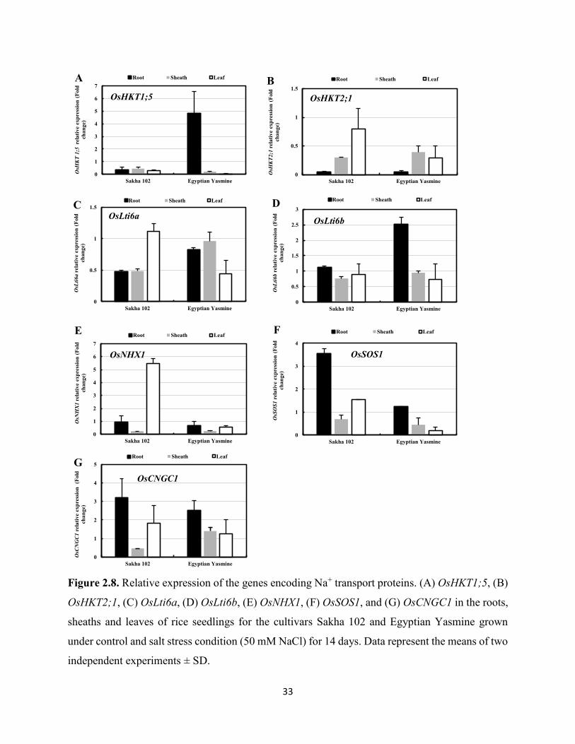
Figure 2.8. Relative expression of the genes encoding Na+ transport proteins. (A) OsHKT1;5, (B)
OsHKT2;1, (C) OsLti6a, (D) OsLti6b, (E) OsNHX1, (F) OsSOS1, and (G) OsCNGC1 in the roots,
sheaths and leaves of rice seedlings for the cultivars Sakha 102 and Egyptian Yasmine grown
under control and salt stress condition (50 mM NaCl) for 14 days. Data represent the means of two
independent experiments ± SD.
0
1
2
3
4
5
6
7
Sakha 102 Egyptian Yasmine
OsH
KT
1;5
rela
tive
expr
essio
n (F
old
chan
ge)
Root Sheath Leaf
0
0.5
1
1.5
Sakha 102 Egyptian Yasmine
OsH
KT2;
1re
lativ
e ex
pres
sion
(Fol
d ch
ange
)
Root Sheath Leaf
OsHKT2;1
B
0
0.5
1
1.5
Sakha 102 Egyptian Yasmine
OsL
ti6a
rela
tive
expr
essio
n (F
old
chan
ge)
Root Sheath Leaf
OsLti6aC
0
0.5
1
1.5
2
2.5
3
Sakha 102 Egyptian Yasmine
OsL
ti6b
rela
tive
expr
essio
n (F
old
chan
ge)
Root Sheath Leaf
OsLti6b
D
0
1
2
3
4
5
6
7
Sakha 102 Egyptian Yasmine
OsN
HX
1re
lativ
e ex
pres
sion
(Fol
d ch
ange
)
Root Sheath Leaf
OsNHX1
E
0
1
2
3
4
Sakha 102 Egyptian Yasmine
OsS
OS1
rel
ativ
e ex
pres
sion
(Fol
d ch
ange
)
Root Sheath Leaf
OsSOS1
F
0
1
2
3
4
5
Sakha 102 Egyptian Yasmine
OsC
NG
C1re
lativ
e ex
pres
sion
(Fol
d ch
ange
)
Root Sheath Leaf
OsCNGC1G
OsHKT1;5
A

34
Figure 2.9. Relative expression of the genes encoding K+ transport proteins. (A) OsAKT1 and (B)
OsHAK7 in the roots, sheaths and leaves of rice seedlings for the cultivars Sakha 102 and Egyptian
Yasmine grown under control and salt stress condition (50 mM NaCl) for 14 days. Data represent
the means of two independent experiments ± SD.
0
1
2
3
4
5
Sakha 102 Egyptian Yasmine
OsA
KT1
rela
tive
expr
essio
n (F
old
chan
ge)
Root Sheath LeafA
0
5
10
15
20
25
30
35
40
Sakha 102 Egyptian Yasmine
OsH
AK7
rel
ativ
e ex
pres
sion
(Fo
ld c
hang
e)
Root Sheath LeafB

35
Figure 2.10. A hypothesized illustration for Na+/K+ uptake and transport in rice plants.
The transporters and channels involved in Na+ influx from soil to the epidermis: OsHKT2;1, HAK7, and
cyclic-nucleotide gated channel (CNGC1). The plasma membrane protein 3 (PMP3) functions to restrict
excess Na+ influx. OsHKT1;5, responsible for Na+ retrieval from the xylem to the xylem parenchyma cells
in the root, and OsHKT1;4 in the shoot. Vacuolar sequestration of Na+ and K+ is mediated by a vacuolar
Na+/H+ antiporter (NHX1). The Na+ /H+ antiporter (SOS1) responsible for efflux of Na+ out of cells either
to external medium or loading into xylem for long-distance Na+ transport and recirculation. AKT1 channel,
functions in K+ up-take. The electrochemical potential is provided by the vacuolar H+-ATPase (V-ATPase)
and the plasma membrane (PM-ATPase). Cell types depicted include: epidermis (EP), exodermis (EX),
cortex (CO), endodermis (EN), casparian stripes (Cs), xylem parenchyma (XP) xylem (X) and phloem (Ph).

36
2.4 Discussion
Two popular Egyptian rice cultivars, Sakha 102 and Egyptian Yasmine, were used in the
present study to elucidate their mode of adaptation to salinity stress through physiological and
transcriptional analyses. The two cultivars showed differential responses to salinity, and Egyptian
Yasmine appeared more tolerant than Sakha 102 in that; it exhibited higher DW, RWC and proline
values and lower ELR, leaf Na+ content, and Na+/K+ ratios.
2.4.1 Mechanisms of Na+ retrieval at the tissue level
Among salt-tolerant traits, the most significant plant adaptation to salinity is the ability to
restrict the transport and accumulation of Na+ in the leaves (Munns and Tester, 2008). Thus plants,
such as Egyptian Yasmine, that exhibit lower leaf Na+ content would be better adapted to salinity.
This restricted transport of Na+ to the leaf blade is often accompanied by a reduced Na+/K+ ratio,
which is relevant for the sustainability of normal metabolic functions (Tester and Davenport,
2003). This is, because high Na+ accumulation often interferes with K+ functions, which results in
impaired metabolic activities. Restricted transport of Na+ or Na+ exclusion to the leaf has been
widely studied and has been shown to be under the control of various Na+ transport proteins
(Munns and Tester, 2008).
To understand the mechanisms underlying limited Na+ transport to the leaves in Egyptian
Yasmine, we analyzed the expression of OsHKT1;5 (Figure 2.8A). Under salt stress conditions,
the gene expression was markedly induced in the roots of Egyptian Yasmine, but was repressed in
the roots of Sakha 102. The OsHKT1;5 transporter is localized in the roots, where it mediates Na+
retrieval from the xylem into xylem parenchyma cells before it is transported in the transpiration
stream to the shoot (Ren et al., 2005; Munns and Tester, 2008). An increase in OsHKT1;5 activity
was observed in the tolerant cultivar Pokkali, but reduced in the sensitive cultivar IR29 (Walia et
37
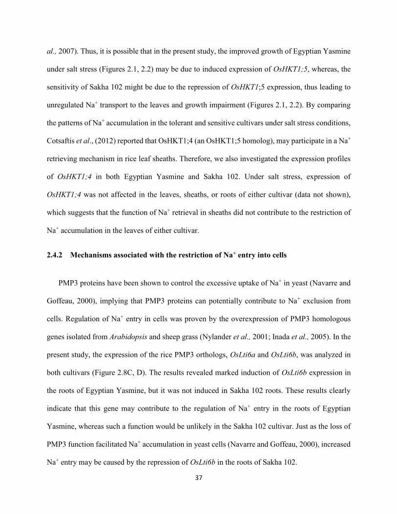
al., 2007). Thus, it is possible that in the present study, the improved growth of Egyptian Yasmine
under salt stress (Figures 2.1, 2.2) may be due to induced expression of OsHKT1;5, whereas, the
sensitivity of Sakha 102 might be due to the repression of OsHKT1;5 expression, thus leading to
unregulated Na+ transport to the leaves and growth impairment (Figures 2.1, 2.2). By comparing
the patterns of Na+ accumulation in the tolerant and sensitive cultivars under salt stress conditions,
Cotsaftis et al., (2012) reported that OsHKT1;4 (an OsHKT1;5 homolog), may participate in a Na+
retrieving mechanism in rice leaf sheaths. Therefore, we also investigated the expression profiles
of OsHKT1;4 in both Egyptian Yasmine and Sakha 102. Under salt stress, expression of
OsHKT1;4 was not affected in the leaves, sheaths, or roots of either cultivar (data not shown),
which suggests that the function of Na+ retrieval in sheaths did not contribute to the restriction of
Na+ accumulation in the leaves of either cultivar.
2.4.2 Mechanisms associated with the restriction of Na+ entry into cells
PMP3 proteins have been shown to control the excessive uptake of Na+ in yeast (Navarre and
Goffeau, 2000), implying that PMP3 proteins can potentially contribute to Na+ exclusion from
cells. Regulation of Na+ entry in cells was proven by the overexpression of PMP3 homologous
genes isolated from Arabidopsis and sheep grass (Nylander et al., 2001; Inada et al., 2005). In the
present study, the expression of the rice PMP3 orthologs, OsLti6a and OsLti6b, was analyzed in
both cultivars (Figure 2.8C, D). The results revealed marked induction of OsLti6b expression in
the roots of Egyptian Yasmine, but it was not induced in Sakha 102 roots. These results clearly
indicate that this gene may contribute to the regulation of Na+ entry in the roots of Egyptian
Yasmine, whereas such a function would be unlikely in the Sakha 102 cultivar. Just as the loss of
PMP3 function facilitated Na+ accumulation in yeast cells (Navarre and Goffeau, 2000), increased
Na+ entry may be caused by the repression of OsLti6b in the roots of Sakha 102.

38
Furthermore, another factor contributing to enhanced adaptation to salinity stress in Egyptian
Yasmine relative to Sakha 102 could reside in the expression of OsCNGC1. This gene product is
involved in Na+ influx in the roots and has been shown to be up-regulated in the sensitive IR29
rice cultivar and down-regulated in the tolerant Pokkali cultivar (Senadheera et al., 2009). Its
induction in the roots of Sakha 102 in the current study indicates another facilitated Na+ uptake
route with subsequent delivery to the leaf. In contrast, its lower expression levels in Egyptian
Yasmine suggest tight control of Na+ uptake at the root level (Figure 2.8G). Although OsHKT2;1
is known to mediate Na+ influx in yeast and plants (Garciadeblás et al., 2003), it was repressed in
both cultivars in the current study (Figure 2.8B). This result is in agreement with the findings of
Horie et al., (2007), which suggested that OsHKT2;1 was down-regulated during salt stress
conditions. Therefore, this indicates that the OsHKT2;1 transporter is important to the restriction
of toxic accumulation of Na+ in both cultivars.
2.4.3 Mechanisms of Na+ exclusion at the cell level
SOS1 antiporter has been shown to be localized at the plasma membrane of Arabidopsis,
where it catalyzes Na+/H+ exchange (Shi et al., 2002). The preferential expression of SOS1 in cells
surrounding the vasculature throughout the plants as demonstrated by the GUS reporter gene,
suggests a role of this transporter in long distance Na+ transport in plants, since Na+ is transported
from the root to the shoot via the xylem. In light of this function, Shi et al., (2002) observed that
high levels (100 mM NaCl) of salt stress, substantially increased the concentration of Na+ in the
xylem sap of both the sos1-mutants and the wild-type plants over time, but the Na+ concentration
was always higher in the sos1 plants. However, Ding and Zhu (1997) showed that, at low levels
(25 mM NaCl) of salt stress, the sos1 mutants accumulated less Na+ than the wild type plants.
These results suggest a dual role of SOS1 in Na+ transport in plants depending on the cellular and

39
extracellular Na+ environments; whereby, under moderate salt stress, SOS1 might function in
loading Na+ into the xylem for controlled delivery to the shoot and storage in leaf mesophyll cells.
Whereas, under high salinity stress, SOS1 would function in Na+ retrieval from the xylem to
prevent over accumulation of Na+ in the transpiration stream (Shi et al., 2002). In addition, the
Oryza sativa SOS1 (OsSOS1) has been shown to complement the function of SOS1 in the sos1
mutant of Arabidopsis, indicating the conservation of the salt overly sensitive pathway in rice as
well (Martínez-Atienza et al., 2007). In the present study, under 50 mM NaCl stress, the expression
of OsSOS1 in the roots of Egyptian Yasmine was unaltered, but was markedly enhanced in the
Sakha 102 roots (Figure 2.8F). This indicates that OsSOS1 may not be involved in the adaptation
of Egyptian Yasmine to salinity, whereas, its induction in Sakha 102 in response to salt treatment
might imply a role in long-distance Na+ transport from root to shoot. Thus, when OsSOS1 is up-
regulated, it facilitates Na+ loading into the xylem, and Na+ is controllably delivered to the leaves
and subsequently compartmentalized into the vacuoles, presumably through the OsNHX1 activity,
leading to high leaf Na+ levels and lower Na+ levels in root (Figure 2.4A,C).
Generally, under high salinity conditions, plants respond by accumulating the excess Na+ in
vacuoles, away from the cytosol, and this compartmentalization is under the control of the vacuolar
Na+/H+ antiporter (NHX) (Munns and Tester, 2008). Arabidopsis AtNHX1 was shown to catalyze
Na+/H+ and K+/H+ exchange with similar affinity (Venema et al., 2002). The dual affinity of
AtNHX1 for Na+ and K+ (Yamaguchi et al., 2005) implies that AtNHX1 mediates the uptake of
K+ from cytosol into vacuoles under regular growth conditions and Na+ sequestration into vacuoles
will also take place because of the rising concentration of this ion in the cytosol under salinity
stress. Overexpression of AtNHX1 in tomato has conferred salt tolerance, although the leaves
accumulated high Na+ concentrations (Zhang and Blumwald, 2001). This observation, together

40
with the fact that the Arabidopsis nhx1 mutant exhibited Na+ sensitivity and significantly less
vacuolar Na+/H+ antiport activity (Apse et al., 2003), strongly supported the role of NHXs in Na+
compartmentalization under salinity stress. However, Leidi et al., (2010) showed that the
overexpression of the AtNHX1 in tomato resulted in enhanced accumulation of K+, but not Na+.
Moreover, Arabidopsis nhx1 nhx2 double mutants displayed reduced K+ concentration in vacuoles,
supporting the role of AtNHX1 and AtNHX2 in mediating H+ efflux coupled to K+ uptake (Bassil
et al., 2011). Fukuda et al. (2004) showed that rice OsNHX1 encodes a vacuolar Na+, K+/H+
antiporter, and suggested that OsNHX1 plays important roles in the compartmentalization of
excess cytosolic Na+ and K+ into the vacuoles. In the present study, under salt stress, OsNHX1
expression was induced in the leaf tissues of Sakha 102 (Figure 2.8E), which had high Na+ (Figure
2.4A) and K+ (Figure 2.5A) contents. This increased accumulation of Na+ and K+ is likely to be a
consequence of the activity of OsNHX1, as the antiporter is supposed to facilitate K+ as well as
Na+ uptake into vacuoles in exchange for H+ into the cytoplasm. However, OsNHX1 was repressed
in Egyptian Yasmine leaves, which had lower Na+ content but high K+ levels. This result indicates
that the induced expression of OsNHX1 would be mainly in response to elevated Na+ levels but
not K+ in Sakha 102.
2.4.4 K+ acquisition under salt stress
OsAKT1 is mapped to chromosome 1 as one of the quantitative trait loci (QTLs) controlling
K+ concentration and the Na+/K+ ratio in salt-stressed plants (Koyama et al., 2001). Our results
showed that salt stress induced the expression of OsAKT1 in both cultivars leaves (Figure 2.9A),
which might enable the maintenance of higher K+ levels in the leaves of both cultivars. Egyptian
Yasmine had abundant OsAKT1 transcripts under salinity stress conditions, and that would explain
the increased K+ accumulation in the leaves and roots as compared to Sakha 102.
41
Although high affinity K+ transporters (HAK) are inhibited by Na+, some may also transport
this ion, and this was shown in barley HvHAK1 and reed plants PhaHAK5 (Santa-Maria et al.,
1997 and Takahashi et al., 2007). In this study, OsHAK7 was induced in the roots and leaves of
both cultivars under stress conditions (Figure 2.9B). There were higher transcript levels of
OsHAK7 in the roots and leaves of Sakha 102 and lower transcript amounts in Egyptian Yasmine,
which may have resulted in the excessive accumulation of Na+ in Sakha 102 leaves and lower Na+
levels in Egyptian Yasmine leaves (Figure 2.4A). These results are consistent with the results of
Senadheera et al., (2009), where OsHAK7 was up-regulated in the salt sensitive rice cultivar IR29
and down-regulated in the tolerant line FL478, which may prevent the overall Na+ load in tolerant
plants.
In summary a hypothesized model depicting the afore-mentioned Na+ /K+ uptake and transport
mechanisms in the studied rice plants can be seen in Figure 2.10.
2.5 Conclusion
In this study, it was demonstrated that Egyptian Yasmine is a relatively salt-tolerant cultivar
compared to Sakha 102, which is due to its ability to restrict Na+ accumulation in leaves under salt
stress. Differences in the mechanisms of salinity tolerance between the two cultivars may be partly
explained by the distinct regulation of gene expression of Na+/K+ transport proteins. This is evident
in the inducible expression of OsHKT1;5 (Na+ retrieval), OsLti6b (restriction of Na+ entry), and
OsAKT1 (K+ uptake), as well as the repressed expression of OsHKT2;1 (Na+ influx). Divergent
regulation of Na+ and K+ transporters may be involved in the maintenance of lower Na+/K+ ratios
in Egyptian Yasmine under salt stress.

42
Chapter 3
Identification of a type 3 metallothionein-like gene
(OsMT-3a) from rice through functional screening
analysis in Escherichia coli, confers tolerance against
salinity and heavy-metal stresses

43
3.1 Introduction
Salinity, drought, temperature, and heavy metals are major factors that reduce plant
productivity and are proving to be an increasing threat to agriculture (Sreenivasulu et al., 2007).
Such stresses have similar consequences: causing ionic imbalance and generating reactive oxygen
species (ROS). Overproduction of ROS damages cell membranes, nucleic acids, and
photosynthetic pigments (Zhang et al., 2007), and therefore, living organisms have developed
cellular ROS detoxifying systems. ROS are scavenged by enzymes such as superoxide dismutase,
catalase, and peroxidase (Apel and Hirt, 2004; Jang et al., 2012), and also by non-enzymatic
components that include low molecular weight antioxidants, such as ascorbate, glutathione,
carotenoids, and metallothioneins (MTs) (Gechev et al., 2006).
MTs are a group of polypeptides that can bind with heavy metals through their thiol group via
chelation. Because of this chelating activity, MTs are involved in the homeostasis of essential
metals (Cu2+ and Zn2+) and cellular detoxification of nonessential metals (Cd2+ and Hg2+) (Hamer,
1986; Huang and Wang, 2010). A structural characteristic of MTs is the cysteine (Cys) residue,
which is the basis of its classification. On the basis of the arrangement of Cys residues, MTs are
divided in two classes (Cobbett and Goldsbrough, 2002). Plant MTs belong to Class II and are
further classified into four types (1-4) based on the position of Cys residues (Cobbett and
Goldsbrough, 2002). Through characterization of biochemical properties of some plant MTs in
heterologous expression system, its potential contribution to abiotic stress tolerance has been
discussed (Chaturvedi et al. , 2014; Dundar et al. , 2015; Turchi et al. , 2012; Xue et al. , 2009;
Yang et al. , 2009).

44
Studies in animals as well as plants showed that MTs are not only involved in maintaining
homeostasis of essential metals and metal detoxification (Cobbett and Goldsbrough, 2002) but are
also implicated in a range of physiological processes, including scavenging ROS (Akashi et al.,
2004; Wong et al., 2004; Kumar et al., 2012; Chaturvedi et al., 2014). The antioxidant function of
MTs has been attributed to the presence of a large number of Cys residues, which besides metal
binding are also capable of scavenging ROS (Hassinen et al., 2011).
MT gene expression is regulated by abiotic stress, including heavy-metal stress, and plays an
important role in metal detoxification and homeostasis (Huang and Wang, 2010; Kim et al., 2014).
Earlier studies of plant MTs focused on their role in maintaining intracellular metal homeostasis
and mediating responses to metal toxicity (Cobbett and Goldsbrough, 2002). However, recent
studies have shown additional roles of plant MTs in development, fruit ripening, senescence, and
defense against oxidative stress; Zhigang et al., (2006) showed that expression of the Brassica
juncea MT2 in Arabidopsis enhanced tolerance to Cu and Cd, but inhibited root elongation. Two
MT-like genes were upregulated during natural leaf senescence in sweet potato (Chen et al., 2003).
Cotton MT3 scavenged ROS and enhanced plant tolerance to oxidative stresses caused by NaCl,
polyethylene glycol, and low temperature (Xue et al., 2009). In rice, most studies of MT isoforms
have focused on the gene expression patterns under different stress conditions (Jin et al., 2006;
Yang et al., 2009) and metal-binding ability of different MT proteins (Nezhad et al., 2013). It is
necessary to investigate the role of rice MTs in response to salinity stress, which could be another
adaptation strategy of rice plants to such stress conditions.
In the previous study, the adaptation mechanisms of the two Egyptian rice cultivars, Egyptian
Yasmine and Sakha 102 to salinity stress has been characterized, and found that the difference in
tolerance between the two lies in the ability to exclude Na+ to the leaf blade. This exclusion

45
mechanism was shown to be under the control of transporters such as the HKT1;5, which functions
in restricting the transport of Na+ to the leaf and hence in stress tolerance. However, it is unclear
whether the tolerance of Egyptian Yasmine is limited only to Na+ exclusion or regulation of Na+
transport protein coding genes. Therefore, the present study was designed to isolate salinity-
inducible genes from Egyptian Yasmine and examine their functional roles which could be
involved in other adaptation processes under salinity stress conditions, in an attempt to further
elucidate the molecular mechanism of salinity stress tolerance in this cultivar.
3.2 Materials and Methods
3.2.1 Plant material, growth conditions, salt treatment, and sample collection
Seeds of the rice cultivars Egyptian Yasmine and Sakha 102 were surface-sterilized via
immersion in a 5% NaClO solution for 30 min, and then thoroughly rinsed with distilled water.
Seeds were subsequently soaked in tap water for 24 h at 28 °C. After germination, the seeds were
transferred to a nylon mesh and allowed to float on 20 L of tap water for two days. Water was then
replaced with half-strength Kimura B solution. Twenty one-day-old seedlings were transferred to
either Kimura B nutrient solution (control) or Kimura B nutrient solution supplemented with 50
mM NaCl (salinity) for 10 days. The solutions were replaced every two days and the pH was daily
adjusted to 5.0–5.5. Seedlings were grown in a growth chamber under the following controlled
environmental conditions: 70% relative humidity, 24 ± 2 °C, and a 16 h photoperiod at a
photosynthetic photon flux density of 250 - 350 μmol m-2 s-1. The root and leaf tissues from
experimental and control plants were separated and frozen in liquid nitrogen, prior to being stored
at -80 °C.

46
3.2.2 Total RNA isolation, cDNA synthesis, and construction of cDNA expression library
Total RNA was extracted from the leaves and roots of the control and stressed plants of the
rice cultivar, Egyptian Yasmine, by using TRIzol reagent (Invitrogen, Carlsbad, CA). cDNA
synthesis was carried out by a PCR-based method using the In-Fusion® SMARTer™ Directional
cDNA library construction kit (Clontech, Takara, Japan) as described by the manufacturer. The
double stranded cDNA was purified using CHROMA SPIN™ + TE-1000 size exclusion column
chromatography (Clontech, Takara, Japan). Three microliters of each fraction was electrophoresed
on a 1.1% agarose/ethidium bromide gel to determine the peak fractions by visualizing the
intensity of the bands under UV light. Fractions containing large, medium, and small-sized cDNA
were pooled, which were then ligated into the pSMART2IFD vector (Clontech Laboratories).
3.2.3 E. coli functional screening
The resulting ligation reactions were transformed into electrocompetent E. coli (KNabc) cells.
The transformed E. coli cells were then selected and inoculated into sterile 96-well microtiter
plates containing L-medium (1% Bacto tryptone, 0.5% yeast extract, and 0.05% NaCl)
supplemented with 0.2 M of NaCl, then incubated at 37 °C for 24 h with gentle shaking (30 rpm).
Subsequently, the individual bacterial transformants were printed on quadruplet selection plates
(L-medium, 100 µg/mL ampicillin, 1.0 mM isopropyl β-D-1-thiogalactopyranoside [IPTG], 1.5%
agar) containing different concentrations of NaCl (0, 0.5, 1.0, 1.5, and 2 M NaCl), and incubated.
The successfully transformed E. coli cells grown on selection plates containing the highest NaCl
concentrations were then isolated and grown in a liquid culture medium supplemented with 100,
200, 500, and 1,000 mM NaCl for 24 h. Growth was verified by measuring the optical density
(OD) at 600 nm (OD600). Then, the inserts of these clones were confirmed by colony PCR using

47
the forward, 5´-TCACACAGGAAACAGCTATGA-3´ and reverse, 5´-
CCTCTTCGCTATTACGCCAGC-3´ screening primers (Clontech Laboratories).
3.2.4 Bacterial strains and salt stress experiment
The bacterial strains E. coli TG1 and its derivative KNabc (ΔnhaA, ΔnhaB, ΔchaA), [a triple
mutant of E. coli, this mutant was disabled in the function of three Na+/H+ antiporters which
exclude Na+ from the cell in the wild-type (TG1)] were used (Nozaki et al., 1996). For bacterial
salinity stress experiment, E. coli mutant (KNabc) expressing a rice cDNA, E. coli KNabc
(negative control carrying the empty vector pUC19) and E. coli WT (TG1/pUC19) strains were
used. A preparatory culture was grown under selective conditions in L-medium plus 100 µg/ml
ampicillin at 37°C under aerobic conditions until a defined cell density was achieved.
Subsequently, equal cell densities were used to inoculate 20 ml of L-medium plus 100 µg/ml
ampicillin. Gene expression was induced by adding IPTG at 1 mM. After a 3 h induction phase,
100 mM of NaCl was added. These bacterial cells were incubated at 37°C with shaking (200 rpm).
Bacterial growth was measured photometrically at OD600 at 2 h intervals for 24 h.
3.2.5 Colony PCR, plasmid DNA extraction and digestion with restriction enzymes
The conditions for colony PCR analysis were as follows: an initial incubation at 95 °C for 2
min, followed by 35 cycles of denaturation at 95 °C for 30 s, annealing at 55 °C for 30 s, and
extension at 68 °C for 30 s. E. coli transformants (representing the plasmid expression vector
containing rice cDNA) were then cultured, and plasmid DNA was extracted as described in the
HiYield Plasmid Mini Kit (RBC Bioscience). Plasmid extraction was followed by digestion with
the EcoRI and BamHI to confirm the size of the insert.
48
3.2.6 Gene sequencing and identification
Sequencing of the rice cDNA conferring salinity tolerance was performed using BigDye
Sequencing Kit (ABI, USA) according to the manufacturer’s instructions, and analyzed using
Genetic Analyzer (ABI 3100, USA). The sequencing was carried out using vector specific primers
(forward and reverse screening primers). Computational search against sequence databases at
NCBI was performed to identify the gene.
3.2.7 Quantification of H2O2 levels in OsMT-3a transformed E. coli under NaCl stress
conditions
The OsMT3a-transformed E. coli cells and those with empty vector were cultured overnight in
L-medium (OD600 = 5.0–6.0). Equal cell densities were used to inoculate 10 mL of L-medium plus
100 µg/mL ampicillin. Gene expression was induced by adding 1 mM IPTG. After a 3-h induction
phase, 200 mM NaCl was added and incubated with shaking for 24 h at 37 °C. Then, 100 µL of
each culture was added to 1 mL of reaction buffer (0.25 mM FeSO4, 0.25 mM (NH4)2SO4, 25 mM
H2SO4, 1.25 mM xylenol orange, and 1 mM sorbitol) and allowed to stand for 1 h at room
temperature. The H2O2 levels were quantified photometrically at 560 nm, and calculated by
reference to standards (Suharsono et al., 2002).
3.2.8 Tolerance of E. coli to H2O2 stress
Overnight cultures of the cells KNabc harboring pSMART2IFD-OsMT-3a, E. coli KNabc/
pUC19 and TG1/pUC19 strains were used. 100 µl from the overnight cultures were added to 10
mL L-medium plus ampicillin, and cultured at 37 °C. When the cells reached OD600 = 0.6, 1 mM
IPTG was added, and culturing was continued for another 12 h. Equal densities of cells (OD600=
49
0.1) were then cultured in the presence of either 0, 1, 3 or 4 mM H2O2 at 37 °C, and the growth
was monitored (OD600) at 2-h intervals for 12 h.
3.2.9 Measurement of growth and heavy metal accumulation in E. coli overexpressing the
OsMT-3a gene
Initially, we run the experiments with the control strain, KNabc/pUC19 under varied
concentrations of CdCl2, CuSO4, or ZnSO4 and detected the used experimental concentration of
each (100 µM of CdCl2, 1.5 mM of CuSO4, 250 µM of ZnSO4) depending on the substantial effect
on growth rate (toxic level). We added 100 µl from the overnight cultures to 10 mL L-medium
plus ampicillin, and cultured at 37 °C. When the cells reached OD600 = 0.6, 1 mM IPTG was added,
and culturing was continued for another 12 h. Equal densities of cells (OD600= 0.05) were then
cultured in the presence of either 1.5 mM CuSO4, 100 µM CdCl2, or 250 µM ZnSO4 at 37 °C, and
the growth was monitored (OD600) at 12-h intervals for 60 h.
To measure the accumulation of heavy metals, E. coli cells were grown in liquid medium
containing 100 µM CdCl2, 250 µM ZnSO4, or 1.5 mM of CuSO4 at 37 °C. When cells reached
OD600 = 0.6, 1 mM IPTG was added and then incubated for another 24 h at 37 °C. After incubation,
the cells were harvested and washed twice with L-medium. Dry weight was determined after 3
days at 70 °C. The cells were then digested using 5 mL concentrated HNO3, incubated at 95 °C
for 2 h. Prior to the measurement, the lysate was diluted using 0.1N HNO3. The Cu, Zn, and Cd
concentrations were determined by using inductively coupled plasma-atomic emission
spectrometry (ICP-AES) (iCAP 6300, Thermo Fisher Scientific Inc., UK).

50
3.2.10 Expression analysis of the OsMT-3a in the salinity-tolerant and salinity-sensitive rice cultivars
Total RNA was extracted from the leaves and roots of control and stressed plants using TRIzol
reagent (Invitrogen, Carlsbad, CA). After digestion with DNaseI, total RNA (1 µg) was reverse-
transcribed to cDNA using a ReverTra Ace qPCR RT kit (Toyobo, Osaka, Japan). Quantitative
polymerase chain reaction (qPCR) was conducted as previously described (Ueda et al., 2013),
using a Thunderbird SYBR qPCR Mix (Toyobo) and an ABI Step One Plus system (Applied
Biosystems). RT-qPCR was performed as described previously. The sequences of the primers used
are as follows: OsMT-3a forward primer, 5´-TGGTTTGTGTGTGTGCTGATT-3´ and reverse
primer, 5´-TAGCACGGGTACATCACATCA-3´.
3.2.11 In vivo localization of O2− in rice plants
Histochemical staining was performed for the in vivo detection of O2− using nitro-blue
tetrazolium (NBT) (Shi et al., 2010). For detecting O2−, the apical 3 cm of the third leaves from
the rice plants (Egyptian Yasmine and Sakha 102) exposed to either NaCl (50 mM) and/or CdCl2
(100 µM) for 10 days were used. The presence of O2− was detected by immersing the leaf samples
in NBT solution (1 mg ml-1 in 10 mM phosphate buffer; pH 7.8) and vacuum infiltrated for 30
min, kept at room temperature for 2 h and then illuminated for 12 h in light until blue spots
appeared. Rice leaves were then decolorized in a bleaching solution (ethanol: acetic acid: glycerol
[3:1:1]), and the tubes with the leaves were placed in 100 °C water bath for about 30 min until the
chlorophyll bleached out, and then were stored in a glycerol-ethanol (1:4) (v/v) solution. The O2−
staining was repeated three times.

51
3.2.12 Quantification of H2O2 in rice leaves under NaCl and CdCl2 stress conditions
The leaf tissues of 10-days treated plants (50 mM NaCl or 100 µM CdCl2) from Egyptian
Yasmine and Sakha 102 were ground in liquid nitrogen, and homogenized with 4 ml cold acetone.
The homogenate was centrifuged at 8,000 g for 15 min at 4 °C. Then, 100 µl of each was added to
1 ml of reaction buffer (0.25 mM FeSO4, 0.25 mM (NH4)2SO4, 25 mM H2SO4, 125 µM xylenol
orange, and 10 mM sorbitol) and allowed to stand for 1 h at room temperature. The H2O2 levels
were quantified photometrically at 560 nm, and calculated by reference to standards (Suharsono,
2002).
3.2.13 Statistical analysis
The collected data were subjected to one-way analysis of variance (ANOVA), using the SPSS
statistics package, version 21 (IBM Inc., USA), and the means (n = 4) were separated using the
Duncan’s multiple range test at p < 0.05.

52
3.3 Results
3.3.1 E. coli functional screening
To further elucidate the molecular mechanisms of salinity tolerance in the rice cultivar
Egyptian Yasmine, we planned to isolate and characterize the salinity-inducible genes from this
plant through their functional validation analysis in E. coli expression system. To achieve that, we
screened a cDNA library that was prepared from salinity-stressed seedlings of Egyptian Yasmine
in the Na+-sensitive E. coli mutant cells KNabc (ΔnhaA, ΔnhaB, ΔchaA), that lack the three Na+/H+
antiporters which exclude Na+ from the cell. Due to deactivation of the major Na+ efflux systems,
the KNabc mutant could not grow in the presence of 0.2 M NaCl. Such genetic backgrounds in the
E. coli mutant would be useful for identification of determinant genes for salinity tolerance in rice
through functional screening.
Successfully transformed E. coli cells were grown on selection plates containing different
concentrations of salt up to 2 M NaCl. We obtained few colonies that were able to grow at the
highest NaCl concentration (Figure 3.1). These colonies were picked out and inoculated in a liquid
culture medium supplemented with different concentrations of salt up to 1 M NaCl to confirm their
ability to grow under more severe salt stress conditions (salt stress is more severe in liquid media).
We identified one such clone, which showed the best growth both at 2 M NaCl on plates (on solid
medium) and at 200 mM NaCl in the liquid medium, for further characterization. The presence of
the insert in that clone was confirmed using colony PCR analysis.
3.3.2 Sequence analysis and multiple alignment of OsMT-3a
From the library screening, one isolated gene shared significant sequence homology with that
of plant type-3 metallothionein (MT-3a). The full-length coding sequence of OsMT-3a is 189 bp

53
long, and encodes a 62-amino acid protein (Figure 3.2A). Similar to other plant type-3 MT
isoforms, this amino acid sequence contains 10 Cys residues arranged in the forms
CXXCXCXXXXC and CXCXXXCXCXXCXC at its N and C termini, respectively (Figure 3.2B).
The genetic similarity of OsMT-3a with other plant MTs was determined using Blastx and
CLUSTAL W. Multiple alignments indicated that OsMT-3a shared high homology with many
MT3-like genes from other plant species (Figure 3.2B).
3.3.3 OsMT-3a confers salinity-stress tolerance to transformed E. coli through the
scavenging of ROS
To assess the role of OsMT-3a in response to salinity stress, E. coli KNabc cells were
transformed with either the empty vector pUC19 or with pSMART2IFD containing OsMT-3a
cDNA. After transformation of the parental wild-type TG1 strain with pUC19 (TG1/pUC19), we
monitored the growth of these three sets of transformants in response to salinity stress (using 100
mM NaCl) (Figure 3.3A). OsMT-3a-transformed E. coli cells had a moderate growth rate between
TG1/pUC19 cells and KNabc/pUC19 cells, and grew more efficiently compared to KNabc/pUC19
cells in media containing 100 mM of NaCl. These results demonstrated that the expression of
OsMT-3a enabled E. coli cells to become more tolerant to NaCl stress. However, this gene did not
completely complement the Na+-sensitivity defect in this bacterial mutant KNabc, as the growth
did not reach the TG1/pUC19 growth rate as seen in Figure 3.3A. This finding implied that OsMT-
3a improved salinity tolerance of KNabc cells through any other mechanisms rather than Na+
exclusion from the cells. To study mechanisms of the potential contribution by OsMT-3a in salinity
tolerance, Na+ concentration was examined in these three cells (Figure 3.3B). In comparison to
Na+ accumulation under the control condition (1.23 µmol/106 cells in TG1/pUC19, 3.51 μmol/106
cells in KNabc/pUC19, and 5.8 μmol/106 cells in KNabc/OsMT-3a), KNabc/OsMT-3a cells

54
accumulated 180 times higher Na+ than TG1/pUC19 cells (2.06 μmol/106 cells in TG1/pUC19,
1300 μmol/106 cells in KNabc/pUC19, and 363.5 μmol/106 cells in KNabc/OsMT-3a), suggesting
that OsMT-3a did not directly participate in Na+ exclusion from the cells (Figure 3.3B).
To investigate whether the increased tolerance to NaCl stress in OsMT-3a-transformed E. coli
cells might be due to the scavenging of ROS by OsMT-3a, we compared the H2O2 concentrations
in KNabc/OsMT-3a, KNabc/pUC19, and TG1/pUC19 under salinity stress. In comparison to
control conditions, H2O2 concentrations were significantly increased in both KNabc/OsMT-3a and
KNabc/pUC19 in the presence of 200 mM NaCl (Figure 3.3C). However, H2O2 concentration in
KNabc/OsMT-3a cells was less than one third of that in KNabc/pUC19 cells (Figure 3.3C),
3.3.4 Tolerance of OsMT-3a-transformed E. coli cells to H2O2 stress
Initial experiments with the KNabc/pUC19 cells revealed that the concentration of H2O2 up to
0.75 mM had no substantial effect on the bacterial growth rate. Therefore, the concentrations of 1,
3, and 4 mM of H2O2 were considered to be used to investigate the effect of OsMT-3a on the
tolerance of the transformed E. coli cells. E. coli cells expressing OsMT-3a showed increased
tolerance to H2O2 stress, and had a similar or even better growth rate as the WT TG1/pUC19 cells
under exposure to high concentrations of H2O2 (Figure 3.4A). The final cell density of
KNabc/OsMT-3a was 33%, 99%, and 98% higher than KNabc/pUC19 in a culture medium
supplemented with 1, 3 and 4 mM H2O2, respectively (Figure 3.4B, C, D), as the higher
concentrations of H2O2 were lethal for KNabc/pUC19 cells. These data suggest that the
heterologous expression of OsMT-3a gave the E.coli cells more tolerance to H2O2, probably
through a scavenging activity.

55
3.3.5 OsMT-3a confers heavy-metal tolerance to transformed E. coli
Previous reports have discussed expressing other classes of plant MTs, such as Brassica juncea
BjMT2 (Zhigang et al., 2006), Salicornia brachiata SbMT-2 (Chaturvedi et al., 2012), sweet potato
IbMT1 and IbMT3 (Kim et al., 2014), and olive OeMT2 (Dundar et al., 2015), in E. coli to assess
if they confer tolerance to heavy metals. Here, we examined the functions of OsMT-3a using E.
coli transfected with a pSMART2IFD-OsMT-3a vector, and assessed cell growth in the presence
of heavy metals (Figure 3.5). Initially, we run the experiments with the control strain,
KNabc/pUC19 under varied concentrations of CdCl2, CuSO4, or ZnSO4 and detected the used
experimental concentration of each (100 µM of CdCl2, 1.5 mM of CuSO4, 250 µM of ZnSO4)
depending on the substantial effect on growth rate (toxic level). In the absence of heavy metals, all
bacterial strains exhibited a normal growth rate; TG1/pUC19 exhibited a higher growth rate
compared to both KNabc/pUC19 and KNabc/OsMT-3a with almost similar growth rate (Figure
3.5A). However, exposure to supplemental CdCl2, CuSO4, or ZnSO4 differentially affected cell
growth. Cell growth of the KNabc/pUC19 was affected by toxic concentrations of heavy metals,
but OsMT-3a overexpression significantly improved growth kinetics, especially under CdCl2 stress
(Figure 3.5B) at which the growth rate of KNabc/OsMT-3a cells until 24 h exposure was
significantly higher compared to that of the TG1/pUC19 and KNabc/pUC19. With respect to
CuSO4, only with 12-h exposure, E. coli cells expressing OsMT-3a grew better than did the
controls, TG1/pUC19 and KNabc/pUC19 (Figure 3.5C). Under ZnSO4 stress conditions, no
significant difference in the growth appeared between the OsMT-3a-transformed cells and the
controls until 12 h exposure, after that time, a decline in the growth rate of the cells,
KNabc/pUC19, was observed (Figure 3.5D).
56
To determine whether the tolerance to heavy metal in E. coli cells induced by OsMT-3a is
associated with accumulation of heavy metals, E. coli cells overexpressing OsMT-3a were cultured
in media containing CdCl2, ZnSO4, or CuSO4, and their metal ion content was measured (Figure
3.6). Under CdCl2 stress, Cd2+ levels were increased in the cells of all the strains, TG1/pUC19,
KNabc/OsMT-3a and KNabc/pUC19. However, E. coli cells overexpressing OsMT-3a contained
a significantly higher concentration of Cd2+ than did those transformed with the empty vector,
KNabc/pUC19 (Figure 3.6A). Under both ZnSO4 and CuSO4 stresses, a similar increase in the
concentrations of Zn2+ and Cu2+ in the cells of all the strains examined, but with no significant
difference in Zn2+ and Cu2+ concentrations between the transformed KNabc/OsMT-3a and
KNabc/pUC19 cells (Figure 3.6B, C), indicating that OsMT-3a may binds heavy metals
selectively.
3.3.6 Tissue-specific expression of OsMT-3a in rice plants
To examine the tissue-specific expression patterns of OsMT-3a in rice, qRT-PCR analysis was
performed using total RNAs extracted from the leaf and root tissues of the rice cultivars Egyptian
Yasmine and Sakha 102. In the previous study, we demonstrated that Egyptian Yasmine has higher
salinity tolerance than does Sakha 102, which is due to its ability to restrict Na+ accumulation in
leaves when under salt stress, mainly through the inducible expression of OsHKT1;5, which
functions in Na+ retrieval (Ren et al., 2005). In the current study, gene expression analysis revealed
that OsMT-3a was highly induced by salinity stress in the leaves of the salinity-tolerant cultivar
Egyptian Yasmine but not in those of the salinity-sensitive, Sakha 102. However, the gene
expression in the roots of both cultivars was repressed under salinity stress conditions, suggesting
that OsMT-3a might function predominantly in the leaves, but not in the roots, under salinity stress
(Figure 3.7).

57
3.3.7 In vivo localization of O2− in rice plants and H2O2 concentration analysis
Under NaCl and CdCl2 stress conditions, O2− production was visualized by a histochemical
method using NBT staining (Figure 3.8). It was observed that leaves of Sakha 102 plants showed
more accumulation of O2− compared to those of Egyptian Yasmine as these leaves showed intense
blue colored polymers accumulation with NBT staining under NaCl and CdCl2 stress condition
(Figure 3.8). Moreover, by analyzing the H2O2 concentrations in the leaves of the two cultivars
under both NaCl and CdCl2 stress conditions, Egyptian Yasmine showed significantly lower H2O2
concentrations under stress conditions compared to those of Sakha 102 (Figure 3.9). Lower
concentrations of H2O2 implied the better ROS scavenging system in Egyptian Yasmine compared
to Sakha 102.

58
Figure 3.1. Rice leaf cDNA library screening in E. coli cells KNabc (ΔnhaA, ΔnhaB, ΔchaA). The
individual bacterial transformants were printed on quadruplet selection plates (L-medium, 100
µg/mL ampicillin, 1.0 mM IPTG, 1.5% agar) as a control (A) or supplemented with 2 M NaCl (B)
and incubated at 37 °C for 24 h.

59
A ATG TCG GAC AAG TGC GGC AAC TGC GAC TGT GCT GAC AAG AGC CAG
M S D K C G N C D C A D K S Q
TGC GTG AAG AAA GGT ACC AGC TAT GGC GTC GTC ATA GTT GAA GCC
C V K K G T S Y G V V I V E A
GAG AAG AGC CAC TTC GAG GAG GTC GCC GCC GGC GAG GAG AAC GGC
E K S H F E E V A A G E E N G
GGC TGC AAG TGC GGC ACC AGC TGC TCC TGC ACC GAC TGC AAG TGC
G C K C G T S C S C T D C K C
GGC AAG TGA
G K *
B
OsMT3 1 MSDKCGNCDCADKSQCVKKGTSYGVVIVEAEKSHFEEVAAGEENG------GCKCGTSCS
HvMT3 1 MADKCGNCDCADKTQCVKKGDSYGIVMVDTEKSHLEVQETAENDD------KCKCGTSCT
ThMT3 1 MSGKCGNCSCADKSQCVQKRNQYGFDLIETQTYAESTVVMDDPPTAAENGGQCKCGDRCA
GhMT3 1 MADKCGNCDCADKSQCVKKGN---SLVIETEESYISTVVVEP---LAENDGKCKCGTSCS
AtMT3 1 MSSNCGSCDCADKTQCVKKGTSYTFDIVETQESYKEAMIMDVGAEENNANCKCKCGSSCS
BjMT3 1 MSDKCGSCDCADKTQCVKKGTSYTFDIVETQESYKEAMFMDVGAEEN--GCQCKCGSTCS
C C C C C C C
OsMT3 55 CTDCKCGK-
HvMT3 55 CTNCTCGH-
ThMT3 61 CVNCTCGSH
GhMT3 55 CTNCTCGSH
AtMT3 61 CVNCTCCPN
BjMT3 59 CVNCTCCPN
C C C
Figure 3.2. (A) Nucleotide and deduced amino acid sequences of OsMT-3a cDNA, (B)
Comparison of the deduced amino acid sequences of Oryza sativa MT-3a with its homologues
from other plant species. Conserved Cys residues are indicated by the letter C. Identical or
conserved amino acid residues are shaded in dark or grey colors, respectively. The accession
numbers in GenBank of the other plant type 3 MTs are as follows: Hordeum vulgare subsp. vulgare
(HvMT3), CAD88266; Tamarix hispida (ThMT3), EH057039; Gossypium hirsutum (GhMT3),
AY857933; Arabidopsis thaliana (AtMT3), NP_566509; and Brassica juncea (BjMT3),
AB057415.

60
0
0.5
1
1.5
2
2.5
3
3.5
4
0 2 4 6 8 10 12 14 16 18 20 22 24
OD
600
Time (h)
TG1/pUC19 KNabc/OsMT-3a KNabc/pUC19A
c c cc
a
b
0
200
400
600
800
1000
1200
1400
1600
TG1/pUC19 KNabc/pUC19 KNabc/OsMT-3a
Na+
conc
entr
atio
n (µ
mol
/10
6 ce
lls)
0 mM NaCl 200 mM NaClB

61
Figure 3.3. (A) Increased tolerance of OsMT-3a-expressing E. coli cells to NaCl stress. At the
beginning of the assay (OD600 = 0.05), 1.0 mM IPTG was added to all three cultures. Bacterial
cells were cultivated in L-medium supplemented with 100 mM NaCl. (B) Na+ concentration in
OsMT-3a-transformed E. coli cells grown in the presence of 200 mM NaCl compared with the
concentration in control cells. (C) H2O2 concentrations in OsMT-3a-transformed E. coli cells
grown in the presence of 200 mM NaCl compared with that in control cells. Data are represented
as the means of three independent experiments ± SE. The same letters indicate no significant
differences (P < 0.05).
b b
c
b
a
b
0
1
2
3
TG1/pUC19 KNabc/pUC19 KNabc/OsMT-3a
Hyd
roge
n pe
roxi
de c
once
ntra
tion
(µm
ol/g
FW
)
0 mM NaCl 200 mM NaClC

62
Figure 3.4. Growth curve of the strains, TG1/pUC19, KNabc/pUC19 and KNabc/OsMT-3a under
H2O2 stress. The medium supplemented with (A) 0 mM, (B) 1 mM, (C) 3 mM, or (D) 4 mM H2O2.
Data are represented as the means of three independent experiments ± SE.
0
1
2
3
4
5
6
0 2 4 6 8 10 12
OD
600
Time (h)
TG1/pUC19 KNabc/pUC19
KNabc/OsMT-3a
C
0
1
2
3
4
5
6
0 2 4 6 8 10 12
OD
600
Time (h)
TG1/pUC19 KNabc/pUC19KNabc/OsMT-3a
B
0
1
2
3
4
5
6
0 2 4 6 8 10 12
OD
600
Time (h)
TG1/pUC19 KNabc/pUC19
KNabc/OsMT-3a
D
0
1
2
3
4
5
6
7
0 2 4 6 8 10 12
OD
600
Time (h)
TG1/pUC19 KNabc/pUC19KNabc/OsMT-3a
A

63
Figure 3.5. Increased tolerance of OsMT-3a-expressing E. coli cells to Cd2+, Cu2+, and Zn2+
stresses. At the beginning of the assay (OD600 = 0.05), 1.0 mM IPTG was added to all three
cultures. Bacterial cells were cultivated either (A) in L-medium only as control or (B)
supplemented with 100 µM CdCl2, (C) 1.5 mM CuSO4, (D) 250 µM ZnSO4. Data are represented
as the means of three independent experiments ± SE.
0
1
2
3
4
5
6
7
0 12 24 36 48 60
OD
600
Time (h)
TG1/pUC19 KNabc/pUC19
KNabc/OsMT-3a
Control
0
1
2
3
4
5
6
0 12 24 36 48 60
OD
600
Time (h)
TG1/pUC19 KNabc/pUC19
KNabc/OsMT-3a
1.5 mM CuSO4
C
A
0
1
2
3
4
5
6
0 12 24 36 48 60
OD
600
Time (h)
TG1/pUC19 KNabc/pUC19
KNabc/OsMT-3a
250µM ZnSO4
0
1
2
3
4
5
6
0 12 24 36 48 60
OD
600
Time (h)
TG1/pUC19 KNabc/pUC19
KNabc/OsMT-3a
100µM CdCl2
B
D

64
Figure 3.6. Metal ions concentration (Cd2+, Zn2+ and Cu2+) in OsMT-3a-expressing E. coli cells
in the presence of (A) 100 µM CdCl2, (B) 250 µM ZnSO4, (C) 1.5 mM CuSO4 compared with that
in control cells. At the beginning of the assay (OD600 = 0.05), 1.0 mM IPTG was added to all three
cultures and they were then grown for 24 h. Data are represented as the means of three independent
experiments ± SE. The same letters indicate no significant differences (P < 0.05).
cd
bab
a a
0
0.05
0.1
0.15
TG1/pUC19 KNabc/pUC19 KNabc/OsMT-3aZn
2+co
ncen
trat
ion
(nm
ol/1
0 9
cells
)
0 µM ZnSO 250 µM ZnSOB
c c c
a
b
a
0
0.2
0.4
0.6
0.8
1
1.2
TG1/pUC19 KNabc/pUC19 KNabc/OsMT-3a
Cd2+
conc
entr
atio
n (n
mol
/10
9ce
lls)
0 µM CdCl 100 µM CdCl
c cc
a
bb
0
0.02
0.04
0.06
0.08
0.1
0.12
0.14
TG1/pUC19 KNabc/pUC19 KNabc/OsMT-3a
Cu2+
conc
entr
atio
n (n
mol
/10
9ce
lls)
0 mM CuSO 1.5 mM CuSO
A
C
2 2 4 4
4 4

65
Figure 3.7. Relative expression of OsMT-3a in the roots and leaves of rice seedlings of the
cultivars Egyptian Yasmine and Sakha 102 grown under salinity stress conditions (50 mM NaCl)
for 10 days. Data are represented as the means of three independent experiments ± SE. The same
letters indicate no significant differences (P < 0.05).
cc
a
b
0
0.5
1
1.5
2
2.5
3
Egyptian Yasmine Sakha 102
OsM
T-3a
rela
tive
expr
essi
on (F
old
chan
ge)
Root Leaf

66
Figure 3.8. Histochemical detection of O2− with NBT in the leaves of rice plants grown under
different stress condition. Egyptian Yasmine and Sakha 102 cultivars were grown in hydroponic
(control) or treated with NaCl (50 mM) and/or CdCl2 (100 µM) for 10 days.
Egyptian Yasmine
Sakha 102
Control 50 mM NaCl 100 µM CdCl 2

67
Figure 3.9. Comparison of H2O2 concentrations in the leaves of the rice cultivars, Egyptian
Yasmine and Sakha 102 grown under control conditions and in the presence of 50 mM NaCl and/or
100 µM CdCl2. Data are represented as the means of four replicates ± SE. The same letters indicate
no significant differences (P < 0.05).
ede
c
cd
b
a
0
0.2
0.4
0.6
0.8
1
1.2
Control 50 mM NaCl 100 µM CdClHyd
roge
n pe
roxi
de c
once
ntra
tion
(µm
ol/g
FW
) Egyptian Yasmine Sakha 102
2

68
Figure 3.10. A hypothetical model for the role of OsMT-3a in ROS scavenging and
phytoremediation under abiotic stress.

69
3.4 Discussion Oxidative stress is a secondary consequence when organisms are challenged with abiotic
stresses, such as high salinity. Oxidative stress results from the disturbance in balance between the
production and scavenging of ROS such as hydrogen peroxide, superoxide anions, and hydroxyl
radicals, which damage cells by destroying lipids, nucleic acids, and proteins (Apel and Hirt,
2004). Living organisms have developed a variety of adaptive mechanisms to cope with various
internal and external stresses. MTs are efficient scavengers of produced ROS in plants (Grennan,
2011; Hassinen et al., 2011).
In the current study, in an effort to further elucidate the molecular mechanisms of salinity stress
tolerance in the rice cultivar Egyptian Yasmine, we set out to isolate salinity-inducible genes and
examine their functional roles which could be involved in other adaptation mechanisms under
salinity stress conditions. To achieve this, we screened a cDNA library that is prepared from
salinity-stressed seedlings of the salinity-tolerant rice cultivar, Egyptian Yasmine, in a Na+-
sensitive E. coli cells KNabc [(ΔnhaA, ΔnhaB, ΔchaA), a triple mutant of E. coli, these mutants
are disabled in the function of three Na+/H+ antiporters which exclude Na+ from the cell in the
wild-type (TG1)] under different salt concentrations of up to 2 M NaCl. From the library screening,
one isolated gene shared significant sequence homology with that of plant type-3 metallothionein
(MT-3a). Quantitative RT-PCR analysis indicated that the expression of OsMT-3a in rice seedlings
was highly induced by salinity stress. Functional screening analysis of the gene in E. coli cells,
and in vivo localization of O2− and quantification of H2O2 in rice plants demonstrated that the
OsMT-3a possibly contribute to the tolerance of rice plants to oxidative stress brought about by
salinity and heavy metal stresses, through its ability to scavenge ROS.
70
There is substantial experimental evidence of the involvement of plant MTs in various
environmental stress resistance through its ability to scavenge ROS, which relieves the oxidative
stress produced by such stresses. Akashi et al. (2004) have demonstrated the strong hydroxyl
radical-scavenging activity of the drought-induced CLMT2 that was isolated from wild
watermelon, and that the hydroxyl radical-catalyzed degradation of genomic DNA was effectively
suppressed by CLMT2 in vitro. Also Wong et al. (2004) showed that the rice recombinant protein
OsMT2b possessed superoxide and hydroxyl radicals scavenging activities in vitro. Xue et al.
(2009) reported that H2O2 levels in transgenic tobacco plants overexpressing GhMT3a, a type 3
MT isolated from cotton (Gossypium hirsutum), were only half of those in wild-type plants when
placed under salinity, drought, and low temperature stresses, suggesting that the higher tolerance
to stress might be due to changes in ROS signaling. In the current study, we found that under 200
mM NaCl stress conditions, H2O2 concentrations in transformed E. coli cells, KNabc/OsMT-3a
were less than one third those in KNabc/pUC19 (Figure 3.3C), indicating a correlation between
the expression of OsMT-3a and the imbalance of ROS production in E. coli cells. Thus, OsMT-3a
may act as an antioxidant to minimize ROS toxicity.
Moreover, a different class of rice MT (rgMT) was shown to improve the salinity tolerance of
E. coli (Jin et al., 2006). In the current study, E. coli cells expressing OsMT-3a were able to tolerate
high salinity levels, and grew more efficiently when compared to KNabc/pUC19, in media
containing a high concentration of NaCl (Figure 3.3A), indicating a potential role of the gene in
enhancing the growth performance of the bacterial cells under such severe salinity stress
conditions. However, OsMT-3a did not completely recover the growth of the bacterial mutants
KNabc when compared to TG1/pUC19 growth rate under salinity stress (Figure 3.3A), and the
KNabc/OsMT-3a accumulated much more Na+ compared to TG1/pUC19 under such conditions

71
(Figure 3.3B), implying that OsMT-3a improved salinity tolerance of KNabc cells through another
mechanism rather than Na+ exclusion from the cells.
The roles of plant MT isoforms in modulation of ROS are well documented (Akashi et al.,
2004; Wong et al., 2004; Yang et al., 2011). Here, we demonstrated that the heterologous
expression of OsMT-3a increased the tolerance of E. coli cells to H2O2 stress (Figures 3.4), in
terms of growth enhancement under higher concentration of H2O2 that was lethal to KNabc/pUC19
cells, suggesting that OsMT-3a is involved in cell protection against an excess of ROS. This result
supports a previous study showing that in transgenic rice plants overexpressing OsMTI-1a, another
rice MT type 1 isoform, the activity of catalase, peroxidase, and ascorbate peroxidase antioxidant
enzymes was significantly increased under H2O2 treatment (Yang et al., 2009). However, further
investigation is needed to study the relationship between MT isoforms and the activities of
antioxidant enzymes.
Many reports have assigned the expression of plant MT genes in E. coli to assess their ability
to confer tolerance to heavy-metal stress (Zhigang et al., 2006; Chaturvedi et al., 2012; Kim et al.,
2014; Dundar et al., 2015). In the current study, we have investigated the growth of the different
bacterial strains in the presence of heavy metals (Figure 3.5). E. coli expressing OsMT-3a grew
better in media containing high concentrations of CdCl2, CuSO4, and ZnSO4, respectively, than
did those without OsMT-3a (Figure 3.5), indicating that OsMT-3a conferred tolerance to heavy-
metal stress.
Several types of plant MTs have been identified, and it was proposed that each MT may have
higher affinity for a specific heavy metal and lower affinity for other metals (Foley et al., 1997).
The results reported by Nezhad et al., (2013) showed that, in vitro analysis of metal-binding
characteristics and spectroscopic behaviors of recombinant OsMT proteins revealed that the two

72
isoforms OsMTI-1b and OsMTII-1a have different ability to bind Cd2+ ions, and that the
heterologous expression of OsMTI-1b, but not OsMTII-1a, augments Cd2+, Ni2+ and Zn2+ tolerance
of E. coli cells by sequestering more metal ions. Type 2 MT from Avicennia marina (AmMT2)
when expressed in E. coli, increased its tolerance to Zn2+, Cu2+, Pb2+, and Cd2+ by binding to these
metals, with the highest affinity for Cd2+ (Huang and Wang, 2010). Our results showed that, under
CdCl2, CuSO4 and ZnSO4 stress conditions, both KNabc/pUC19 and KNabc/OsMT-3a cells
accumulated metal ions (Figure 3.6); however, OsMT-3a-expressing cells accumulated a
significantly higher levels of Cd2+, than did KNabc/pUC19 (Figure 3.6A), while no significant
difference in the concentrations of Zn2+ and Cu2+ between KNabc/OsMT-3a and KNabc/pUC19.
These results indicate that, OsMT-3a has greater affinity to bind Cd2+ (about two times higher than
that of control cells) (Figure 3.6A) than for Zn2+ and Cu2+, suggesting that OsMT-3a may protect
E. coli cells against the toxic effect of Cd2+ and play a role in detoxification by sequestering more
metal ions. Similarly, in a previous study, Arabidopsis MT3 was shown to enhance Cd2+ tolerance
when expressed in Vicia faba guard cells (Lee et al., 2004) and also, the Tamarix hispida MT-like
ThMT3 protein was proved to enhance tolerance to Cd2+, Zn2+, Cu2+, and NaCl stress when
expressed in yeast cells (Yang et al., 2011).
MT expression is under spatial and temporal control in plants, and plant MTs display tissue-
specific expression. Plant type 1 MTs are more abundantly expressed in roots than in leaves,
whereas type 2 MTs are expressed primarily in leaves, and at a lower level in roots (Cobbett and
Goldsbrough, 2002). Type 3 MTs are expressed in leaves or in ripening fruit (Ledger and Gardner,
1994), whereas expression of type 4 MTs appear to be restricted to developing seeds (Kawashima
et al., 1992). Similarly, the current study found that OsMT-3a was expressed more abundantly in
the leaves than in the roots of both assessed cultivars (Figure 3.7). Previous studies reported that
73
MT expression was induced by heavy metals and abiotic stresses in various plants (Lee et al., 2004;
Zhigang et al., 2006; Xue et al., 2009; Kim et al., 2014). In particular, the rice rgMT expression
has been shown to be induced by several abiotic stresses, including salt (NaCl) and alkali
(NaHCO3) stresses, drought (polyethylene glycol), and metal ions (CuCl2, ZnCl2, and CdCl2) (Jin
et al., 2006). Moreover, expression of the rice type 1 OsMT1a gene was induced by dehydration
and Zn2+ treatment (Yang et al., 2009), and the expression of the Pokkali rice OsMT1e-P gene was
induced in response to salinity stress (Kumar et al., 2012). In the current study, we found that
OsMT-3a exhibited expression patterns similar to those of these two MT genes and was strongly
induced in response to salinity stress (Figure 3.7). However, the expression level of OsMT-3a was
higher in the salinity-tolerant Egyptian Yasmine than in the salinity-sensitive Sakha 102. These
results clearly show that OsMT-3a is of importance in the tolerance mechanism of Egyptian
Yasmine under salinity stress conditions.
Because heterologous expression of OsMT-3a improved the salinity tolerance of E. coli cells
probably through scavenging ROS, we examined that proposed function of this gene in rice plants
by determining the ROS levels in the two contrasting rice genotypes. For this purpose, leaves of
the rice cultivars Egyptian Yasmine and Sakha 102, subjected to NaCl and CdCl2 stresses for 10
days were stained with NBT to reveal the accumulation of O2−. Histochemical staining showed
that under both salinity and heavy-metal stresses, notable increase in ROS levels in Sakha 102 as
these leaves showed intense blue colored polymers accumulation (Figure 3.8). However under the
same condition, the leaves of Egyptian Yasmine showed remarkably less accumulation of blue
colored polymers implying less ROS production. As ROS levels during stress greatly relies on the
homeostasis between ROS generation and removal (Miller et al., 2010), accumulation of less ROS
in Egyptian Yasmine seems to indicate that ROS scavenging systems in this cultivar might work

74
more effectively as compared to Sakha 102. MTs can protect plants by participating in signaling
(early response) or adaptation (late response). Increased production of the antioxidant thiol protein,
MT during late phases of oxidative stress, helps in mitigating stress by scavenging the ROS
probably through the oxidation of cysteine thiols (Chubatsu and Meneghini, 1993). Based on this
analysis, we propose that OsMT-3a may enhance plant stress tolerance through the reduction of
ROS. A hypothetical model summarizing the role of OsMT-3a in modulating ROS
scavenging/detoxification during stress conditions is shown in Figure 3.10.
Although the functions of plant MTs in ROS removal or signaling and the interplay between
the metal ions and ROS scavengers remain unclear, it is likely that plant MTs provide an efficient
antioxidant system that enables plants to cope with oxidative stress (Yang et al., 2009). Because
salinity and metal stress can both induce the production of ROS and consequently damage (Li and
Trush, 1993), it is possible that the ROS scavenging activity of OsMT-3a enhanced the tolerance
of the transformed E. coli to salt and heavy metals.
3.5 Conclusion
In conclusion, we cloned a MT gene, classified as a type 3 MT, from rice leaves (OsMT-3a).
OsMT-3a expression was highly induced in the leaves of the salinity-tolerant rice cultivar Egyptian
Yasmine, and expression was strongly induced by NaCl stress. Heterologous expression of OsMT-
3a in E. coli reduced the deleterious effects of H2O2 stress and conferred tolerance to NaCl and
heavy metals in E. coli cells, suggesting that OsMT-3a is important not only for metal
homoeostasis/detoxification but also for imparting tolerance to oxidative stress, and that OsMT-
3a can potentially contribute to the tolerance of rice plants to oxidative stress brought about by
salinity stress, through its ability to scavenge ROS. Therefore, this gene (OsMT-3a) was shown to

75
be of great importance for the adaptation to salinity stress and might be the key gene for salinity
tolerance in the cultivar Egyptian Yasmine.

76
Chapter 4
General discussion

77
This study was conducted to: (1) investigate the physiological responses and mode of
adaptation to salinity stress in the Egyptian rice cultivars, Sakha 102 and Egyptian Yasmine by
comparing the growth criteria, Na+ and K+ accumulation patterns and other physiological
parameters under salinity stress conditions; (2) elucidate differences in the mechanisms of salinity
tolerance and the molecular basis for Na+ accumulation patterns in the salinity-tolerant, Egyptian
Yasmine and the salinity-sensitive, Sakha 102 cultivars through the analysis of gene expression
profiles of some genes encoding Na+ and K+ transport protein; and (3) further elucidate the
molecular mechanisms of salinity stress tolerance in the salinity-tolerant rice cultivar, through the
isolation and characterization of candidate genes that may function in adaptation to other stress
conditions such as osmotic or oxidative stresses, which could reveal other strategies of adaptation
of this cultivar in response to environmental stresses.
4.1 Growth, physiological adaptation and gene expression analysis of two Egyptian rice cultivars under salt stress
To explore the mode of adaptation under salinity stress, the physiological parameters of two
local Egyptian rice (Oryza sativa L.) cultivars, Sakha 102 and Egyptian Yasmine were grown
under 50 mM NaCl stress for 14 days. The results indicated that Egyptian Yasmine was relatively
salt tolerant compared Sakha 102, and this was evident in its higher dry mass production, lower
leaf Na+ levels, and enhanced water conservation under salt stress conditions. Reduction in plant
growth under salinity stress is often associated with salt-induced osmotic effect, nutrient deficiency
or ion toxicity (Munns, 2002). Moreover, Egyptian Yasmine exhibited lower Na+/K+ ratios in all
tissues examined under salinity stress. In contrast, Sakha 102 appeared salt sensitive and
accumulated much higher Na+ in the leaves, with reduced growth and higher Na+/K+ ratio

78
especially in the leaves. Low cytosolic Na+ contents and high K+/Na+ ratios aid in maintaining an
osmotic and biochemical equilibrium in the cells (Tester and Davenport, 2003). Therefore, the
reduced Na+ accumulation in the leaves of Egyptian Yasmine might be due to a mechanism which
excludes Na+ from shoot, and this mechanism may not be operating in Sakha 102. Such an
exclusion mechanism could be operating through, efflux of excess Na+ out of cells, retrieval of
excess Na+ from the xylem stream or compartmentalization of excess Na+ into the cell vacuoles
(Munns, 2002). These processes are under the control of a number of transporters that have been
implicated in leaf Na+ exclusion including, the plasma membrane Na+/H+ antiporter SOS1, the
high-affinity K+ transporter (HKT 1), and the tonoplast Na+/H+ antiporter (NHX1) (Shi et al., 2002;
Ren et al., 2005; Horie et al., 2009; Venema et al., 2002). However, this mechanism of Na+
exclusion needs to be elucidated, in order to understand the basis for the differences in Na+
accumulation between the two cultivars. We analyzed the transcript levels of genes encoding Na+
and K+ transport proteins in different tissues of Egyptian Yasmine and Sakha 102. In response to
salinity stress, Egyptian Yasmine showed induction of expression of some membrane
transporter/channel genes that may contribute to Na+ exclusion from the shoots including
OsHKT1;5, which mediates Na+ retrieval from the xylem into xylem parenchyma cells before it is
transported in the transpiration stream to the shoot (Ren et al., 2005). Thus, it is possible that in
the present study, the improved growth of Egyptian Yasmine under salt stress may be due to
induced expression of OsHKT1;5, whereas, the sensitivity of Sakha 102 might be due to the
repression of OsHKT1;5 expression, leading to unregulated Na+ transport to the leaves and hence
growth impairment. Also, PMP3 proteins can potentially contribute to Na+ exclusion from cells
(Navarre and Goffeau, 2000). The marked induction of OsLti6b (the rice PMP3 ortholog)
expression in the roots of Egyptian Yasmine indicated a role in limiting excess Na+ entry into the

79
roots of this cultivar, whereas such a function would be unlikely in the Sakha 102 cultivar.
Furthermore, Egyptian Yasmine had abundant OsAKT1 transcripts under salinity stress conditions,
and that would explain the increased K+ accumulation in the leaves and roots as compared to Sakha
102. In contrast to Na+ exclusion, OsHKT2;1 is known to mediate Na+ influx in yeast and plants
(Garciadeblás et al., 2003). Repressed expression of OsHKT2;1 in both cultivars indicated that the
OsHKT2;1 transporter is important for the restriction of toxic accumulation of Na+ in both
cultivars. Therefore, the active regulation of genes related to Na+ transport at the transcription level
may be involved in salt tolerance mechanisms of Egyptian Yasmine.
4.2 Identification of a type 3 metallothionein-like gene (OsMT-3a) from rice through functional screening analysis in Escherichia coli, confers tolerance against salinity and heavy-metal stresses
Screening using microorganisms like Escherichia coli and yeast, which can efficiently express
heterologous cDNA clones of a library, is a powerful tool for identifying genes with specific
functions (Serrano and Gaxiola, 1994; Kumari et al., 2009). E. coli or yeast cells expressing plant
cDNA clones have been previously screened to identify genes involved in enhanced stress
tolerance (Rausell et al., 2003; Yamada et al., 2003; Ezawa and Tada, 2009; Joshi et al., 2009).
In the previous study, the adaptation mechanisms of two Egyptian rice cultivars to salinity
stress have been characterized. The difference in tolerance between the two cultivars lies in the
ability to exclude Na+ to the leaf blade. This exclusion mechanism was shown to be under the
control of transporters such as the HKT1;5, which functions in restriction the transport of Na+ to
the leaf and hence in stress tolerance. However, it is unclear whether the tolerance of Egyptian
Yasmine is limited only to Na+ exclusion or expression of Na+ transport genes. Therefore, the

80
present study was designed to isolate and characterize some genes from Egyptian Yasmine that
could be involved in other adaptation processes, for example against osmotic or oxidative stresses
under salt stress conditions, which could reveal other adaptation strategies of this cultivar to
environmental stresses.
A metallothionein-like type 3 (OsMT-3a) gene was identified from a rice (Oryza sativa L.) leaf
cDNA library prepared from plants grown under NaCl stress, through functional screening in
Escherichia coli cells. Expression analysis of OsMT-3a in rice seedlings revealed that OsMT-3a
was highly upregulated by high salinity in the leaves of the salinity-tolerant cultivar Egyptian
Yasmine, but not in those of the salinity-sensitive one, Sakha 102. Previous studies have reported
that MT expression was induced by heavy metals and abiotic stresses in various plants (Lee et al.,
2004; Zhigang et al., 2006; Xue et al., 2009; Kim et al., 2014). In particular, the rice rgMT
expression has been shown to be induced by several abiotic stresses, including salt (NaCl) and
alkali (NaHCO3) stresses, drought (polyethylene glycol), and metal ions (CuCl2, ZnCl2, and CdCl2)
(Jin et al., 2006). E. coli cells expressing OsMT-3a showed higher tolerance to NaCl, H2O2 and
heavy metal (Cd2+) stresses than did control cells. Transformed E. coli cells also accumulated
higher amounts of Cd2+ than other metal ions. The main action mechanism of MTs is in ROS
scavenging, which relieves the oxidative stress produced by high salt concentrations (Nishiuchi et
al., 2007; Grennan, 2011; Akashi et al., 2004; Hassinen et al., 2011). In this study, H2O2 levels
under NaCl stress conditions in transformed E. coli were less than one third those in control cells.
In vivo localization of O2− and quantification of H2O2 in rice plants demonstrated that the OsMT-
3a possibly contribute to the tolerance of rice plants to oxidative stress brought about by salinity
and heavy metal stresses, through its ability to scavenge ROS. Also, the reduced ELR observed
for Egyptian Yasmine in chapter 2 can be attributed to the activity of this MT in protecting
81
biological membranes against oxidative damage. Together, these results provided evidence that
the rice OsMT-3a could function as a reactive oxygen species scavenger that could improve
salinity tolerance.
4.3 Conclusion
This study aimed at characterizing the physiological responses of the Egyptian rice cultivars,
Sakha 102 and Egyptian Yasmine to salinity stress and to elucidate differences in the mechanisms
of salinity tolerance between them. By comparing the growth parameters, Na+ and K+
accumulation and the analysis of gene expression of Na+ and K+ transport protein coding genes, it
was demonstrated that Egyptian Yasmine is relatively salt tolerant compared to Sakha 102, and
this was evident in its higher dry mass production, lower leaf Na+ levels, and enhanced water
conservation under salt stress conditions. Moreover, Egyptian Yasmine exhibited lower Na+/K+
ratios in all tissues examined under salinity stress. Furthermore, its ability to restrict Na+
accumulation in leaves under salt stress is believed to be one of its main adaptation strategies,
which may be partly explained by the distinct regulation of gene expression of Na+/K+ transport
proteins. This was evident in the inducible expression of OsHKT1;5 (Na+ retrieval), OsLti6b
(restriction of Na+ entry), and OsAKT1 (K+ uptake), as well as the repressed expression of
OsHKT2;1 (Na+ influx). Divergent regulation of Na+ and K+ transporters may be involved in the
maintenance of lower Na+/K+ ratios in Egyptian Yasmine under salt stress. Further evaluation of
salinity stress-induced genes in Egyptian Yasmine yielded the OsMT-3a gene, whose
overexpression in E. coli showed that it is important in mediating ROS scavenging. Overall, these
studies have shown that the tolerance of Egyptian Yasmine to salt stress lies in its ability to exclude
Na+ in the leaf and to scavenge ROS.

82
Summary
Soil salinity is one of the most severe problems in agriculture. Absorption of excessive salt
inhibits both root and shoot growth, reduces reproductive activity and affects viability of plants.
To counter salinity stress, plant cells have several adaptive mechanisms. However, the molecular
mechanisms regulating biochemical and physiological changes in response to salinity stress are
not well understood. Rice, Oryza sativa L., is one of the most important crop species and the major
food crop for much of the world’s population. Since it is a relatively salt-sensitive crop species, it
is important to understand the mode of adaptation of the plant to salinity stress in order to produce
new salinity tolerant rice varieties with increased productivity. In Egypt, soil salinization is
becoming increasingly challenging for agriculture, and the response of many rice cultivars to
salinity stress has not been clearly established. Thus, our study was conducted to investigate the
physiological responses of two important local rice cultivars to salinity stress and to elucidate
differences in the mechanisms of salinity tolerance between them by comparing the growth
parameters, Na+ and K+ accumulation and the expression profiles of some genes encoding Na+ and
K+ transport protein. Also, to elucidate the further molecular mechanisms of stress tolerance in the
salinity-tolerant cultivar, through the isolation and characterization of genes which could be
involved in other tolerance pathways that might reveal other adaptation strategies of this cultivar
in response to salinity stress.

83
1. Growth, physiological adaptation and gene expression analysis of two Egyptian rice cultivars under salt stress
To investigate the mode of adaptation under salinity stress, the physiological parameters of
two local Egyptian rice cultivars, Sakha 102 and Egyptian Yasmine were examined under 50 mM
NaCl stress for 14 days. The results indicated that Egyptian Yasmine was relatively salt tolerant
compared Sakha 102, and this was evident in its higher dry mass production, lower leaf Na+ levels,
and enhanced water conservation under salt stress conditions. Moreover, Egyptian Yasmine
exhibited lower Na+/K+ ratios in all tissues under salinity stress. In contrast, Sakha 102 appeared
salt sensitive and accumulated much higher Na+ in the leaves, with reduced growth and higher
Na+/K+ ratio especially in the leaves. Therefore, the adaptation of Egyptian Yasmine to salt stress
involves reduced Na+ accumulation in the leaves, which might be due to a mechanism which
excludes Na+ from shoot, and that this mechanism is not operating in Sakha 102. However, this
mechanism of Na+ exclusion needs to be elucidated, in order to understand the basis for the
differences in Na+ accumulation between the two cultivars. Na+/K+ transport proteins have been
shown to control the transport of Na+ and/or K+ across membranes and regulate ion homeostasis
in cells. We analyzed the transcript levels of some key genes encoding Na+ and K+ transport
proteins in different tissues of Egyptian Yasmine and Sakha 102. Moreover, in response to salinity
stress, Egyptian Yasmine showed induction of expression of some membrane transporter/channel
genes that may contribute to Na+ exclusion from the shoots (OsHKT1;5), limiting excess Na+ entry
into the roots (OsLti6b), K+ uptake (OsAKT1), and reduced expression of a Na+ transporter gene
(OsHKT2;1). Therefore, the active regulation of genes related to Na+ transport at the transcription
level might be involved in salt tolerance mechanisms of Egyptian Yasmine. Differences in the
mechanisms of salinity tolerance between the two cultivars may be partly explained by the distinct

84
regulation of gene expression of Na+ and K+ transport proteins, and these mechanisms offer the
promise of improved salinity stress tolerance in local Egyptian rice genotypes.
2. Identification of a type 3 metallothionein-like gene (OsMT-3a) from rice through functional screening analysis in Escherichia coli, confers tolerance against salinity and heavy-metal stresses
To further elucidate the molecular mechanism of stress tolerance in Egyptian Yasmine, we set
out to isolate and characterize the salinity-inducible genes which could be involved in other
adaptation processes under salinity stress conditions. A metallothionein-like type 3 (OsMT-3a)
gene was identified from rice plants (cv. Egyptian Yasmine) grown under NaCl stress, through
cDNA library screening in Escherichia coli cells. Heterologous expression of OsMT-3a in E. coli
cells has induced their tolerance to NaCl and heavy metals, Cd2+, Zn2+, and Cu2+ (mostly Cd2+)
than did control cells, in terms of growth performance. Under high concentrations of H2O2, OsMT-
3a-overexpressing E. coli cells showed enhanced growth, while the growth of control cells was
completely inhibited. Hydrogen peroxide levels under NaCl stress conditions in OsMT-3a-
transformed cells were less than one third those in control cells. Quantitative real-time PCR
analysis revealed that expression of OsMT-3a was highly induced by salinity stress in the leaves
of the salinity-tolerant cultivar Egyptian Yasmine, but not in those of the salinity-sensitive one,
Sakha 102. In vivo localization of oxygen superoxide (O2−) and quantification of H2O2 in the leaves
of the rice plants grown under NaCl and CdCl2 stress conditions showed that, Egyptian Yasmine
maintained lower levels of O2− and H2O2 concentrations compared to those in Sakha 102. Taken
together, these results suggested that, heterologous expression of OsMT-3a has conferred tolerance
to NaCl and heavy metals and reduced the deleterious effects of H2O2 stress in E. coli cells, and
that OsMT-3a can potentially contribute to the tolerance of rice plants to oxidative stress brought
about by salinity and heavy metal stresses, through its ability to scavenge ROS.

85
In conclusion, the divergent regulation of Na+ and K+ transporters may be involved in the
maintenance of lower Na+/K+ ratios in Egyptian Yasmine under salt stress. Moreover, further
evaluation of salinity stress-induced genes in Egyptian Yasmine yielded the OsMT-3a gene, whose
overexpression in E. coli showed that it is important in mediating ROS scavenging. Overall, these
studies have shown that the tolerance of Egyptian Yasmine to salinity stress lies in its ability to
exclude Na+ in the leaf and to scavenge ROS.

86
References
Abd Allah, A.A., Amar, M.H., Badawi, A.T. 2010. Screening rice genotypes for drought resistance in Egypt. J. Plant Breed. Crop Sci., 7, 205–215.
Abebe, T., Guenzi, A. C., B, M., Cushman, J. C. 2003. Tolerance of mannitol accumulating transgenic wheat to water stress and salinity. Plant Physiol., 131, 1748–1755.
Akashi, K., Nishimura, N., Ishida, Y., Yokota, A. 2004. Potent hydroxyl radical-scavenging activity of drought-induced type-2 metallothionein in wild watermelon. Biochem. Biophys. Res. Commun., 323, 72–78.
Apel, K., Hirt, H. 2004. Reactive oxygen species: metabolism, oxidative stress, and signal
transduction. Annu. Rev. Plant Biol., 55, 373–399. Apse, M. P., Aharon, G. S., Snedden, W. A., Blumwald, E. 1999. Salt tolerance conferred by
overexpression of a vacuolar Na+/H+ anitport in Arabidopsis. Science, 285, 1256–1258.
Apse, M.P., Sottosanto, J.B., Blumwald, E. 2003. Vacuolar cation/H+ exchange, ion homeostasis, and leaf development are altered in a T-DNA insertional mutant of AtNHX1, the Arabidopsis vacuolar Na+/H+ antiporter. Plant J., 36, 229–239.
Arafat, S., Afify, A., Aboelghar, M., Belal, A. 2010. Rice crop monitoring in Egyptian Nile delta using EGYPTSAT-1 data. Joint U.S.-Egypt workshop for space technology and geo-information for sustainable development, NARSS-Cairo.
Arzani, A. 2008. Improving salinity tolerence in crop plants: a biotechology view. In Vitro Cell. Dev. Biol., Plant, 44, 373–383.
Assaha, D.V.M., Ueda, A., Saneoka, H., 2013. Comparison of growth and mineral accumulation of two solanaceous species, Solanumm scabrum Mill. (huckleberry) and S. melongena L. (eggplant), under salt stress. Soil Sci. Plant Nutr., 59, 912–920.
Badawi, A.T. 2005. Sustainability of rice productivity in Egypt. Egypt J. Agirc. Res., 83 (5A).
Bassil, E., Tajima, H., Liang, Y.C., Ohto, M.A., Ushijima, K., Nakano, R., Esumi, T., Coku, A., Belmonte, M., Blumwald, E. 2011. The Arabidopsis Na+/H+ antiporters NHX1 and NHX2 control vacuolar pH and K+ homeostasis to regulate growth, flower development, and reproduction. Plant Cell, 23, 3482–3497.
Bates, L.S., Waldren, R.P., Teare, I.D. 1973. Rapid determination of free proline for water stress studies. Plant Soil, 39, 205–207.

87
Berthomieu, P., Conejero, G., Nublat, A., Brackenbury, W.J., Lambert, C., Savio, C., Uozumi, N., Oiki, S., Yamada, K., Cellier, F., Gosti, F., Simonneau, T., Essah, P.A., Tester, M., Very, A.A., Sentenac, H., Casse, F. 2003. Functional analysis of AtHKT1 in Arabidopsis shows that Na+ recirculation by the phloem is crucial for salt tolerance. EMBO J., 22, 2004–2014.
Blumwald, E. 2000. Sodium transport and salt tolerance in plants. Curr. Opin. Cell Biol., 12, 431–434.
Carden, D.E., Walker, D.J., Flowers, T.J., Miller, A. J. 2003. Single-cell measurements of the contributions of cytosolic Na+ and K+ to salt tolerance. Plant Physiol., 131, 676–683.
Chaturvedi, A.K., Mishra, A., Tiwari, V., Jha, B. 2012. Cloning and transcript analysis of type 2 metallothionein gene (SbMT-2) from extreme halophyte Salicornia brachiata and its heterologous expression in E. coli. Gene, 499, 280–287.
Chaturvedi, A.K., Patel, M.K., Mishra, A., Tiwari, V., Jha, B. 2014. The SbMT-2 gene from a
halophyte confers abiotic stress tolerance and modulates ROS scavenging in transgenic tobacco. PLoS One, 9, e111379.
Chen, H.J., Hou, W.C., Yang, C.Y., Huang, D.J., Liu, J.S., Lin, Y.H. 2003. Molecular cloning of
two metallothionein-like protein genes with differential expression patterns from sweet potato (Ipomoea batatas) leaves. J. Plant Physiol., 160, 547–555.
Chubatsu, L.S., Meneghini, R. 1993. Metallothionein protects DNA from oxidative damage.
Biochem. J., 291, 193–198. Cobbett, C., Goldsbrough, P. 2002. Phytochelatins and metallothioneins: roles in heavy metal
detoxification and homeostasis. Annu. Rev. Plant Biol., 53, 159–182. Cotsaftis, O., Plett, D., Shirley, N., Tester, M., Hrmova, M. 2012. A two-staged model of Na+
exclusion in rice explained by 3D modeling of HKT transporters and alternative splicing. PLoS One, 7, e39865.
Cuartero, J., Bolarin, M.C., Asins, M.J., Moreno, V. 2006. Increasing salt tolerance in the tomato. J. Exp. Bot., 57, 1045–1058.
Cuin, T.A., Miller, A.J., Laurie, S.A., Leigh, R.A. 2003. Potassium activities in cell compartments of salt-grown barley leaves. J. Exp. Bot., 54, 657–661.
Darwish, E., Testerink, C., Khalil, M., El-Shihy, O., Munnik, T. 2009. Phospholipid signaling responses in salt-stressed rice leaves. Plant Cell Physiol., 50, 986–997.

88
Davenport, R.J., Tester, M. 2000. A weakly voltage-dependent, nonselective cation channel mediates toxic sodium influx in wheat. Plant Physiol., 122, 823–834.
Ding, L., Zhu, J.K. 1997. Reduced Na+ uptake in the NaCl hypersensitive sos1 mutant of Arabidopsis thaliana. Plant Physiol., 113, 795–799.
Dundar, E., Sonmez, G.D., Unver, T. 2015. Isolation, molecular characterization and functional analysis of OeMT2, an olive metallothionein with a bioremediation potential. Mol. Genet. Genom., 290, 187–99.
Ezawa, S., Tada, Y. 2009. Identification of salt tolerance genes from the mangrove plant Bruguiera
gymnorrhiza using Agrobacterium functional screening. Plant Sci., 176, 272–278.
FAO. 2008. FAO Land and plant nutrition management service. http://www.fao.org/ ag/agl/agll/spush.
Flowers, T.J. 1999. Salinization and horticultural production. Sci. Hortic. 78, 1–4.
Flowers, T.J. 2004. Improving crop salt tolerance. J. Exp. Bot., 55, 307–319.
Flowers, T.J., Troke, P.F., Yeo, A.R. 1977. The mechanism of salt tolerance in halophytes. Annu. Rev. Plant Physiol., 28, 89–121.
Foley, R.C., Liang, Z.M., Karam, B.S. 1997. Analysis of type 1 metallothionein cDNAs in Vicia faba. Plant Mol. Biol., 33, 583–591.
Foyer, C.H., Lelandais, M., Kunert, K.J. 1994. Photooxidative stress in plants. Physiol. Plant., 92, 696–717.
Fukuda, A., Nakamura, A., Tagiri, A., Tanaka, H., Miyao, A., Hirochika, H., Tanaka, Y. 2004.
Function, intracellular localization and the importance in salt tolerance of a vacuolar Na+/H+ antiporter from rice. Plant Cell Physiol., 45, 146–159.
Fukuda, A., Nakamura, A., Tanaka, Y. 1999. Molecular cloning and expression of the Na+/H+ exchanger gene in Oryza sativa. Biochim. Biophys. Acta., 1446, 149–155.
Fukuda, A., Yazaki, Y., Ishikawa, T., Koike, S., Tanaka, Y. 1998. Na+/H+ antiporter in tonoplast vesicles from rice roots. Plant Cell Physiol., 39, 196–201.
Gao, X.H., Ren, Z.H., Zhao, Y.X. and Zhang, H. 2003. Overexpression of SOD2 increases salt tolerance of Arabidopsis. Plant Physiol., 133, 1873–1881.
Garciadeblás, B., Senn, M.E., Bañuelos, M.A., Rodríguez-Navarro, A. 2003. Sodium transport and HKT transporters: the rice model. Plant J., 34, 788–801.

89
Gechev, T.S., Van Breusegem, F., Stone, J.M., Denev, I., Laloi, C. 2006. Reactive oxygen species as signals that modulate plant stress responses and programmed cell death. Bioessays, 28, 1091–1101.
Golldack, D., Quigley, F., Michalowski, C.B., Kamasani, U.R., Bohnert, H.J. 2003. Salinity stress-
tolerant and -sensitive rice (Oryza sativa L.) regulates AKT1-type potassium channel transcripts differently. Plant Mol. Biol., 51, 71–81.
Golldack, D., Su, H., Quigley, F., Kamasani, U.R., Munoz-Garay, C., Balderas, E., Popova, O.V., Bennett, J., Bohnert, H.J., Pantoja, O. 2002. Characterization of a HKT-type transporter in rice as a general alkali cation transporter. Plant J., 31, 529–542.
Grennan, A.K. 2011. Metallothioneins, a diverse protein family. Plant Physiol., 155, 1750–1751. Hamer, D.H. 1986. Metallothionein. Annu. Rev. Biochem., 55, 913–951. Hassinen, V.H., Tervahauta, A.I., Schat, H., Karenlampi, S.O. 2011. Plant metallothioneins- metal
chelators with ROS scavenging activity? Plant Biol., 13, 225–232. Hong, Z., Lakkineni, K., Zhang, Z., Verma, D. P. S. 2000. Removal of feedback inhibition of Δ1-
pyrroline-5-carboxylate systhetase results in increased proline accumulation and protection of plants from osmotic stress. Plant Physiol., 122, 1119–1136.
Horie, T., Costa, A., Kim, T.H., Han, M.J., Horie, R., Leung, H.Y., Miyao, A., Hirochika, H., Schroeder, J.I. 2007. Rice OsHKT2;1 transporter mediates large Na+ influx component into K+-starved roots for growth. EMBO J., 26, 3003–3014.
Horie, T., Hauser, F., Schroeder, J.I. 2009. HKT transporter-mediated salinity resistance mechanisms in Arabidopsis and monocot crop plants. Trends Plant Sci., 14, 660–668.
Horie, T., Yoshida, K., Nakayama, H., Yamada, K., Oiki, S., Shinmyo, A. 2001. Two types of HKT transporters with different properties of Na+ and K+ transport in Oryza sativa. Plant J., 27, 129–138.
Huang, G.Y., Wang, Y.S. 2010. Expression and characterization analysis of type 2 metallothionein from grey mangrove species (Avicennia marina) in response to metal stress. Aquat. Toxicol., 99, 86–92.
Inada, M., Ueda, A., Shi, W., Takabe, T. 2005. A stress-inducible plasma membrane protein 3
(AcPMP3) in a monocotyledonous halophyte, Aneurolepidium chinense, regulates cellular Na+ and K+ accumulation under salt stress. Planta, 220, 395–402.

90
Jain, M., Nijhawan, A., Tyagi, A.K., Khurana, J.P. 2006. Validation of housekeeping genes as internal control for studying gene expression in rice by quantitative real-time PCR. Biochem. Biophys. Res. Commun., 345, 646–651.
Jang, S.J., Wi, S.J., Choi, Y.J., An, G., Park, K.Y. 2012. Increased polyamine biosynthesis enhances stress tolerance by preventing the accumulation of reactive oxygen species: T-DNA mutational analysis of Oryza sativa lysine decarboxylase-like protein 1. Mol. Cells, 34, 251–262.
Jin, S., Cheng, Y., Guan, Q., Liu, D., Takano, T., Liu, S. 2006. A metallothionein-like protein of
rice (rgMT) functions in E. coli and its gene expression is induced by abiotic stresses. Biotechnol. Lett., 28, 1749–1753.
Joshi, A., Dang, H.Q., Vaid, N., Tuteja, N. 2009. Isolation of high salinity stress tolerant genes
from Pisum sativum by random overexpression in Escherichia coli and their functional validation. Plant Signal Behav., 4, 400–412.
Kaiser, W.M. 1979. Reversible inhibition of the Calvin cycle and activation of oxidative pentose phosphate cycle in isolated intact chloroplast by hydrogen peroxide. Planta 145, 377–382.
Kandil, A.A., Sharief, A.E., Nassar, E.S. E. 2012. Response of some rice (Oryza sativa L.)
cultivars to germination under salinity stress. Int. J. Agric. Sci., 4, 272–277.
Karahara, I., Ikeda, A., Kondo, T., Uetake, Y. 2004. Development of the Casparian strip in primary roots of maize under salt stress. Planta, 219, 41–47.
Kawashima, I., Kennedy, T.D., Chino, M., Lane, B.G. 1992. Wheat Ec metallothionein genes. Like mammalian Zn2+ metallothionein genes, wheat Zn2+ metallothionein genes are conspicuously expressed during embryogenesis. Eur. J. Biochem., 209, 971–976.
Kim, S.H., Jeong, J.C., Ahn, Y.O., Lee, H.S., Kwak, S.S. 2014. Differential responses of three
sweet potato metallothionein genes to abiotic stress and heavy metals. Mol. Biol. Rep., 41, 6957–66.
Koyama, M.L., Levesley, A., Koebner, R.M.D., Flowers, T.J., Yeo, A.R. 2001. Quantitative trait
loci for component physiological traits determining salt tolerance in rice. Plant Physiol., 125, 406–422.
Kumar, G., Kushwaha, H.R., Panjabi-Sabharwal, V., Kumari, S., Joshi, R., Karan, R., Mittal, S., Pareek, S.L., Pareek, A. 2012. Clustered metallothionein genes are co-regulated in rice and ectopic expression of OsMT1e-P confers multiple abiotic stress tolerance in tobacco via ROS scavenging. BMC Plant Biol., 12, 107.

91
Kumari, S., Singh, P., Singla-Pareek, S.L., Pareek, A. 2009. Heterologous expression of a salinity
and developmentally regulated rice cyclophilin gene (OsCyp2) in E. coli and S. cerevisiae confers tolerance towards multiple abiotic stresses. Mol. Biotechnol., 42, 195–204.
Kushizaki, M. 1968. An extraction procedure of plant materials for the rapid determination of Mn, Cu, Zn, and Mg by the atomic absorption analysis. J. Sci. Soil Manure, 39, 489–490.
Ledger, S.E., Gardner, R.C. 1994. Cloning and characterization of five cDNAs for genes differentially expressed during fruit development of kiwifruit (Actinidia deliciosa var. deliciosa). Plant Mol. Biol., 25, 877–886.
Lee, J., Shim, D., Song, W.Y., Hwang, I., Lee, Y. 2004. Arabidopsis metallothioneins 2a and 3
enhance resistance to cadmium when expressed in Vicia faba guard cells. Plant Mol. Biol., 54, 805–815.
Lefévre, I., Gratia, E., Lutts, S. 2001. Discrimination between the ionic and osmotic components
of salt stress in relation to free polyamine level in rice (Oryza sativa). Plant Sci., 161, 943–952.
Leidi, E. O., Barragán, V., Rubio, L., El-Hamdaoui, A., Ruiz, M. T., Cubero, B., Fernández, J. A., Bressan, R. A., Hasegawa, P. M., Quintero, F. J., Pardo, J. M. 2010. The AtNHX1 exchanger mediates potassium compartmentation in vacuoles of transgenic tomato. Plant J., 61, 495–506.
Li, Y., Trush, M.A. 1993. DNA damage resulting from the oxidation of hydroquinone by copper: role for a Cu (II)/Cu (I) redox cycle and reactive oxygen generation. Carcinogenesis. 7, 1303–1311.
Livak, K.J., Schmittgen, T. D. 2001. Analysis of relative gene expression data using real-time quantitative PCR and the 2-∆∆CT method. Methods, 25, 402–408.
Maathuis, F. J. M., Sanders, D. 2001. Sodium uptake in Arabidopsis thaliana is regulated by cyclic nucleotides. Plant Physiol., 127, 1617–1625.
Mandhania, S., Madan, S., Sawhney, V. 2006. Antioxidant defense mechanism under salt stress in wheat seedling. Biol. Plant., 227, 227–231.
Martínez-Atienza, J., Jiang, X., Garciadeblás, B., Mendoza, I., Zhu, J. K., Pardo, J. M., Quintero, F. J. 2007. Conservation of the salt overly sensitive pathway in rice. Plant Physiol., 143, 1001–1012.
Miller, G., Suzuki, N., Ciftci-Yilmaz, S., Mittler, R. 2010. Reactive oxygen species homeostasis and signaling during drought and salinity stresses. Plant Cell Environ., 33, 453–467.
92
Mittler, R. 2002. Oxidative stress, antioxidants and stress tolerance. Trends Plant Sci., 7, 405–410. Mohanty, A., Kathuria, H., Ferjani, A., Sakamoto, A., Mohanty, P., Murata, N., Tyagi, A. 2002.
Transgenics of an elite indica rice variety Pusa Basmati 1 harboring the codA gene are highly tolerant to salt stress. Theor. Appl. Genet., 106, 51–57.
Munns, R. 2002. Comparative physiology of salt and water stress. Plant Cell Environ., 25, 239–250.
Munns, R., Tester, M. 2008. Mechanisms of salinity tolerance. Annu. Rev. Plant Biol., 59, 651–681.
Munns, R., James, A. J., Läuchli, A. 2006. Approaches to increasing the salt tolerance of wheat and other cereals. J. Exp. Bot., 57, 1025–1043.
Navarre, C., Goffeau, A. 2000. Membrane hyperpolarization and salt sensitivity induced by deletion of PMP3, a highly conserved small protein of yeast plasma membrane. EMBO J., 19, 2515–2524.
Nezhad, R. M., Shahpiri, A., Mirlohi, A. 2013. Heterologous expression and metal-binding characterization of a type 1 metallothionein isoform (OsMTI-1b) from rice (Oryza sativa). Protein J., 32, 131–137.
Nishiuchi, S., Liu, S., Takano, T. 2007. Isolation and characterization of a metallothionein-1
protein in Chloris virgata Swartz that enhances stress tolerances to oxidative, salinity and carbonate stress in Saccharomyces cerevisiae. Biotechnol. Lett., 29, 1301–1305.
Noctor, G., Foyer, C. 1998. Ascorbate and glutathione: keeping active oxygen under control.
Annu. Rev. Plant Physiol. Plant Mol. Biol. 49, 49–279. Nozaki, K., Inaba, K., Kuroda, T., Tsuda, M., Tsuchiya, T. 1996. Cloning and sequencing of the
gene for Na+/H+ antiporter of Vibrio parahaemolyticus. Biochem. Biophys. Res. Commun., 222, 774–779.
Nylander, M., Heino, P., Helenius, E., Palva, E.T., Ronne, H., Welin, B.V. 2001. The low-
temperature and salt-induced RCI2A gene of Arabidopsis complements the sodium sensitivity caused by a deletion of the homologous yeast gene SNA1. Plant Mol. Biol., 45, 341–352.
Pardo, J. M. 2010. Biotechnology of water and salinity stress tolerance. Curr. Opin. Biotechnol., 21, 185–196.

93
Polle, A. 2001. Dissecting the superoxide dismutase-ascorbate-glutathione pathway in chloroplasts by metabolic modeling, Computer simulations as a step towards flux analysis. Plant Physiol. 126, 445–462.
Rausell, A., Kanhonou, R., Yenush, L., Serrano, R., Ros, R. 2003. The translation initiation factor
eIF1A is an important determinant in the tolerance to NaCl stress in yeast and plants. Plant J., 34, 257–267.
Ren, Z.H., Gao, J.P., Li, L.G., Cai, X.L., Huang, W., Chao, D.Y., Zhu, M.Z., Wang, Z.Y., Luan, S., Lin, H.X. 2005. A rice quantitative trait locus for salt tolerance encodes a sodium transporter. Nature Genet., 37, 1141–1146.
Roberts, S.K., Tester, M., 1997. Patch clamp study of Na+ transport in maize roots. J. Exp. Bot., 48, 431–440.
Rodríguez-Navarro, A., Rubio, F. 2006. High-affinity potassium and sodium transport systems in plants. J. Exp. Bot., 57, 1149–1160.
Santa-Maria, G.E., Rubio, F., Dubcovsky, J., Rodríguez-Navarroa, A. 1997. The HAK 1 gene of barley is a member of a large gene family and encodes a high-affinity potassium transporter. Plant Cell, 9, 2281–2289.
Senadheera, P., Singh, R.K., Maathuis, F.J.M. 2009. Differentially expressed membrane transporters in rice roots may contribute to cultivar dependent salt tolerance. J. Exp. Bot., 60, 2553–2563.
Sentenac, H., Bonneaud, N., Minet, M., Lacroute, F., Salmon, J.M., Gaymard, F. Grignon, C. 1992. Cloning and expression in yeast of a plant potassium ion transport system. Science, 256, 663–665.
Serrano, R., Gaxiola, R. 1994. Microbial models and salt stress tolerance in plants. CRC Crit. Rev. Plant Sci., 13, 121–138.
Shehata, S.M., Abd Allah, A.A., Zayed, B.A. 2009. Development of salt tolerant rice lines through mutation breeding. J. Agric. Res. Kafrelsheikh Univ., 35 (4).
Shi, H., Lee, B. H., Wu, S. J., Zhu, J. K. 2003. Overexpression of a plasma membrane Na+/H+
antiporter gene improves salt tolerance in Arabidopsis thaliana. Nature Biotechnol., 21, 81–85.
Shi, H., Quintero, F.J., Pardo, J.M., Zhu, J.K. 2002. The putative plasma membrane Na+/H+ antiporter SOS1 controls long-distance Na+ transport in plants. Plant Cell, 14, 465–477.

94
Sivakumar, P., Sharmila, P., Pardha Saradhi, P. 2000. Proline alleviates salt stress induced enhancement in ribulose-1,5-bisphosphate oxygenase activity. Biochem. Biophys. Res. Commun., 279, 512–515.
Sondergaard, T.E., Schulz, A., Palmgren, M.G. 2004. Energization of transport processes in plants.
Roles of the plasma membrane H+-ATPase. Plant Physiol., 136, 2475–2482.
Sreenivasulu, N., Sopory, S. K., Kavi Kishor, P. B. 2007. Deciphering the regulatory mechanisms of abiotic stress tolerance in plants by genomic approaches. Gene, 388, 1–13.
Subbarao, G.V., Ito, O., Berry, W.L., Wheeler, R.M. 2003. Sodium – A functional plant nutrient.
Crit. Rev. Plant Sci., 22, 391–416.
Suharsono, U., Fujisawa, Y., Kawasaki, T., Iwasaki, Y., Satoh, H., Shimamoto, K. 2002. The heterotrimeric G protein alpha subunit acts upstream of the small GTPase Rac in disease resistance of rice. Proc. Natl. Acad. Sci. USA, 99, 13307–13312.
Sunarpi, Horie, T., Motoda, J., Kubo, M., Yang, H., Yoda, K., Horie, R., Chan, W.Y., Leung,
H.Y., Hattori, K., Konomi, M., Osumi, M., Yamagami, M.,Schroeder, J.I., Uozumi, N. 2005. Enhanced salt tolerance mediated by AtHKT1 transporter-induced Na+ unloading from xylem vessels to xylem parenchyma cells. Plant J., 44, 928–938.
Takahashi, R., Nishio, T., Ichizen, N., Takano, T. 2007. High-affinity K+ transporter PhaHAK5 is expressed only in salt-sensitive reed plants and shows Na+ permeability under NaCl stress. Plant Cell Rep., 26, 1673–9.
Tester, M., Davenport, R.J. 2003. Na+ tolerance and Na+ transport in higher plants. Ann. Bot., 91, 503–527.
Turchi, A., Tamantini, I., Camussi, A.M., Racchi, M.L. 2012. Expression of a metallothionein A1 gene of Pisum sativum in white poplar enhances tolerance and accumulation of zinc and copper. Plant Sci., 183, 50–56.
Ueda, A., Kanechi, M., Uno, Y., Inagaki, N. 2003. Photosynthetic limitations of a halophyte sea
aster (Aster tripolium L.) under water stress and NaCl stress. J. Plant Res., 116, 65–70.
Ueda, A., Kathiresan, A., Bennett, J., Takabe, T. 2006. Comparative transcriptome analyses of barley and rice under salt stress. Theor. Appl. Genet., 112, 1286–1294.
Ueda, A., Kathiresan, A., Inada, M., Narita, Y., Nakamura, T., Shi, W., Takabe, T., Bennett, J. 2004. Osmotic stress in barley roots regulates expression of a different set of genes than salt stress does. J. Exp. Bot., 406, 2213–2218.

95
Ueda, A., Shi, W., Nakamura, T., Takabe, T. 2002. Analysis of salt inducible genes in barley roots by differential display. J. Plant Res., 115, 119–130.
Ueda, A., Yahagi, H., Fujikawa, Y., Nagaoka, T., Esaka, M., Calcaño, M., González, M.M., Martich, J.D.H., Saneoka, H. 2013. Comparative physiological analysis of salinity tolerance in rice. Soil Sci. Plant Nutr., 59, 896–903.
USDA-ARS. 2008. Research Databases. Bibliography on Salt Tolerance. George E. Brown, Jr. Salinity Lab. US Dep. Agric., Agric. Res. Serv. Riverside, CA. http://www. ars.usda.gov/Services/docs.htm?docid=8908
Venema, K., Quintero, F.J., Pardo, J.M., Donaire, J.P. 2002. The Arabidopsis Na+/H+ exchanger AtNHX1 catalyzes low affinity Na+ and K+ transport in reconstituted liposomes. J. Biol. Chem., 277, 2413–2418.
Walia, H., Wilson, C., Zeng, L., Ismail, A.M., Condamine, P., Close, T.J. 2007. Genome-wide transcriptional analysis of salinity-stressed japonica and indica rice genotypes during panicle initiation stage. Plant Mol. Biol., 63, 609–623.
Wang, J., Zuo, K., Wu, W., Song, J., Sun, X., Lin, J. 2004. Expression of a novel antiporter gene from Brassica napus resulted in enhanced salt tolerance in transgenic tobacco plants. Plant Biol., 48, 509–515.
Wang, W.B., Vinocur, A., Altman, A. 2003. Plant responses to drought, salinity and extreme temperatures: towards genetic engineering for stress tolerance. Planta, 218, 1–14.
Weatherly, P.E. 1950. Studies in water relations of cotton plants, the field measurement of water deficit in leaves. New Phytol., 49, 81–87.
White, P.J. 2001. The pathways of calcium movement to the xylem. J. Exp. Bot., 52, 891–899.
Wong H. L., Sakamoto T., Kawasaki T., Umemura K., Shimamoto K. 2004. Down-regulation of metallothionein, a reactive oxygen scavenger, by the small GTPase OsRac1 in rice. Plant Physiol., 135, 1447–1456.
Xue, T., Li, X., Zhu, W., Wu, C., Yang, G., Zheng, C. 2009. Cotton metallothionein GhMT3a, a
reactive oxygen species scavenger, increased tolerance against abiotic stress in transgenic tobacco and yeast. J. Exp. Bot., 60, 339–349.
Xue, Z. Y., Zhi, D. Y., Xue, G. P., Zhang, H., Zhao, Y. X., Xia, G. M. 2004. Enhanced salt
tolerance of transgenic wheat (Triticum aesativum L.) expressing a vacuolar Na+/H+
antiporter gene with improved grain yields in saline soils in the field and a reduced level of leaf Na+. Plant Sci., 167, 849–859.

96
Yamada, A., Tsutsumi, K., Tanimoto, S., Ozeki, Y. 2003. Plant RelA/ SpoT homolog confers salt tolerance in Escherichia coli and Saccharomyces cerevisiae. Plant Cell Physiol., 44, 3–9.
Yamaguchi, T., Aharon, G.S., Sottosanto, J.B., Blumwald, E. 2005. Vacuolar Na+/H+ antiporter cation selectivity is regulated by calmodulin from within the vacuole in Ca2+ and pH-dependent manner. Proc. Natl. Acad. Sci. USA, 102, 16107–16112.
Yan, Y. X., Fang, Y. A., Wei, Z. K., Ren, Z. J. 2004. Production and analysis of transgenic maize with improved salt tolerance by the introduction of AtNHX1 gene. J. Integr. Plant Biol., 46, 854–861.
Yang, J., Wang, Y., Liu, G., Yang, C., Li, C. 2011. Tamarix hispida metallothionein-like ThMT3, a reactive oxygen species scavenger, increases tolerance against Cd2+, Zn2+, Cu2+, and NaCl in transgenic yeast. Mol. Biol. Rep., 38, 1567–1574.
Yang, Z., Wu, Y., Li, Y., Ling, H.Q., Chu, C. 2009. OsMT1a, a type 1 metallothionein, plays the
pivotal role in zinc homeostasis and drought tolerance in rice. Plant Mol. Biol., 70, 219–229.
Yao, X., Horie, T., Xue S., Leung, H.Y., Katsuhara, M., Brodsky, D.E., Wu, Y., Schroeder, J.I.
2010. Differential sodium and potassium transport selectivities of the rice OsHKT2;1 and OsHKT2;2 transporters in plant cells. Plant Physiol., 152, 341–355.
Zhang, F.Q., Wang, Y.S., Lou, Z.P., Dong, J.D. 2007. Effect of heavy metal stress on antioxidative enzymes and lipid peroxidation in leaves and roots of two mangrove plant seedlings (Kandelia candel and Bruguiera gymnorrhiza). Chemosphere, 67, 44–50.
Zhang, H., Blumwald, E. 2001. Transgenic salt-tolerant tomato plants accumulate salt in foliage
but not in fruit. Nature Biotechnol., 19, 765–768. Zhang, H. X., Hodson, J. N., William, J. P., Blumwald, E. 2001. Engineering salt tolerant Brassica
plants: Characterization of yield and seed oil quality in transgenic plants with increased vacuolar sodium accumulation. Proc. Natl. Acad. Sci. USA, 98, 12832–12836.
Zhigang, A., Cuijie, L., Yuangang, Z., Yejie, D., Wachter, A., Gromes, R. and Rausch, T. 2006. Expression of BjMT2, a metallothionein 2 from Brassica juncea, increases copper and cadmium tolerance in Escherichia coli and Arabidopsis thaliana, but inhibits root elongation in Arabidopsis thaliana seedlings. J. Exp. Bot., 57, 3575–3582.
Zhu J.K., Liu J, Xiong L. 1998. Genetic analysis of salt tolerance in Arabidopsis: evidence for a
critical role to potassium nutrition. Plant Cell, 10, 1181–1191.

97
Zhu, J.K. 2001. Plant salt tolerance. Trends Plant Sci., 6, 66–71. Zhu, J.K. 2002. Salt and drought stress signal transduction in plants. Annu. Rev. Plant Biol., 53,
247–273. Zhu, J.K. 2003. Regulation of ion homeostasis under salt stress. Curr. Opin. Plant Biol., 6, 441–
445.

98
Acknowledgments
Many people have made important contributions to my research. Without their help and
encouragement, it would not have been possible to complete this research.
First, I must reserve a special place for Professor Dr. Hirofumi Saneoka and Dr. Akihiro Ueda,
my advisors and mentors. Joining their research group was one of the best decisions I have made.
It has been incredible experience learning from brilliant scientists, principled and fair managers of
students and projects, and a source of uncommonly insightful, actionable guidance. They have
been a relentlessly positive force in my research career, supportive of new ideas and methods, and
strong proponent of the essential foundations of integrity, curiosity, and hard work. I cannot thank
them enough.
Dr. Akihiro Ueda deserves special mention as an energetic and dedicated mentor in
experimental research; we worked together on a daily basis for years, and my time working with
him provided an excellent running start to my graduate career. Special thanks to him for the great
role in supporting me with a scientifically way and with encouraging me from time to time.
I would like to thank Professor Dr. Muneharu Esaka, Professor Dr. Naoki Sakurai and Dr.
Toshinori Nagaoka for all their suggestions that were critical in improving the quality of this work.
In addition, I would like to take this opportunity to express my deep appreciation to Dr.
Dekoum Assaha, who was great and I can say that he was the person I have to owe respectfully to
him.
I also have much gratefulness to wife, colleagues, my longtime friends, roommates, and family
for getting me to this point.
Finally, I thank the Egyptian ministry of higher education for offering the scholarship and
Hiroshima University for providing the resources to carry out this research.
I ask the help from Allah who is omnipotent, omnipresent and omniscient to enhance my
steps and to push me onward for the sake our nation. Ahmad Mohammad Mohammad Mekawy