Embed Size (px)
Citation preview
AD______________ Award Number: W81XWH-10-1-0603 TITLE: A Novel RNA Helicase Inhibitor to Treat Breast Cancer PRINCIPAL INVESTIGATOR: Venu Raman CONTRACTING ORGANIZATION: The Johns Hopkins University Baltimore, MD 21218 REPORT DATE: August 2011 TYPE OF REPORT: Annual PREPARED FOR: U.S. Army Medical Research and Materiel Command Fort Detrick, Maryland 21702-5012 DISTRIBUTION STATEMENT: Approved for public release; distribution unlimited The views, opinions and/or findings contained in this report are those of the author(s) and should not be construed as an official Department of the Army position, policy or decision unless so designated by other documentation.
REPORT DOCUMENTATION PAGE Form Approved
OMB No. 0704-0188 Public reporting burden for this collection of information is estimated to average 1 hour per response, including the time for reviewing instructions, searching existing data sources, gathering and maintaining the data needed, and completing and reviewing this collection of information. Send comments regarding this burden estimate or any other aspect of this collection of information, including suggestions for reducing this burden to Department of Defense, Washington Headquarters Services, Directorate for Information Operations and Reports (0704-0188), 1215 Jefferson Davis Highway, Suite 1204, Arlington, VA 22202-4302. Respondents should be aware that notwithstanding any other provision of law, no person shall be subject to any penalty for failing to comply with a collection of information if it does not display a currently valid OMB control number. PLEASE DO NOT RETURN YOUR FORM TO THE ABOVE ADDRESS. 1. REPORT DATE (DD-MM-YYYY) 2. REPORT TYPE 3. DATES COVERED (From - To)
4. TITLE AND SUBTITLE 5a. CONTRACT NUMBER
5b. GRANT NUMBER
5c. PROGRAM ELEMENT NUMBER
6. AUTHOR(S) 5d. PROJECT NUMBER
5e. TASK NUMBER
E-Mail: 5f. WORK UNIT NUMBER 7. PERFORMING ORGANIZATION NAME(S) AND ADDRESS(ES) 8. PERFORMING ORGANIZATION REPORT NUMBER
9. SPONSORING / MONITORING AGENCY NAME(S) AND ADDRESS(ES) 10. SPONSOR/MONITOR’S ACRONYM(S) U.S. Army Medical Research and Materiel Command
Fort Detrick, Maryland 21702-5012 11. SPONSOR/MONITOR’S REPORT NUMBER(S) 12. DISTRIBUTION / AVAILABILITY STATEMENT Approved for Public Release; Distribution Unlimited
13. SUPPLEMENTARY NOTES 14. ABSTRACT
15. SUBJECT TERMS
16. SECURITY CLASSIFICATION OF:
17. LIMITATION OF ABSTRACT
18. NUMBER OF PAGES
19a. NAME OF RESPONSIBLE PERSON USAMRMC
a. REPORT U
b. ABSTRACT U
c. THIS PAGE U
UU
19b. TELEPHONE NUMBER (include area code)
Standard Form 298 (Rev. 8-98) Prescribed by ANSI Std. Z39.18
W81XWH-10-1-0603
1 AUG 2010 - 31 JUL 2011Annual01-08-2011
A Novel RNA Helicase Inhibitor to Treat Breast Cancer
Venu Raman
The Johns Hopkins University Baltimore, MD 21218
During the current funding period, we have done extensive work to prepare and characterize PLGA nanoparticles containing the dual-contrast agent, which will be used to detect delivery and release of NZ51 within the tumor environment. The nanoparticles generated will be used in orthotopic models of breast cancer to visualize the release kinetics of NZ51 and the effect on tumor growth with MRI. In addition, we have generated data to indicate that NZ51 when administered i.p can significantly reduce the growth of primary orthotopic mammary tumors in SCID mice. Additionally, we have demonstrated that in the presence of NZ51, DDX3 is functionally inactive in two breast cancer cell lines, MCF-7 and MDA-MB-231 as demonstrated by functional reporter assays, even though NZ51 decreases turnover of DDX3. This could be due to the fact that binding of NZ51 to DDX3 may have structurally altered DDX3, thus abrogating its functional activity. Finally, we have shown that NZ51 is active under both hypoxic and normoxic conditions, an essential criterion for effective chemotherapy.
Breast cancer, RNA Helicase, DDX3, NZ-51
8
Table of Contents
Page
Introduction 1
Body 1-5
Key Research Accomplishments 5
Reportable Outcomes 5
Conclusion 5
References 5
1
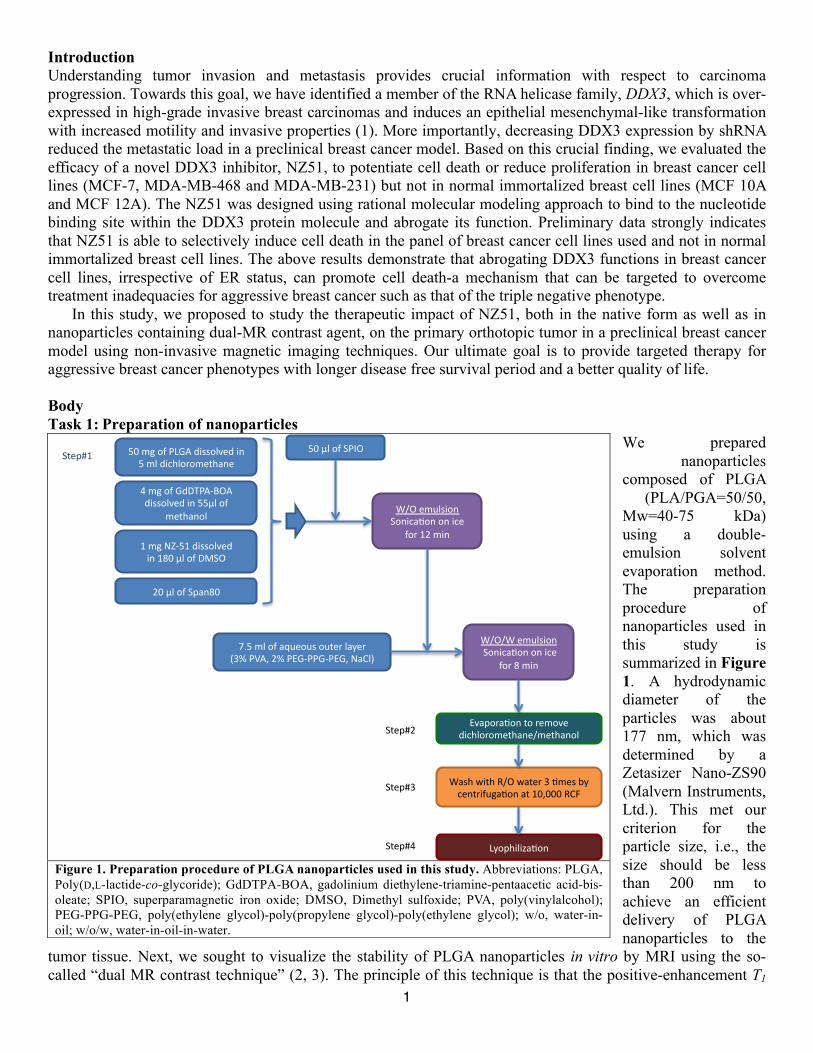
Introduction Understanding tumor invasion and metastasis provides crucial information with respect to carcinoma progression. Towards this goal, we have identified a member of the RNA helicase family, DDX3, which is over-expressed in high-grade invasive breast carcinomas and induces an epithelial mesenchymal-like transformation with increased motility and invasive properties (1). More importantly, decreasing DDX3 expression by shRNA reduced the metastatic load in a preclinical breast cancer model. Based on this crucial finding, we evaluated the efficacy of a novel DDX3 inhibitor, NZ51, to potentiate cell death or reduce proliferation in breast cancer cell lines (MCF-7, MDA-MB-468 and MDA-MB-231) but not in normal immortalized breast cell lines (MCF 10A and MCF 12A). The NZ51 was designed using rational molecular modeling approach to bind to the nucleotide binding site within the DDX3 protein molecule and abrogate its function. Preliminary data strongly indicates that NZ51 is able to selectively induce cell death in the panel of breast cancer cell lines used and not in normal immortalized breast cell lines. The above results demonstrate that abrogating DDX3 functions in breast cancer cell lines, irrespective of ER status, can promote cell death-a mechanism that can be targeted to overcome treatment inadequacies for aggressive breast cancer such as that of the triple negative phenotype. In this study, we proposed to study the therapeutic impact of NZ51, both in the native form as well as in nanoparticles containing dual-MR contrast agent, on the primary orthotopic tumor in a preclinical breast cancer model using non-invasive magnetic imaging techniques. Our ultimate goal is to provide targeted therapy for aggressive breast cancer phenotypes with longer disease free survival period and a better quality of life. Body Task 1: Preparation of nanoparticles
We prepared nanoparticles
composed of PLGA (PLA/PGA=50/50,
Mw=40-75 kDa) using a double-emulsion solvent evaporation method. The preparation procedure of nanoparticles used in this study is summarized in Figure 1. A hydrodynamic diameter of the particles was about 177 nm, which was determined by a Zetasizer Nano-ZS90 (Malvern Instruments, Ltd.). This met our criterion for the particle size, i.e., the size should be less than 200 nm to achieve an efficient delivery of PLGA nanoparticles to the
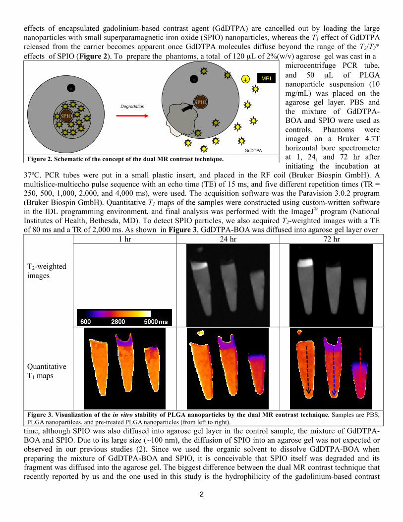
tumor tissue. Next, we sought to visualize the stability of PLGA nanoparticles in vitro by MRI using the so-called “dual MR contrast technique” (2, 3). The principle of this technique is that the positive-enhancement T1
Figure 1. Preparation procedure of PLGA nanoparticles used in this study. Abbreviations: PLGA, Poly(D,L-lactide-co-glycoride); GdDTPA-BOA, gadolinium diethylene-triamine-pentaacetic acid-bis-oleate; SPIO, superparamagnetic iron oxide; DMSO, Dimethyl sulfoxide; PVA, poly(vinylalcohol); PEG-PPG-PEG, poly(ethylene glycol)-poly(propylene glycol)-poly(ethylene glycol); w/o, water-in-oil; w/o/w, water-in-oil-in-water.
!"#$%#&'#()*+#,-..&/01,#-2#
!#$/#,-34/&5&$164721#
8#$%#&'#*,9:(+;<=+#
,-..&/01,#-2#!!>/#&'#
$16472&/#
?#$%#@A;!?#,-..&/01,#
-2#?B"#>/#&'#9CD=#
E"#>/#&'#DF72B"#
GH!#$/#&'#7IJ1&J.#&J615#/7K15#
LMN#(O+P#EN#(Q*;((*;(Q*P#@7R/S#
TU=#1$J/.-&2#
D&2-37V&2#&2#-31#
'&5#?E#$-2#
TU=UT#1$J/.-&2#
#D&2-37V&2#&2#-31#
'&5#B#$-2##
Q07F&57V&2#63$&01#
,-34/&5&$164721U$16472&/#
T7.4#W-64#XU=#W7615#M#V$1.#YK#
31265-'J%7V&2#76#?"P"""#XRZ#
)K&F4-/-[7V&2#
D61F\M#
D61F\8#
D61F\E#
D61F\?#!"#>/#&'#D(]=#
2
effects of encapsulated gadolinium-based contrast agent (GdDTPA) are cancelled out by loading the large nanoparticles with small superparamagnetic iron oxide (SPIO) nanoparticles, whereas the T1 effect of GdDTPA released from the carrier becomes apparent once GdDTPA molecules diffuse beyond the range of the T2/T2* effects of SPIO (Figure 2). To prepare the phantoms, a total of 120 µL of 2%(w/v) agarose gel was cast in a
microcentrifuge PCR tube, and 50 µL of PLGA nanoparticle suspension (10 mg/mL) was placed on the agarose gel layer. PBS and the mixture of GdDTPA-BOA and SPIO were used as controls. Phantoms were imaged on a Bruker 4.7T horizontal bore spectrometer at 1, 24, and 72 hr after initiating the incubation at
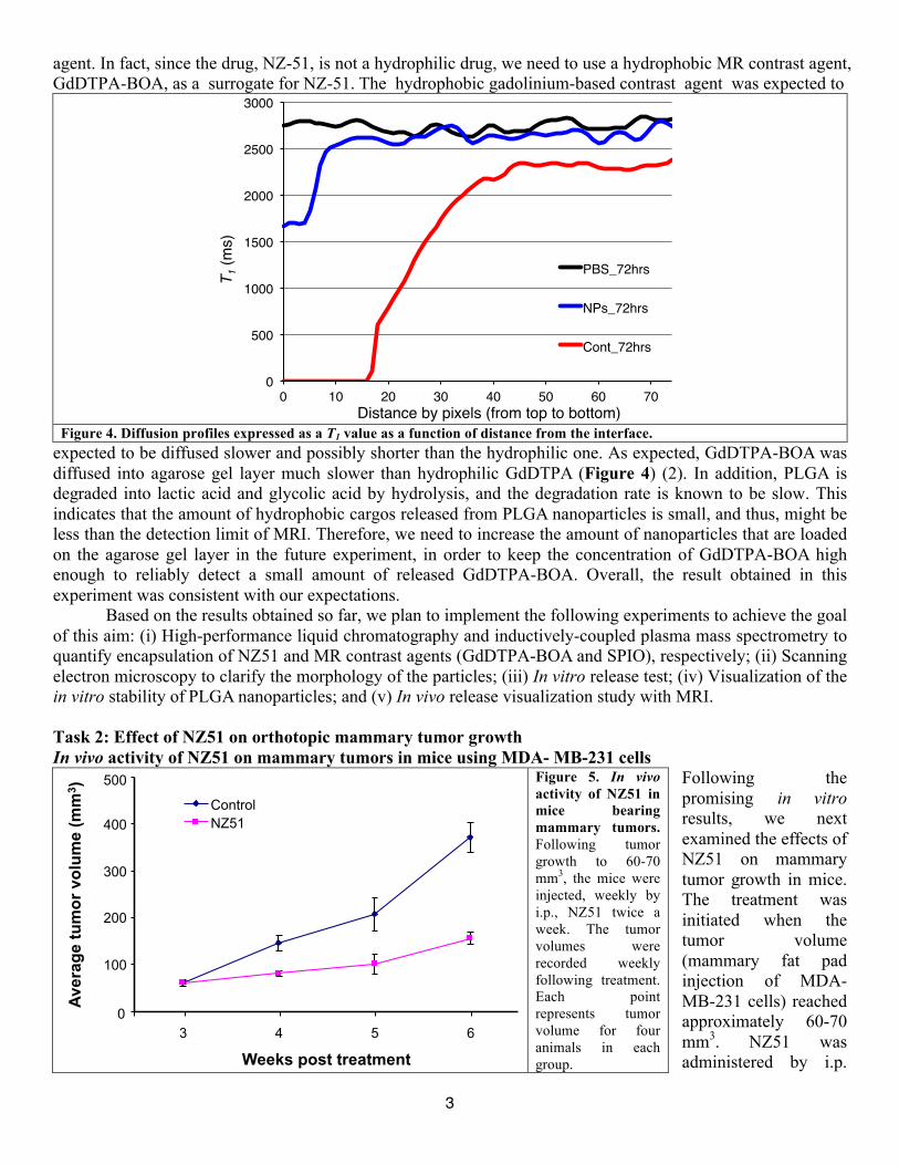
37ºC. PCR tubes were put in a small plastic insert, and placed in the RF coil (Bruker Biospin GmbH). A multislice-multiecho pulse sequence with an echo time (TE) of 15 ms, and five different repetition times (TR = 250, 500, 1,000, 2,000, and 4,000 ms), were used. The acquisition software was the Paravision 3.0.2 program (Bruker Biospin GmbH). Quantitative T1 maps of the samples were constructed using custom-written software in the IDL programming environment, and final analysis was performed with the ImageJ® program (National Institutes of Health, Bethesda, MD). To detect SPIO particles, we also acquired T2-weighted images with a TE of 80 ms and a TR of 2,000 ms. As shown in Figure 3, GdDTPA-BOA was diffused into agarose gel layer over T2-weighted images Quantitative T1 maps
1 hr 24 hr 72 hr
Figure 3. Visualization of the in vitro stability of PLGA nanoparticles by the dual MR contrast technique. Samples are PBS, PLGA nanopartilces, and pre-treated PLGA nanoparticles (from left to right).
time, although SPIO was also diffused into agarose gel layer in the control sample, the mixture of GdDTPA-BOA and SPIO. Due to its large size (~100 nm), the diffusion of SPIO into an agarose gel was not expected or observed in our previous studies (2). Since we used the organic solvent to dissolve GdDTPA-BOA when preparing the mixture of GdDTPA-BOA and SPIO, it is conceivable that SPIO itself was degraded and its fragment was diffused into the agarose gel. The biggest difference between the dual MR contrast technique that recently reported by us and the one used in this study is the hydrophilicity of the gadolinium-based contrast
Figure 2. Schematic of the concept of the dual MR contrast technique.
SPIO!
SPIO!
+!
Degradation!
-!-!
MRI!
GdDTPA!
3
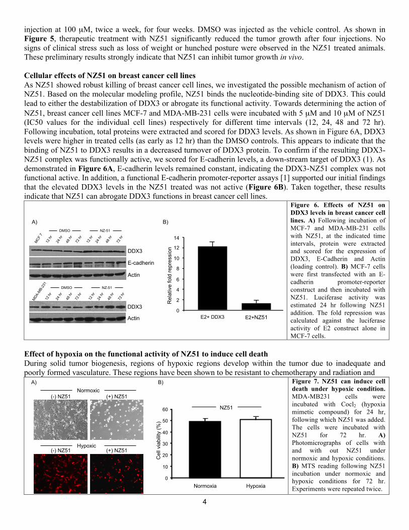
agent. In fact, since the drug, NZ-51, is not a hydrophilic drug, we need to use a hydrophobic MR contrast agent, GdDTPA-BOA, as a surrogate for NZ-51. The hydrophobic gadolinium-based contrast agent was expected to
Figure 4. Diffusion profiles expressed as a T1 value as a function of distance from the interface.
expected to be diffused slower and possibly shorter than the hydrophilic one. As expected, GdDTPA-BOA was diffused into agarose gel layer much slower than hydrophilic GdDTPA (Figure 4) (2). In addition, PLGA is degraded into lactic acid and glycolic acid by hydrolysis, and the degradation rate is known to be slow. This indicates that the amount of hydrophobic cargos released from PLGA nanoparticles is small, and thus, might be less than the detection limit of MRI. Therefore, we need to increase the amount of nanoparticles that are loaded on the agarose gel layer in the future experiment, in order to keep the concentration of GdDTPA-BOA high enough to reliably detect a small amount of released GdDTPA-BOA. Overall, the result obtained in this experiment was consistent with our expectations.
Based on the results obtained so far, we plan to implement the following experiments to achieve the goal of this aim: (i) High-performance liquid chromatography and inductively-coupled plasma mass spectrometry to quantify encapsulation of NZ51 and MR contrast agents (GdDTPA-BOA and SPIO), respectively; (ii) Scanning electron microscopy to clarify the morphology of the particles; (iii) In vitro release test; (iv) Visualization of the in vitro stability of PLGA nanoparticles; and (v) In vivo release visualization study with MRI.
Task 2: Effect of NZ51 on orthotopic mammary tumor growth In vivo activity of NZ51 on mammary tumors in mice using MDA- MB-231 cells
Following the promising in vitro results, we next examined the effects of NZ51 on mammary tumor growth in mice. The treatment was initiated when the tumor volume (mammary fat pad injection of MDA-MB-231 cells) reached approximately 60-70 mm3. NZ51 was administered by i.p.
0!
500!
1000!
1500!
2000!
2500!
3000!
0! 10! 20! 30! 40! 50! 60! 70!
T 1 (m
s)!
Distance by pixels (from top to bottom)!
PBS_72hrs!
NPs_72hrs!
Cont_72hrs!
Figure 5. In vivo activity of NZ51 in mice bearing mammary tumors. Following tumor growth to 60-70 mm3, the mice were injected, weekly by i.p., NZ51 twice a week. The tumor volumes were recorded weekly following treatment. Each point represents tumor volume for four animals in each group. Weeks post treatment
Control NZ51
Ave
rage
tum
or v
olum
e (m
m3 )
0
100
200
300
400
500
3 4 5 6
4
injection at 100 µM, twice a week, for four weeks. DMSO was injected as the vehicle control. As shown in Figure 5, therapeutic treatment with NZ51 significantly reduced the tumor growth after four injections. No signs of clinical stress such as loss of weight or hunched posture were observed in the NZ51 treated animals. These preliminary results strongly indicate that NZ51 can inhibit tumor growth in vivo. Cellular effects of NZ51 on breast cancer cell lines As NZ51 showed robust killing of breast cancer cell lines, we investigated the possible mechanism of action of NZ51. Based on the molecular modeling profile, NZ51 binds the nucleotide-binding site of DDX3. This could lead to either the destabilization of DDX3 or abrogate its functional activity. Towards determining the action of NZ51, breast cancer cell lines MCF-7 and MDA-MB-231 cells were incubated with 5 µM and 10 µM of NZ51 (IC50 values for the individual cell lines) respectively for different time intervals (12, 24, 48 and 72 hr). Following incubation, total proteins were extracted and scored for DDX3 levels. As shown in Figure 6A, DDX3 levels were higher in treated cells (as early as 12 hr) than the DMSO controls. This appears to indicate that the binding of NZ51 to DDX3 results in a decreased turnover of DDX3 protein. To confirm if the resulting DDX3-NZ51 complex was functionally active, we scored for E-cadherin levels, a down-stream target of DDX3 (1). As demonstrated in Figure 6A, E-cadherin levels remained constant, indicating the DDX3-NZ51 complex was not functional active. In addition, a functional E-cadherin promoter-reporter assays [1] supported our initial findings that the elevated DDX3 levels in the NZ51 treated was not active (Figure 6B). Taken together, these results indicate that NZ51 can abrogate DDX3 functions in breast cancer cell lines.
Figure 6. Effects of NZ51 on DDX3 levels in breast cancer cell lines. A) Following incubation of MCF-7 and MDA-MB-231 cells with NZ51, at the indicated time intervals, protein were extracted and scored for the expression of DDX3, E-Cadherin and Actin (loading control). B) MCF-7 cells were first transfected with an E-cadherin promoter-reporter construct and then incubated with NZ51. Luciferase activity was estimated 24 hr following NZ51 addition. The fold repression was calculated against the luciferase activity of E2 construct alone in MCF-7 cells.
Effect of hypoxia on the functional activity of NZ51 to induce cell death During solid tumor biogenesis, regions of hypoxic regions develop within the tumor due to inadequate and poorly formed vasculature. These regions have been shown to be resistant to chemotherapy and radiation and
Figure 7. NZ51 can induce cell death under hypoxic condition. MDA-MB231 cells were incubated with Cocl2 (hypoxia mimetic compound) for 24 hr, following which NZ51 was added. The cells were incubated with NZ51 for 72 hr. A) Photomicrographs of cells with and with out NZ51 under normoxic and hypoxic conditions. B) MTS reading following NZ51 incubation under normoxic and hypoxic conditions for 72 hr. Experiments were repeated twice.
MCF
7
DMSO
12 h
r 24
hr
48 h
r 72
hr
12 h
r 24
hr
48 h
r
72 h
r
NZ-51
DDX3
E-cadherin
Actin
MDA
-MB-
231
DMSO
12 h
r 24
hr
48 h
r 72
hr
12 h
r 24
hr
48 h
r 72
hr
NZ-51
DDX3
Actin E2+ DDX3 E2+NZ51 0 2 4 6 8
10 12 14
Rel
ativ
e fo
ld re
pres
sion
A) B)
Normoxia Hypoxia 0
10 20 30 40 50 60
Cel
l via
bilit
y (%
)
NZ51
Normoxic
Hypoxic
(-) NZ51
(+) NZ51 (-) NZ51
(+) NZ51
A) B)
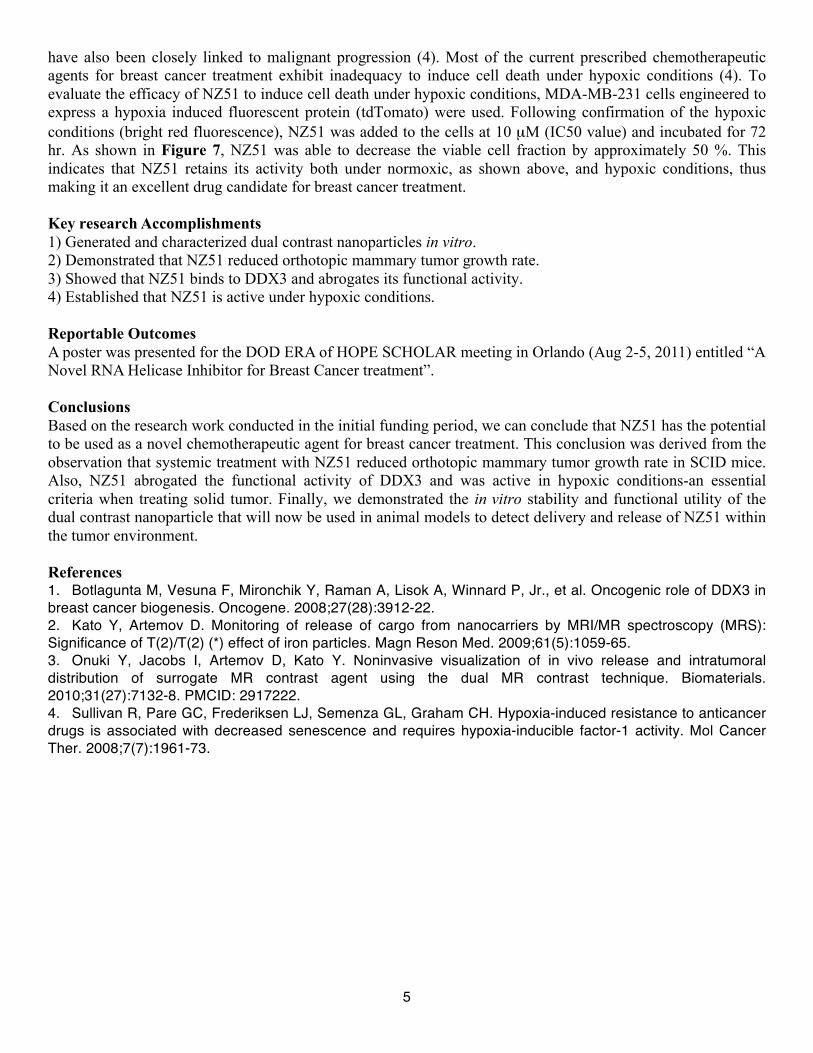
5
have also been closely linked to malignant progression (4). Most of the current prescribed chemotherapeutic agents for breast cancer treatment exhibit inadequacy to induce cell death under hypoxic conditions (4). To evaluate the efficacy of NZ51 to induce cell death under hypoxic conditions, MDA-MB-231 cells engineered to express a hypoxia induced fluorescent protein (tdTomato) were used. Following confirmation of the hypoxic conditions (bright red fluorescence), NZ51 was added to the cells at 10 µM (IC50 value) and incubated for 72 hr. As shown in Figure 7, NZ51 was able to decrease the viable cell fraction by approximately 50 %. This indicates that NZ51 retains its activity both under normoxic, as shown above, and hypoxic conditions, thus making it an excellent drug candidate for breast cancer treatment. Key research Accomplishments 1) Generated and characterized dual contrast nanoparticles in vitro. 2) Demonstrated that NZ51 reduced orthotopic mammary tumor growth rate. 3) Showed that NZ51 binds to DDX3 and abrogates its functional activity. 4) Established that NZ51 is active under hypoxic conditions. Reportable Outcomes A poster was presented for the DOD ERA of HOPE SCHOLAR meeting in Orlando (Aug 2-5, 2011) entitled “A Novel RNA Helicase Inhibitor for Breast Cancer treatment”. Conclusions Based on the research work conducted in the initial funding period, we can conclude that NZ51 has the potential to be used as a novel chemotherapeutic agent for breast cancer treatment. This conclusion was derived from the observation that systemic treatment with NZ51 reduced orthotopic mammary tumor growth rate in SCID mice. Also, NZ51 abrogated the functional activity of DDX3 and was active in hypoxic conditions-an essential criteria when treating solid tumor. Finally, we demonstrated the in vitro stability and functional utility of the dual contrast nanoparticle that will now be used in animal models to detect delivery and release of NZ51 within the tumor environment. References 1. Botlagunta M, Vesuna F, Mironchik Y, Raman A, Lisok A, Winnard P, Jr., et al. Oncogenic role of DDX3 in breast cancer biogenesis. Oncogene. 2008;27(28):3912-22. 2. Kato Y, Artemov D. Monitoring of release of cargo from nanocarriers by MRI/MR spectroscopy (MRS): Significance of T(2)/T(2) (*) effect of iron particles. Magn Reson Med. 2009;61(5):1059-65. 3. Onuki Y, Jacobs I, Artemov D, Kato Y. Noninvasive visualization of in vivo release and intratumoral distribution of surrogate MR contrast agent using the dual MR contrast technique. Biomaterials. 2010;31(27):7132-8. PMCID: 2917222. 4. Sullivan R, Pare GC, Frederiksen LJ, Semenza GL, Graham CH. Hypoxia-induced resistance to anticancer drugs is associated with decreased senescence and requires hypoxia-inducible factor-1 activity. Mol Cancer Ther. 2008;7(7):1961-73.