Embed Size (px)
Citation preview
Does Gross Appearance Indicate Prognosisin Intrahepatic Cholangiocarcinoma?
MASAKAZU YAMAMOTO, MD,1* KEN TAKASAKI, MD,1 TATSUYA YOSHIKAWA, MD,1
KEIKO UENO, MD,2 AND MASAYUKI NAKANO, MD3
1Department of Gastrointestinal Surgery, Institute of Gastroenterology, Tokyo Women’sMedical College, Tokyo, Japan
2Division of Radiology, Tokyo Women’s Medical College, Tokyo, Japan3Division of Pathology, Chiba University Hospital, Chiba, Japan
Background and Objectives:Survival after surgery for intrahepatic chol-angiocarcinoma (ICC) is usually poor. The objective of this study was toinvestigate whether the gross appearance of ICC indicates postoperativeprognosis.Methods: Seventy patients with ICC underwent hepatectomy, with a 50%curative resection rate. Tumors were classified according to gross appear-ance [mass-forming (n4 28), periductal-infiltrating (n4 14), intraductalgrowth (n4 10), and mass-forming plus periductal-infiltrating (n4 18)],and the presence of lymph node or intrahepatic metastasis was studiedmicroscopically.Results: The incidence of positive lymph nodes was significantly higherin the patients with mass-forming plus periductal-infiltrating tumors thanin those with intraductal growth tumors (P 4 0.0089). The curative re-section rate was significantly lower in patients with mass-forming plusperiductal-infiltrating tumors than in those with either mass-forming orintraductal growth tumors (P 4 0.0001,P 4 0.0048, respectively). The5-year survival rate after surgery in patients with mass-forming plus peri-ductal-infiltrating tumors (0%) was significantly lower than that in pa-tients with mass-forming tumors (39%) or intraductal growth tumors(69%) (P 4 0.0036,P 4 0.0011, respectively). Multivariate analysisusing Cox’s hazards model revealed that lymph node metastasis (P 40.0109) and curative resection (P 4 0.0315) were statistically significantindependent prognostic factors; however, macroscopic types were not.Conclusions:Patients with mass-forming plus periductal-infiltrating ICCshave a poor prognosis; however, the macroscopic types may not be astatistically significant independent prognostic factor.J. Surg. Oncol. 1998;69:162–167. © 1998 Wiley-Liss, Inc.
KEY WORDS: intrahepatic cholangiocarcinoma; gross appearance;postoperative survival
INTRODUCTION
Intrahepatic cholangiocarcinoma (ICC) is a rare tumorassociated with poor survival after resection. Becausemost cases of ICC are detected at an advanced stage,curative resection is often impossible. Although patientshave survived for more than 5 years after surgery [1–4],survival rates after surgery have not yet been reported fora large series of patients treated at a single institution
[5,6]. Over the past two decades, we have performedhepatic resection in more than 1,200 patients with pri-mary hepatic carcinoma, including 83 with ICC. Mor-phologic features and metastasis have been studied in
*Correspondence to: Masakazu Yamamoto, MD, Tokyo MetropolitanEbara Hospital, 4-5-10, Higashi-Yukigaya Ohta-ku, Tokyo, 145-0065Japan. Fax No.: (81)3-5734-8023.Accepted 22 September 1998
Journal of Surgical Oncology 1998;69:162–167
© 1998 Wiley-Liss, Inc.
detail. The aim of this study was to determine whethermacroscopic features indicate prognosis after surgery.
MATERIALS AND METHODS
Eighty-three patients with ICC underwent surgical re-section from November 27, 1980, through February, 15,1996, at the Institute of Gastroenterology, Tokyo Wom-en’s Medical College, Tokyo. Cases of hilar cholangio-carcinoma were excluded. Peritoneal dissemination wasalso present in five patients. Intrahepatic metastases werepresent in the residual liver in eight patients. These 13patients were excluded from the study. The remaining 70patients were studied. Patients did not receive any che-motherapy during the preoperative or postoperative pe-riod.
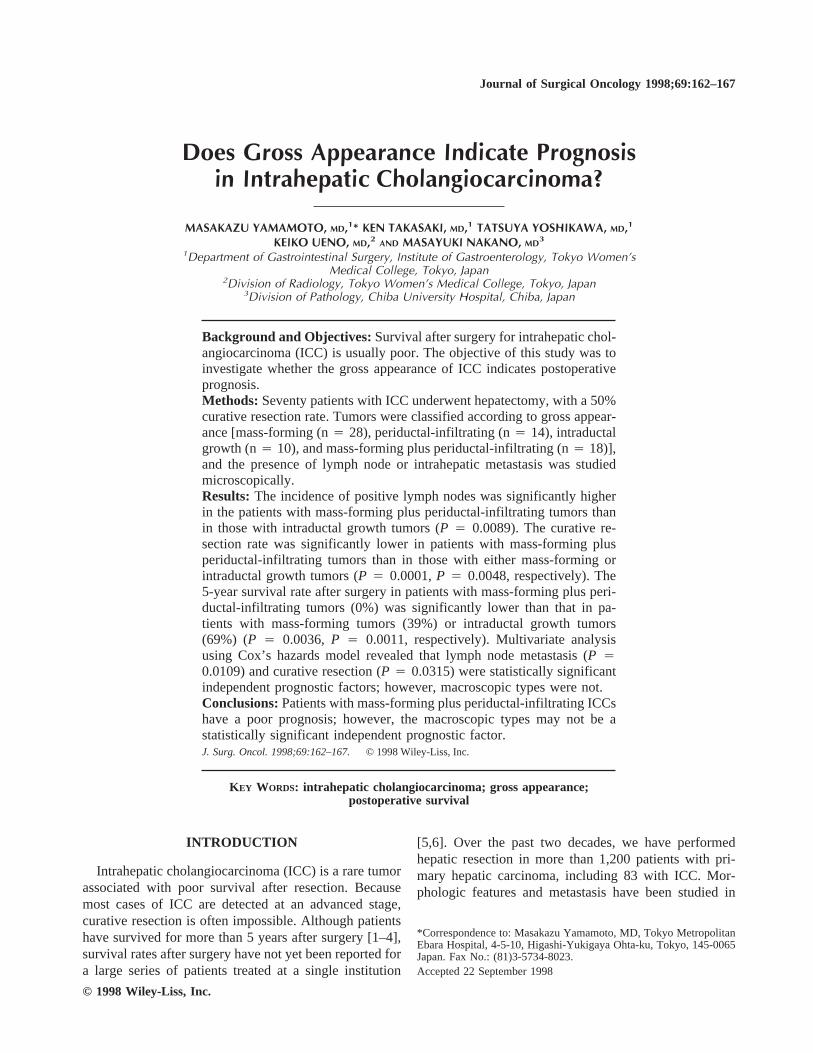
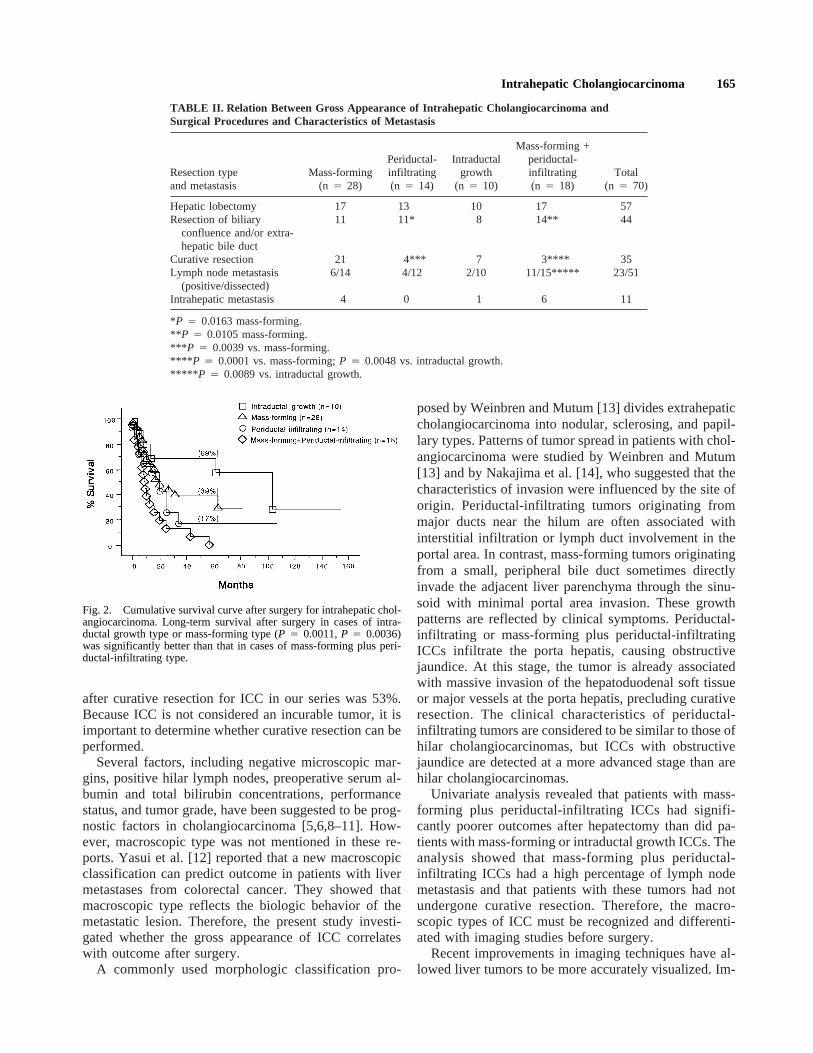
ICCs were classified on the basis of gross appear-ance—mass-forming, periductal-infiltrating, intraductalgrowth, or mass-forming plus periductal-infiltratingtypes—as proposed by the Liver Cancer Study Group ofJapan [7]. Mass-forming tumors (n4 28) are character-ized by the presence of a nodular mass in the liver pa-renchyma, with a distinct border (Fig. 1A). Periductal-infiltrating tumors (n4 14) are characterized by tumorinfiltration along the bile duct; they occasionally involve
the surrounding blood vessels or hepatic parenchyma(Fig. 1B). Intraductal growth tumors (n4 10) are char-acterized by papillary growth or granular growth or bothwithin the bile duct lumen (Fig. 1C). Mass-forming plusperiductal-infiltrating tumors (n4 18) have both mass-forming and periductal-infiltrating features (Fig. 1D).
Lymph node metastasis and intrahepatic metastasiswere also noted and confirmed histopathologically.
Cumulative survival rates of all patients were calcu-lated with the Kaplan-Meier method; survival rates werecompared using the log-rank test. The duration of sur-vival was defined as the time from the first liver surgeryto the date of death or last contact. For comparisonsbetween the macroscopic subgroups, the chi squared testwas used. Several clinicopathologic factors, includingthose found to be associated with patient survival byunivariate analysis, were subjected to multivariate analy-sis using Cox’s proportional hazards model. The signifi-cance level was set atP < 0.05.
RESULTSCharacteristics of Patients
Of the 70 patients in this study, 43 were men and 27were women, with a mean age of 59.9 years (range 30 to
Fig. 1. Gross appearance of intrahepatic cholangiocarcinoma (maximum diameter).A: Mass-forming type (5.7 cm).B: Periductal-infiltratingtype (3.5 cm).C: Intraductal growth type (2.5 cm).D: Mass-forming plus periductal-infiltrating type (4.5 cm).
Intrahepatic Cholangiocarcinoma 163
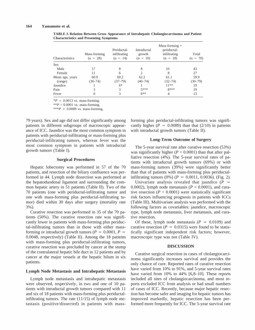
79 years). Sex and age did not differ significantly amongpatients in different subgroups of macroscopic appear-ance of ICC. Jaundice was the most common symptom inpatients with periductal-infiltrating or mass-forming plusperiductal-infiltrating tumors, whereas fever was themost common symptom in patients with intraductalgrowth tumors (Table I).
Surgical Procedures
Hepatic lobectomy was performed in 57 of the 70patients, and resection of the biliary confluence was per-formed in 44. Lymph node dissection was performed atthe hepatoduodenal ligament and surrounding the com-mon hepatic artery in 51 patients (Table II). Two of the70 patients (one with periductal-infiltrating tumor andone with mass-forming plus periductal-infiltrating tu-mor) died within 30 days after surgery (mortality rate3%).
Curative resection was performed in 35 of the 70 pa-tients (50%). The curative resection rate was signifi-cantly lower in patients with mass-forming plus periduc-tal-infiltrating tumors than in those with either mass-forming or intraductal growth tumors (P 4 0.0001,P 40.0048, respectively) (Table II). Among the 18 patientswith mass-forming plus periductal-infiltrating tumors,curative resection was precluded by cancer at the stumpof the contralateral hepatic bile duct in 12 patients and bycancer at the major vessels at the hepatic hilum in sixpatients.
Lymph Node Metastasis and Intrahepatic Metastasis
Lymph node metastasis and intrahepatic metastasiswere observed, respectively, in two and one of 10 pa-tients with intraductal growth tumors compared with 11and six of 18 patients with mass-forming plus periductal-infiltrating tumors. The rate (11/15) of lymph node me-tastasis (positive/dissected) in patients with mass-
forming plus periductal-infiltrating tumors was signifi-cantly higher (P 4 0.0089) than that (2/10) in patientswith intraductal growth tumors (Table II).
Long-Term Outcome of Surgery
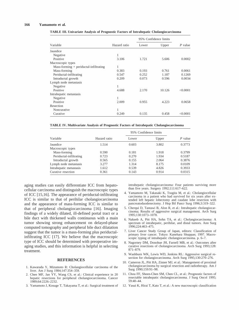
The 5-year survival rate after curative resection (53%)was significantly higher (P < 0.0001) than that after pal-liative resection (4%). The 5-year survival rates of pa-tients with intraductal growth tumors (69%) or withmass-forming tumors (39%) were significantly betterthan that of patients with mass-forming plus periductal-infiltrating tumors (0%) (P 4 0.0011, 0.0036). (Fig. 2).
Univariate analysis revealed that jaundice (P 40.0002), lymph node metastasis (P < 0.0001), and cura-tive resection (P < 0.0001) were statistically significantrisk factors influencing prognosis in patients with ICCs(Table III). Multivariate analysis was performed with thefollowing factors as covariables: jaundice, macroscopictype, lymph node metastasis, liver metastasis, and cura-tive resection.
Of these, lymph node metastasis (P 4 0.0109) andcurative resection (P 4 0.0315) were found to be statis-tically significant independent risk factors; however,macroscopic type was not (Table IV).
DISCUSSION
Curative surgical resection in cases of cholangiocarci-noma significantly increases survival and provides theonly chance of cure. Reported rates of curative resectionhave varied from 10% to 91%, and 5-year survival rateshave varied from 10% to 44% [6,8–10]. These reportsincluded all sites of cholangiocarcinoma, and most re-ports excluded ICC from analysis or had small numbersof cases of ICC. Recently, because major hepatic resec-tion has become safer and imaging for hepatic tumors hasimproved markedly, hepatic resection has been per-formed more frequently for ICC. The 5-year survival rate
TABLE I. Relation Between Gross Appearance of Intrahepatic Cholangiocarcinoma and PatientCharacteristics and Presenting Symptoms
CharacteristicsMass-forming
(n 4 28)
Periductal-infiltrating(n 4 14)
Intraductalgrowth
(n 4 10)
Mass-forming +periductal-infiltrating(n 4 18)
Total(n 4 70)
SexMale 17 8 8 10 43Female 11 6 2 8 27
Mean age, years 60.9 60.2 62.2 61.1 59.9(range) (30–74) (37–79) (40–74) (32–74) (30–79)
Jaundice 1 6* 1 11** 19Pain 3 3 5*** 8*** 19Fever 0 3 6** 4 13
*P 4 0.0013 vs. mass-forming.** P < 0.0001 vs. mass-forming.*** P 4 0.0089 vs. mass-forming.
164 Yamamoto et al.
after curative resection for ICC in our series was 53%.Because ICC is not considered an incurable tumor, it isimportant to determine whether curative resection can beperformed.
Several factors, including negative microscopic mar-gins, positive hilar lymph nodes, preoperative serum al-bumin and total bilirubin concentrations, performancestatus, and tumor grade, have been suggested to be prog-nostic factors in cholangiocarcinoma [5,6,8–11]. How-ever, macroscopic type was not mentioned in these re-ports. Yasui et al. [12] reported that a new macroscopicclassification can predict outcome in patients with livermetastases from colorectal cancer. They showed thatmacroscopic type reflects the biologic behavior of themetastatic lesion. Therefore, the present study investi-gated whether the gross appearance of ICC correlateswith outcome after surgery.
A commonly used morphologic classification pro-
posed by Weinbren and Mutum [13] divides extrahepaticcholangiocarcinoma into nodular, sclerosing, and papil-lary types. Patterns of tumor spread in patients with chol-angiocarcinoma were studied by Weinbren and Mutum[13] and by Nakajima et al. [14], who suggested that thecharacteristics of invasion were influenced by the site oforigin. Periductal-infiltrating tumors originating frommajor ducts near the hilum are often associated withinterstitial infiltration or lymph duct involvement in theportal area. In contrast, mass-forming tumors originatingfrom a small, peripheral bile duct sometimes directlyinvade the adjacent liver parenchyma through the sinu-soid with minimal portal area invasion. These growthpatterns are reflected by clinical symptoms. Periductal-infiltrating or mass-forming plus periductal-infiltratingICCs infiltrate the porta hepatis, causing obstructivejaundice. At this stage, the tumor is already associatedwith massive invasion of the hepatoduodenal soft tissueor major vessels at the porta hepatis, precluding curativeresection. The clinical characteristics of periductal-infiltrating tumors are considered to be similar to those ofhilar cholangiocarcinomas, but ICCs with obstructivejaundice are detected at a more advanced stage than arehilar cholangiocarcinomas.
Univariate analysis revealed that patients with mass-forming plus periductal-infiltrating ICCs had signifi-cantly poorer outcomes after hepatectomy than did pa-tients with mass-forming or intraductal growth ICCs. Theanalysis showed that mass-forming plus periductal-infiltrating ICCs had a high percentage of lymph nodemetastasis and that patients with these tumors had notundergone curative resection. Therefore, the macro-scopic types of ICC must be recognized and differenti-ated with imaging studies before surgery.
Recent improvements in imaging techniques have al-lowed liver tumors to be more accurately visualized. Im-
TABLE II. Relation Between Gross Appearance of Intrahepatic Cholangiocarcinoma andSurgical Procedures and Characteristics of Metastasis
Resection typeand metastasis
Mass-forming(n 4 28)
Periductal-infiltrating(n 4 14)
Intraductalgrowth
(n 4 10)
Mass-forming +periductal-infiltrating(n 4 18)
Total(n 4 70)
Hepatic lobectomy 17 13 10 17 57Resection of biliary
confluence and/or extra-hepatic bile duct
11 11* 8 14** 44
Curative resection 21 4*** 7 3**** 35Lymph node metastasis
(positive/dissected)6/14 4/12 2/10 11/15***** 23/51
Intrahepatic metastasis 4 0 1 6 11
*P 4 0.0163 mass-forming.** P 4 0.0105 mass-forming.*** P 4 0.0039 vs. mass-forming.**** P 4 0.0001 vs. mass-forming;P 4 0.0048 vs. intraductal growth.***** P 4 0.0089 vs. intraductal growth.
Fig. 2. Cumulative survival curve after surgery for intrahepatic chol-angiocarcinoma. Long-term survival after surgery in cases of intra-ductal growth type or mass-forming type (P 4 0.0011,P 4 0.0036)was significantly better than that in cases of mass-forming plus peri-ductal-infiltrating type.
Intrahepatic Cholangiocarcinoma 165
aging studies can easily differentiate ICC from hepato-cellular carcinoma and distinguish the macroscopic typesof ICC [15,16]. The appearance of periductal-infiltratingICC is similar to that of perihilar cholangiocarcinomaand the appearance of mass-forming ICC is similar tothat of peripheral cholangiocarcinoma [16]. Imagingfindings of a widely dilated, ill-defined portal tract or abile duct with thickened walls continuous with a maintumor showing strong enhancement on delayed-phasecomputed tomography and peripheral bile duct dilatationsuggest that the tumor is a mass-forming plus periductal-infiltrating ICC [17]. We believe that the macroscopictype of ICC should be determined with preoperative im-aging studies, and this information is helpful in selectingtreatment.
REFERENCES
1. Kawarada Y, Mizumoto R: Cholangiocellular carcinoma of theliver. Am J Surg 1984;147:354–359.
2. Chen MF, Jan YY, Wang CS, et al.: Clinical experience in 20hepatic resections for peripheral cholangiocarcinoma. Cancer1989;64:2226–2232.
3. Yamamoto J, Kosuge T, Takayama T, et al.: Surgical treatment of
intrahepatic cholangiocarcinoma: Four patients surviving morethan five years. Surgery 1992;111:617–622.
4. Yamamoto M, Takasaki K, Tsugita M, et al.: Cholangiocellularcarcinoma in a patient who had survived for six years after ex-tended left hepatic lobectomy and caudate lobe resection withpancreatoduodenectomy. J Hep Bil Pancr Surg 1996;3:319–322.
5. Cherqui D, Tantawi B, Alon R, et al.: Intrahepatic cholangiocar-cinoma; Results of aggressive surgical management. Arch Surg1995;130:1073–1078.
6. Nakeeb A, Pitt HA, Sohn TA, et al.: Cholangiocarcinoma: Aspectrum of intrahepatic, perihilar, and distal tumors. Ann Surg1996;224:463–475.
7. Liver Cancer Study Group of Japan, editors: Classification ofprimary liver cancer. Tokyo: Kanehara Shuppan; 1997. Macro-scopic typing of intrahepatic cholangiocarcinoma. p. 6–7.
8. Nagorney DM, Donohue JH, Farnell MB, et al.: Outcomes aftercurative resections of cholangiocarcinoma. Arch Surg 1993;128:871–879.
9. Washburn WK, Lewis WD, Jenkins RL: Aggressive surgical re-section for cholangiocarcinoma. Arch Surg 1995;130:270–276.
10. Cameron JL, Pitt HA, Zinner MJ, et al.: Management of proximalcholangiocarcinoma by surgical resection and radiotherapy. Am JSurg 1990;159:91–98.
11. Chou FF, Sheen-Chen SM, Chen CL, et al.: Prognostic factors ofresectable intrahepatic cholangiocarcinoma. J Surg Oncol 1995;59:40–44.
12. Yasui K, Hirai T, Kato T, et al.: A new macroscopic classification
TABLE III. Univariate Analysis of Prognostic Factors of Intrahepatic Cholangiocarcinoma
Variable Hazard ratio
95% Confidence limits
P valueLower Upper
JaundiceNegative 1Positive 3.106 1.721 5.606 0.0002
Macroscopic typesMass-forming + periductal-infiltrating 1Mass-forming 0.383 0.193 0.761 0.0061Periductal-infiltrating 0.547 0.252 1.187 0.1269Intraductal growth 0.209 0.073 0.596 0.0034
Lymph node metastasisNegative 1Positive 4.688 2.170 10.126 <0.0001
Intrahepatic metastasisNegative 1Positive 2.009 0.955 4.223 0.0658
ResectionNoncurative 1Curative 0.249 0.135 0.458 <0.0001
TABLE IV. Multivariate Analysis of Prognostic Factors of Intrahepatic Cholangiocarcinoma
Variable Hazard ratio
95% Confidence limits
P valueLower Upper
Jaundice 1.514 0.603 3.802 0.3773Macroscopic types
Mass-forming 0.590 0.181 1.918 0.3799Periductal-infiltrating 0.723 0.270 1.934 0.5187Intraductal growth 0.565 0.155 2.064 0.3876
Lymph node metastasis 3.277 1.314 8.175 0.0109Intrahepatic metastasis 1.612 0.539 4.826 0.3933Curative resection 0.361 0.143 0.914 0.0315
166 Yamamoto et al.
predicts prognosis for patients with liver metastases from colo-rectal cancer. Ann Surg 1997;226:582–586.
13. Weinbren K, Mutum SS: Pathological aspects of cholangiocarci-noma. J Pathol 1982;139:217–238.
14. Nakajima T, Kondo Y, Miyazaki M, et al.: A histopathologicstudy of 102 cases of intrahepatic cholangiocarcinoma: Histologicclassification and modes of spreading. Hum Pathol 1988;19:1228–1234.
15. Nesbit GM, Johnson CD, James EM, et al.: Cholangiocarcinoma:Diagnosis and evaluation of resectability by CT and sonography
as procedures complementary to cholangiography. Am J Roent-genol 1988;151:933–938.
16. Choi BI, Han JK, Kim TK: Diagnosis and staging of cholangio-carcinomas by computed tomography. In: Meyers MA (ed): Neo-plasms of digestive tract. Imaging, staging, and management.Philadelphia: Lippincott-Raven; 1998:508–516.
17. Honda H, Onitsuka H, Yasumori K, et al.: Intrahepatic peripheralcholangiocarcinoma: Two-phased dynamic incremental CT andpathologic correlation. J Comput Assist Tomogr 1993;17:397–402.
Intrahepatic Cholangiocarcinoma 167