Embed Size (px)
Citation preview

RESEARCH ARTICLE Open Access
Does stress perfusion imaging improve thediagnostic accuracy of late gadoliniumenhanced cardiac magnetic resonance forestablishing the etiology of heart failure?Gaurav S. Gulsin1,2*, Abishek Shetye1,2, Jeffrey Khoo1,2, Daniel J. Swarbrick1,2, Eylem Levelt1,2, Florence Y. Lai1,2,Iain B. Squire1,2, Jayanth R. Arnold1,2 and Gerry P. McCann1,2
Abstract
Background: Late gadolinium enhanced cardiovascular magnetic resonance (LGE-CMR) has excellent specificity,sensitivity and diagnostic accuracy for differentiating between ischemic cardiomyopathy (ICM) and non-ischemicdilated cardiomyopathy (NICM). CMR first-pass myocardial perfusion imaging (perfusion-CMR) may also play role indistinguishing heart failure of ischemic and non-ischemic origins, although the utility of additional of stress perfusionimaging in such patients is unclear. The aim of this retrospective study was to assess whether the addition of adenosinestress perfusion imaging to LGE-CMR is of incremental value for differentiating ICM and NICM in patients with severe leftventricular systolic dysfunction (LVSD) of uncertain etiology.
Methods: We retrospectively identified 100 consecutive adult patients (median age 69 years (IQR 59–73)) with severeLVSD (mean LV EF 26.6 ± 7.0%) referred for perfusion-CMR to establish the underlying etiology of heart failure. The causeof heart failure was first determined on examination of CMR cine and LGE images in isolation. Subsequent examination ofcomplete adenosine stress perfusion-CMR studies (cine, LGE and perfusion images) was performed to identify whetherthis altered the initial diagnosis.
Results: On LGE-CMR, 38 patients were diagnosed with ICM, 46 with NICM and 16 with dual pathology. With perfusion-CMR, there were 39 ICM, 44 NICM and 17 dual pathology diagnoses. There was excellent agreement in diagnosesbetween LGE-CMR and perfusion-CMR (κ 0.968, p<0.001). The addition of adenosine stress perfusion images toLGE-CMR altered the diagnosis in only two of the 100 patients.
Conclusion: The addition of adenosine stress perfusion-CMR to cine and LGE-CMR provides minimal incrementaldiagnostic yield for determining the etiology of heart failure in patients with severe LVSD.
Keywords: Cardiovascular magnetic resonance, Heart failure, Late gadolinium enhancement, Adenosine stress perfusion,Non-ischemic cardiomyopathy
* Correspondence: [email protected] of Cardiovascular Sciences, University of Leicester, GlenfieldHospital, Groby Road, Leicester LE3 9QP, UK2The NIHR Leicester Cardiovascular Biomedical Research Unit, GlenfieldHospital, Leicester, UK
© The Author(s). 2017 Open Access This article is distributed under the terms of the Creative Commons Attribution 4.0International License (http://creativecommons.org/licenses/by/4.0/), which permits unrestricted use, distribution, andreproduction in any medium, provided you give appropriate credit to the original author(s) and the source, provide a link tothe Creative Commons license, and indicate if changes were made. The Creative Commons Public Domain Dedication waiver(http://creativecommons.org/publicdomain/zero/1.0/) applies to the data made available in this article, unless otherwise stated.
Gulsin et al. BMC Cardiovascular Disorders (2017) 17:98 DOI 10.1186/s12872-017-0529-y

BackgroundIdentifying the etiology of heart failure has importantmanagement and prognostic implications [1]. Thera-peutic strategies for ischemic cardiomyopathy (ICM)include revascularisation and/or secondary preventionmeasures such as antiplatelet and lipid lowering therap-ies. Conversely, the management of non-ischemic dilatedcardiomyopathy (NICM) relies predominantly on pharma-cological agents and device therapy [2]. Rarer causes ofNICM (e.g. sarcoid, amyloid, HIV) require treatment of theunderlying condition [3]. Importantly survival rates arepoorer in those patients with heart failure of ischemicorigin [4]. Clinical guidelines suggest echocardiography asthe initial investigation of choice for evaluation of chronicheart failure [1, 5]. In many instances, echocardiographymay reliably establish the underlying cause of heart failure.Myocardial regional wall motion abnormalities alone, how-ever, may not distinguish heart failure of ischemic originfrom NICM, since segmental wall motion abnormalitiesmay accompany both [6]. Where echocardiography doesnot clearly identify the etiology of chronic heart failure, car-diac magnetic resonance imaging (CMR) is advantageous[1], owing to its capacity to detect evidence of myocardialinfarction and non-ischemic fibrosis and assess ischemiaand viability in a single examination [7].Late gadolinium enhanced CMR (LGE-CMR) has been
shown to be non-inferior (and indeed may be superior)to coronary angiography in differentiating heart failuredue to coronary artery disease (CAD) from NICM [8, 9].Subendocardial LGE is present in CAD, whereas patientswith NICM have either no LGE or mid-wall late en-hancement that does not correspond to a coronary ar-tery territory (Fig. 1) [9]. Patients with heart failure anda history of ischemic heart disease have a high preva-lence of LGE visible on CMR (88% in one report) [10].This suggests a high likelihood that LGE-CMR will yielda diagnosis of ICM by hyperenhancement patterns alone,without the need for additional perfusion imaging. Fol-lowing myocardial infarction, the presence of LGEaffecting ≤50% of the thickness of the myocardiumpredicts the likelihood of functional recovery in responseto revascularisation of the affected coronary territory
[11, 12]. In NICM, the presence of mid-wall fibrosis isalso of prognostic value, being an independent predictorof cardiovascular and all-cause mortality [13].CMR first-pass myocardial perfusion imaging (perfusion-
CMR) has been shown in several large studies to haveexcellent sensitivity and specificity for detection of CADand may thus play a role in distinguishing heart failure ofischemic and non-ischemic origins [14, 15]. However, theadded value of adenosine stress perfusion-CMR in subjectswith severe left ventricular systolic dysfunction (LVSD) hasnot been investigated previously. Given that LGE-CMRalone has excellent specificity (96%), sensitivity (100%) anddiagnostic accuracy (95%) for differentiating between ICMand NICM [8], the utility of additional of stress perfusionimaging in such patients is questionable.Adenosine stress perfusion-CMR imaging adds time
(approximately 15 min added time for the perfusion-CMR protocol) and expense to the CMR protocol, dueto the need for additional electrocardiograms, adenosinepreparations, infusion pumps and lines, as well asphysician-supervision. Undesirable symptoms and po-tentially serious complications may occur with adenosineinfusion, albeit infrequently [16–19]. These includewheeze secondary to bronchospasm, Mobitz II 2nd or3rd degree atrioventricular block, and angina requiringsublingual nitrates [17]. Furthermore, in patients withheart failure, there is impairment of adenosine receptorexpression and signal-transduction [20], which maydiminish the hemodynamic response for stress perfusionassessment, compromising diagnostic confidence.In our regional cardiac centre, over 1000 clinical perfu-
sion-CMR studies are performed annually. Approximately10% of these are to identify the cause of heart failure, anddespite a lack of evidence of additional benefit, stress per-fusion is usually requested and performed.The aim of this retrospective study was to assess
whether the addition of adenosine stress perfusion im-aging to LGE-CMR is of incremental value for differenti-ating ICM and NICM in patients with severe LVSD ofuncertain etiology.
MethodsStudy populationWe retrospectively identified 100 consecutive adultpatients with severe LVSD on echocardiography, re-ferred for perfusion-CMR to ascertain the underlyingetiology of heart failure. Patients were scanned betweenApril 2015 and March 2016. Severe LVSD was definedas a left ventricular ejection fraction (LV EF) ≤35% andwas confirmed by CMR volumetric and functional ana-lysis prior to inclusion in the study. Participants’ pastmedical history, medications, electrocardiographic data,resting pulse and blood pressure were recorded at thetime of the CMR.
Fig. 1 Illustration of typical patterns of LGE seen in NICM and ICM.White areas within the myocardium represent LGE. a Mid-wall LGE iscommonly seen in NICM, whereas b a subendocardial distribution ofLGE is typical in ICM
Gulsin et al. BMC Cardiovascular Disorders (2017) 17:98 Page 2 of 7
The study was approved as a clinical audit and ethicsapproval was deemed unnecessary.
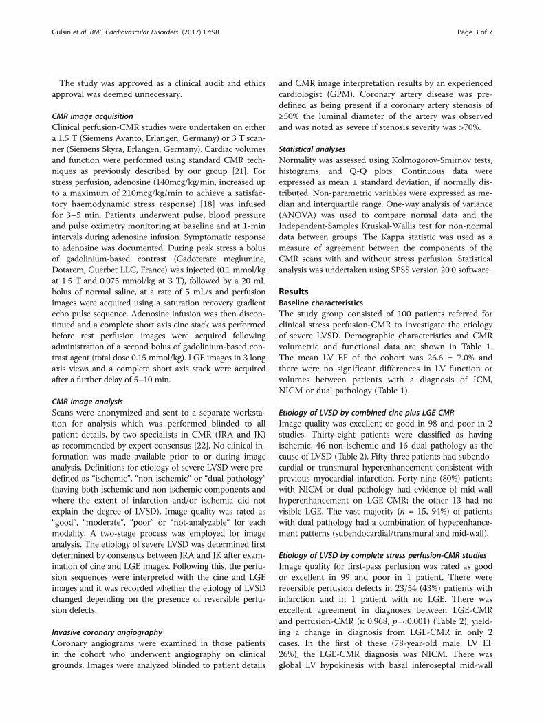
CMR image acquisitionClinical perfusion-CMR studies were undertaken on eithera 1.5 T (Siemens Avanto, Erlangen, Germany) or 3 T scan-ner (Siemens Skyra, Erlangen, Germany). Cardiac volumesand function were performed using standard CMR tech-niques as previously described by our group [21]. Forstress perfusion, adenosine (140mcg/kg/min, increased upto a maximum of 210mcg/kg/min to achieve a satisfac-tory haemodynamic stress response) [18] was infusedfor 3–5 min. Patients underwent pulse, blood pressureand pulse oximetry monitoring at baseline and at 1-minintervals during adenosine infusion. Symptomatic responseto adenosine was documented. During peak stress a bolusof gadolinium-based contrast (Gadoterate meglumine,Dotarem, Guerbet LLC, France) was injected (0.1 mmol/kgat 1.5 T and 0.075 mmol/kg at 3 T), followed by a 20 mLbolus of normal saline, at a rate of 5 mL/s and perfusionimages were acquired using a saturation recovery gradientecho pulse sequence. Adenosine infusion was then discon-tinued and a complete short axis cine stack was performedbefore rest perfusion images were acquired followingadministration of a second bolus of gadolinium-based con-trast agent (total dose 0.15 mmol/kg). LGE images in 3 longaxis views and a complete short axis stack were acquiredafter a further delay of 5–10 min.
CMR image analysisScans were anonymized and sent to a separate worksta-tion for analysis which was performed blinded to allpatient details, by two specialists in CMR (JRA and JK)as recommended by expert consensus [22]. No clinical in-formation was made available prior to or during imageanalysis. Definitions for etiology of severe LVSD were pre-defined as “ischemic”, “non-ischemic” or “dual-pathology”(having both ischemic and non-ischemic components andwhere the extent of infarction and/or ischemia did notexplain the degree of LVSD). Image quality was rated as“good”, “moderate”, “poor” or “not-analyzable” for eachmodality. A two-stage process was employed for imageanalysis. The etiology of severe LVSD was determined firstdetermined by consensus between JRA and JK after exam-ination of cine and LGE images. Following this, the perfu-sion sequences were interpreted with the cine and LGEimages and it was recorded whether the etiology of LVSDchanged depending on the presence of reversible perfu-sion defects.
Invasive coronary angiographyCoronary angiograms were examined in those patientsin the cohort who underwent angiography on clinicalgrounds. Images were analyzed blinded to patient details
and CMR image interpretation results by an experiencedcardiologist (GPM). Coronary artery disease was pre-defined as being present if a coronary artery stenosis of≥50% the luminal diameter of the artery was observedand was noted as severe if stenosis severity was >70%.
Statistical analysesNormality was assessed using Kolmogorov-Smirnov tests,histograms, and Q-Q plots. Continuous data wereexpressed as mean ± standard deviation, if normally dis-tributed. Non-parametric variables were expressed as me-dian and interquartile range. One-way analysis of variance(ANOVA) was used to compare normal data and theIndependent-Samples Kruskal-Wallis test for non-normaldata between groups. The Kappa statistic was used as ameasure of agreement between the components of theCMR scans with and without stress perfusion. Statisticalanalysis was undertaken using SPSS version 20.0 software.
ResultsBaseline characteristicsThe study group consisted of 100 patients referred forclinical stress perfusion-CMR to investigate the etiologyof severe LVSD. Demographic characteristics and CMRvolumetric and functional data are shown in Table 1.The mean LV EF of the cohort was 26.6 ± 7.0% andthere were no significant differences in LV function orvolumes between patients with a diagnosis of ICM,NICM or dual pathology (Table 1).
Etiology of LVSD by combined cine plus LGE-CMRImage quality was excellent or good in 98 and poor in 2studies. Thirty-eight patients were classified as havingischemic, 46 non-ischemic and 16 dual pathology as thecause of LVSD (Table 2). Fifty-three patients had subendo-cardial or transmural hyperenhancement consistent withprevious myocardial infarction. Forty-nine (80%) patientswith NICM or dual pathology had evidence of mid-wallhyperenhancement on LGE-CMR; the other 13 had novisible LGE. The vast majority (n = 15, 94%) of patientswith dual pathology had a combination of hyperenhance-ment patterns (subendocardial/transmural and mid-wall).
Etiology of LVSD by complete stress perfusion-CMR studiesImage quality for first-pass perfusion was rated as goodor excellent in 99 and poor in 1 patient. There werereversible perfusion defects in 23/54 (43%) patients withinfarction and in 1 patient with no LGE. There wasexcellent agreement in diagnoses between LGE-CMRand perfusion-CMR (κ 0.968, p=<0.001) (Table 2), yield-ing a change in diagnosis from LGE-CMR in only 2cases. In the first of these (78-year-old male, LV EF26%), the LGE-CMR diagnosis was NICM. There wasglobal LV hypokinesis with basal inferoseptal mid-wall
Gulsin et al. BMC Cardiovascular Disorders (2017) 17:98 Page 3 of 7

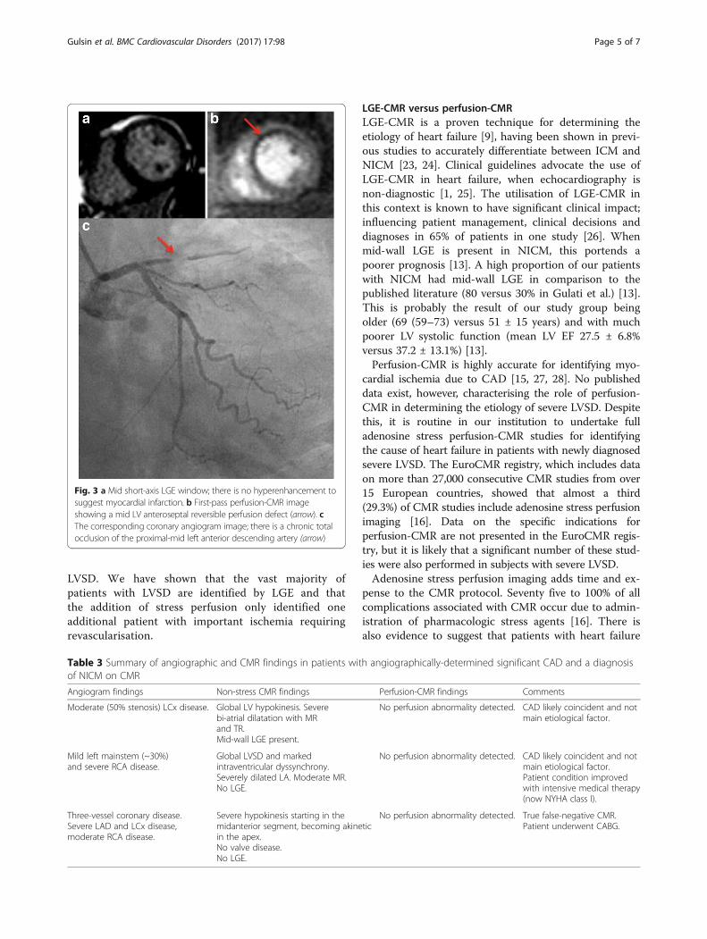
LGE (Fig. 2a). The perfusion-CMR diagnosis was dualpathology as an inferolateral subendocardial perfusionabnormality was visualised, suggestive of ischemia(Fig. 2b), but not severe enough to account for thedegree of LV impairment observed. In the second case(a 77-year-old female), the LGE-CMR diagnosis wasNICM: image quality for LGE imaging was rated aspoor (though still considered diagnostic), and no enhance-ment was visualised (Fig. 3a). The diagnosis by perfusion-CMR was ICM with demonstration of a subendocardialbasal and mid LV anteroseptal perfusion defect (Fig. 3b).Subsequently coronary angiography revealed a significantLAD stenosis (Fig. 3c) and the patient underwentrevascularisation.
Angiographic findingsThirty-two of the patients underwent coronary angiog-raphy after CMR. The vast majority (n = 30, 93.8%) ofthese showed significant CAD. Both patients whounderwent angiography that did not reveal significantCAD had LGE-CMR and perfusion-CMR diagnoses ofNICM. LGE-CMR had a sensitivity of 87% for
predicting significant CAD in subjects with severeLVSD, identifying ICM or dual pathology in 26/30patients with significant CAD on angiography. Sensitiv-ity of perfusion-CMR for predicting significant CADwas 90% (27/30 patients with significant CAD identifiedas having ICM or dual pathology). Only three cases ofCAD identified by angiography were not detected byeither LGE-CMR or perfusion-CMR (Table 3). In twoof these, the degree of coronary disease was notdeemed severe enough to alone account for the degreeof LV impairment.
DiscussionThis study is the first to evaluate the incrementalvalue of perfusion-CMR over LGE-CMR in identifyingthe etiology of heart failure in patients with severe
Table 1 Baseline characteristics of the 100 study participants
All patients (n = 100) ICM (n = 39) NICM (n = 44) Dual pathology (n = 17) P-value
Median age (years) 69 (59–73) 69 (54–84) 69 (50–88) 69 (57–81) 0.984
Gender 77% M, 33% F 77% M, 33% F 70% M, 30% F 94% M, 6% F
SBP (mmHg) 134.7 ± 23.7 135.9 ± 24.4 132.0 ± 19.8 134.5 ± 30.7 0.715
DBP (mmHg) 79.9 ± 14.8 79.7 ± 13.2 80.3 ± 14.5 77.6 ± 20.2 0.824
Pulse rate (beats/min) 72.3 ± 14.1 69.9 ± 12.4 75.1 ± 16.3 71.9 ± 11.4 0.263
ACEI (%) 73 72 71 83 0.68
ARB (%) 13 19 9 8 0.426
Beta blocker (%) 82 84 76 92 0.419
Loop diuretic (%) 51 50 53 50 0.967
Thiazide diuretic (%) 1 0 3 0 0.519
Aldosterone antagonist (%) 28 38 24 17 0.283
Calcium channel antagonist (%) 5 3 3 17 0.144
Digoxin (%) 13 9 15 17 0.738
Ivabradine (%) 3 0 3 8 0.292
Creatinine (umol/L) 93 ± 24 96 ± 30 89 ± 19 95 ± 22 0.334
LVEF (%) 26.6 ± 7.0 27.2 ± 7.1 28.2 ± 6.4 23.5 ± 6.5 0.095
LVEDVi (mL/m2) 139 ± 35 137.2 ± 33.3 135.0 ± 40.5 152.0 ± 29.5 0.406
LVESVi (mL/m2) 104 ± 34 101.5 ± 33.8 97.5 ± 36.1 117.6 ± 29.2 0.133
Table 2 Cause of LVSD diagnosed by LGE-CMR and perfusion-CMR
LGE-CMR PERFUSION-CMR
Cause of LVSD (n)
Ischemic 38 39 K = 0.968, p < 0.001
Non-ischemic 46 44
Dual pathology 16 17
Fig. 2 a Two-chamber LGE image with inferior LV mid-wallhyperenhancement (arrow). b First-pass perfusion-CMR imagedemonstrating an inferolateral subendocardial perfusionabnormality (arrow)
Gulsin et al. BMC Cardiovascular Disorders (2017) 17:98 Page 4 of 7
LVSD. We have shown that the vast majority ofpatients with LVSD are identified by LGE and thatthe addition of stress perfusion only identified oneadditional patient with important ischemia requiringrevascularisation.
LGE-CMR versus perfusion-CMRLGE-CMR is a proven technique for determining theetiology of heart failure [9], having been shown in previ-ous studies to accurately differentiate between ICM andNICM [23, 24]. Clinical guidelines advocate the use ofLGE-CMR in heart failure, when echocardiography isnon-diagnostic [1, 25]. The utilisation of LGE-CMR inthis context is known to have significant clinical impact;influencing patient management, clinical decisions anddiagnoses in 65% of patients in one study [26]. Whenmid-wall LGE is present in NICM, this portends apoorer prognosis [13]. A high proportion of our patientswith NICM had mid-wall LGE in comparison to thepublished literature (80 versus 30% in Gulati et al.) [13].This is probably the result of our study group beingolder (69 (59–73) versus 51 ± 15 years) and with muchpoorer LV systolic function (mean LV EF 27.5 ± 6.8%versus 37.2 ± 13.1%) [13].Perfusion-CMR is highly accurate for identifying myo-
cardial ischemia due to CAD [15, 27, 28]. No publisheddata exist, however, characterising the role of perfusion-CMR in determining the etiology of severe LVSD. Despitethis, it is routine in our institution to undertake fulladenosine stress perfusion-CMR studies for identifyingthe cause of heart failure in patients with newly diagnosedsevere LVSD. The EuroCMR registry, which includes dataon more than 27,000 consecutive CMR studies from over15 European countries, showed that almost a third(29.3%) of CMR studies include adenosine stress perfusionimaging [16]. Data on the specific indications forperfusion-CMR are not presented in the EuroCMR regis-try, but it is likely that a significant number of these stud-ies were also performed in subjects with severe LVSD.Adenosine stress perfusion imaging adds time and ex-
pense to the CMR protocol. Seventy five to 100% of allcomplications associated with CMR occur due to admin-istration of pharmacologic stress agents [16]. There isalso evidence to suggest that patients with heart failure
Fig. 3 a Mid short-axis LGE window; there is no hyperenhancement tosuggest myocardial infarction. b First-pass perfusion-CMR imageshowing a mid LV anteroseptal reversible perfusion defect (arrow). cThe corresponding coronary angiogram image; there is a chronic totalocclusion of the proximal-mid left anterior descending artery (arrow)
Table 3 Summary of angiographic and CMR findings in patients with angiographically-determined significant CAD and a diagnosisof NICM on CMR
Angiogram findings Non-stress CMR findings Perfusion-CMR findings Comments
Moderate (50% stenosis) LCx disease. Global LV hypokinesis. Severebi-atrial dilatation with MRand TR.Mid-wall LGE present.
No perfusion abnormality detected. CAD likely coincident and notmain etiological factor.
Mild left mainstem (~30%)and severe RCA disease.
Global LVSD and markedintraventricular dyssynchrony.Severely dilated LA. Moderate MR.No LGE.
No perfusion abnormality detected. CAD likely coincident and notmain etiological factor.Patient condition improvedwith intensive medical therapy(now NYHA class I).
Three-vessel coronary disease.Severe LAD and LCx disease,moderate RCA disease.
Severe hypokinesis starting in themidanterior segment, becoming akineticin the apex.No valve disease.No LGE.
No perfusion abnormality detected. True false-negative CMR.Patient underwent CABG.
Gulsin et al. BMC Cardiovascular Disorders (2017) 17:98 Page 5 of 7
exhibit a diminished response to adenosine due to downregulation of adenosine receptors in the failing myocar-dium [16]. Clearly the role of perfusion-CMR in deter-mining the etiology of heart failure should be subject toscrutiny before routine implementation in clinicalpractice.We found excellent agreement between the causes of
LVSD diagnosed by LGE-CMR and by perfusion-CMR,suggesting that perfusion-CMR is of limited additionalbenefit over LGE-CMR for the specific indication ofidentifying the etiology of heart failure. In only two ofour 100 patients did perfusion-CMR alter the diagnosisestablished by LGE-CMR (and led to a meaningful changein patient management, i.e. revascularisation, in only onepatient). Perfusion-CMR did, however, identify reversibleischemia in 43% of patients with a non-stress-CMR diag-nosis of ICM/dual pathology. Whilst this did not alter thediagnosis in these patients it may have implications onclinical management. Generally both LGE-CMR andperfusion-CMR had excellent sensitivity for detection ofCAD. Importantly these analyses were made entirelyblinded to patient details and medical history, which inclinical practice would ordinarily guide risk stratificationand decision-making and raise suspicion of CAD.
Clinical implicationsOur study demonstrates that stress perfusion-CMR is ofminimal incremental benefit in diagnosing the cause ofsevere LVSD. It is therefore contentious whether stresstesting should be routinely performed in this context.Patients with severe LVSD attributed to previous infarc-tion on LGE-CMR with a likelihood of functionalrecovery often undergo coronary angiography and revas-cularisation. However the role of revascularization inpatients with ICM in the absence of symptoms is con-troversial. In the STITCH trial, there was no significantdifference in outcomes when patients with heart failureand coronary artery disease underwent surgical revascu-larization versus medical therapy alone [29]. In such in-stances perfusion-CMR is therefore of limited addedbenefit. In cases of severe LVSD where LGE-CMR ex-cludes ICM by patterns of hyperenhancement specific toNICM, exposing the patient to risks of invasive angiog-raphy could be unwarranted as the likelihood of identify-ing significant coronary disease is low. On the otherhand, we found that perfusion-CMR identified additionalischemia in half our patients diagnosed with ICM. Inthese cases, perfusion-CMR may influence subsequentmanagement, although the benefit of routine revasculari-sation in the absence of angina over medical manage-ment is far from clear [29].The present study challenges the incremental role of
perfusion-CMR over LGE-CMR for diagnosing the eti-ology of heart failure in severe LVSD. Clinical guidelines
do not specifically recommend perfusion-CMR for thispurpose [1, 5]. However, observations from our ownclinical practice and EuroCMR registry data suggest thatperfusion-CMR is routinely utilised to determine thecause of severe LVSD when echocardiography is non-diagnostic [16]. Exposing patients in these cases to theadded risks of adenosine infusion, together with increas-ing MR scanning times and expense are probably unjus-tified at present.
LimitationsThe retrospective and single-centre design limits thestrength of this study, as does the relatively small samplesize. Coronary angiography, as the reference standardfor CAD, was not performed on all subjects in the co-hort to exclude CAD. Computed tomography coronaryangiography can be used to reliably exclude the presenceof CAD but will be difficult to interpret in high risk pa-tients with coronary calcium and does not provide prog-nostic information related to LGE [30].
ConclusionsAdenosine stress perfusion-CMR is of minimal add-itional benefit to cine and LGE-CMR for determiningthe etiology of heart failure in patients with severeLVSD. Prospective studies are required to define the roleof perfusion-CMR in heart failure and identify thosepatients most likely to benefit from the addition of per-fusion imaging to LGE-CMR.
AbbreviationsCAD: Coronary artery disease; CMR: Cardiac magnetic resonance;ICM: Ischemic cardiomyopathy; LGE: Late gadolinium enhancement;LVEF: Left ventricular ejection fraction; LVSD: Left ventricular systolicdysfunction; NICM: Non-ischemic cardiomyopathy
AcknowledgementsNone.
FundingNone.
Availability of data and materialsThe datasets during and/or analyzed during the current study available fromthe corresponding author on reasonable request.
Authors’ contributionsGSG, GPM, JRA and IS conceived the idea for the study and developed theinitial protocol. GSG, AS, EL and DJS developed study documents, retrospectivelyidentified patients and managed the study. JRA and JK performed the CMRanalyses. GPM performed the coronary angiography analysis. GSG and FLperformed statistical analysis. GSG wrote the paper, which all authors criticallyreviewed. All authors read and approved the final manuscript.
Competing interestsThe authors declare that they have no competing interests.
Consent for publicationNot applicable.
Gulsin et al. BMC Cardiovascular Disorders (2017) 17:98 Page 6 of 7

Ethics approval and consent to participateThis study was approved as an audit by our institution’s clinical audit boardand ethical approval was deemed unnecessary.
Publisher’s NoteSpringer Nature remains neutral with regard to jurisdictional claims inpublished maps and institutional affiliations.
Received: 20 December 2016 Accepted: 31 March 2017
References1. Ponikowski P, Voors AA, Anker SD, Bueno H, Cleland JG, Coats AJ, et al. 2016
ESC Guidelines for the diagnosis and treatment of acute and chronic heartfailure: The Task Force for the diagnosis and treatment of acute and chronicheart failure of the European Society of Cardiology (ESC) Developed withthe special contribution of the Heart Failure Association (HFA) of the ESC.Eur Heart J. 2016.
2. Wu AH. Management of patients with non-ischaemic cardiomyopathy.Heart. 2007;93(3):403–8.
3. Cerrato E, D'Ascenzo F, Biondi-Zoccai G, Calcagno A, Frea S, Grosso Marra W, etal. Cardiac dysfunction in pauci symptomatic human immunodeficiency viruspatients: a meta-analysis in the highly active antiretroviral therapy era. EurHeart J. 2013;34(19):1432–6.
4. Bart BA, Shaw LK, McCants Jr CB, Fortin DF, Lee KL, Califf RM, et al. Clinicaldeterminants of mortality in patients with angiographically diagnosed ischemicor nonischemic cardiomyopathy. J Am Coll Cardiol. 1997;30(4):1002–8.
5. Yancy CW, Jessup M, Bozkurt B, Butler J, Casey Jr DE, Drazner MH, et al.2013 ACCF/AHA guideline for the management of heart failure: executivesummary: a report of the American College of Cardiology Foundation/American Heart Association task force on practice guidelines. Circulation.2013;128(16):1810–52.
6. Wallis DE, O'Connell JB, Henkin RE, Costanzo-Nordin MR, Scanlon PJ. Segmentalwall motion abnormalities in dilated cardiomyopathy: a common finding andgood prognostic sign. J Am Coll Cardiol. 1984;4(4):674–9.
7. Karamitsos TD, Neubauer S. Cardiovascular magnetic resonance in heartfailure. Curr Cardiol Rep. 2011;13(3):210–9.
8. Assomull RG, Shakespeare C, Kalra PR, Lloyd G, Gulati A, Strange J, et al. Roleof cardiovascular magnetic resonance as a gatekeeper to invasive coronaryangiography in patients presenting with heart failure of unknown etiology.Circulation. 2011;124(12):1351–60.
9. McCrohon JA, Moon JC, Prasad SK, McKenna WJ, Lorenz CH, Coats AJ, et al.Differentiation of heart failure related to dilated cardiomyopathy andcoronary artery disease using gadolinium-enhanced cardiovascularmagnetic resonance. Circulation. 2003;108(1):54–9.
10. Bourantas CV, Nikitin NP, Loh HP, Lukaschuk EI, Sherwi N, de Silva R, et al.Prevalence of scarred and dysfunctional myocardium in patients with heartfailure of ischaemic origin: a cardiovascular magnetic resonance study.J Cardiovasc Magn Reson. 2011;13:53.
11. Kim RJ, Wu E, Rafael A, Chen EL, Parker MA, Simonetti O, et al. The use ofcontrast-enhanced magnetic resonance imaging to identify reversiblemyocardial dysfunction. N Engl J Med. 2000;343(20):1445–53.
12. Selvanayagam JB, Kardos A, Francis JM, Wiesmann F, Petersen SE, Taggart DP,et al. Value of delayed-enhancement cardiovascular magnetic resonanceimaging in predicting myocardial viability after surgical revascularization.Circulation. 2004;110(12):1535–41.
13. Gulati A, Jabbour A, Ismail TF, Guha K, Khwaja J, Raza S, et al. Association offibrosis with mortality and sudden cardiac death in patients withnonischemic dilated cardiomyopathy. JAMA. 2013;309(9):896–908.
14. Greenwood JP, Motwani M, Maredia N, Brown JM, Everett CC, Nixon J, et al.Comparison of cardiovascular magnetic resonance and single-photonemission computed tomography in women with suspected coronary arterydisease from the clinical evaluation of magnetic resonance imaging incoronary heart disease (CE-MARC) trial. Circulation. 2014;129(10):1129–38.
15. Schwitter J, Wacker CM, van Rossum AC, Lombardi M, Al-Saadi N,Ahlstrom H, et al. MR-IMPACT: comparison of perfusion-cardiac magneticresonance with single-photon emission computed tomography for thedetection of coronary artery disease in a multicentre, multivendor, randomizedtrial. Eur Heart J. 2008;29(4):480–9.
16. Bruder O, Wagner A, Lombardi M, Schwitter J, van Rossum A, Pilz G, et al.European cardiovascular magnetic resonance (EuroCMR) registry–multi nationalresults from 57 centers in 15 countries. J Cardiovasc Magn Reson. 2013;15:9.
17. Karamitsos TD, Arnold JR, Pegg TJ, Cheng AS, van Gaal WJ, Francis JM, et al.Tolerance and safety of adenosine stress perfusion cardiovascular magneticresonance imaging in patients with severe coronary artery disease. Int JCardiovasc Imaging. 2009;25(3):277–83.
18. Karamitsos TD, Ntusi NA, Francis JM, Holloway CJ, Myerson SG, Neubauer S.Feasibility and safety of high-dose adenosine perfusion cardiovascularmagnetic resonance. J Cardiovasc Magn Reson. 2010;12:66.
19. Khoo JP, Grundy BJ, Steadman CD, Sonnex EP, Coulden RA, McCann GP. Stresscardiovascular MR in routine clinical practice: referral patterns, accuracy,tolerance, safety and incidental findings. Br J Radiol. 2012;85(1018):e851–7.
20. Asakura M, Asanuma H, Kim J, Liao Y, Nakamaru K, Fujita M, et al. Impact ofadenosine receptor signaling and metabolism on pathophysiology inpatients with chronic heart failure. Hypertens Res. 2007;30(9):781–7.
21. Steadman CD, Jerosch-Herold M, Grundy B, Rafelt S, Ng LL, Squire IB, et al.Determinants and functional significance of myocardial perfusion reserve insevere aortic stenosis. JACC Cardiovasc Imaging. 2012;5(2):182–9.
22. Schulz-Menger J, Bluemke DA, Bremerich J, Flamm SD, Fogel MA, Friedrich MG,et al. Standardized image interpretation and post processing in cardiovascularmagnetic resonance: Society for Cardiovascular Magnetic Resonance (SCMR)board of trustees task force on standardized post processing. J CardiovascMagn Reson. 2013;15:35.
23. Casolo G, Minneci S, Manta R, Sulla A, Del Meglio J, Rega L, et al.Identification of the ischemic etiology of heart failure by cardiovascularmagnetic resonance imaging: diagnostic accuracy of late gadoliniumenhancement. Am Heart J. 2006;151(1):101–8.
24. Soriano CJ, Ridocci F, Estornell J, Jimenez J, Martinez V, De Velasco JA.Noninvasive diagnosis of coronary artery disease in patients with heart failureand systolic dysfunction of uncertain etiology, using late gadolinium-enhancedcardiovascular magnetic resonance. J Am Coll Cardiol. 2005;45(5):743–8.
25. NICE. Chronic heart failure: management of chronic heart failure in adults inprimary and secondary care. 2010.
26. Abbasi SA, Ertel A, Shah RV, Dandekar V, Chung J, Bhat G, et al. Impact ofcardiovascular magnetic resonance on management and clinical decision-making in heart failure patients. J Cardiovasc Magn Reson. 2013;15:89.
27. Greenwood JP, Maredia N, Younger JF, Brown JM, Nixon J, Everett CC, et al.Cardiovascular magnetic resonance and single-photon emission computedtomography for diagnosis of coronary heart disease (CE-MARC): aprospective trial. Lancet. 2012;379(9814):453–60.
28. Nandalur KR, Dwamena BA, Choudhri AF, Nandalur MR, Carlos RC. Diagnosticperformance of stress cardiac magnetic resonance imaging in the detection ofcoronary artery disease: a meta-analysis. J Am Coll Cardiol. 2007;50(14):1343–53.
29. Velazquez EJ, Lee KL, Jones RH, Al-Khalidi HR, Hill JA, Panza JA, et al.Coronary-artery bypass surgery in patients with ischemic Cardiomyopathy.N Engl J Med. 2016;374(16):1511–20.
30. Vavere AL, Arbab-Zadeh A, Rochitte CE, Dewey M, Niinuma H, Gottlieb I, etal. Coronary artery stenoses: accuracy of 64-detector row CT angiography insegments with mild, moderate, or severe calcification–a subanalysis of theCORE-64 trial. Radiology. 2011;261(1):100–8.
• We accept pre-submission inquiries
• Our selector tool helps you to find the most relevant journal
• We provide round the clock customer support
• Convenient online submission
• Thorough peer review
• Inclusion in PubMed and all major indexing services
• Maximum visibility for your research
Submit your manuscript atwww.biomedcentral.com/submit
Submit your next manuscript to BioMed Central and we will help you at every step:
Gulsin et al. BMC Cardiovascular Disorders (2017) 17:98 Page 7 of 7