Embed Size (px)
Citation preview
Tinoco et al., J Clin Case Rep 2013, 3:4 DOI: 10.4172/2165-7920.1000265
Volume 3 • Issue 4 • 1000265J Clin Case RepISSN: 2165-7920 JCCR, an open access journal
Open AccessCase Report
Acute Reversible Tetraplegia Induced by Hyperkalemia in a Patient with Paravertebral Mass due to LymphomaGabriel Tinoco1*, Rehan Kanji2, Izidore S Lossos3,4* and Peter J Hosein3 1Department of Medicine, Division of Hospital Medicine, University of Miami Miller School of Medicine, USA2American University of Antigua, USA3Department of Medicine, Division of Hematology/Oncology, University of Miami Miller School of Medicine, USA4Department of Molecular and Cellular Pharmacology, University of Miami Miller School of Medicine, USA
*Corresponding authors: Gabriel Tinoco, MD, Department of Medicine, Division of Hospital Medicine, University of Miami Miller School of Medicine, 1120 NW 14th street, suite 1185 CRB, Miami, FL, USA, 33136, Tel: 305-243-1960; Fax: 305-243-5546; E-mail: [email protected]
Izidore S Lossos, MD, Department of Medicine, Division of Hematology/Oncology, University of Miami Miller School of Medicine,1475 NW 12th Ave, D8-4, Miami, FL, USA, 33136, Tel: 305-243-4785; Fax: 305-243-4787; E-mail: [email protected]
Received February 16, 2013; Accepted March 18, 2013; Published March 20, 2013
Citation: Tinoco G, Kanji R, Lossos IS, Hosein PJ (2013) Acute Reversible Tetraplegia Induced by Hyperkalemia in a Patient with Paravertebral Mass due to Lymphoma. J Clin Case Rep 3: 265. doi:10.4172/2165-7920.1000265
Copyright: © 2013 Tinoco G, et al. This is an open-access article distributed under the terms of the Creative Commons Attribution License, which permits unrestricted use, distribution, and reproduction in any medium, provided the original author and source are credited.
AbstractAcute onset of tetraplegia is a medical emergency. Hyperkalemia has been described as a very uncommon
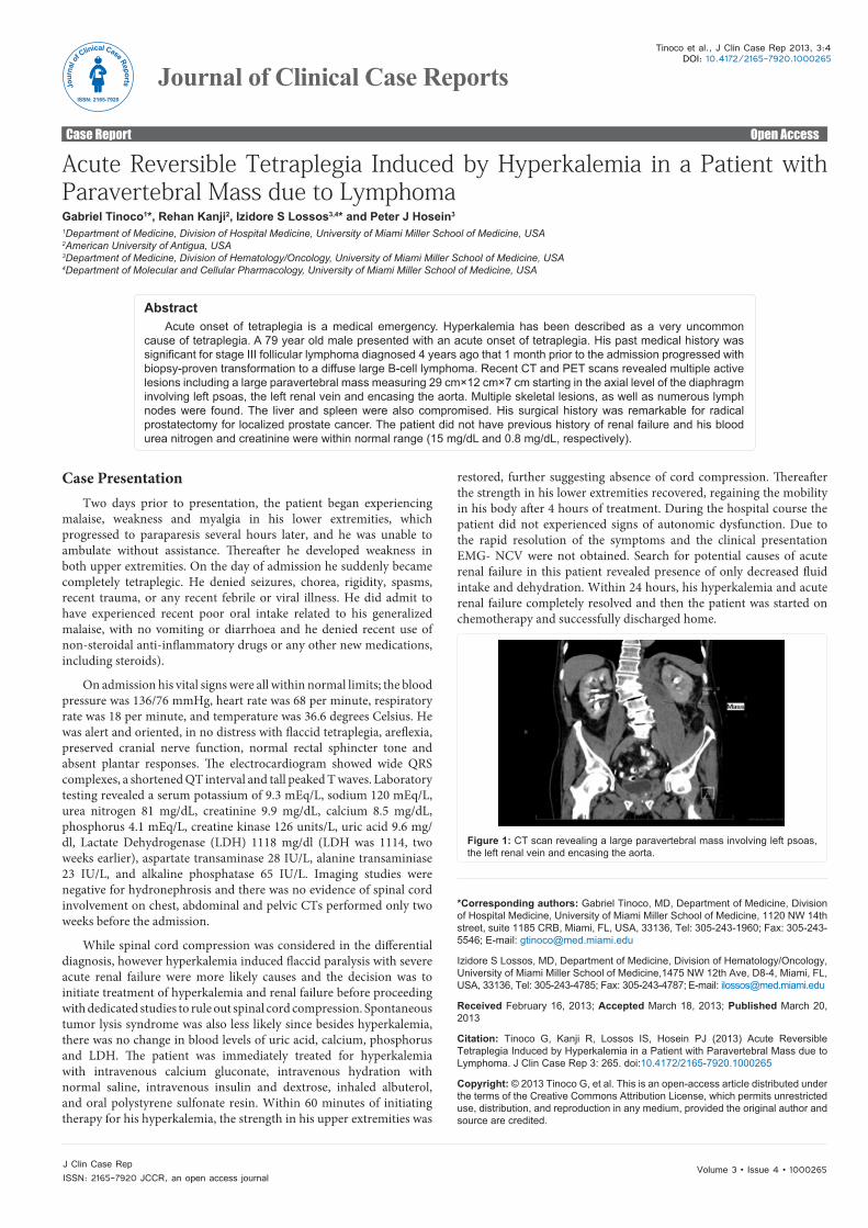
cause of tetraplegia. A 79 year old male presented with an acute onset of tetraplegia. His past medical history was significant for stage III follicular lymphoma diagnosed 4 years ago that 1 month prior to the admission progressed with biopsy-proven transformation to a diffuse large B-cell lymphoma. Recent CT and PET scans revealed multiple active lesions including a large paravertebral mass measuring 29 cm×12 cm×7 cm starting in the axial level of the diaphragm involving left psoas, the left renal vein and encasing the aorta. Multiple skeletal lesions, as well as numerous lymph nodes were found. The liver and spleen were also compromised. His surgical history was remarkable for radical prostatectomy for localized prostate cancer. The patient did not have previous history of renal failure and his blood urea nitrogen and creatinine were within normal range (15 mg/dL and 0.8 mg/dL, respectively).
Case PresentationTwo days prior to presentation, the patient began experiencing
malaise, weakness and myalgia in his lower extremities, which progressed to paraparesis several hours later, and he was unable to ambulate without assistance. Thereafter he developed weakness in both upper extremities. On the day of admission he suddenly became completely tetraplegic. He denied seizures, chorea, rigidity, spasms, recent trauma, or any recent febrile or viral illness. He did admit to have experienced recent poor oral intake related to his generalized malaise, with no vomiting or diarrhoea and he denied recent use of non-steroidal anti-inflammatory drugs or any other new medications, including steroids).
On admission his vital signs were all within normal limits; the blood pressure was 136/76 mmHg, heart rate was 68 per minute, respiratory rate was 18 per minute, and temperature was 36.6 degrees Celsius. He was alert and oriented, in no distress with flaccid tetraplegia, areflexia, preserved cranial nerve function, normal rectal sphincter tone and absent plantar responses. The electrocardiogram showed wide QRS complexes, a shortened QT interval and tall peaked T waves. Laboratory testing revealed a serum potassium of 9.3 mEq/L, sodium 120 mEq/L, urea nitrogen 81 mg/dL, creatinine 9.9 mg/dL, calcium 8.5 mg/dL, phosphorus 4.1 mEq/L, creatine kinase 126 units/L, uric acid 9.6 mg/dl, Lactate Dehydrogenase (LDH) 1118 mg/dl (LDH was 1114, two weeks earlier), aspartate transaminase 28 IU/L, alanine transaminiase 23 IU/L, and alkaline phosphatase 65 IU/L. Imaging studies were negative for hydronephrosis and there was no evidence of spinal cord involvement on chest, abdominal and pelvic CTs performed only two weeks before the admission.
While spinal cord compression was considered in the differential diagnosis, however hyperkalemia induced flaccid paralysis with severe acute renal failure were more likely causes and the decision was to initiate treatment of hyperkalemia and renal failure before proceeding with dedicated studies to rule out spinal cord compression. Spontaneous tumor lysis syndrome was also less likely since besides hyperkalemia, there was no change in blood levels of uric acid, calcium, phosphorus and LDH. The patient was immediately treated for hyperkalemia with intravenous calcium gluconate, intravenous hydration with normal saline, intravenous insulin and dextrose, inhaled albuterol, and oral polystyrene sulfonate resin. Within 60 minutes of initiating therapy for his hyperkalemia, the strength in his upper extremities was
restored, further suggesting absence of cord compression. Thereafter the strength in his lower extremities recovered, regaining the mobility in his body after 4 hours of treatment. During the hospital course the patient did not experienced signs of autonomic dysfunction. Due to the rapid resolution of the symptoms and the clinical presentation EMG- NCV were not obtained. Search for potential causes of acute renal failure in this patient revealed presence of only decreased fluid intake and dehydration. Within 24 hours, his hyperkalemia and acute renal failure completely resolved and then the patient was started on chemotherapy and successfully discharged home.
Figure 1: CT scan revealing a large paravertebral mass involving left psoas, the left renal vein and encasing the aorta.
Journal of Clinical Case ReportsJour
nal o
f Clinical Case Reports
ISSN: 2165-7920
Citation: Tinoco G, Kanji R, Lossos IS, Hosein PJ (2013) Acute Reversible Tetraplegia Induced by Hyperkalemia in a Patient with Paravertebral Mass due to Lymphoma. J Clin Case Rep 3: 265. doi:10.4172/2165-7920.1000265
Page 2 of 2
Volume 3 • Issue 4 • 1000265J Clin Case RepISSN: 2165-7920 JCCR, an open access journal
Secondary hyperkalemic paralysis is extremely rare and potentially life threatening condition. Although the precise mechanism of secondary hyperkalemic paralysis remains unclear, the persistent state of depolarization in the neurons is believed to be responsible for the reversible neuromuscular paralysis. It manifests clinically with an ascending pattern, from the lower extremities followed by a progression into the trunk and arms closely resembling the Guillian Barré syndrome [1,2]. In our patient, as well as most of other cases in the literature, there was no compromise of respiratory muscles, cranial nerves or sphincter tone [3]. Hyperkalemia induced flaccid paralysis has
been described mostly in the setting of end-stage renal disease, typically affecting lower extremities, and sometimes associated with ascending paralysis, but also focal neurological deficits have been described [4]. Shortening of the QT interval and peaked T waves (Figures 3 and 4), as well as other typical ECG changes are often observed in these patients [5-7]. If treatment for hyperkalemia is instituted promptly, complete recovery of the hyperkalemia–induced tetraplegia is achieved in 89% of patients [8]. However, the recovery of the symptoms is not related to potassium level or the concentration to which it is corrected [8,9].
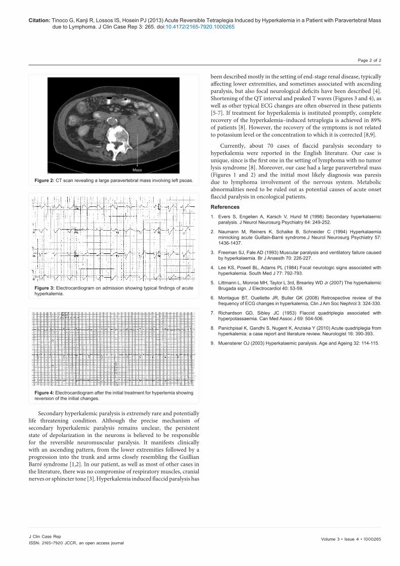
Currently, about 70 cases of flaccid paralysis secondary to hyperkalemia were reported in the English literature. Our case is unique, since is the first one in the setting of lymphoma with no tumor lysis syndrome [8]. Moreover, our case had a large paravertebral mass (Figures 1 and 2) and the initial most likely diagnosis was paresis due to lymphoma involvement of the nervous system. Metabolic abnormalities need to be ruled out as potential causes of acute onset flaccid paralysis in oncological patients.
References
1. Evers S, Engelien A, Karsch V, Hund M (1998) Secondary hyperkalaemic paralysis. J Neurol Neurosurg Psychiatry 64: 249-252.
2. Naumann M, Reiners K, Schalke B, Schneider C (1994) Hyperkalaemia mimicking acute Guillain-Barré syndrome.J Neurol Neurosurg Psychiatry 57: 1436-1437.
3. Freeman SJ, Fale AD (1993) Muscular paralysis and ventilatory failure caused by hyperkalaemia. Br J Anaesth 70: 226-227.
4. Lee KS, Powell BL, Adams PL (1984) Focal neurologic signs associated with hyperkalemia. South Med J 77: 792-793.
5. Littmann L, Monroe MH, Taylor L 3rd, Brearley WD Jr (2007) The hyperkalemic Brugada sign. J Electrocardiol 40: 53-59.
6. Montague BT, Ouellette JR, Buller GK (2008) Retrospective review of the frequency of ECG changes in hyperkalemia, Clin J Am Soc Nephrol 3: 324-330.
7. Richardson GD, Sibley JC (1953) Flaccid quadriplegia associated with hyperpotassaemia. Can Med Assoc J 69: 504-506.
8. Panichpisal K, Gandhi S, Nugent K, Anziska Y (2010) Acute quadriplegia from hyperkalemia: a case report and literature review. Neurologist 16: 390-393.
9. Muensterer OJ (2003) Hyperkalaemic paralysis. Age and Ageing 32: 114-115.
Figure 2: CT scan revealing a large paravertebral mass involving left psoas.
Figure 3: Electrocardiogram on admission showing typical findings of acute hyperkalemia.
Figure 4: Electrocardiogram after the initial treatment for hyperlemia showing reversion of the initial changes.