Embed Size (px)
Citation preview
Dopamine release from the locus coeruleus to thedorsal hippocampus promotes spatial learningand memoryKimberly A. Kempadooa,1, Eugene V. Mosharovb,c,d,e, Se Joon Choib,c,d,e, David Sulzerb,c,d,e, and Eric R. Kandela,f,g,1
aDepartment of Neuroscience, Columbia University, New York, NY 10032; bDepartment of Neurology, Columbia University, New York, NY 10032;cDepartment of Psychiatry, Columbia University, New York, NY 10032; dDepartment of Pharmacology, Columbia University, New York, NY 10032; eNewYork State Psychiatric Institute, New York, NY 10032; fKavli Institute for Brain Science, New York, NY 10032; and gHoward Hughes Medical Institute, NewYork, NY 10032
Contributed by Eric R. Kandel, November 9, 2016 (sent for review October 5, 2016; reviewed by Tom Abrams and John H. Byrne)
Dopamine neurotransmission in the dorsal hippocampus is criticalfor a range of functions from spatial learning and synaptic plasticityto the deficits underlying psychiatric disorders such as attention-deficit hyperactivity disorder. The ventral tegmental area (VTA) isthe presumed source of dopamine in the dorsal hippocampus.However, there is a surprising scarcity of VTA dopamine axonsin the dorsal hippocampus despite the dense network of dopaminereceptors. We have explored this apparent paradox using optoge-netic, biochemical, and behavioral approaches and found thatdopaminergic axons and subsequent dopamine release in the dorsalhippocampus originate from neurons of the locus coeruleus (LC).Photostimulation of LC axons produced an increase in dopaminerelease in the dorsal hippocampus as revealed by high-performanceliquid chromatography. Furthermore, optogenetically induced releaseof dopamine from the LC into the dorsal hippocampus enhancedselective attention and spatial object recognition via the dopamineD1/D5 receptor. These results suggest that spatial learning andmemory are energized by the release of dopamine in the dorsalhippocampus from noradrenergic neurons of the LC. The presentfindings are critical for identifying the neural circuits that enableproper attention selection and successful learning and memory.
dopamine | locus coeruleus | hippocampus | memory | attention
Dopamine is a neurotransmitter released throughout the brainto encode salience and facilitate the formation of associative
memory (1, 2). When released into the dorsal hippocampus, do-pamine binds to D1/D5 receptors to promote attention, episodicmemory formation, spatial learning, and synaptic plasticity (3–5).Successful spatial learning requires that hippocampal place cells,location-encoding pyramidal neurons (6), display consistent andstable patterns of neural activity, a process that can be enhancedby selective attention to spatial cues and by dopamine agonists(7, 8). Conversely, dopamine receptor blockade attenuates theability of spatial attention to stabilize the firing pattern of hip-pocampal place cells (8). The role of dopamine in driving atten-tional processes is highlighted by the fact that methylphenidate,one of the most common treatments for attention-deficit hyper-activity disorder (ADHD), improves attention by increasing syn-aptic availability of dopamine in the hippocampus, as well as in theprefrontal cortex and striatum (9–11). These findings suggest thatdopamine is critical for the selective attention underlying spatiallearning and memory.For decades, the ventral tegmental area (VTA) has been the
presumed source of hippocampal dopamine. However, in recentyears, the source of dopamine in the dorsal hippocampus hasbecome less clear. McNamara et al. (12) argued that a dopa-minergic projection from the VTA to the dorsal hippocampuspromoted hippocampal reactivation during sleep and stabilizedmemory. However, only 10% of the sparse projection from theVTA to the hippocampus contains dopamine, raising the questionof whether this weak VTA projection could be solely responsible
for activating the dense network of dopamine receptors found inthe dorsal hippocampus (13–16). Moreover, whereas the ventralaspect of the hippocampus, an area associated with learned fearand anxiety (17), receives significant dopaminergic projectionsfrom the VTA (13, 14), the dorsal region of the hippocampus,which houses the place cells required for spatial memory (7), hasrelatively sparse dopaminergic innervation from the VTA. Wetherefore set out to identify the neurons that provide dopami-nergic tone to the dorsal hippocampus and to assess theirinvolvement in spatial learning and attention.Dopamine is removed from hippocampal synapses by the
norepinephrine transporter (18), which suggests that dopaminemay also be released from norepinephrine-containing neuronsoutside of the VTA. Although indirect action via the VTA hadnot been ruled out, electrical and pharmacological stimulation ofthe noradrenergic locus coeruleus (LC) increased dopaminelevels in the prefrontal cortex and modulated hippocampal syn-aptic transmission (19, 20). Also, dopamine-mediated enhance-ment of excitatory transmission in the hippocampus can bereversed by tyrosine hydroxylase (TH) knockdown in the LC, butnot in the VTA (21). Smith and Greene (21) therefore suggestedthat dopamine may be released from the LC into the dorsalhippocampus. A major caveat of the study is that the synaptictransmission effects are produced in vitro by amphetamine, a
Significance
Successful completion of daily activities relies on the ability toselect the relevant features of the environment to pay atten-tion to and remember. Disruptions of these processes can leadto disorders, such as attention-deficit hyperactivity disorderand age-related memory loss. To devise therapeutic strategies,we must understand the neural circuits underlying normalcognition. One important pathway is the signaling of dopa-mine, a reinforcement-related neurotransmitter, in the hippo-campus, a spatial learning and memory center. Surprisingly,the brain region supplying dopamine to the dorsal hippocam-pus is unclear. This study provides direct evidence that thenoradrenergic locus coeruleus coreleases dopamine in thedorsal hippocampus and provides insight into dopamine func-tion in selective attention and spatial learning and memory.
Author contributions: K.A.K., E.V.M., D.S., and E.R.K. designed research; K.A.K., E.V.M.,and S.J.C. performed research; K.A.K. and E.V.M. analyzed data; and K.A.K. and E.R.K.wrote the paper.
Reviewers: T.A., University of Maryland School of Medicine; and J.H.B., University of TexasMedical School at Houston.
The authors declare no conflict of interest.
Freely available online through the PNAS open access option.1To whom correspondence may be addressed. Email: [email protected] or [email protected].
This article contains supporting information online at www.pnas.org/lookup/suppl/doi:10.1073/pnas.1616515114/-/DCSupplemental.
www.pnas.org/cgi/doi/10.1073/pnas.1616515114 PNAS | December 20, 2016 | vol. 113 | no. 51 | 14835–14840
NEU
ROSC
IENCE
Dow
nloa
ded
by g
uest
on
Mar
ch 1
7, 2
020
drug known to increase extracellular dopamine concentrations(22). The finding provides no insight as to whether dopamine isreleased from the LC under more physiological, nondrug con-ditions, a question we test directly in our study.Taken together, there are some groups who argue that the
VTA is the main source of dopamine to the dorsal hippocampus(12–14) and others who support the idea that dopamine is re-leased from LC neurons (19–21). These conflicting hypothesesleave unanswered the question of which brain region suppliesthe dorsal hippocampus with dopaminergic tone. We combinedoptogenetic, biochemical, and behavioral methods to identify themajor source of dopamine in the dorsal hippocampus and thenexamined the involvement of this dopaminergic pathway in se-lective attention and spatial learning and memory.
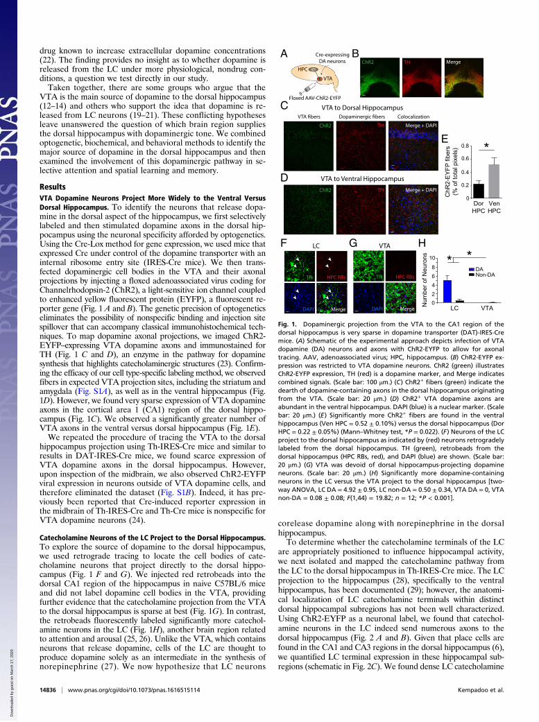
ResultsVTA Dopamine Neurons Project More Widely to the Ventral VersusDorsal Hippocampus. To identify the neurons that release dopa-mine in the dorsal aspect of the hippocampus, we first selectivelylabeled and then stimulated dopamine axons in the dorsal hip-pocampus using the neuronal specificity afforded by optogenetics.Using the Cre-Lox method for gene expression, we used mice thatexpressed Cre under control of the dopamine transporter with aninternal ribosome entry site (IRES-Cre mice). We then trans-fected dopaminergic cell bodies in the VTA and their axonalprojections by injecting a floxed adenoassociated virus coding forChannelrhodopsin-2 (ChR2), a light-sensitive ion channel coupledto enhanced yellow fluorescent protein (EYFP), a fluorescent re-porter gene (Fig. 1 A and B). The genetic precision of optogeneticseliminates the possibility of nonspecific binding and injection sitespillover that can accompany classical immunohistochemical tech-niques. To map dopamine axonal projections, we imaged ChR2-EYFP–expressing VTA dopamine axons and immunostained forTH (Fig. 1 C and D), an enzyme in the pathway for dopaminesynthesis that highlights catecholaminergic structures (23). Confirm-ing the efficacy of our cell type-specific labeling method, we observedfibers in expected VTA projection sites, including the striatum andamygdala (Fig. S1A), as well as in the ventral hippocampus (Fig.1D). However, we found very sparse expression of VTA dopamineaxons in the cortical area 1 (CA1) region of the dorsal hippo-campus (Fig. 1C). We observed a significantly greater number ofVTA axons in the ventral versus dorsal hippocampus (Fig. 1E).We repeated the procedure of tracing the VTA to the dorsal
hippocampus projection using Th-IRES-Cre mice and similar toresults in DAT-IRES-Cre mice, we found scarce expression ofVTA dopamine axons in the dorsal hippocampus. However,upon inspection of the midbrain, we also observed ChR2-EYFPviral expression in neurons outside of VTA dopamine cells, andtherefore eliminated the dataset (Fig. S1B). Indeed, it has pre-viously been reported that Cre-induced reporter expression inthe midbrain of Th-IRES-Cre and Th-Cre mice is nonspecific forVTA dopamine neurons (24).
Catecholamine Neurons of the LC Project to the Dorsal Hippocampus.To explore the source of dopamine to the dorsal hippocampus,we used retrograde tracing to locate the cell bodies of cate-cholamine neurons that project directly to the dorsal hippo-campus (Fig. 1 F and G). We injected red retrobeads into thedorsal CA1 region of the hippocampus in naive C57BL/6 miceand did not label dopamine cell bodies in the VTA, providingfurther evidence that the catecholamine projection from the VTAto the dorsal hippocampus is sparse at best (Fig. 1G). In contrast,the retrobeads fluorescently labeled significantly more catechol-amine neurons in the LC (Fig. 1H), another brain region relatedto attention and arousal (25, 26). Unlike the VTA, which containsneurons that release dopamine, cells of the LC are thought toproduce dopamine solely as an intermediate in the synthesis ofnorepinephrine (27). We now hypothesize that LC neurons
corelease dopamine along with norepinephrine in the dorsalhippocampus.To determine whether the catecholamine terminals of the LC
are appropriately positioned to influence hippocampal activity,we next isolated and mapped the catecholamine pathway fromthe LC to the dorsal hippocampus in Th-IRES-Cre mice. The LCprojection to the hippocampus (28), specifically to the ventralhippocampus, has been documented (29); however, the anatomi-cal localization of LC catecholamine terminals within distinctdorsal hippocampal subregions has not been well characterized.Using ChR2-EYFP as a neuronal label, we found that catechol-amine neurons in the LC indeed send numerous axons to thedorsal hippocampus (Fig. 2 A and B). Given that place cells arefound in the CA1 and CA3 regions in the dorsal hippocampus (6),we quantified LC terminal expression in these hippocampal sub-regions (schematic in Fig. 2C). We found dense LC catecholamine
D
F VTA
TH HPC RBs
MergeDAPI
TH HPC RBs
MergeDAPI
GLC
VTA to Dorsal Hippocampus
VTA to Ventral Hippocampus
H
C
**
LC VTANum
ber o
f Neu
rons
2468
10
0
DANon-DA
VTA fibers Dopaminergic fibers Colocalization
A
VTA
HPC
BTH MergeChR2
Floxed AAV-ChR2-EYFP
Cre-expressing
DA neurons
0.2
0.4
0.6
0.8
0
*
ChR
2-E
YFP
fibe
rs(%
of t
otal
pix
els)
DorHPC
VenHPC
E
ChR2 Merge + DAPITH
ChR2 TH Merge + DAPI
Fig. 1. Dopaminergic projection from the VTA to the CA1 region of thedorsal hippocampus is very sparse in dopamine transporter (DAT)-IRES-Cremice. (A) Schematic of the experimental approach depicts infection of VTAdopamine (DA) neurons and axons with ChR2-EYFP to allow for axonaltracing. AAV, adenoassociated virus; HPC, hippocampus. (B) ChR2-EYFP ex-pression was restricted to VTA dopamine neurons. ChR2 (green) illustratesChR2-EYFP expression, TH (red) is a dopamine marker, and Merge indicatescombined signals. (Scale bar: 100 μm.) (C) ChR2+ fibers (green) indicate thedearth of dopamine-containing axons in the dorsal hippocampus originatingfrom the VTA. (Scale bar: 20 μm.) (D) ChR2+ VTA dopamine axons areabundant in the ventral hippocampus. DAPI (blue) is a nuclear marker. (Scalebar: 20 μm.) (E) Significantly more ChR2+ fibers are found in the ventralhippocampus (Ven HPC = 0.52 ± 0.10%) versus the dorsal hippocampus (DorHPC = 0.22 ± 0.05%) (Mann–Whitney test, *P = 0.022). (F) Neurons of the LCproject to the dorsal hippocampus as indicated by (red) neurons retrogradelylabeled from the dorsal hippocampus. TH (green), retrobeads from thedorsal hippocampus (HPC RBs, red), and DAPI (blue) are shown. (Scale bar:20 μm.) (G) VTA was devoid of dorsal hippocampus-projecting dopamineneurons. (Scale bar: 20 μm.) (H) Significantly more dopamine-containingneurons in the LC versus the VTA project to the dorsal hippocampus [two-way ANOVA, LC DA = 4.92 ± 0.95, LC non-DA = 0.50 ± 0.34, VTA DA = 0, VTAnon-DA = 0.08 ± 0.08; F(1,44) = 19.82; n = 12; *P < 0.001].
14836 | www.pnas.org/cgi/doi/10.1073/pnas.1616515114 Kempadoo et al.
Dow
nloa
ded
by g
uest
on
Mar
ch 1
7, 2
020

fiber expression throughout the dorsal CA1 region (Fig. 2D).This finding suggests that LC catecholamine fibers terminate inclose proximity to the dendritic branches of hippocampal placecells, neurons that encode position in space and are tunedby selective attention to environmental cues (7, 8). We nextcompared the density of LC catecholamine axons with thedensity of VTA dopamine axons in the dorsal CA1 region andfound that LC axons are substantially denser than VTA fibers(Fig. 2E).Previous studies support the hypothesis that LC neurons are
capable of coreleasing dopamine in the dorsal hippocampus.Electrical stimulation of the LC produces increased tissue con-tent and pharmacological action of dopamine in the hippocam-pus (20, 30). However, these studies did not rule out networkaction via the VTA and failed to demonstrate release of dopa-mine from noradrenergic LC axons into the dorsal hippocampus.An increase in dopamine tissue content may suggest intracellu-lar accumulation of norepinephrine synthesis products, butdoes not provide evidence that these neurons are capable ofdopamine release. We attempted to address these unresolvedquestions using the genetic and anatomical specificity affordedby optogenetics.
Dopamine Is Released from Axons of the LC in the Dorsal Hippocampus.To assess directly whether and to what extent dopamine is releasedfrom axons of the LC, we measured extracellular dopamine con-centrations in acute hippocampal slices using high-performanceliquid chromatography (HPLC). Dorsal hippocampus coronal brainsections (250 μm) were prepared from Th-IRES-Cre mice in whichLC catecholamine neurons and fibers were infected with the
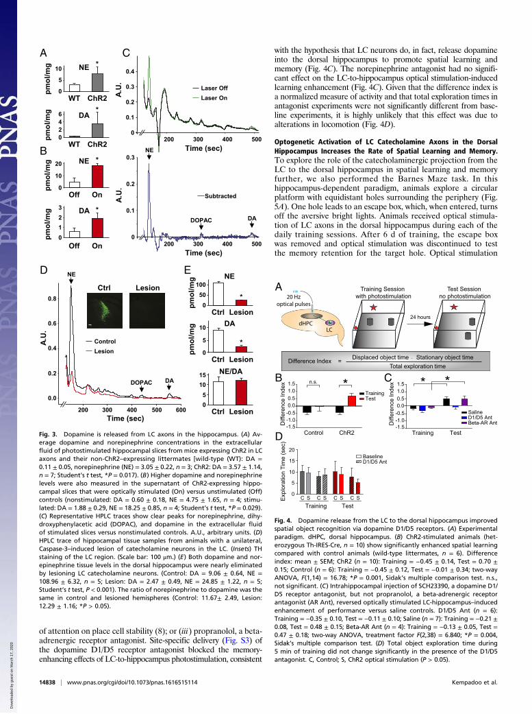
floxed ChR2-EYFP virus. We then photostimulated LC axonsin the hippocampus ex vivo and compared dopamine release inthe extracellular fluid of stimulated hippocampal slices withidentically handled wild-type controls lacking ChR2 expression.Optical stimulation of LC catecholamine terminals in thedorsal hippocampus evoked significantly higher levels of bothdopamine and norepinephrine release compared with controls(Fig. 3A). Similarly, optically stimulated slices (laser on)showed significantly higher extracellular concentrations of dopa-mine and norepinephrine than nonstimulated slices expressingChR2-EYFP (laser off; Fig. 3 B and C). These findings confirmthat both neurotransmitters were released from LC axons intothe dorsal hippocampus.We next lesioned catecholamine neurons of the LC selectively
using a floxed Caspase-3 virus (University of North CarolinaVector Core) (31) and measured a 72.7% reduction of dopamineand a 77.2% reduction of norepinephrine tissue levels in thehippocampus (Fig. 3 D and E). The ratio of norepinephrine todopamine remained the same in lesioned and control samples(Fig. 3E). We then stimulated nonspecific neurotransmitter re-lease into the extracellular fluid by incubating slices in potassiumchloride to promote release of dopamine and norepinephrinefrom all axons in the dorsal hippocampus. A selective lesion ofLC catecholamine neurons reduced the extracellular dopamineconcentration by 67.9% and norepinephrine by 83.8% (Fig. S2).This finding suggests that the majority of the dopaminergic tonepresent in dorsal hippocampal tissue and actively released intothe extracellular space originated from neurons of the LC. To-gether, these results provide a direct demonstration that dopa-mine is released from LC axons into the dorsal hippocampus.
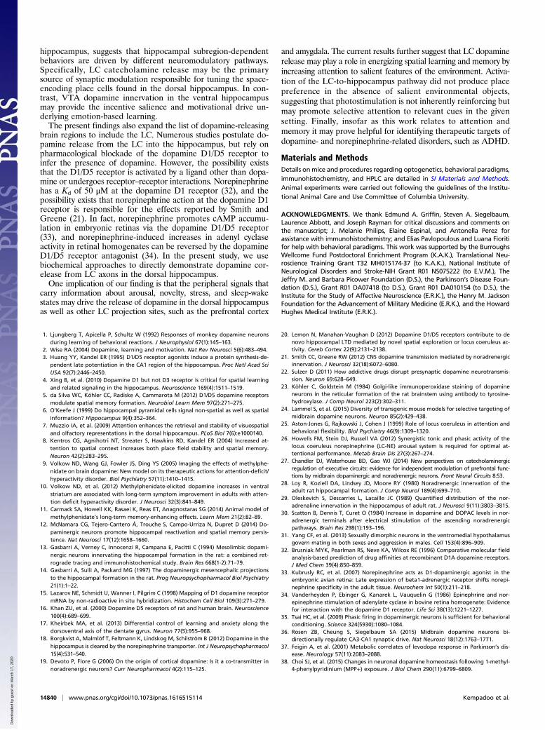
Optogenetic Activation of LC Catecholamine Axons in the DorsalHippocampus Promotes Selective Attention and Spatial Learning viathe Dopamine D1/D5 Receptor. To assess the behavioral signifi-cance of the dopamine pathway from the LC to the hippocam-pus, we examined mice in a spatial object recognition task whileactivating ChR2-EYFP+ LC axons in the dorsal hippocampus(Fig. 4A). We allowed mice to explore a square arena with dis-tinct walls containing two identical objects during a 5-mintraining session. One day later, we challenged these mice toexplore the arena during a 5-min test session in which the leastpreferred object was moved to another position. During thetraining session for this task, both ChR2-EYFP–expressingTh-IRES-Cre animals and their wild-type controls receivedintrahippocampal optical stimulation. We then assessed per-formance by determining the difference index, a normalizedmeasure indicating the relative amount of time spent exploringthe displaced object. Increased attention to the displaced ob-ject during the test session generates a higher difference indexand correlates positively with the degree of successful spatiallearning and memory. In ChR2-EYFP–expressing animals,optical activation of hippocampal LC catecholamine terminalsduring training significantly increased the average differenceindex in the test session. This enhancement of spatial learningonly occurred in optically stimulated animals, and not in wild-typecontrols (Fig. 4B). These results demonstrate that photostimulationof the LC-to-hippocampus catecholamine pathway promotedspatial learning and converted nonlearners into learners.The LC-to-hippocampus projection was previously thought to
be strictly noradrenergic (28); therefore, the function of dopaminereleased from LC fibers into the dorsal hippocampus was notknown. Given the dearth of catecholamine innervation from theVTA, we now asked whether the LC actively releases behav-iorally relevant dopamine concentrations into the dorsal hippo-campus. To address this question, before the optical stimulationtraining session, we infused into the dorsal hippocampus one ofthree compounds: (i) saline; (ii) SCH23390, a dopamine D1/D5receptor antagonist we have previously used to block the effects
ChR2: viral expression
DAPI: nuclear marker TH: catecholamines
Merge
A
DC
Injection Site: LC
Hippocampal Subregion
Axo
n de
nsity
(#/im
age)
0
5
10
15
20
25
SO
CA1 CA3 DGSR SLM SO SR SLM MOL PL
SOSRSLM
MOLPL
CA1
CA3 DG
B Projection Site: Dorsal CA1
ChR2: LC fibers
DAPI: nuclear marker TH: catecholamines
Merge
10
20
30
40
0
ChR
2+TH
fibe
rs (%
TH
)
VTA LC
E*
Fig. 2. Distribution of LC catecholamine axons in the dorsal hippocampus ofTh-IRES-Cre mice. (A) ChR2 successfully infected catecholamine-containingneurons in the LC. (Scale bar: 100 μm.) (B) ChR2+ LC catecholamine axons arefound in the dorsal hippocampus. (Scale bar: 20 μm.) (C) Dorsal hippocampalsubregions. (D) LC catecholamine fiber density in hippocampal subregions(one-way ANOVA, Kruskal–Wallis test, P = 0.024; n = 4). MOL, molecularlayer of the dentate gyrus (DG); PL, polymorphic layer of the DG; SLM,stratum lacunosum-moleculare; SO, stratum oriens; SR, stratum reticulatum.DAPI (blue) is a nuclear marker, ChR2 (green) indicates ChR2+ LC axons, TH(red) indicates catecholamine-containing fibers, and Merge indicates com-bined signals. (E) LC catecholamine axon density is significantly greater thanthe density of VTA dopamine axons in the dorsal hippocampus (LC: 32.53 ±5.48%, VTA: 7.20 ± 1.32%, Mann–Whitney test, *P < 0.001).
Kempadoo et al. PNAS | December 20, 2016 | vol. 113 | no. 51 | 14837
NEU
ROSC
IENCE
Dow
nloa
ded
by g
uest
on
Mar
ch 1
7, 2
020
of attention on place cell stability (8); or (iii) propranolol, a beta-adrenergic receptor antagonist. Site-specific delivery (Fig. S3) ofthe dopamine D1/D5 receptor antagonist blocked the memory-enhancing effects of LC-to-hippocampus photostimulation, consistent
with the hypothesis that LC neurons do, in fact, release dopamineinto the dorsal hippocampus to promote spatial learning andmemory (Fig. 4C). The norepinephrine antagonist had no signifi-cant effect on the LC-to-hippocampus optical stimulation-inducedlearning enhancement (Fig. 4C). Given that the difference index isa normalized measure of activity and that total exploration times inantagonist experiments were not significantly different from base-line experiments, it is highly unlikely that this effect was due toalterations in locomotion (Fig. 4D).
Optogenetic Activation of LC Catecholamine Axons in the DorsalHippocampus Increases the Rate of Spatial Learning and Memory.To explore the role of the catecholaminergic projection from theLC to the dorsal hippocampus in spatial learning and memoryfurther, we also performed the Barnes Maze task. In thishippocampus-dependent paradigm, animals explore a circularplatform with equidistant holes surrounding the periphery (Fig.5A). One hole leads to an escape box, which, when entered, turnsoff the aversive bright lights. Animals received optical stimula-tion of LC axons in the dorsal hippocampus during each of thedaily training sessions. After 6 d of training, the escape boxwas removed and optical stimulation was discontinued to testthe memory retention for the target hole. Optical stimulation
600500400300200Time (sec)
0.8
0.6
0.4
0.2
0.0
A.U
.
NE
DADOPAC
NE
LesionCtrl
LesionCtrl
050
100
DA
LesionCtrl0
5
10
NE/DA
LesionCtrl05
1015
pmol
/mg
pmol
/mg
0
0.1
0.2
0.3
0.4
A.U
.
0
0.1
0.2
0.3
A.U
.
Laser OnLaser Off
LesionControl
200 300 400 500
Subtracted
DADOPAC
Time (sec)
200 300 400 500Time (sec)
CANE
ChR2WT0
5
10
pmol
/mg
NE
Off On0
10
DA
01
pmol
/mg
pmol
/mg
B
ED
DA
ChR2WT024
pmol
/mg 6
20
23
NE
Off On
*
*
*
*
*
*
Fig. 3. Dopamine is released from LC axons in the hippocampus. (A) Av-erage dopamine and norepinephrine concentrations in the extracellularfluid of photostimulated hippocampal slices from mice expressing ChR2 in LCaxons and their non-ChR2–expressing littermates [wild-type (WT): DA =0.11 ± 0.05, norepinephrine (NE) = 3.05 ± 0.22, n = 3; ChR2: DA = 3.57 ± 1.14,n = 7; Student’s t test, *P = 0.017). (B) Higher dopamine and norepinephrinelevels were also measured in the supernatant of ChR2-expressing hippo-campal slices that were optically stimulated (On) versus unstimulated (Off)controls (nonstimulated: DA = 0.60 ± 0.18, NE = 4.75 ± 1.65, n = 4; stimu-lated: DA = 1.88 ± 0.29, NE = 18.25 ± 0.85, n = 4; Student’s t test, *P = 0.029).(C) Representative HPLC traces show clear peaks for norepinephrine, dihy-droxyphenylacetic acid (DOPAC), and dopamine in the extracellular fluidof stimulated slices versus nonstimulated controls. A.U., arbitrary units. (D)HPLC trace of hippocampal tissue samples from animals with a unilateral,Caspase-3–induced lesion of catecholamine neurons in the LC. (Insets) THstaining of the LC region. (Scale bar: 100 μm.) (E) Both dopamine and nor-epinephrine tissue levels in the dorsal hippocampus were nearly eliminatedby lesioning LC catecholamine neurons. (Control: DA = 9.06 ± 0.64, NE =108.96 ± 6.32, n = 5; Lesion: DA = 2.47 ± 0.49, NE = 24.85 ± 1.22, n = 5;Student’s t test, P < 0.001). The ratio of norepinephrine to dopamine was thesame in control and lesioned hemispheres (Control: 11.67± 2.49, Lesion:12.29 ± 1.16; *P > 0.05).
A Training Sessionwith photostimulation
Test Sessionno photostimulation
TrainingTest
Saline
Beta-AR AntD1/D5 Ant
n.s.
Control ChR2
1.51.00.50.0
-0.5-1.0-1.5
B C
Difference Index = Displaced object time – Stationary object timeTotal exploration time
24 hours
Diff
eren
ce In
dex 1.5
1.00.50.0
-0.5-1.0-1.5D
iffer
ence
Inde
x
D
BaselineD1/D5 Ant
Training Test
Training Test
15
10
20
5
0C S C S C S C SE
xplo
ratio
n Ti
me
(sec
)
dHPC
20 Hz
optical pulses
LC
* **
Fig. 4. Dopamine release from the LC to the dorsal hippocampus improvedspatial object recognition via dopamine D1/D5 receptors. (A) Experimentalparadigm. dHPC, dorsal hippocampus. (B) ChR2-stimulated animals (het-erozygous Th-IRES-Cre, n = 10) show significantly enhanced spatial learningcompared with control animals (wild-type littermates, n = 6). Differenceindex: mean ± SEM; ChR2 (n = 10): Training = −0.45 ± 0.14, Test = 0.70 ±0.15; Control (n = 6): Training = −0.45 ± 0.12, Test = −0.01 ± 0.34; two-wayANOVA, F(1,14) = 16.78; *P = 0.001, Sidak’s multiple comparison test. n.s.,not significant. (C) Intrahippocampal injection of SCH23390, a dopamine D1/D5 receptor antagonist, but not propranolol, a beta-adrenergic receptorantagonist (AR Ant), reversed optically stimulated LC-hippocampus–inducedenhancement of performance versus saline controls. D1/D5 Ant (n = 6):Training = −0.35 ± 0.10, Test = −0.11 ± 0.10; Saline (n = 7): Training = −0.21 ±0.08, Test = 0.48 ± 0.15; Beta-AR Ant (n = 4): Training = −0.13 ± 0.05, Test =0.47 ± 0.18; two-way ANOVA, treatment factor F(2,38) = 6.840; *P = 0.004,Sidak’s multiple comparison test. (D) Total object exploration time during5 min of training did not change significantly in the presence of the D1/D5antagonist. C, Control; S, ChR2 optical stimulation (P > 0.05).
14838 | www.pnas.org/cgi/doi/10.1073/pnas.1616515114 Kempadoo et al.
Dow
nloa
ded
by g
uest
on
Mar
ch 1
7, 2
020

resumed for the next 6 d of training to the same target hole,followed by a second probe day. Animals were connected to afiber optic cable that did not deliver photostimulation on probedays. LC-to-hippocampus–stimulated animals learned this tasksignificantly better than controls during probe test 1, as indicatedby a fewer number of errors, shorter latency to the target hole,and more time spent in the target quadrant (Fig. 5B). By probetest 2, control animals had sufficiently learned the task, yieldingno significant difference between the two groups (Fig. 5C).These findings demonstrate that in a task learned by controlanimals, activation of LC axons in the dorsal hippocampus ofChR2-expressing mice significantly increased the rate of learn-ing. These results reinforce earlier findings and support theidea that dopamine release from the LC to the dorsal hippo-campus is a significant component of the spatial learning andmemory circuit.
Optogenetic Activation of LC Catecholamine Axons in the DorsalHippocampus Had No Effect on a Modified Conditioned PlacePreference Task. To assess whether photostimulation of the LC-to-hippocampus catecholamine pathway is inherently reinforc-ing, we performed a conditioned place preference task modifiedto increase involvement of hippocampus-dependent spatiallearning circuits. Animals explored a circular chamber with cuessurrounding the walls for spatial orientation. We delivered pho-tostimulation each time the animal explored an unmarked targetquadrant of the arena (Fig. 6A). This spatial version of condi-tioned place preference was designed to assay whether the LC-to-
hippocampus pathway provided reinforcement in the absence ofselective attention to a specific object or goal. Optically stimu-lated animals did not display a significant change in the amountof time spent in the unmarked target region versus wild-typecontrols (Fig. 6B). This finding is consistent with the argumentthat LC-to-hippocampus stimulation enhances selective attentionto specific stimuli, thereby energizing spatial learning and mem-ory. This result demonstrates that dopamine signaling in the dorsalhippocampus did not produce an inherently rewarding or rein-forcing signal but, instead, promoted selective attention to salientenvironmental cues.
DiscussionThese findings provide direct evidence that efferents from theLC are not purely noradrenergic, but corelease dopamine in thedorsal hippocampus. The LC dopamine signal drives the selec-tive attention underlying spatial learning and memory. Numer-ous reports suggest that the VTA is the main source of dopamineto the dorsal hippocampus (12–14), whereas others suggest thatthe LC supplies the hippocampus with dopamine (19–21). Thepresent results reconcile the discrepancy between dense dopa-mine receptor expression and sparse VTA dopamine axon net-works in the dorsal hippocampus and support the hypothesis thatdopaminergic tone in the dorsal hippocampus arises from neu-rons of the LC.Previous work by McNamara et al. (12) demonstrated that
activation of VTA dopamine axons in the dorsal hippocampuspromoted spatial memory for a reward location. Our work addsto these findings by suggesting that dopamine release from LCneurons energizes the attention component of the spatial envi-ronment in the absence of reward and supports the acquisition ofspatial memory. Given that catecholamine neurons of the LCcontribute 73% of all measurable dopaminergic tone in thedorsal hippocampus, we argue that dopamine release from theLC may have more significant influence over dorsal hippocam-pus neuronal activity than the VTA.Our findings that LC neurons corelease dopamine in the
dorsal hippocampus are consistent with the earlier finding thatLC neurons also corelease dopamine in the prefrontal cortex anddrive dopamine-dependent activity in the hippocampus (19, 21).Moreover, the current findings provide anatomical support forthe documented functional distinctions between the dorsal andventral hippocampus (17). The density of VTA dopamine termi-nals in the ventral hippocampus, but sparse expression in the dorsal
A
B
C
Inco
rrec
t Hol
es (#
)
Control ChR2
6
4
10
8
2
0
20
40
60
80
100
0
20
40
60
80
100
0
Barnes Maze Probe Test 1
Barnes Maze Probe Test 2
Late
ncy
to T
arge
t (se
c)
Tim
e in
Tar
get Q
uadr
ant
(% o
f tot
al ti
me)
Control ChR2 Control ChR2
150
100
200
50
0
* * *
Inco
rrec
t Hol
es (#
)
Control ChR2
6
4
10
8
2
0 Late
ncy
to T
arge
t (se
c)
Tim
e in
Tar
get Q
uadr
ant
(% o
f tot
al ti
me)
Control ChR2 Control ChR2
150
100
200
50
0
Training days withphotos�mula�on
Training days withphotos�mula�on
Probe Test 1Escape
chamberProbe Test 2
Fig. 5. Optogenetic activation of LC catecholamine axons in the dorsalhippocampus increased the rate of learning in the Barnes Maze task. (A) Aschematic of the Barnes Maze and training protocol is shown. Small lightgray circles represent empty holes, and the dark gray circle indicates theposition of the hole leading to a goal box in which the animal can escape toturn the lights off. Memory retention was tested by probe sessions duringwhich the escape box and optical stimulation were removed. (B) On probeday 1, optically stimulated animals learned the task significantly better thanwild-type controls [Control (n = 6), ChR2 (n = 7): incorrect holes visited be-fore reaching target hole: *P = 0.030, latency to reach target hole from trialstart: *P = 0.005, time spent in target quadrant: *P = 0.015]. (C) By probe day2, both controls and optically stimulated animals learned the task well, asindicated by no significant difference between the two groups (P > 0.05).
A B
3
2
5
4
1
32 541BL NS0
Photos�mula�on inunmarked quadrant
Session
ChR2Control
Tim
e in
Tar
get Q
uadr
ant (
min
)
Fig. 6. Optogenetic activation of LC catecholamine axons in the dorsalhippocampus had no effect on a modified conditioned place preferencetask. (A) Schematic of the circular chamber in which animals received pho-tostimulation of the catecholaminergic pathway from the LC to the dorsalhippocampus when traversing through the unmarked target quadrant.(B) ChR2-stimulated animals (n = 10) did not spend significantly more timein the target quadrant than wild-type controls (n = 6), two-way ANOVA,F(1,14) = 0.00, P > 0.05. The numbers 1–5 represent 5 d of exploration withoptical stimulation in the target region. BL, baseline session with no opticalstimulation; NS, no stimulation day.
Kempadoo et al. PNAS | December 20, 2016 | vol. 113 | no. 51 | 14839
NEU
ROSC
IENCE
Dow
nloa
ded
by g
uest
on
Mar
ch 1
7, 2
020
hippocampus, suggests that hippocampal subregion-dependentbehaviors are driven by different neuromodulatory pathways.Specifically, LC catecholamine release may be the primarysource of synaptic modulation responsible for tuning the space-encoding place cells found in the dorsal hippocampus. In con-trast, VTA dopamine innervation in the ventral hippocampusmay provide the incentive salience and motivational drive un-derlying emotion-based learning.The present findings also expand the list of dopamine-releasing
brain regions to include the LC. Numerous studies postulate do-pamine release from the LC into the hippocampus, but rely onpharmacological blockade of the dopamine D1/D5 receptor toinfer the presence of dopamine. However, the possibility existsthat the D1/D5 receptor is activated by a ligand other than dopa-mine or undergoes receptor–receptor interactions. Norepinephrinehas a Kd of 50 μM at the dopamine D1 receptor (32), and thepossibility exists that norepinephrine action at the dopamine D1receptor is responsible for the effects reported by Smith andGreene (21). In fact, norepinephrine promotes cAMP accumu-lation in embryonic retinas via the dopamine D1/D5 receptor(33), and norepinephrine-induced increases in adenyl cyclaseactivity in retinal homogenates can be reversed by the dopamineD1/D5 receptor antagonist (34). In the present study, we usebiochemical approaches to directly demonstrate dopamine cor-elease from LC axons in the dorsal hippocampus.One implication of our finding is that the peripheral signals that
carry information about arousal, novelty, stress, and sleep-wakestates may drive the release of dopamine in the dorsal hippocampusas well as other LC projection sites, such as the prefrontal cortex
and amygdala. The current results further suggest that LC dopaminerelease may play a role in energizing spatial learning and memory byincreasing attention to salient features of the environment. Activa-tion of the LC-to-hippocampus pathway did not produce placepreference in the absence of salient environmental objects,suggesting that photostimulation is not inherently reinforcing butmay promote selective attention to relevant cues in the givensetting. Finally, insofar as this work relates to attention andmemory it may prove helpful for identifying therapeutic targets ofdopamine- and norepinephrine-related disorders, such as ADHD.
Materials and MethodsDetails onmice and procedures regarding optogenetics, behavioral paradigms,immunohistochemistry, and HPLC are detailed in SI Materials and Methods.Animal experiments were carried out following the guidelines of the Institu-tional Animal Care and Use Committee of Columbia University.
ACKNOWLEDGMENTS. We thank Edmund A. Griffin, Steven A. Siegelbaum,Laurence Abbott, and Joseph Rayman for critical discussions and comments onthe manuscript; J. Melanie Philips, Elaine Espinal, and Antonella Perez forassistance with immunohistochemistry; and Elias Pavlopoulous and Luana Fioritifor help with behavioral paradigms. This work was supported by the BurroughsWellcome Fund Postdoctoral Enrichment Program (K.A.K.), Translational Neu-roscience Training Grant T32 MH015174-37 (to K.A.K.), National Institute ofNeurological Disorders and Stroke-NIH Grant R01 NS075222 (to E.V.M.), TheJeffry M. and Barbara Picower Foundation (D.S.), the Parkinson’s Disease Foun-dation (D.S.), Grant R01 DA07418 (to D.S.), Grant R01 DA010154 (to D.S.), theInstitute for the Study of Affective Neuroscience (E.R.K.), the Henry M. JacksonFoundation for the Advancement of Military Medicine (E.R.K.), and the HowardHughes Medical Institute (E.R.K.).
1. Ljungberg T, Apicella P, Schultz W (1992) Responses of monkey dopamine neuronsduring learning of behavioral reactions. J Neurophysiol 67(1):145–163.
2. Wise RA (2004) Dopamine, learning and motivation. Nat Rev Neurosci 5(6):483–494.3. Huang YY, Kandel ER (1995) D1/D5 receptor agonists induce a protein synthesis-de-
pendent late potentiation in the CA1 region of the hippocampus. Proc Natl Acad SciUSA 92(7):2446–2450.
4. Xing B, et al. (2010) Dopamine D1 but not D3 receptor is critical for spatial learningand related signaling in the hippocampus. Neuroscience 169(4):1511–1519.
5. da Silva WC, Köhler CC, Radiske A, Cammarota M (2012) D1/D5 dopamine receptorsmodulate spatial memory formation. Neurobiol Learn Mem 97(2):271–275.
6. O’Keefe J (1999) Do hippocampal pyramidal cells signal non-spatial as well as spatialinformation? Hippocampus 9(4):352–364.
7. Muzzio IA, et al. (2009) Attention enhances the retrieval and stability of visuospatialand olfactory representations in the dorsal hippocampus. PLoS Biol 7(6):e1000140.
8. Kentros CG, Agnihotri NT, Streater S, Hawkins RD, Kandel ER (2004) Increased at-tention to spatial context increases both place field stability and spatial memory.Neuron 42(2):283–295.
9. Volkow ND, Wang GJ, Fowler JS, Ding YS (2005) Imaging the effects of methylphe-nidate on brain dopamine: New model on its therapeutic actions for attention-deficit/hyperactivity disorder. Biol Psychiatry 57(11):1410–1415.
10. Volkow ND, et al. (2012) Methylphenidate-elicited dopamine increases in ventralstriatum are associated with long-term symptom improvement in adults with atten-tion deficit hyperactivity disorder. J Neurosci 32(3):841–849.
11. Carmack SA, Howell KK, Rasaei K, Reas ET, Anagnostaras SG (2014) Animal model ofmethylphenidate’s long-term memory-enhancing effects. Learn Mem 21(2):82–89.
12. McNamara CG, Tejero-Cantero Á, Trouche S, Campo-Urriza N, Dupret D (2014) Do-paminergic neurons promote hippocampal reactivation and spatial memory persis-tence. Nat Neurosci 17(12):1658–1660.
13. Gasbarri A, Verney C, Innocenzi R, Campana E, Pacitti C (1994) Mesolimbic dopami-nergic neurons innervating the hippocampal formation in the rat: a combined ret-rograde tracing and immunohistochemical study. Brain Res 668(1-2):71–79.
14. Gasbarri A, Sulli A, Packard MG (1997) The dopaminergic mesencephalic projectionsto the hippocampal formation in the rat. Prog Neuropsychopharmacol Biol Psychiatry21(1):1–22.
15. Lazarov NE, Schmidt U, Wanner I, Pilgrim C (1998) Mapping of D1 dopamine receptormRNA by non-radioactive in situ hybridization. Histochem Cell Biol 109(3):271–279.
16. Khan ZU, et al. (2000) Dopamine D5 receptors of rat and human brain. Neuroscience100(4):689–699.
17. Kheirbek MA, et al. (2013) Differential control of learning and anxiety along thedorsoventral axis of the dentate gyrus. Neuron 77(5):955–968.
18. Borgkvist A, Malmlöf T, Feltmann K, Lindskog M, Schilström B (2012) Dopamine in thehippocampus is cleared by the norepinephrine transporter. Int J Neuropsychopharmacol15(4):531–540.
19. Devoto P, Flore G (2006) On the origin of cortical dopamine: Is it a co-transmitter innoradrenergic neurons? Curr Neuropharmacol 4(2):115–125.
20. Lemon N, Manahan-Vaughan D (2012) Dopamine D1/D5 receptors contribute to denovo hippocampal LTD mediated by novel spatial exploration or locus coeruleus ac-tivity. Cereb Cortex 22(9):2131–2138.
21. Smith CC, Greene RW (2012) CNS dopamine transmission mediated by noradrenergicinnervation. J Neurosci 32(18):6072–6080.
22. Sulzer D (2011) How addictive drugs disrupt presynaptic dopamine neurotransmis-sion. Neuron 69:628–649.
23. Köhler C, Goldstein M (1984) Golgi-like immunoperoxidase staining of dopamineneurons in the reticular formation of the rat brainstem using antibody to tyrosine-hydroxylase. J Comp Neurol 223(2):302–311.
24. Lammel S, et al. (2015) Diversity of transgenic mouse models for selective targeting ofmidbrain dopamine neurons. Neuron 85(2):429–438.
25. Aston-Jones G, Rajkowski J, Cohen J (1999) Role of locus coeruleus in attention andbehavioral flexibility. Biol Psychiatry 46(9):1309–1320.
26. Howells FM, Stein DJ, Russell VA (2012) Synergistic tonic and phasic activity of thelocus coeruleus norepinephrine (LC-NE) arousal system is required for optimal at-tentional performance. Metab Brain Dis 27(3):267–274.
27. Chandler DJ, Waterhouse BD, Gao WJ (2014) New perspectives on catecholaminergicregulation of executive circuits: evidence for independent modulation of prefrontal func-tions by midbrain dopaminergic and noradrenergic neurons. Front Neural Circuits 8:53.
28. Loy R, Koziell DA, Lindsey JD, Moore RY (1980) Noradrenergic innervation of theadult rat hippocampal formation. J Comp Neurol 189(4):699–710.
29. Oleskevich S, Descarries L, Lacaille JC (1989) Quantified distribution of the nor-adrenaline innervation in the hippocampus of adult rat. J Neurosci 9(11):3803–3815.
30. Scatton B, Dennis T, Curet O (1984) Increase in dopamine and DOPAC levels in nor-adrenergic terminals after electrical stimulation of the ascending noradrenergicpathways. Brain Res 298(1):193–196.
31. Yang CF, et al. (2013) Sexually dimorphic neurons in the ventromedial hypothalamusgovern mating in both sexes and aggression in males. Cell 153(4):896–909.
32. Brusniak MYK, Pearlman RS, Neve KA, Wilcox RE (1996) Comparative molecular fieldanalysis-based prediction of drug affinities at recombinant D1A dopamine receptors.J Med Chem 39(4):850–859.
33. Kubrusly RC, et al. (2007) Norepinephrine acts as D1-dopaminergic agonist in theembryonic avian retina: Late expression of beta1-adrenergic receptor shifts norepi-nephrine specificity in the adult tissue. Neurochem Int 50(1):211–218.
34. Vanderheyden P, Ebinger G, Kanarek L, Vauquelin G (1986) Epinephrine and nor-epinephrine stimulation of adenylate cyclase in bovine retina homogenate: Evidencefor interaction with the dopamine D1 receptor. Life Sci 38(13):1221–1227.
35. Tsai HC, et al. (2009) Phasic firing in dopaminergic neurons is sufficient for behavioralconditioning. Science 324(5930):1080–1084.
36. Rosen ZB, Cheung S, Siegelbaum SA (2015) Midbrain dopamine neurons bi-directionally regulate CA3-CA1 synaptic drive. Nat Neurosci 18(12):1763–1771.
37. Feigin A, et al. (2001) Metabolic correlates of levodopa response in Parkinson’s dis-ease. Neurology 57(11):2083–2088.
38. Choi SJ, et al. (2015) Changes in neuronal dopamine homeostasis following 1-methyl-4-phenylpyridinium (MPP+) exposure. J Biol Chem 290(11):6799–6809.
14840 | www.pnas.org/cgi/doi/10.1073/pnas.1616515114 Kempadoo et al.
Dow
nloa
ded
by g
uest
on
Mar
ch 1
7, 2
020