Embed Size (px)
Citation preview

MQP-BIO-DSA-0603
DOPAMINE TRANSPORTER INTERNALIZES BY MEANS OF A DYNAMIN-INDEPENDENT ENDOCYTIC
PATHWAY
A Major Qualifying Project Report
Submitted to the Faculty of the
WORCESTER POLYTECHNIC INSTITUTE
in partial fulfillment of the requirements for the
Degree of Bachelor of Science
in
Biology and Biotechnology
by
_________________________ Patrick Kearney
April 26, 2012
APPROVED:
_________________________ _________________________ Haley Melikian, PhD David Adams, PhD Psychiatry Biology and Biotechnology UMass Medical School WPI Project Advisor MAJOR ADVISOR

2
ABSTRACT
The Dopamine Transporter (DAT) is a trans-membrane protein that binds
dopamine and transports it into the cell, removing it from the synapse and terminating the
signal to the post-synaptic cell. The endocytic pathway by which DAT internalizes is yet
unknown. Dynamin is a GTPase that is known to be involved in the budding of vesicles
during some endocytic pathways. The present study was performed to determine if DAT
internalizes via a dynamin-dependent endocytic pathway. Data shows that DAT
internalization is not dynamin-dependent and implicates that dynamin may be involved in
recycling DAT to the cell surface.

3
TABLE OF CONTENTS
Signature Page ………………………………………………………………………. 1 Abstract ……………………………………………………………………………… 2 Table of Contents ……………………………………………………………….…… 3 Acknowledgements ………………………………………………………………….. 4 Background ………………………………………………………………………….. 5 Project Purpose ………………………………………………………………………. 11 Methods ……………………………………………………………………………… 12 Results ……………………………………………………………………………….. 14 Discussion …………………………………………………………………………… 19 Bibliography ………………………………………………………………………… 21

4
ACKNOWLEDGEMENTS
I would like to thank Dr. Haley Melikian for welcoming me into her laboratory
and for her insight, guidance and patience throughout the year. I would also like to thank
Luke Gabriel, Zack Stevens, and Sijia Wu for their assistance with my project. Finally, I
would like to thank Dr. David Adams for his advice and guidance throughout the MQP
and my time at WPI. The experiments for this project were carried out in Dr. Melikian’s
lab in the Department of Psychiatry at the University of Massachusetts Medical School.
All cell lines, materials and reagents were provided by Dr. Melikian’s lab.

5
BACKGROUND
The Dopamine Transporter
The Dopamine Transporter (DAT) is a trans-membrane protein that binds
dopamine and transports it into the cell, removing it from the synaptic cleft and
terminating the dopaminergic signal to the post-synaptic cell. Dopamine is an important
neurotransmitter necessary in the central nervous system for correct motor function and
rewarding behaviors. As such, the dopamine transporter has been implicated in a number
of neurological disorders including attention deficit hyperactivity disorder, schizophrenia,
and Parkinson’s disease. DAT has also been linked to drug addiction because it is the
target of many psychostimulants such as cocaine and amphetamines, which competitively
inhibit the transporter’s ability to remove DA from the synapse and cause an increase in
the duration and magnitude of the synaptic signal. Fully understanding the activity and
trafficking of DAT is vital for the potential treatment of these neurological disorders and
addictions.
DAT is a member of the SLC6 transporter gene family that is characterized by the
transporters’ dependency on sodium and chloride ions for paired transport of dopamine
into the cell. Other members of the SLC6 family include serotonin, norepinephrine and
GABA transporters. All members of this gene family are highly homologous, having
twelve transmembrane domains and a conserved endocytic signal (Figure 1).

6
Dopamine Transporter Internalization and Recycling
Protein trafficking via internalization and recycling plays a vital role in neuronal
function. In the synapse, neurotransmitters are released by exocytosis and removed again
by transporters. These transporters and the synaptic signal are regulated through
transporter internalization and recycling. Dopamine reuptake by DAT terminates
dopaminergic neurotransmission and is mediated by DA transporters. A careful balance
must be maintained between the release and reuptake of dopamine to ensure proper
dopaminergic neurotransmission.
Several studies have demonstrated that phorbal ester activation of protein kinase
C (PKC) results in a decreased capacity of DAT to reuptake DA into the cell due to a
decrease in DAT on the cell surface (Melikian & Buckley, 1999). The redistributing of
DAT from the cell surface to endosomal compartments is what causes DA transport
down-regulation following PKC activation (Loder & Melikian, 2003). This down-
regulation is achieved by simultaneously increasing basal DAT endocytosis and slowing
DAT delivery to the cell-surface (Holton et al, 2005). PKC relies on the endocytic signal
that is conserved in the SLC6 family members at residues 587-596 “FREKLAYAIA”
(Boudanova et al, 2008). Although the endocytic signal is known, the mechanism by
Figure 1: Structure of the Dopamine Transporter (DAT) DAT is an SLC6 transporter with 12 transmembrane domains and the endocytic signal “FREKLAYAIA” (Melikian, 1999).

7
which DAT is endocytosed still remains unclear. A study using bafilomycin A1 to
facilitate recycling blockade has shown however that the DAT endocytic mechanism is
distinct from the clathrin- and dynamin- dependent mechanism utilized by the transferrin
receptor (Loder & Melikian, 2003).
The GTPase Dynamin
Dynamin is a large GTPase protein with a molecular weight of ~100kDa. As a
GTPase, dynamin’s main catalytic activity is the hydrolysis of GTP to GDP. The
Dynamin I isoform is found primarily in neuronal cells and is the main isoform of
interest. In its purified form, the protein is observed to spontaneously oligermerize in the
form of rings and spirals.
Dynamin has been shown to be essential for vesicle budding in clathrin-dependent
as well as other endocytic pathways, specifically at a late stage during the transition from
a fully formed pit on the cell surface to pinched-off vesicle. This was first demonstrated
by observations of a temperature sensitive mutation in the Drosophila shibire gene,
which is a homologue to the human dynamin-I gene. In this study, flies observed at the
nonpermissive temperature revealed a loss of synaptic vesicles and an accumulation of
both coated and uncoated invaginations at the synaptic membrane (Damke et al, 1994).
Furthermore, studies of human dynamin using a mutant that is defective in GTP binding
and hydrolysis demonstrated that coated vesicles at the surface of cells expressing the
mutant failed to constrict or bud from the membrane, indicating that dynamin is
specifically required for coated vesicle endocytosis, in particular its GTP binding and
hydrolysis activities (Damke et al, 1994).

8
In the mechanochemical models of activity, dynamin assembles as a collar on the
neck of a budding vesicular pit, and the cooperative conformational change that
accompanies GTP-hydrolysis leads to neck constriction and scission of the vesicle from
the plasma membrane (Figure 2) (Macia et al, 2006). Additionally, it has been determined
that when dynamin catalyzes GTP hydrolysis, it undergoes a conformation change in
which it expands in length and contracts in width, resulting in the fission of the vesicle
(Hill et al, 2009; Takei et al, 2005).
In addition to clathrin-mediated endocytosis, dynamin has been linked to transport
from the trans-Golgi network, free vesicle formation by fission of caveolae from the
plasma membrane and tubule fission from early endosomal compartments (Kirchhausen
et al, 2006; Mesaki et al, 2011)
.
Dynamin Inhibition
In recent years, several small molecule inhibitors of dynamin activity have been
discovered and synthesized. Such small molecule inhibitors are useful because they allow
Figure 2: Mechanochemical Model of Dynamin-Dependent Endocytosis Illustration of dynamin’s role in membrane vesicle budding (Doherty & McMahon, 2009).

9
researchers to freeze biological processes, permitting them to observe transient
phenomena, such as membrane trafficking, that were nearly impossible to view when
using prior methods of temperature sensitive mutants or permanent and nonspecific
inhibition of GTP binding (Kirchhausen et al, 2008). Small molecule inhibitors are also
useful because they are highly cell permeable and their effects can be reversed through
washout (Hill et al, 2009; Kirchhausen et al, 2006).
Two important small molecule inhibitors of dynamin that were used in this study
are Dynasore and Dynole 34-2. Dynasore is a noncompetitive inhibitor of dynamin I, II,
and mitochondrial dynamin (Macia et al, 2006). Studies using Dynasore have shown it to
be a potent inhibitor of known dynamin-dependent endocytic pathways by blocking
coated vesicle formation within the first 1-2 minutes of exposure (Macia et al, 2006). The
inhibitor Dynole 34-2 was discovered and synthesized more recently than Dynasore and
is the most potent member of an indole-based inhibitor family known as dynoles (Hill et
al, 2009). Dynole 34-2 inhibition of dynamin is also noncompetitive and studies suggest
that the molecule must bind to an allosteric site that may become available upon binding
of GTP to the enzyme’s active site (Hill et al, 2009). The creators of Dynole 34-2 were
also able to conclude that it was a superior dynamin inhibitor to Dynasore because it was
15-fold more active against dynamin I than Dynasore and it is more membrane permeable
than Dynasore due to the lipophilic nature of the whole dynole family (Hill et al, 2009).
There have been previous studies using small molecule inhibitors to test the role
of dynamin in synaptic vesicle endocytosis. Some of these studies claim that dynamin
plays an undisputed role in synaptic vesicle recycling. One particular study concluded
that dynamin is essential for all forms of synaptic vesicle endocytosis and that inhibition

10
of dynamin by Dynasore had no alternative effect on exocytosis (Newton et al, 2006).
However, this claim is not consistent to data obtained during the experiments for this
project. Another study demonstrated that the inhibition of LDL cholesterol trafficking by
Dynasore led to an abnormal accumulation of the internalized cholesterol in the
endolysosomal network (Girard et al, 2011). An additional study has demonstrated that
dynamin inhibition with Dynasore prevents tubule fission from early endosome
compartments, sequestering 63.2% of internalized transferrin within the compartment and
impairing recycling to the cell surface (Mesaki et al, 2011). This sequestering of
cholesterol and transferrin is more consistent with the apparent sequestration of DAT
observed in this project.

11
PROJECT PURPOSE
This project was completed to experimentally determine the potential role of the
GTPase dynamin in DAT endocytosis. While previous studies had shown that DAT did
not internalize via the same clathrin-mediated endocytic pathway as transferrin, the
particular mechanism utilized by DAT and whether this mechanism relied on dynamin
for vesicular budding remained unknown. Characterization of the effects of dynamin
inhibition on DA uptake in cultured cell lines would help elucidate the endocytic pathway
by which DAT internalizes. The hypothesis being tested states that DAT internalization
occurs via a dynamin-dependent but clathrin-independent pathway.

12
METHODS
Cell Lines and Culture
The cell line utilized in this study was of PC12 cells stably expressing hDAT.
PC12 cells are derived from pheochromocytoma of the rat adrenal medulla. The cells
were cultured at 37°C 10%CO2 in DMEM supplemented with 5% Horse Serum, 5%
Bovine Calf Serum, 2mM Glutamine, 102 units/ml Penicillin/Streptomycin antibiotic and
0.2mg/ml G418 to ensure expression of DAT.
Plasma Membrane Transport Assays
DAT PC12 cells were maintained in cell culture as described above. Cells were
plated at a density of 2.5x10⁵ cells per well in 24 well scintillation plates. Before plating,
each well was washed with 0.5 mg/ml poly-D-lysine to provide a layer for the PC12 cells
to adhere to and prevent loss of cells during later washes of the assay. After 15 minutes
the wells were washed with PBS to remove excess poly-D-lysine. Cells were plated and
allowed to incubate for 24 hours. Following incubation, the cells were washed three times
with KRH buffer (120 nM NaCl, 4.7mM KCl, 1.2mM MgSO4, 1.2mM KH2PO4, 2.2mM
CaCl2, and 10mM HEPES, pH 7.4). A 0.18% glucose in KRH solution was used in the
preparation of drugs for the membrane transport assays. All assays had a total volume of
250μl. Dynamin inhibiting drugs, Dynasore or Dynole, were added to their
respective wells and allowed to pre-‐incubate for an allotted time. Following the pre-‐
incubation, the cells were subsequently incubated with any other drugs being
tested. After all drug incubations, [3H]DA in a 10μM pargyline and 10μM ascorbic
acid supplemented KRH/glucose solution was added to each well. The specific

13
activity of the [3H]DA cocktail was diluted 20x by adding nonradioactive DA to
obtain the desired concentration. Endogenous NE transporter activity was blocked
by adding 0.1μM DMI to each well. Non-‐specific counts were obtained by using GBR
12909 to block DAT activity. In some assays, PMA was used to observe rapid DAT
down-‐regulation. After incubating with [3H]DA for 10 minutes, the cells were
washed three times with ice cold KRH buffer to halt transporter activity. Cells were
then lysed in 250μl scintillation fluid while shaking for 15 minutes before the plate
was counted in a scintillation counter.
Plasma membrane transport assays were also performed to observe [3H] Alanine
uptake. These assays followed nearly the same procedure as those for [3H]DA with the
exception that non-specific counts were obtained by removing Na+ from any solutions
that the cells in the NS condition were treated with. This was accomplished by making a
KRH solution with Choline in place of Sodium Chloride (120 nM Choline, 4.7mM KCl,
1.2mM MgSO4, 1.2mM KH2PO4, 2.2mM CaCl2, and 10mM HEPES, pH 7.4). Alanine
uptake assays also lacked DMI because endogenous NE transport was irrelevant or
pargyline and ascorbic acid in the [3H]Ala cocktails.

14
RESULTS
The purpose of this study was to experimentally determine the role of the GTPase
dynamin in DAT endocytosis. Previously, dynamin had been shown to be involved in the
budding of vesicles during several endocytic mechanisms, including clathrin-mediated
endocytosis. However, additional studies have determined that DAT does not internalize
via the same clathrin-mediated endocytic pathway as transferrin, and the particular
mechanism utilized by DAT and whether this mechanism relies on dynamin for vesicular
budding remains unknown. The original hypothesis being tested was that DAT
internalization utilizes a dynamin-dependent endocytic pathway.
Studies of DA Uptake with Dynamin Inhibition
In order to determine whether dynamin is involved in DAT trafficking, dopamine
uptake assays were performed on PC12 cells that stably expressed DAT. DA uptake
assays were performed ±dynamin inhibitor ± the phorbal ester, PMA. Incubation with
PMA results in DAT down-regulation through rapid internalization of the transporter. If
Dynamin were involved in DAT endocytosis, incubation of DAT PC12 cells with a
Dynamin inhibitor, either Dynasore or Dynole, would prevent or reduce this down-
regulation. Incubation with PMA alone exhibited a loss in DA uptake characteristic of
phorbal ester down-regulation; a 75% loss of specific DA uptake with Dynole (Figure
3A) and 70% loss with Dynasore (Figure 3B). In the case of both dynamin inhibitors,
there was also a loss of specific DA uptake when incubated with the dynamin inhibitor
alone. There was a 65% loss seen with Dynole (Figure 3A) and a 41% loss with
15
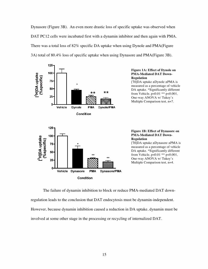
Dynasore (Figure 3B). An even more drastic loss of specific uptake was observed when
DAT PC12 cells were incubated first with a dynamin inhibitor and then again with PMA.
There was a total loss of 82% specific DA uptake when using Dynole and PMA(Figure
3A) total of 80.4% loss of specific uptake when using Dynasore and PMA(Figure 3B).
The failure of dynamin inhibition to block or reduce PMA-mediated DAT down-
regulation leads to the conclusion that DAT endocytosis must be dynamin-independent.
However, because dynamin inhibition caused a reduction in DA uptake, dynamin must be
involved at some other stage in the processing or recycling of internalized DAT.
** **
Figure 1A: Effect of Dynole on PMA-Mediated DAT Down-Regulation [3H]DA uptake ±Dynole ±PMA is measured as a percentage of vehicle DA uptake. *Significantly different from Vehicle, p<0.01 ** p<0.001, One-way ANOVA w/ Tukey’s Multiple Comparison test, n=7.
Figure 1B: Effect of Dynasore on PMA-Mediated DAT Down-Regulation [3H]DA uptake ±Dynasore ±PMA is measured as a percentage of vehicle DA uptake. *Significantly different from Vehicle, p<0.01 ** p<0.001, One-way ANOVA w/ Tukey’s Multiple Comparison test, n=4.

16
DA Uptake Kinetics Following Dynamin Inhibition
After it was observed that dynamin inhibition caused a loss of specific DA uptake
on its own, it was decided that the kinetics of DAT’s interaction with DA in the presence
of a dynamin inhibitor should be studied to determine how the addition of a dynamin
inhibitor reduced DA transport. In the kinetics assays, uptake was recorded ±Dynasore
with varying concentrations of [3H]DA. These were performed to determine any changes
caused by dynamin inhibition in either the maximum velocity of DA binding at saturation
(Vmax) or the concentration at which the velocity was half of Vmax (Km) of DA transport.
Data from theses assays showed a drastic decrease in the Vmax following Dynasore
treatment as can be seen in a representative Michaelis-Menton curve in Figure 4A.
Figure 4A: Michaelis-Menton Kinetics of DA Uptake ±Dynasore Velocity of [3H]DA uptake ±Dynole is measured at DA concentrations of 0.1, 0.3, 0.6, 1.0, 3.0 and 6.0μM. Graph is representative of at least 6 independent experiments.
Figure 4B: Effect of Dynasore on the Vmax of DA Uptake Kinetics Vmax ±Dynasore is measured as a percentage of the vehicle Vmax. *Significantly different from Vehicle, p<0.05 Unpaired t test, n=6.

17
Over the course of six kinetics experiments, it was observed that there was a
significant decrease in Vmax following dynamin inhibition, but no significant change in
Km. On average, the Vmax in the presence of Dynasore was only 48.3% of the vehicle Vmax
(Figure 4B).
[3H] Alanine Uptake Following Dynamin Inhibition
To test if the loss in DA uptake and decrease in Vmax that occurred following
dynamin inhibition was the result of any disruption by the dynamin inhibiting drugs to
either the cell membrane or the Na+ gradient that DAT relies on for transport, [3H]
Alanine uptake assays were performed ±Dynole. Alanine transport in PC12 cells is Na+
dependent and an altering of the Na+ gradient would be reflected in [3H]Ala uptake.
Concurrently, a disruption of the dynamics of the plasma membrane could also be
reflected in [3H] Alanine uptake.
The 3H Alanine studies showed that there was no significant loss in 3H Alanine
uptake in the presence of Dynole and if anything, there was a trend that 3H Alanine
uptake increased in its presence (Figure 5). Additionally, it suggested that the loss in DA
Figure 5: Effect of Dynole on Na+ Dependent Alanine Uptake [3H]Alanine uptake ±Dynole is measured as a percentage of vehicle Alanine uptake. There is no significant difference between conditions p>0.05.

18
uptake following dynamin inhibition was not the result of any Na+ gradient change or a
disruption of the cell membrane that would cause interrupted transporter functions.
Dynamin Inhibition Time Course
When it was determined that DAT internalization was dynamin-independent,
further studies were performed to attempt to determine what point in DAT recycling was
dynamin-dependent. To test this, time course assays were performed. In the time course,
DAT PC12 cells were incubated with the dynamin-inhibitor Dynole for 5, 10, 15, 20, 30
and 45 minute intervals to determine how long of an incubation was needed for an effect.
The assays revealed that there was a drastic loss in specific DA uptake after only
5 minutes incubation with Dynole (Figure 6). Overall this was observed to be an average
65% loss of specific uptake and that Dynole reduced DA uptake regardless of the
incubation period.
Figure 6: Effect of Dynole on DA Uptake [3H]DA uptake ±Dynole is measured as a percentage of vehicle DA uptake. Cells were incubated with Dynole for 5, 10, 15, 20, 30 and 45 minutes. n=3.

19
DISCUSSION
The overall conclusion that can be made from these experiments is that the
dopamine transporter is internalized via a dynamin-independent endocytic pathway. The
failure of dynamin inhibition to block or reduce PMA-mediated DAT down-regulation
leads to this conclusion.
Furthermore, because there was an observed loss in DA uptake when cells were
treated solely with a dynamin inhibitor and an additional loss in DA uptake when cells
were treated with PMA following dynamin inhibition, the study suggests that dynamin
must be involved at some subsequent stage in DAT trafficking. According to Michaelis-
Menton kinetics, the observed reduction in the Vmax of DA uptake following treatment
with dynamin indicates that the transporter has either become inactivated or there has
been a loss of total transporter from the system. Data from [3H] Alanine uptake assays
can be used to infer that the loss in DA uptake from the dynamin inhibitor Dynole is not
the result of a Na+ gradient change or a disruption of the plasma membrane that would
cause interrupted transporter functions. Therefore, the decrease in Vmax must be due to a
subtraction of DAT from the cell surface and the inhibition of dynamin must sequester
DAT in some compartment within the cell, suggesting that dynamin is involved at some
point in the processing or recycling of internalized DAT.
Further studies on the internalization and recycling pathways utilized by DAT are
needed to fully understand how exactly dynamin is involved in DAT trafficking. Cell
surface biotinylation experiments could be used to quantitatively measure changes in
DAT levels on the cell surface following dynamin inhibition. Immunocytochemistry

20
experiments are needed to observe where DAT is sequestered in the presence of a
dynamin inhibitor and whether it co-localizes in any particular compartment, such as
early endosomes, endosomal recycling compartments or lysosomes. Additionally, further
experiments should be performed to determine the nature of the apparently dynamin- and
clathrin-independent endocytic pathway utilized by DAT.
The dopamine transporter has been implicated in a number of neurological
disorders including Bipolar Disorder, Attention Deficit Disorder, and drug addiction.
Fully understanding the activity and trafficking of DAT is vital for the potential treatment
of such disorders. The data from this MQP can help narrow down the possible endocytic
pathways that could be employed in DAT endocytosis by eliminating any that are
dynamin-dependent. Furthermore, the data raises new questions into how many other
pathways there may be for the internalization of plasma membrane vesicles.

21
BIBLIOGRAPHY
Boudanova, E., Navaroli, D.M., Stevens, Z.H., & Melikian, H.E. (2008). “ Dopamine
transporter endocytic determinants: carboxy terminal residues critical for basal and PKC-stimulated internalization”. Mollec. & Cellular Neurosci. 39: 211-217.
Boudanova, E., Navaroli, D.M., & Melikian, H.E. (2008). “Amphetamine-induced
decreases in dopamine transporter surface expression are protein kinase C-independent”. Neuropharmacology. 54: 605-612.
Buckley, K.M., Melikian, H.E., Provoda, C.J., & Waring, M.T. (2000). “Regulation of
neuronal function by protein trafficking: a role for the endosomal pathway”. J. Physiol. 525.1: 11-19.
Damke, H., Baba, T., Warnock, D.E., & Schmid, S.L. (1994). “Induction of mutant
dynamin specifically blocks endocytic coated vesicle formation”. J. Cell Biology. 127 (4): 915-934.
Daniels, G. M., & Amara, S.G. (1999). “Regulated trafficking of the human dopamine
transporter”. J. Biol. Chem. 274(50): 35791-35801. Doherty, G.J., & McMahon, H.T. (2009). “Mechanisms of endocytosis”. Annu. Rev.
Biochem. 78: 31.1-31.46. Girard, E., Paul J.L., Fournier, N., Beaune, P., Johannes, L., et al. (2011). “The dynamin
chemical inhibitor Dynasore impares cholesterol trafficking and sterol-sensitive genes transcription in human HeLa cells and macrophages”. PLoS ONE. 6(12): e29042. Doi:10:1371/journal.pone.0029042.
Hill, T.A., Gordon, C.P., McGeachie, A.B., et al. (2009). “Inhibition of dynamin
mediated endocytosis by the dynoles - synthesis and functional activity of a family of indoles”. J. Med. Chem. 52: 3762-3773.
Holton, K.L., Loder, M.K., & Melikian, H.E. (2005). “Nonclassical, distinct endocytic
signals dictate constitutive and PKC-regulated neurotransmitter transporter internalization”. Nature Neurosci. 8(7): 881-888.
Kirchhausen, T., Macia, E., & Pelish, H.E. (2008). “Use of dynasore, the small molecule
inhibitor of dynamin, in the regulation of endocytosis”. Methods Enzymol. 438: 77-93.
Kong, M.M.C., Hasbi, A., Mattocks, M., Fan, T., O’Dowd, B.F., & George, S.R. (2007).
“Regulation of D1 dopamine receptor trafficking and signaling by caveolin-1”. Mol. Pharmacol. 72:1157-1170.

22
Loder, M.K., & Melikian, H.E. (2003). “The dopamine transporter constitutively internalizes and recycles in a protein kinase C-regulated manner in stably transfected PC12 cell lines”. J. Biol. Chem. 278(24): 221368-22174.
Macia, E., Ehrlich, M., Massol, R., Boucrot, E., Brunner, C., & Kirchhausen, T. (2006).
“Dynasore, a cell-permeable inhibitor of dynamin”. Developmental Cell. 10: 839-850.
Melikian, H.E., & Buckley, M.K. (1999). “Membrane trafficking regulates the activity of
the human dopamine transporter”. J. Neurosci. 19(18): 7699-7710. Melikian, H.E., (2004). “Neurotransmitter transporter trafficking: endocytosis, recycling
and regulation”. Pharmacol. & Therapeutics. 104: 17-27. Mesaki, K., Tanabe, K., Obayashi, M., Oe, N., & Takei, K. (2011). “Fission of tubular
endosomes triggers endosomal acidification and movement”. PLoS ONE. 6(5):e19764. Doi:10.1371/journal.pone.0019764.
Navaroli, D.M., Stevens, Z.H., Uzelac, Z., Gabriel, L., et al. (2011). “The plasma
membrane-associated GTPase Rin interacts with the dopamine transporter and is required for protein kinase C-regulated dopamine transporter trafficking”. J. Neurosci. 31(39): 13758-13770.
Newton A.J., Kirchhausen, T., & Murthy, V.N. (2006). “Inhibition of dynamin
completely blocks compensatory synaptic vesicle endocytosis”. PNAS. 103 (47): 17955-17960.
Saunders, C., Ferrer, J.V., Shi, L., Chen, J., et al. (2000). “Amphetamine-induced los of
human dopamine transporter activity: an internalization-dependent and cocaine sensitive mechanism”. PNAS. 97(12): 6850-6855.
Sorkina, T., Miranda, M., Dionne, K.R., Hoover, B.R., Zahniser, N.R., & Sorkin, A.
(2006). “RNA interference screen reveals an essential role of Nedd4-2 in dopamine transporter ubiquitination and endocytosis”. J. Neurosci. 26(31):8195-8205.
Takei, K., Yoshida, Y., & Yamada, H. (2005). “Regulatory mechanisms of dynamin-
dependent endocytosis”. J. Biochem. 137: 243-247. Zahniser, N.R., & Sorkin, A. (2004). “Rapid regulation of the dopamine transporter: role
in stimulant addiction?”. Neuropharmacology. 47: 80-91. Zahniser, N.R., & Sorkin, A. (2009). “Trafficking of dopamine transporters in
psychostimulant actions”. Sem. Cell & Dev. Bio. 20:411-417.