Embed Size (px)
Citation preview

Original
Rev. Argent. Radiol. 2014;78(3): 149-155 149
Doppler Ultrasound in the diagnosis of placenta percreta: our experienceP. García Saraví, N.K. Patiño, M.L. Juana, J. Mariano*, E. Reyna and R. Tizzano
Hospital Interzonal General de Agudos ‘‘General José de San Martín’’, La Plata, Buenos Aires, Argentina
IntroductionPlacenta accreta (PA) results from abnormal placentation characterized by the invasion of trophoblastic villi beyond the decidua1. In placenta accreta vera, chorionic villi are attached to the surface of the myometrium; placenta increta occurs when there is a deep invasion of the villi into the myometrum and in placenta percreta the entire myometrum, including the serosa, is penetrated by chorionic villi. The latter is the least common, but it carries the highest maternal and perina-tal morbidity and mortality because of its difficult diagnosis2.The main risk factors include placenta previa and Cesarean section. Owing to the increasing rate of Cesarean section in recent years, there has been a rise in the prevalence and inci-dence of this condition1. Prenatal diagnosis of placenta percreta is extremely difficult. Diagnosis relies on ultrasound and color Doppler, but may be supplemented with magnetic resonance imaging (MRI), cystoscopy and biochemical markers2. If the condition is not detected before the time of delivery, it may be a devastat-ing obstetric condition that can be life-threatening for both
mother and fetus. Therefore, prenatal diagnosis is essential for adequate planning and multidisciplinary management1,3.The aim of our study is to demonstrate the usefulness of Doppler ultrasound in the detection of bladder invasion in cases of placenta percreta.
Materials and methods
Between November 2011 and May 2013, 21 patients aged 20-44 years with a surgical and histopathological diagnosis of placenta accreta were evaluated by ultrasound. All patients were examined in the third trimester of pregnancy (weeks 32 through 40 of pregnancy) and in 100% of cases bladder ul-trasound was performed by the suprapubic approach with a sufficient bladder repletion, considered to be adequate when it allowed correct visualization of the entire bladder wall.Different ultrasound systems were used for this study (Toshi-ba Nemio, Philips HD7 and Sonosite Micromax) equipped with a convex transducer of 3.5 MHz and a lineal transducer of 7.5 MHz.
AbstractPurpose: To demonstrate the usefulness of Doppler ultrasound in the detection of bladder invasion in cases of placenta percreta.Materials and methods: Twenty-one patients, aged 20-44 years old, with surgical and histopathological diagnosis of placenta accreta were evaluated by ultrasound between November 2011 and May 2013. The presence of increased vascularity on the bladder wall on Doppler ultrasound was classified as bladder invasion, while the presence of other sonographic findings on grayscale imaging with negative Doppler signal was classified as probable invasion. Results: Of the 21 patients included in the study with placenta accreta, 7 had bladder invasion on histopathological examination. Out of these 7, 5 had a diagnosis and ultrasound report of bladder invasion (due to the identification of vascular structures on color Doppler examination) and the remaining two were considered to have probable invasion. Of the 14 patients with no bladder invasion detected on histopathological examination, 7 had normal ultrasound reports and 7 were reported as probable invasion.Conclusion: Doppler ultrasound is a very reliable method for the detection of bladder invasion in placenta percreta, seen as increased vascularity of the uterine-bladder interface on color Doppler examination.© 2013 Sociedad Argentina de Radiología. Published by Elsevier España, S.L.U. All rights reserved.
Keywords: Placenta accreta; placenta percreta; ultrasound
Rev. Argent. Radiol. 2014;78(3): 149-155
Doppler Ultrasound in the diagnosis of placenta percreta: our experience
150
Findings were assessed using grayscale ultrasound (loss of the echogenic line between the bladder wall and the uterus, ir-regularity and/or bulging of the bladder wall and exophytic masses within the bladder) as well as color Doppler (irregular-ity of the bladder wall because of serpiginous structures with positive flow on Doppler evaluation).Sagittal imaging was used to assess the depth of placental tis-sue, its vascularity and its relationship to the bladder wall, while coronal imaging was used to assess the extent of invasion.The presence of vascular structures on the bladder wall on Dop-pler ultrasound was classified as bladder invasion and the pres-ence of other sonographic findings on grayscale imaging with negative Doppler was classified as probable bladder invasion.All patients included in our study underwent scheduled Ce-sarean section.
Final diagnosis of bladder invasion was performed post-sur-gery with a histopathologic examination.
Results
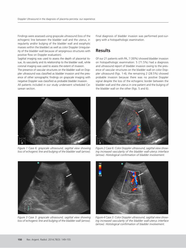
Of our 21 patients with PA, 7 (30%) showed bladder invasion on histopathologic examination: 5 (71.5%) had a diagnosis and ultrasound report of bladder invasion owing to the pres-ence of vascular structures on the bladder wall on color Dop-pler ultrasound (figs. 1-4); the remaining 2 (28.5%) showed probable invasion because there was no positive Doppler signal despite the loss of the echogenic border between the bladder wall and the uterus in one patient and the bulging of the bladder wall on the other (figs. 5 and 6).
Figure 1 Case 6: grayscale ultrasound, sagittal view showing loss of echogenic line and bulging of the bladder wall (arrow).
Figure 2 Case 6: Color Doppler ultrasound, sagittal view show-ing increased vascularity of the bladder wall-uterus interface (arrow). Histological confirmation of bladder involvement.
Figure 3 Case 3: grayscale ultrasound, sagittal view showing loss of echogenic line and bulging of the bladder wall (arrow).
Figure 4 Case 3: Color Doppler ultrasound, sagittal view show-ing increased vascularity of the bladder wall-uterus interface (arrow). Histological confirmation of bladder involvement.

Doppler Ultrasound in the diagnosis of placenta percreta: our experience
Rev. Argent. Radiol. 2014;78(3): 149-155
P. García Saraví et al.
151
Figure 5 Case 2: grayscale ultrasound, sagittal view showing loss of echogenic line (arrow).
Figure 6 Case 2: Color Doppler ultrasound, sagittal view not showing increased vascularity of the bladder wall (negative Doppler) (arrow). Histological confirmation of placenta ac-creta without bladder involvement.
Figure 7 Case 16: grayscale ultrasound, sagittal view showing regular and intact echogenic line of the bladder wall (arrow).
Figure 8 Case 16: Color Doppler ultrasound showing standard vascularity of the placental base, with no interface vascularity (arrow). Histological confirmation of placenta accreta without bladder involvement.
Figure 9 Case 12: grayscale ultrasound, sagittal view showing apparent loss of echogenic line at the upper region of the bladder wall (arrow).
Figure 10 Case 12: Color Doppler ultrasound, sagittal view showing no increased vascularity of the bladder wall (nega-tive Doppler) (arrow). Histological confirmation of placenta accreta without bladder involvement.
Rev. Argent. Radiol. 2014;78(3): 149-155
Doppler Ultrasound in the diagnosis of placenta percreta: our experience
152
Table 1: Sonographic findings in 21 cases of placenta accreta
Case/ Pregnancies- GA Placental Sonographic Sonographic HistopathologicalAge miscarriages- location findings of findings of findings deliveries- bladder invasion probable bladder Cesarean (positive Doppler) invasion sections (negative Doppler)
1/33 P8 M4 CS3 37 Total occlusive Yes - PA with bladder invasion2/32 P6 M1 CS4 35 Total occlusive - Yes PA with bladder invasion3/39 P8 M1 D5 CS1 40 Total occlusive Yes - PA with bladder invasion4/33 P4 CS3 35 Total occlusive Yes - PA with bladder invasion5/44 P14 M1 D9 CS3 38 Total occlusive - Yes PA with bladder invasion6/32 P4 M1 CS2 35 Total occlusive Yes - PA with bladder invasion7/37 P3 M1 CS1 37 Partial occlusive Yes - PA with bladder invasion8/29 P5 D4 34 Total occlusive - Yes PA with no bladder invasion9/25 P4 D2 CS1 35 Total occlusive - Yes PA with no bladder invasion10/28 P6 D1 CS4 37 Total occlusive - Yes PA with no bladder invasion11/38 P8 D4 CS3 32 Partial occlusive - Yes PA with no bladder invasion12/40 P9 M1 D6 CS1 37 Total occlusive - Yes PA with no bladder invasion13/42 P5 M1 CS3 37 Total occlusive - Yes PA with no bladder invasion14/27 P6 M1 CS4 33 Partial occlusive - Yes PA with no bladder invasion15/31 P5 M1 D2 CS1 35 Total occlusive - - PA with no bladder invasion16/40 P4 M2 CS1 36 Total occlusive - - PA with no bladder invasion17/36 P3 CS2 36 Total occlusive - - PA with no bladder invasion18/24 P4 CS3 35 Total occlusive - - PA with no bladder invasion19/33 P3 CS2 33 Marginal - - PA with no bladder invasion20/37 P8 M2 D5 37 Partial occlusive - - PA with no bladder invasion21/28 P4 M1 CS2 38 Total occlusive - - PA with no bladder invasion
P: pregnancies; M: miscarriages; D: deliveries; CS: Cesarean sections; PA: placenta accreta; GA: gestational age
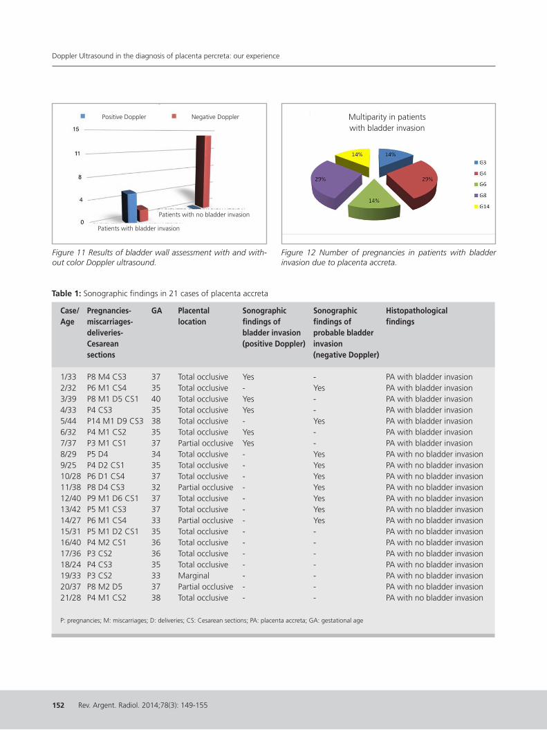
Figure 11 Results of bladder wall assessment with and with-out color Doppler ultrasound.
Patients with bladder invasion
Patients with no bladder invasion
Positive Doppler Negative Doppler
Figure 12 Number of pregnancies in patients with bladder invasion due to placenta accreta.
Multiparity in patients with bladder invasion
Doppler Ultrasound in the diagnosis of placenta percreta: our experience
Rev. Argent. Radiol. 2014;78(3): 149-155
P. García Saraví et al.
153
Of the 14 patients (70%) with no bladder invasion on histo-pathological examination, 7 had normal ultrasound reports, with an intact bladder wall (figs. 7 and 8), and 7 cases were classified as probable bladder invasion based on the presence of findings on grayscale ultrasound only (figs. 9 and 10).Sonographic findings for each case are detailed in Table 1, while results obtained with and without color Doppler are reported in Figure 11.
As regards risk factors for PA, there were 16 cases of placenta previa with total occlusion, 4 cases of placenta previa with partial occlusion and one case of marginal placenta. Of the 7 patients with bladder invasion, 6 had total occlusive placenta and 1 had partial occlusive placenta.With the number of previous cesarean deliveries, 19 patients had undergone one or more Cesarean sections, 1 reported two curettage procedures and 1 had no history of surgery. All 7 patients with bladder invasion had undergone Cesarean section.All patients were multiparous women, with 3 or more preg-nancies. For patients with bladder invasion, the number of pregnancies is summarized in Figure 12.
Discussion
Placenta accreta (PA) results from abnormal placentation characterized by the invasion of trophoblastic villi beyond the deciduas. The deciduas basalis is fully or partially absent due to a defect in the development of the fibrinoid layer of Nita-buch, separating the deciduas basalis from the villous portion of the placenta4.The current incidence of placenta accreta ranges between 1/540 and 1/93000 births2. Risk factors for the development of this condition include uterine instrumentation, intrauterine scarring and prior Cesarean delivery, all of which may be as-sociated with damage to or absence of the decidua basalis, as well as placenta previa, smoking, endometritis, maternal age over 35, grand multiparity and recurrent miscarriage5. The combination of prior Cesarean section and placenta pre-via represents the highest contribution to risk.Our findings were consistent with the literature as regards the aforementioned factors. From this perspective, it is worth mentioning that over the last 50 years, the frequency of ab-normal placentation has increased 10-fold and that, accord-ing to the World Health Organization (WHO) statistics, in our setting over one-third of all deliveries occur by Cesarean section (with higher rates associated with prepaid healthcare systems)4.Depending on the depth of penetration, there are three forms of abnormal placentation:
• Accreta: theplacenta isattached to,butdoesnotpene-trate, the myometrium (78% of cases).
• Increta:theplacentalpenetratesthroughthemyometrium(17% of cases)
• Percreta:theplacentapenetratesthefullthicknessofthemyometrium to the uterine serosa, and may invade adja-cent organs (5% of cases).
Depending on the extension, abnormal placentation may be classified into6:
• Focal:whenonlysmallplacentalareasareinvolved.• Partial:whenoneormorecotyledonsareinvolved.• Total:whentheentiresurfaceoftheplacentaisabnormally
attached.Our study revealed the presence of placenta percreta with bladder invasion in 30% of cases evaluated, in contrast with current statistics reported in the literature. Nevertheless, this finding may probably be related to the nature of our service, which belongs to a hospital that is a referral center for pla-centa accreta in the province of Buenos Aires.As regards the forms of presentation, there is no specific clini-cal syndrome for placenta accreta. A large number of cases are asymptomatic; therefore, PA should be suspected when-ever risk factors are present. Symptoms depend mainly on the features of the placenta previa and the invasion to other organs and the resulting complications. The main manifesta-tion is hemorrhage, which may occur before, during or after delivery.In cases of bladder invasion, placenta percreta may manifest with gross hematuria (20% of cases) or with microscopic he-maturia. The latter is the most common sign and should raise suspicion and alert the urologist2. In our series, none of the 7 patients with bladder invasion exhibited this finding.Furthermore, placenta percreta may become evident with re-tained placenta at delivery or as complications (such as shock and disseminated intravascular coagulation).The importance of making the diagnosis of PA before delivery is that it allows for multidisciplinary planning with the aim of minimizing potential maternal or neonatal morbidity and mortality.For placenta percreta with bladder invasion, the rate of ma-ternal mortality has been reported to be 20% and the rate of perinatal mortality has been reported to be 30%, as it is difficult to diagnose this condition prior to delivery7. In our 7 patients with bladder invasion, no deaths occurred.The diagnosis is usually established by ultrasound and occa-sionally supplemented by MRI8. Some authors have detected elevated serum concentrations of alpha-fetoprotein (AFP) in patients with placenta accreta, which suggests that placen-tal-uterine interface abnormalities might result in leakage of AFP into the maternal circulation. For this reason, in pregnant women the presence of elevated AFP concentrations and risk

Rev. Argent. Radiol. 2014;78(3): 149-155
Doppler Ultrasound in the diagnosis of placenta percreta: our experience
154
factors should raise suspicion for placenta accreta9.In order to establish diagnosis, more than one sonographic criterion should be met. Sonographic diagnostic criteria in-clude: 1,10
• Placentaprevia• Placentalvascularlacunae• AbnormalcolorDopplerpatterns• Lossoftheretroplacentalclearspace• Thinningorlossofthemyometriallayer• Disruption or irregularity of the uterine-bladder cleavage
plane• Placentaltissuewithmasseffect(bulging)• Bladderwallirregularity
The uterus is separated from the bladder wall by the uter-ine-bladder cleavage plane, also known as uterine-bladder complex. This complex appears as a smooth hyperechoic lin-ear band representing the uterine serosa, the fat tissue and the posterior bladder wall1. On grayscale ultrasound exami-nation, the bladder wall appears thin, smooth and regular, while on color Doppler ultrasound no vascular structures are detected.Special care should be taken when examining the bladder wall since in order to make a correct and clear assessment and mini-mize potential errors, adequate repletion is required and the transducer should be positioned at different Doppler angles. It is much easier to diagnose placenta accreta than to make the subsequent step of determining if trophoblast has grown through the uterine wall into other structures. Although it would be ideal to identify percreta with certainty, no one has yet been able to do that reliably11. Unfortunately, disruption of the echogenic line between the bladder and the uterus and bulging are non-specific signs for placenta percreta in-volving the bladder, and do not always predict this condition. Comstock 11 reported that in the three cases in his series in which these signs were present, two had a placenta percreta and one had a simple accreta. Kirkinen et al 12, in turn, have demonstrated similar findings, in agreement with our study.Bladder involvement should be suspected in the event of bladder wall irregularity caused not only by placental tissue invasion but also by the existence of a large number of as-sociated vascular structures1.Comstock11 reports a problem in patients who had a previous Cesarean section: these women usually develop increased vascularity in the space between the myometrium and the bladder, probably because the bladder flap is retracted be-fore the incision is made into the uterus and because this area is exposed to blood products. Therefore, it is important to differentiate between bulging due to increased number of preexisting vessels and actual neovascularization from the
placenta through the myometrium.In cases of placenta percreta, Doppler examination for as-sessment of vascularization of the uterine serosa-bladder in-terface shows an extensive hypervascular appearance with densely confluent anarchic vessels that occasionally protrude into the bladder lumen13.In our experience, of the 7 patients with a histopathologi-cal diagnosis of placenta percreta with bladder invasion, 5 (71.5%) had positive color Doppler findings.The sensitivity and specificity of grayscale ultrasound for diag-nosing placenta accreta vary according to different authors, but sensitivity is estimated to be approximately 77-87% and specificity approximately 96-98%. The use of Doppler in-creases diagnostic sensitivity, reaching --according to Lerner et al14-- 100% sensitivity and 94% specificity. Levine15, in turn, has reported a sensitivity of 86% and a specificity of 92% for Doppler imaging, while the American College of Obstetricians and Gynecologists8 has not seen significant changes in these percentages with the use of color Doppler. In our case, with color Doppler we obtained 77% sensitivity and 100% Specificity.The diagnostic efficacy of ultrasound allows limitation of the use of MRI (whose diagnostic value is still a subject of debate) as well as of the administration of gadolinium in the prena-tal stage8,13,16. In addition, ultrasound is a simple, low-cost, reproducible, noninvasive and accessible method, thus being ideal for patients with suspected placenta accreta.Our data, which agree with those of other authors11,13,17, suggest the possibility of diagnosing bladder invasion using a combination of grayscale and color Doppler ultrasound find-ings. However, based on the results obtained, color Doppler examination became more relevant, as it enabled confirma-tion of 5 out of 7 cases with bladder involvement and al-lowed us to rule out bladder invasion in 14 patients.
Conclusion
Placenta accreta/percreta is an obstetric complication that is potentially ominous for the mother and that is currently developing the characteristics of an epidemic. This condition is one of the main causes of peripartum hysterectomy and maternal and perinatal morbidity and mortality.Diagnosis of this condition is based on two essential pillars: a high suspicion for disease based on the presence of risk fac-tors, and both grayscale and color Doppler ultrasound find-ings (because they have a high specificity).Doppler ultrasound allows for diagnosis of bladder invasion in placenta percreta, seen as positive vascularization of the bladder wall on color Doppler.
Doppler Ultrasound in the diagnosis of placenta percreta: our experience
Rev. Argent. Radiol. 2014;78(3): 149-155
P. García Saraví et al.
155
Conflicts of interest The authors declare no conflicts of interest, except for Dr. Mariano who declares a possible conflict of interest as junior reviewer of the Argentine Journal of Radiology.
References 1. Stoisa D, Sánchez NO, Villavicencio RL. Hallazgos imagenológicos en el
acretismo placentario. Imágenes. 2012;1: 17---26.2. Vera E, Lattus J, Bermúdez H, Espinoza L, Ibá˜nez C, Herrera A, et al. Pla-
centa percreta con invasión vesical: reporte de 2 casos. Rev Chil Obstet Ginecol. 2005;70:404---10.
3. Martin F, Corbetta JP, Urday N, Grippo L. Percretismo placentario. Com-promiso vesical. Rev Arg Urol. 2003;68: 99---102.
4. Martínez M. Protocolo para el tratamiento y la prevención de las hemor-ragias obstétricas graves. En: Nú˜nez de Pierro A, Vinacur J, Voto L, edi-tores. Programa de Actualización en Ginecología y Obstetricia (PROAGO). Buenos Aires: Médica Panamericana; 2008. p. 147---88.
5. Esakoff TF, Sparks TN, Kaimal AJ, Kim LH, Feldstein VA, Goldstein RB, et al. Diagnosis and morbidity of placenta accreta. Ultrasound Obstet Gynecol. 2011;37:324---7.
6. Due˜nas O, Rico H, Rodriguez M. Actualidad en el diagnóstico y manejo del acretismo placentario. Rev Chil Obstet Ginecol. 2007;72:266---71.
7. Abehsera D, González C, López S, Sancha M, Magdaleno F. Placenta per-creta, experiencia en 20 a˜nos del Hospital Universitario La Paz, 76. Ma-
drid: Espa˜na; 2011. p. 127---31. Rev Chil Obstet Ginecol.8. Committee on Obstetric Practice. Committee opinion No 529: placenta
accreta. Obstet Gynecol. 2012;120:207---11.9. Haghenbeck-Altamirano FJ, Leis-Márquez T, Ayala-Yá˜nez R, Juárez-García
LC, García-Moreno C. Diagnóstico antenatal de acretismo-percretismo placentario. Ginecol Obstet Mex. 2013;81:259---71.
10. Baughman WC, Corteville JE, Shah RR. Placenta accreta: spectrum of US and MR imaging findings. Radiographics. 2008;28:1905---16.
11. Comstock CH. Antenatal diagnosis of placenta accreta: a review. Ultra-sound Obstet Gynecol. 2005;26:89---96.
12. Kirkinen P, Helin-Martikainen HL, Vanninen R, Partanen K. Placenta accre-ta: imaging by gray-scale and contrast-enhanced color Doppler sonogra-phy and magnetic resonance imaging. J Clin Ultrasound. 1998;26:90---4.
13. Calí G, Giambanco L, Puccio G, Forlani F. Morbidly adherent placenta: evaluation of ultrasound diagnostic criteria and differentiation of placenta accreta from percreta. Ultrasound Obstet Gynecol. 2013;41:406---12.
14. Lerner JP, Deane S, Timor-Tritsch IE. Characterization of placenta accreta using transvaginal sonography and color Doppler imaging. Ultrasound Obstet Gynecol. 1995;5:198---201.
15. Levine D, Hulka CA, Ludmir J, Li W, Edelman RR. Placenta accreta: evalu-ation with color Doppler US, power Doppler US, and MR imaging. Radiol-ogy. 1997;205:773---6.
16. Kanal E, Barkovich AJ, Bell C, Borgstede JP, Bradley Jr WG, Froelich JW, et al. ACR guidance document for safe MR practices: 2007. AJR Am J Roentgenol. 2007;188:1447---74.
17. Chou MM, Ho ES, Lee YH. Prenatal diagnosis of placenta previa accreta by transabdominal color Doppler ultrasound. Ultrasound Obstet Gynecol. 2000;15:28---35.