Embed Size (px)
Citation preview

DOPPLER VELOCITY ASSESSMENT
OF VENOUS RETURN
IN THE HUMAN FETUS

Met dank aan Hitachi Nederland b.v. en Schering Nederland b.v. voor hun financi81e bijdrage aan de drukkosten.
The work presented in this thesis was periormed in the Department of Obstetrics and Gynaecology. University Hospital Dijkzigt. Erasmus University, Rotterdam, The Netherlands and supported by the Dutch Foundation for Medical Research MEDIGON (grant nr. 900-568-215).
No part of this book may be reproduced in any form, by print, photoprint, microfilm or any other means without written permission from the publisher.
Niets uit deze uitgave mag worden verveelvoudigd en/of openbaar gemaakt worden door middel van druk, fotocopie, microfilm of op welke andere wijze oak zonder voorafgaande schriftelijke toestemming van de uitgever.
© T.W.A. Huisman ISBN 90-9006484-2
Printed by Pasmans Offsetdrukkerij b.v., The Hague

DOPPLER VELOCITY ASSESSMENT
OF VENOUS RETURN
IN THE HUMAN FETUS
Evaluatie van de veneuze return in de humane foetus
met behulp van Doppler bloedsnelheidsmetingen
PROEFSCHRIFT
TER VERKRIJGING VAN DE GRAAD VAN DOCTOR
AAN DE ERASMUS UNIVERSITEIT ROTTERDAM
OP GEZAG VAN DE RECTOR MAGNIFICUS
PROF.DR. P.W.C. AKKERMANS M.Lit.
EN VOLGENS BESLUIT VAN HET COLLEGE VAN DEKANEN.
DE OPENBARE VERDEDIGING ZAL PLAATSVINDEN OP
WOENSDAG 15 SEPTEMBER 1993 OM 15.45 UUR
DOOR
T JEERD WILL EM ALEXANDER HUISMAN
GEBOREN TE AMSTERDAM

PROMOTIE-COMMISSIE
PROMOTOR:
OVERIGE LEDEN
Prof.Jhr.Dr. J.W. Wladimiroff
Prof. S.H. Eik-Nes M.D., Ph.D.
Prof.Dr. AC. Gittenberger-de Groot
Prof.Or. J. Hess

"Some who do not know~ and especially those who have experience~
are more practical than others who know".
Aristoteles
Aan mijn ouders

Contents
Chapter 1 Introduction and definition of objectives
1.1
1.2
Introduction
Definition of objectives
Chapter 2 The fetal circulation with emphasis on venous return
2.1 Introductory remarks
2.2 Historical background
2.3 Embryology and anatomy
2.4 Preload physiology
2.5 Animal experiments
2.6 Doppler techniques; transabdominal versus transvaginal Doppler ultrasound
Part of this chapter was published in: TWA Huisman, JW Wladimiroff.
The ductus venosus. Fetal Matern Med Rev 1993;5:45-55.
Chapter 3 Doppler assessment of venous return relative to cardiac performance
and afterload in early pregnancy
3.1 Introductory remarks
3.2 Doppler flow velocity waveforms in late first and early second trimester fetuses;
9
11
15
15
18
21
24
28
41
reproducibility of waveform recordings (Ultrasound Obstet Gynecol 1993;3:260-263) 42
3.3 Evaluation of fetal cardiac performance by cardiac Doppler flow velocity
recording in early pregnancy
3.3.1 Fetal cardiac flow velocities in the rate first trimester of pregnancy;
a transvaginal Doppler study (JAm Coil Cardiel 1991·,17:1357-1359)
3.4 Evaluation of fetal afterload by arterial Doppler flow velocity recording
in early pregnancy
3.4.1 Fetal and umbllical flow velocity waveforms between 10 -16 weeks'
gestation: a preliminary study (Obstet Gynecol 1991;78:812-814)
3.4.2 Intracerebral, aortic and umbilical artery flow velocity waveforms
in the late first trimester fetus (Am J Obstet Gynecol 1992;166:46-49)
3.5 Evaluation of fetal preload by venous Doppler flow velocity recording in early pregnancy
3.5.1 Normal fetal Doppler inferior vena cava, transtricuspid and umbilical
artery flow velocity waveforms between 11 and 16 weeks' gestation
(Am J Obstet Gynecol 1992;166:921-924)
3.5.2 Flow velocity waveforms in the ductus venosus, umbilical vein and
inferior vena cava in normal human fetuses at 12- 15 weeks of gestation
(Ultrasound Med Bioi 1993;19:441-445)
50
57
63
68
76
6

Chapter 4 Doppler assessment of venous return during the second half of pregnancy
4.1 Introductory remarks 91
4.2 Anatomy of the venous inflow vasculature
4.2.1 Recognition of a fetal subdiaphragmatic venous vestibulum essential
for fetal venous Doppler assessment (Pediatr Res 1992;32:338-341) 91
4.3 Venous Doppler flow velocity waveforms during the second half of pregnancy;
reproducibility of waveform recording
4.3.1 Reproducibility of fetal inferior vena cava and ductus venosus
waveform recording
4.4 Normal flow velocity waveforms from fetal venous inflow during
the second half of pregnancy
4.4.1 Flow velocity waveforms in the fetal inferior vena cava during the
101
second half of normal pregnancy (Ultrasound Med Bioi 1991;17:679-682) 108
4.4.2 Ductus venosus blood flow velocity waveforms in the human fetus:
a Doppler study (Ultrasound Med Bioi 1992;18:33-37)
Chapter 5 Influence of fetal variables on venous Doppler waveforms
5.1 Introductory remarks
5.2 Fetal breathing movements and venous inflow
5.2.1 Changes in inferior vena cava blood flow and diameter during fetal breathing
movements in the human fetus (Ultrasound Obstet Gynecol 1993;3:26-30)
5.2.2 Changes in ductus venosus, hepatic veins, distal part of the
inferior vena cava and umbilical vein flow velocities during
fetal breathing movements in the human fetus.
5.3 Fetal behavioral states and venous inflow
5.4
5.3.1 Ductus venosus flow velocity waveforms relative to fetal behavioral states
in normal term pregnancy (Br J Obstet Gynaecol 1993;in press)
5.3.2 Inferior vena cava flow velocity waveforms relative to fetal
behavioral states and sample site in normal term pregnancy
(submitted in extended form to Early Human Development).
Fetal arrhythmias and venous inflow
5.4.1 Doppler evaluation of venous return during fetal arrhythmias
(submitted)
Chapter 6 General conclusions
Summary
Samenvatting
Dankwoord
Curriculum Vitae
115
127
127
138
144
154
161
178
183
187
192
195

8

Chapter 1 Introduction and objectives 9
Chapter 1
INTRODUCTION AND DEFINITION OF OBJECTIVES
1.1 Introductory remarks
Studies in the human fetus are limited by the methods available for investigation.
Pressure and volume flow measurements in the fetal cardiovascular system
require invasive techniques that are not performed at present. However, infor
mation on fetal circulatory performance may be helpful in the evaluation of
pathologic conditions. With the introduction of Doppler ultrasound non-invasive
examination of the fetal vessels became possible. In the last decade cardio
vascular research in the human fetus has focused on the study of arterial,
cardiac and umbilical blood flows. Four factors mainly determine cardiac perfor
mance: (i) afterload (ii) cardiac contraction force (iii) heart rate {iv) preload.
Examination of the factor afterload in the fetal circulation has been characterized
by Doppler studies of the fetal descending aorta and umbilical artery in the
second half of pregnancy (Marsal et al. 1984; Trudinger et al. 1985;Tonge 1987).
The second factor, cardiac contraction force, is even more difficult to study in the
fetus. Efforts have been made by a number of investigators (Maulik et al. 1985;
Kenny et al. 1986; Reed et al. 1986; Allan et al. 1987), who all tried to quantify
cardiac stroke volume and force by means of Doppler velocimetry at the level of
the atrioventricular valves and in the outflow tracts. However, it was pointed out
that the reproducibility of these data is disappointingly low and large within and
between variation was documented (Beeby et al. 1991).
The third factor is the fetal heart rate, which is relatively easy to obtain. Studies
have shown that as a result of the Frank-Starling mechanism fetal heart rate
changes within the normal heart rate range do not seem to considerably influen
ce fetal cardiac output (Kenny et al. 1987; van der Mooren et al. 1991).
Finally, little is known about the factor preload and the hemodynamics of the fetal
venous vasculature, although it has become clear from animal experimental work

Chapter 1 Introduction and objectives 10
(Rudolph and Heymann 1967; Rudolph 1983) that venous return is an important
factor in cardiac functioning. With the presence of three shunts (foramen ovale,
ductus arteriosus and ductus venosus) and the placenta as a third circulation
venous blood flow and pressures in the normally developing fetus are significant
ly different from the physiologic situation in adults.
In this thesis data are presented o~ Doppler venous cardiac inflow, in particular
from the umbilical vein, ductus venosus and inferior vena cava in (i) the late first
and early second trimester fetus and (ii) the late second and third trimester fetus.
The inclusion of early pregnancy flow studies was based on the significant
changes occurring at placental level around 13 to 14 weeks of gestation with
emphasis on the process of trophoblast invasion, resulting in low resistance
placental vascular dynamics (Pijnenburg et al. 1980; Jauniaux et al. 1991). The
transvaginal approach now allows fetal flow velocity waveform recording as early
as 9 to 10 weeks.
Late pregnancy studies were mainly performed to establish the effect of fetal
variables such as breathing movements, behavioural states and cardiac arrhyth
mias on fetal venous cardiac inflow.
Obviously, proper interpretation of venous inflow velocity parameters is only
feasible when related to other parameters of cardiovascular performance, such
as afterload and heart rate.

Chapter 1 Introduction and objectives 11
1.2 Definition of objectives
The first objective was to review the available literature on fetal venous return
and to assess the various methods of investigating human fetal venous hemody
namics (Chapter 2).
The second objective was to collect data on the nature and reproducibility of fetal
fiow velocity waveforms in late first and early second trimester pregnancies. To
appreciate venous infiow velocities in relation to total cardiovascular dynamics,
attention was also focused on waveform patterns at cardiac and arterial level
(Chapter 3).
The third objective of the study was (i) to ascertain the exact anatomical relation
ship between vessels responsible for fetal venous return in late second and third
trimester pregnancies; (ii) to establish reproducibility and normal values during
this period of pregnancy (Chapter 4).
The fourth and last objective was to determine the influence of fetal internal
variables such as breathing movements, behavioural states and cardiac arrhyth
mias on venous fiow velocity waveforms in late pregnancy (Chapter 5).
REFERENCES
Allan LD, Chita SK, AI-Ghazali W, Crawford DC, Tynan M. Doppler echocardio
graphic evaluation of the normal fetal heart. Br Heart J 1987;57:528-533.
Beeby AR, Dunlop W, Heads A, Hunter S. Reproducibility of ultrasonic measure
ment of fetal cardiac haemodynamics. Br J Obstet Gynaecol 1991;98:807-814.

Chapter 1 Introduction and objectives 12
Jauniaux E, Jurkovic D, Campbell S. In vivo investigations of the anatomy and
the physiology of early human placental circulations.
Ultrasound Obstet Gynecol 1991 ;1 :435-445.
Kenny J, Plappert T, Doubilet P, Saltzman DH, Cartier M, Zollars L, Leatherman
GF, St.John Sutton MG. Changes in intracardiac blood flow velocities and right
and left ventricular stroke volumes with gestational age in the normal fetus: a
prospective Doppler echocardiographic study. Circulation 1986;74:1208-1216.
Kenny J, Plappert T, Doubilet P, Saltzman DH, St.John Sutton MG. Effects of
heart rate on ventricular size, stroke volume and output in the normal human
fetus; a prospective Doppler echocardiographic study. Circulation 1987;76:52-58.
Marsal K, Eik-Nes SH, Lindblad A, Lingman G. Blood flow in the fetal descending
aorta; intrinsic factors affecting fetal blood flow, i.e. fetal breathing movements
and cardiac arrhythmia. Ultrasound Med Bioi 1984;1 0:339-348.
Maulik D, Nanda NC, Saini VD. Fetal Doppler echocardiography: methods and
characterisation of normal and abnormal hemodynamics.
Am J Cardiel 1985;53:572-578.
van der Mooren K, Barendregt LG, Wladimiroff JW. Fetal atrioventricular and
outflow tract flow velocity waveforms during the normal second half of pregnancy.
Am J Obstet Gynecol 1991;165:668-674.
Pijnenburg R, Dixon G, Robertson WB, Brosens I. Trophoblastic invasion of
human decidua from 8 to 18 weeks of pregnancy. Placenta 1980;1 :3-19.
Reed KL, Meijboom EJ, Sahn OJ, Scagnelli SA, Valdes Cruz LM, Shenker L.
Doppler flow velocities in human fetuses. Circulation 1986;73:41-6.

Chapter 1 Introduction and objectives l3
Rudolph AM, Heymann MA. The circulation of the fetus in utero; methods for
studying distribution of blood flow, cardiac output and organ blood flow.
Circ Res 1967;21:163-184.
Rudolph AM. Hepatic and ductus venosus blood flows during fetal life.
Hepatology 1983;3:254-258.
Tonge HM. A Doppler ultrasound study of human fetal vascular dynamics.
Thesis. Erasmus University Rotterdam, the Netherlands.
Promotor: Prof.J.W.Wiadimiroff.
Trudinger BJ, Giles WB, Cook CM, Bombardieri J, Collins L. Fetal umbilical
artery flow velocity waveforms and placental resistance: clinical significance.
Br J Obstet Gynaecol 1985;92:23-30.

Chapter 1 Introduction and objectives 14

Chapter 2 Fetal circulation and venous return: a review
Chapter 2
THE FETAL CIRCULATION WITH EMPHASIS
ON VENOUS RETURN
2.1 Introductory remarks
l5
Knowledge of the fetal circulation has been acquired over the centuries with
remarkable slowness. Most investigations were performed either on bought,
stolen or exhumed corpses or on animals. Invasive studies on the human fetus
are limited. Introduction of non-invasive methods like ultrasonography and
Doppler measurements has enormously accelerated and increased our insights
into the normal and abnormal fetal circulation. This chapter will describe the
acquisition of knowledge on fetal hemodynamics, in particular the embryological,
anatomical and physiological aspects of fetal venous vasculature and blood fiow
with emphasis on venous return. Finally, the Doppler techniques available for
obtaining this information will be discussed.
2.2 Historical background
The understanding of the normal circulation in adults was developed during the
Egyptian and Greek period, in which famous physicians like Aristotle and
Herophilus tried to explain the function of the vascular system. The uniqueness of
the fetal circulation and its anatomical structures was first recognized by Galen
(130-200 AD) who probably studied the anatomy of the cardiovascular system in
animal preparations. Nevertheless Galen gives the first description of the foramen
ovale and a vessel that could only be the ductus arteriosus in De usu partium, a
part of his immense opus (200 books) (Harris 1973). Although he accurately
observed the postnatal obliteration of these structures, Galen also fitted them into
his wrong perception of 'lifegiving pneuma as the source of vitality". Moreover, he

Chapter 2 Fetal circulation and venous return: a review 16
assumed that blood passes directly from the right to the left ventricle through a
porous interventricular septum. This was disputed by the Arabic physician Ibn ai
Naphis (± 1210 - 1288), who discovered the lung circulation (Shampo and Kyle
1 987)_
The many anatomical drawings and sketches from Leonardo da Vinci (1452 -
151 9) were only published more than 400 years after his death, but they feature
among other interesting anatomical structures the ductus arteriosus/ligamentum
arteriosum (Franklin 1941a)_ In 1561 Gabriele Falloppio (1523- 1562) published
his book Observationes anatomicae, in which the ductus arteriosus is mentioned
for the first time since Galen_ In this book the word "placenta" (Latin for cake) is
given to the organ that had, until that period, been called in Italian and vulgar
Latin "secundinas" (what comes second)_ His teacher was the famous Flemish
anatomist Andreas Vesalius (1514 - 1564), who wrote in 1543 his famous work
De humani corporis fabrica libri septem, often referred to as "the Fabrica"_ In it is
an accurate description of the junctions of the portal and hepatic venous bran
ches, but no mention of the third fetal shunt, the ductus venosus. This vessel
was first recognized by Vesalius in 1561 in a critical response to Falloppio's
work, which was only published three years later (1564) as Anatomicarum Ga
brie!is Falloppii Observationum Examen_ The year 1564 saw also two other
important contributions for fetal circulation research: De humano fetu libel/us from
Giulio Cesare Arantius (1534 - 1589) and a note from Leonardo Botallus (±
1530- 1600). Arantius used another term for Falloppio's "placenta", namely "uteri
iecur" (uterine liver) regarding the placenta as a vascular centre from which blood
passed to the fetal organs for their nutrition_ Description of the ductus venosus,
which carries Arantius' name, was only published in the third, enlarged edition of
his book in 1579, 18 years after Vesalius' contribution (Franklin 1941b).
A similar misattribution occured with the ductus arteriosus and foramen ovale. In
a short work, published as an appendix to another manuscript, Botallus described
a persistent foramen ovale in a calf, a structure already extensively described by
Galen, Vesalius and Arantius (Franklin 1941a)_ Through a number of coinciden
ces he ended up being credited with having discovered both the foramen ovale

Chapter 2 Fetal circulation and venous return: a review 17
(trou de Botal in the French literature) and the ductus arteriosus (ductus Botallii in
the Basel Nomenclature). Hieronymus Fabrizius ab Aquapendente (1533 - 1619)
published in 1600 in his book De formatu fetu for the first time precise illu
strations of the fetal shunts and the cardiovascular system (Mani 1967).
In conclusion, one should call the ductus venosus "ductus Vesalii" and the ductus
arteriosus "ductus Galeni"(Franklin 1941a; Mani 1967).
It should be emphasized that none of the above-mentioned anatomists or physi
cians appreciated the true physiology of the fetal circulation and the differences
from the adult situation. This older era of circulatory speculation ended with the
publication in 1628 of William Harvey's Exercitatio anatomica de mot cordis et
sanguinis in anima/ibus. He perceived that the fetal ductus arteriosus does not
bring blood from the aorta to the lungs "for their nourishment" as had been postu
lated previously, but that the right ventricle pumps blood through the ductus to
the aorta, bypassing the lungs, which were "motionless and useless".
After him a number of investigators (Hailer , Sabatier, Wolff, Bichat and Kilian)
refined Harvey's perceptions of the fetal circulation to a detailed concept of
venous blood returning from the placenta and its distribution at cardiac level
(Franklin 1941a). Finally, Barclay and colleagues were able to demonstrate for
the first time in 1939, using cineangiography with X-ray techniques, the hemody
namics of the (lamb) fetal circulation (Barclay et al. 1939). Wihereas the functional
role of the foramen ovale and ductus arteriosus had been well established, the
prenatal role of the ductus venosus was still unclear. Similar to the observations
in fetal lamb, it has been proposed by Edelstone and Rudolph that the ductus
venosus acts as a passive shunt of well-oxygenated umbilical venous blood to
maintain a stable oxygen delivery to the vital fetal organs (brain and heart)
(Edelstone and Rudolph 1979).
The historic development of Doppler ultrasound is much shorter. Christian
Johann Doppler (1842) described shifts in red light from binary stars (Dop
plersterne), which was called after him the Doppler effect. It was the great
contribution of the Dutch physicist Buys Ballot (1845), which lead to the appli-

Chapter 2 Fetal circulation and venous return: a review 18
cation of the Doppler effect to sound. He used a moving trainwagon with an
orchestra and documented the changes in their music tones by observers along
the railway. Technology needed more than a century to develop systems that
could use these observations. Satomura (1956 and 1959) first realised that red
blood cells can reflect ultrasound waves, thus changing the frequency in accor
dance with the Doppler effect. Following the application of these findings in adult
cardiology, the fetal circulation was studied by a combination of two-dimensional
ultrasound and Doppler systems (FitzGerald and Drumm 1977; Gill and Kossoff
1979; Eik-Nes et al. 1980).
2.3 Embryology and Anatomy
In the embryonic period the liver rudiment can be distinguished as early as the
third week of gestation (Barry 1963). Blood from the yolk sac is transported
through the hepatic sinusoids via the vitelline (or omphalomesenteric) vessels
into the sinus venosus, whereas blood from the chorionic villi bypasses the liver
to empty into the sinus via the right and left umbilical veins (Edelstone 1980;
Chacko and Reynolds 1953). During the fifth week of gestation the right umbilical
vein and the proximal portion of the left umbilical vein degenerate. The rest of the
left umbilical vein forms anastomoses with the hepatic sinusoids to create a new
channel, which is called the ductus venosus (Moore 1977; Gilbert 1989). Flow
through these early anastomoses is considered to play an important role in the
organogenesis of the venous structures and segmentation in the human liver
(lassau and Bastian 1983). The ductus venosus is localised in the fetal liver,
approximately between the right and left lobe (Fig.1 ). Its course is from caudal to
cranial, from ventral to dorsal and sometimes slightly oblique to the left or right
side. Its origin is at the ventral side of the umbilical sinus, thus resembling
macroscopically a continuation of the intra-abdominal part of the umbilical vein
(Huisman et al. 1992a). Its end has been reported to be variable. most often in
the terminal portion of the inferior vena cava (Balique et al. 1984).

Chapter 2 Fetal circulation and venous return: a review l9
Figure 1: Schematic illustration of anatomical relationships of the venous vasculature in the
fetal liver. Anterior view (upper panel) and sagittal view (lower panel).
RA
us right atrium
umbilical sinus
GB = gallbladder
RPV = right portal vein
MHV = middle hepatic vein
LHV = left hepatic vein
VEST = vestibulum
SVC = superior vena cava
UV = umbilical vein
PV = portal vein
LPV = left portal vein
RHV = right hepatic vein
DV = ductus venosus
lVC = inferior vena cava

Chapter 2 Fetal circulation and venous return: a review 20
The fate of the sinus venosus is insignificant as compared with its embryologic
role: it is incorporated in the dorsal heart waiL The proximal portion of the right
vitelline vein persists as a hepatocardiac connection, forming the part of the
inferior vena cava which extends from the liver to the heart (Gilbert 1989). This
termination of the inferior vena cava at the level of the diaphragm is complex;
hepatic veins together with the ductus venosus conjoin approximately just below
the entrance into the right atrium (Figure 1). Recently, it was reported that the
dilatation of the terminal part of the inferior vena cava is, in fact, a funnel-like
cavity or vestibulum which contains the orifices of the three hepatic veins, the
Inferior vena cava, a phrenic vein and the ductus venosus (Huisman et aL
1992a). The vestibulum continues through the diaphragm where it connects the
right atrium as the thoracic part of the inferior vena cava. From the position of the
foramen ovale flap (mobile septum primum) and the crista dividens (rigid septum
secundum) in the right atrium two functional blood fiow pathways can be discer
ned (Amoroso et aL 1942; Kiserud et al. 1992b ): a left ductus venosus-foramen
ovale pathway that delivers blood directly to the foramen ovale circumventing the
right atrium, and a right inferior vena cava-right atrium pathway that delivers
blood into the right atrium through the right portion of the proximal inferior vena
cava. The left and medial hepatic veins enter the left ductus venosus-foramen
ovale pathway, while the right hepatic vein enters the right inferior vena cava
right atrium pathway (Kiserud et aL 1992b).
An anatomical variant with the ductus venosus terminating into the left hepatic
vein has been documented (Balique et aL 1984), as well as anomalous pulmona
ry vein connections (Duff et aL 1977; Rammos et aL 1990). A congenital arterio
venous fistula between the left internal mammary artery and the ductus venosus
has also been reported (Stanford et aL 1970). Finally, congenital absence of this
shunt is possible and can cause neonatal portal hypertension (Paltauf 1888;
MacMahon 1960; Blanc 1960; Leonidas and Fellows 1976).
In human term fetuses and newborn the ductus venosus is approximately 2 em
long and its length is a linear function of gestational age (Chacko and Reynolds
1953; Meyer and lind 1966). It forms the connection between the umbilical sinus

Chapter 2 Fetal circulation and venous return: a review 2l
and the inferior vena cava; its diameter is a little smaller than that of the inferior
vena cava, but about one half of its origin, the umbilical sinus. At this point the
ductus venosus displays its smallest diameter and has been the subject to many
investigations with respect to the existence of a muscular sphincter (Barron 1942;
Chacko and Reynolds 1953; Meyer and Lind 1966). A thickening at the level of
the umbilical-portal sinus conjunction has been described, consisting of oblique,
circular and longitudinal smooth muscle fibers together with elastic tissue. The
scarcity of muscle fibers has lead to the conclusion, that this conjunction does
not represent a functional sphincter but it rather plays a role in the postnatal
closure (Meyer and Lind 1965 and 1966; Salzer 1970; Ferraz de Carvalho and
Rodrigues 1975).
2.4 Preload physiology
It is remarkable to notice that most published reports have been concentrating on
the physiology of the left heart and arterial vascular system. This is also reflected
in the data available on fetal cardiology and on venous return.
One of the pioneers in the field of venous return in adults is Arthur C. Guyton
(1989), who was the first to determine cardiac output by equating venous return
curves with cardiac response curves. It was pointed out that the heart could only
perform properly under conditions of normal venous return. Many situations which
result in cardiac compromise have their origin in an abnormal preload.
Under normal conditions cardiac output is mainly controlled by the following
variables:
(i) right atrial pressure, which exerts a backward force on the veins to impede
blood fiow into the right atrium;
(ii) the mean systemic filling pressure, which forces systemic blood flow towards
the heart and is related to blood volume;
(iii) muscular movement, which together with the presence of venous valves is
part of the force which enables the return of blood from the extremities. Whether
this factor plays an important role in fetal venous return, is unknown.

Chapter 2 Fetal circulation and venous return: a review 22
(iv) negative intrathoracic pressure during inspiration, which also acts with the
venous valves to aid return of blood to the thorax (Scher 1989). Breathing move
ments have been recognized in fetuses as early as 11 weeks of gestation, but an
explanation for their occurrence has yet to be provided. It is clear, however, that
fetal breathing movements increase blood flow velocities at the level of the
venous entrance into the right atrium, probably as a result of both a reduction in
vessel diameter and increased inflow of blood (Huisman et al. 1993);
(v) peripheral resistance or afterload, which is important in determining venous
return on its way to the right atrium. This last factor is completely different in the
neonate and adult when compared with the fetal situation, since in the latter the
placental circulation is situated between the arterial and venous vascular system.
Not only the different sites of oxygenation imply different hemodynamics, but also
different venous blood pressures have significant implications regarding the
physiology of venous return. For instance, in fetal sheep the umbilical venous
pressure is 15 mm Hg (Dawes 1984), whereas the human fetal umbilical venous
pressure is reported to be only 5 mm Hg (Weiner et al. 1989). Umbilical venous
pressures have been measured through cordocentesis and would allow calcula
tion of central venous pressure based on umbilical venous pressure recording
and Doppler velocimetry in the umbilical vein, ductus venosus (Kiserud et al.
1992a) or inferior vena cava. The knowledge of central venous pressure would
shed new light on fetal hemodynamics.
The function of veins mainly depend on their specific physical properties. Since
veins have a large cross-sectional area, blood flow experiences low resistance
resulting in a relatively small pressure drop from venous capillaries to the right
heart as compared with the reduction of blood pressure from the aorta towards
the arterial capillaries (Scher 1989). Another important factor is the venous
compliance. Increases in volume and pressure impose very little stretch on its
thin-walled, elastic vessel structure. Because of its cross-section , compliance
and length, the veins can contain a large proportion (60 % to 80 % in adults) of
the circulating blood volume. Their capacity and compliance are optimally utilized
in the role of a variable blood reservoir that receives or releases blood volume

Chapter 2 Fetal circulation and venous return: a review 23
with only small changes in pressure.
Whereas the functional role of the foramen ovate and ductus arteriosus shunt has
been reasonably well-established, the role of the ductus venosus is less clear.
The foramen ovate and ductus arteriosus act to bypass the ineffective pulmonary
circulation ensuring quick delivery of well-oxygenated blood to the fetal brain and
heart (van Eyck 1990). It has been suggested that the ductus venosus serves as
a bypass of the hepatic microcirculation for well-oxygenated umbilical venous
blood (Peltonen and Hirvonen 1965; Rudolph 1983). From anatomical and
angiographic data it was postulated that using its sphincter the ductus venosus
could actively regulate umbilical venous blood pressure (Chacko and Reynolds
1953; Reynolds and Mackie 1962). A change in vessel diameter would tend to
keep umbilical and portal venous pressures equal while reacting to physiological
fluctuations (Edelstone 1980). Edelstone et aL (1978) demonstrated that these
pressures are approximately equal in sheep, but can reach large differences in
the healthy term fetus in the process of diminishing umbilical perfusion. It is likely
that the ductus venosus fiow is affected by both umbilical and portal venous
pressure (Brinkman et aL 1970; Edelstone et aL 1980a), also because congenital
absence of this vessel can lead to substantial portal hypertension and ascites
(Paltauf 1888; MacMahon 1960; Blanc 1960; Leonidas and Fellows 1976). In this
respect the report by Amoroso et aL (1955) surprisingly suggested that prolonged
ductus venosus occlusion in fetal lamb did not alter carotid arterial 0 2 saturation
nor systemic arterial blood pressure_ Therefore, they speculated that the ductus
venosus may be of no importance in the mature fetal lamb. The anatomical rela
tionship of the distal end of the inferinr vena cava, ductus venosus and foramen
ovale, however, implies that similar to the observations in fetal lamb, well
oxygenated umbilical venous blood in the human fetus seems to follow a
preferential pathway through the ductus venosus towards the foramen ovale and
left heart (Edelstone and Rudolph 1979; Kiserud et aL 1992b).

Chapter 2 Fetal circulation and venous return: a review 24
2.5 Animal experiments
Since the first cineangiographic studies in newborn lambs by Barclay and co
workers (1939) this method has been used by other investigators to study the
neonatal circulation (Peltonen and Hirvonen 1964 and 1965). These studies
showed that highly variable portions of umbilical venous blood flow entered the
ductus venosus during fetal life. It was also demonstrated that ductus venosus
blood flow is pulsatile (Lind and Wegelius 1954; Peltonen and Hirvonen 1965).
Barclay et al. suggested that the umbilical vein supplied the left and central
portions of the liver, while the portal vein supplied the right liver lobe (Barclay et
al. 1944). Lind presumed a similar mechanism of blood flow distribution in the
human fetus (Lind and Wegelius 1954). Information from these exteriorized
sheep fetuses or human fetuses after caesarean section, however, may not
represent the actual situation in-utero. Umbilical blood flow will be decreased and
thus will effect ductus venosus shunting. A more reliable method is the radio
active-labelled microsphere technique, which enables accurate measurements in
chronically-catheterised animal fetuses in-utero (Rudolph and Heymann 1967).
Calculation of the ductus venosus shunt fraction has been performed in the fetal
lamb. A considerable variation in shunt fraction ranging from 34 to 91 % was
demonstrated. In another fetal lamb study it was demonstrated that 9 % of the
portal venous blood flow passed through the ductus venosus together with an
average umbilical venous portion of 53 % (Edelstone et al. 1978). This portion
was not correlated with the umbilical venous blood flow nor was it correlated with
gestational age.
Although Barron demonstrated the presence of nerve fibres innervating the
smooth muscle cells around the ductus venosus in fetal sheep (Barron 1942 and
1944), a mechanism involving innervated muscle fibres remains considerably
controversial in the human fetus. Meyer and Lind (1966) found little smooth
muscle and neural fibres in the ductus venosus vessel wall, while Chacko and
Reynolds (1953) observed a muscular sphincter with nerve fibres entering its
adventitial layer. Pearson and Sauter (1968, 1969 and 1971) also identified nerve

Chapter 2 Fetal circulation and venous return: a review 25
fibres originating from the anterior and posterior vagal trunks and terminating in a
slight muscular thickening at the origin of the ductus venosus. Vagal stimulation
in exteriorized fetal lambs was reported to result in no changes in the ductus
venosus by one group (Edelstone et al. 1980b), but to lead to vasodilatation of
the ductus with simultaneous contraction of other intrahepatic vasculature
according to others(Arstila 1965). Finally, Ehinger et al. (1968) reported a distinct
accumulation of alpha-adrenergic nerves at the junction of the umbilical sinus and
the ductus venosus in the human fetus, while Coceani et al. (1984) observed the
same in the fetal lamb. Administration of phentolamine (an alpha-adrenergic
antagonist), however, did not change either umbilical venous or ductus venosus
blood flow (Edelstone et al. 1980b). Only a minimal increase in umbilical venous
flow was observed after administration of atropine, a cholinergic antagonist, but
no effect was seen on ductus venosus flow. It seems likely, therefore, that the
autonomic nervous system wields little influence on the regulation of ductus
venosus blood flow (Edelstone 1980).
The influence of various vasoactive substances on the ductus venosus blood flow
have been studied in-vitro and in-vivo in animal experiments. Peltonen and Arstila
et al. (1964 and 1965) demonstrated in some lambs a dilating effect of epine
phrine, norepinephrine and acetylcholine on the ductus venosus, but their fin
dings should be considered with scepticism due to many methodological inaccu
racies. It was pointed out that in vitro administration of these agents produced
contraction of the ductus venosus and umbilical sinus (Ehinger et al. 1968). This
controversy was explained by the observation that these vasoactive substances
also had a considerable impact on the liver and, systemic circulation (Edelstone
1980). McCuskey (1966) showed that epinephrine and norepinephrine admini
stration to fetal rabbits resulted in hepatic circulatory vasoconstriction, although
acetylcholine caused hepatic vasodilatation. Already in 1956, Dawes reported
that injections of epinephrine and norepinephrine into fetal lambs produced an
increase in umbilical blood flow proportional to a rise in blood pressure. This was
confirmed by others (Zink and van Petten 1980). Examination of blood flow
distribution at the level of the portal vein demonstrated that acetylcholine,

Chapter 2 Fetal circulation and venous return: a review 26
norepinephrine and epinephrine reduced liver blood flow and subsequently
increased ductus venosus flow in the fetal lamb (Edelstone 1980). Animal studies
also demonstrate that alpha-adrenergic venoconstriction has a relatively minor
effect on mobilization of blood and on cardiac output. Responses that have been
ascribed to venomotor changes may be due to the actions of vasoactive drugs or
reflexes on arteriolar rather than on venous vessels (Benett et al. 1984; Scher
1989). A few reports have appeared on the effect of prostaglandins (or eicosa
noids) and their inhibitors on the vessel wall of the ductus venosus in fetal lamb.
Firstly, rt was reported that the cyclooxygenase inhibitor indomethacin contracted
both the fetal and neonatal ductus venosus in vitro (Adeagbo et al. 1982; Sideris
et al. 1982). Prostaglandin E2 and 12 , both products of the cycle-oxygenase
reaction, relaxed the ductus venosus, thus suggesting a prostaglandin-mediated
relaxing mechanism (Adeagbo et al. 1982; Coceani and Olley 1988). Conversely,
a prostaglandin endoperoxide analog and PGF2, were contractile. Later, the
PGH2 relaxing and thromboxane TXA2 constricting action was described, suppor
ting the influence of prostaglandin on patency and closure of the ductus venosus
in fetal lambs (Coceani et al. 1983; Adeagbo et al. 1985).
Moreover, PGE1 and PGE, were reported to reopen the ductus venosus in the
newborn lamb (Sideris et al. 1982; Morin 1987) and to induce vasoconstriction in
the placental bed (Navy et al. 1974). Finally, Adeagbo, Coceani et al. (1988 and
1990) discovered that the lamb ductus venosus sphincter, like the ductus
arteriosus, relies on an intramural cytochrome P-450 mechanism to develop its
contractile tone. It should be emphasized that all experiments reflect the situation
in fetal and neonatal sheep, which in our opinion is not completely similar to the
human situation. There is only one report of PGE 1 application in a human
neonate with total anomalous pulmonary venous connection to the portal system.
in which the use of a prostaglandin seemed to have induced improved shunting
through the ductus venosus (Bullaboy et al. 1984). Moreover, the dosage of
prostaglandins which was administered to the neonatal lamb was ten times the
dose used to open the ductus arteriosus in newborn infants with congenital heart
disease (Morin 1987). Therefore, it seems likely that there is an effect of prosta-

Chapter 2 Fetal circulation and venous return: a review 27
glandins on the ductus venosus tone, but not to an extent that it causes alter
ations in blood fiow.
The effect of several experimental manipulations to simulate various distressing
perinatal conditions probably bears more clinical relevance. Reduction in umbi
lical venous return by reduction in fetal blood volume or compression of the
umbilical cord and induction of fetal hypoxemia have been investigated. A
decrease in umbilical blood fiow by 25 - 50 %, as a result of partial clamping of
the fetal descending aorta did not alter hepatic arterial or portal venous blood
fiow to liver or ductus venosus (Edelstone et al. 1980a). In exteriorized fetal
lambs it was shown that occlusion of the ductus venosus for five minutes did not
change arterial 0 2 saturation or systemic arterial blood pressure (Amoroso et al.
1955). Fetal hypoxemia induced by administration of a low oxygen gas mixture to
the maternal sheep, resulted in a considerable increase in vascular resistance
across the umbilical sinus and ductus venosus in association with a 24 %
decrease in umbilical venous blood fiow (Brinkman et al. 1970). It was speculated
that this decrease lead to reduction in the umbilical to ductus venosus fraction. In
contrast, Behrman and coworkers (1970) reported that in fetal monkeys the
ductus venosus portion increased from 53 % to 90 % when umbilical vein blood
fiow was reduced by 41 %. Edelstone et al. (1980a) attempted to identify the
alterations in the different hepatic branches under the same experimental
conditions. They demonstrated that induction of fetal hypoxemia had no effect on
the blood fiow fractions towards the liver or ductus venosus. However, a 50 %
reduction in umbilical blood fiow by partial cord compression increased the
portion of umbilical venous blood shunted througn the ductus venosus from 44 to
72 % (ltskovitz et al. 1987). At the same time preferential ductus venosus fiow
through the foramen ovale was also enhanced (29 to 47 %). With 75 % reduced
umbilical venous fiow, there was a progressive fall in fetal oxygen consumption,
but also a significant increase in oxygen extraction -(ltskovitz et al. 1983).
Altogether, it was suggested that cord compression modified the distribution of
venous return and cardiac output with the purpose of maintaining optimal oxygen
delivery to the vital organs.

Chapter 2 Fetal circulation and venous return: a review 28
Re-establishment of ductus venosus patency would be interesting in the light of
treatment of severe portal hypertension in adults. Asuncion and Silva (1971 and
1979) demonstrated in adult cadavers that dilatation of the obliterated vascular
structure is possible, but the absence of endothelial lining and the difficulty
associated with an anterior approach of the liver prevents the use of the ductus
venosus in surgical treatments. However, Balique and coworkers (1984) challen
ged this view and reported a successful grafting of a patent ductus venosus in a
rabbit. In their opinion, it may be possible to use this technique in man once the
problems of long term resistance and patency have been solved.
2.6 Doppler techniques; transabdominal versus transvaginal Doppler
· ultrasound
Particularly during the late first and early second trimester of pregnancy marked
developmental changes occur both at fetal and placental level which should have
an impact on fetal cardiovascular performance. Fetal heart rate changes from
170-180 bpm to 140-150 bpm with appearance of beat to beat variation most
likely resulting from parasympathetic nerve development (Wiadimiroff and Seelen
1972). At the same time there is a remarkable differentiation in fetal movement
patterns (de Vries et al. 1982). Furthermore, around 14 weeks a continuous inter
villous flow pattern has been observed (Jauniaux et al. 1991 ). This is associated
with an abrupt increase of the mean uterine blood fiow velocity, which possibly
corresponds to the complete dislocation of the trophoblast plugs allowing unin
hibited blood supply to the intervillous space (Jauniaux et al. 1992).
Transvaginal images under matched conditions are in general superior in quality
to transabdominal sonograms. The dominant factor for this difference is the
amount of overlying tissues. Transvaginal sonography in early pregnancy will
allow higher emission frequencies, more strongly focused beams and a closer
approach of the fetus than the transabdominal approach resulting in better image
resolution (Schats 1991; Kossoff et al. 1991). Following a more detailed visualiza-

Chapter 2 Fetal circulation and venous return: a review 29
lion of fetal cardiac or extra-cardiac vessel structures Doppler waveform recor
ding became possible as early as the late first trimester of pregnancy (Schaaps
and Soyeur 1989; Wladimiroff et al. 1991). Colour Doppler or velocity imaging will
be helpful in locating fetal arterial and maternal vessels, but less so in estab
lishing fetal intra-cardiac and venous blood flow in early pregnancy (Kurjak et al.
1990; Huisman and Wladimiroff 1992).
When applying ultrasound and Doppler techniques energy levels should be taken
into account (Miller 1991). The most often used standard to describe the intensity
from a certain system is the Spatial Peak Temporal Average Intensity (lsPTA)
Energy output levels from the transvaginal Doppler transducer are clearly
situated in the lower regions for acoustic output of Japanese and American
diagnostic ultrasound equipment (Ide 1989). This is determined by the fact that
the fetus is closer to the transducer with the transvaginal approach than with the
abdominal approach and needs to reflect less energy to be detected. Studies
have demonstrated that the energy exposure on the surface of the fetus (lsPTA:
1.2 - 1.9 mW/cm') is well within the Food and Drug Administration (FDA)
guidelines of 94 mW/cm2. (Hussain et al. 1992). Moreover, the physical and
psychomotor development of children exposed to transvaginal ultrasonography
did not differ from that of non-exposed infants (Gershoni-Baruch et al. 1991 ).
The acceptance of the transvaginal method by patients in early pregnancy is
excellent (Schats 1991; Huisman unpublished data). The preference of transvagi
nal to transabdominal ultrasound was mainly due to the fact that a full bladder
was not necessary using the first approach. Next to the discomfort experienced
from pressure of the abdominal transducer on a tense bladder, this method also
means that the patients have to drink about one liter of fluid one hour before the
abdominal scan is performed (Schats 1991). This disruption of the daily routine
together with the possibility of quick diagnosis (without waiting for a full bladder)
makes transvaginal sonography the method of choice in ultrasound assessment
in early pregnancy.
Whereas under 13-14 weeks of gestation the superiority of the transvaginal
approach is unchallenged, the growing fetus will render this technique increa-

Chapter 2 Fetal circulation and venous return: a review 30
singly difficult beyond that stage of pregnancy (table 1). Beyond 14 weeks fetal
flow velocity waveforms will nearly always be obtained by means of transab
dominal Doppler ultrasound.
Transvaginal fetal echocardiography has shown to be effective in the visualization
of normal early fetal cardiac anatomy and, therefore, suggested to have a
significant potential for the diagnosis of gross fetal cardiac anomalies during the
late first and early second trimester of pregnancy (Dolkart and Reimers 1991;
Johnson et al. 1992). Present transvaginal Doppler techniques allow detailed
information on fetal waveform characteristics and velocities as early as 8-9
weeks of gestation (Huisman et al. 1992b).
Table 1 : The success rate of transvaginal and transabdominal Doppler ultrasound in obtaining
fetal flow velocity waveforms relative to gestational age.
transvaginal
scan (%)
transabdominal
scan (%)
10
100
0
gestational age (weeks)
11 12
100 100
0 0
13 14 15 16
60 20 0 0
40 80 100 100

Chapter 2 Fetal circulation and venous return: a review 3l
REFERENCES
Adeagbo AS, Coceani F, Olley PM. The response of the lamb ductus venosus to prostaglandins
and inhibitors of prostaglandin and thromboxane synthesis. Circ Res 1982;51:580-586.
Adeagbo AS, Bishai I, Lees J, Olley PM, Coceani F. Evidence for a role of prostaglandin 12 and
thromboxane A2. in the ductus venosus of the lamb. Can J Physiol Pharmacal 1985;63: 1101-1105.
Adeagbo AS, Breen CA, Cutz E, Lees JG, Olley PM, Coceani F. Lamb ductus venosus: evidence
of a cytochrome P-450 mechanism in its contractile tension. J Pharmacal Exp Ther 1990;252-
:875-879.
Amoroso EC, Barclay AE, Franklin KJ, Prichard MML. The bifurcation of the eutherian foetal
heart. J Anat 1942;76:240-247.
Amoroso EC, Dawes GS, Matt JC, Rennick BR. Occlusion of the ductus venosus in the mature
foetal lamb. J Physiol (Land) 1955;129:64P-65P.
Arantius GC. de humane foetu libellus. Bologna: Sacris Medicorum,ac Philosophorum, Collegiis
Bonon. Bononiae, ex officina Joannis Rubrii ad insigne Mercurii. 1564.
Arstila M, Hirvonen L, Peltonen T Further studies on the response of the venous duct of the lamb
to various stimuli. Ann Paediatr Fenn 1965;11 :65-70.
Asuncion ZG, Silva Y J. Surgical significance of the ductus venosus Arantii.
Am J Surg 1971;122:109-111.
Balique JG, Regairaz C, Lemeur P, Espalieu P, Hugonnier G, Cuilleret J. Anatomical and
experimental study of the ductus venosus. Anat Clln 1984;6:311-316.
Barclay AE, Barcroft J, Barron DH, Franklin KJ. A radiographic demonstration of the circulation
through the heart in the adult and in the foetus and the identification of the ductus arteriosus. Br J
Radial 1939;12:505-518.
Barclay AE, Franklin KJ, Prichard MML. The foetal circulation and cardiovascular system, and the
changes that they undergo at birth. Springfield, Illinois: C.C.Thomas. 1944.

Chapter 2 Fetal circulation and venous return: a review 32
Barron DH. The "sphincter" of the ductus venosus. Anat Rec 1942;82:398.
Barron DH. The changes in the fetal circulation at birth. Physic! Rev 1944;24:277-295.
Barry A. The development of hepatic vascular structures. Ann NY Acad Sci 1963;111:105-109.
Behrman RE, Lees MH, Peterson EN, de Lannoy CW, Seeds AE. Distribution of the circulation in
the normal and asphyxiated fetal primate. Am J Obstet Gynecol 1970;108:956-969.
Bennett TD, Wyss CR, Scher AM. Changes in vascular capacity in awake dogs in response to
carotid sinus occlusion and administration of catecholamines. Circ Res 1984;55:440-453.
Blanc WB. Premature closure of the ductus venosus. Am J Dis Child 1960;100:572.
Botallus L.(1564) Original not accessible. ln:(1660) Observatio anatomica Ill.
Vena arteriarum nutrix, a nullo antea notata. pp. 66-70; in: Opera omnia medica & chirurgica, ed.
van Horne J. Lugduni Batavorum, Danielis & Abrahami a Gaasbeck.
Brinkman Ill CR, Kirschbaum TH, Assali NS. The role of the umbilical sinus in the regulation of
placental vascular resistance. Gynec Invest 1970;1:115-127.
Bul!aboy CA, Johnson DH, Azar H, Jennings RB. Total anomalous pulmonary venous connection
to portal system: a new therapeutic role for PGE1 ? Pediatr Cardiel 1984;5:115-116.
Buys Ballot CHD. Akustische Versuche auf der Niederlandischen Eisenbahn, nebst gelegentlichen
Bemerkungen zur Theorie des Hrn. Prof. Doppler. ~:n: Annalen der Physik und Chemie. Ed.
Poggendorff JC. Leipzig 1845, vol.66:321-351.
Chacko AW, Reynolds SRM. Embryonic development in the human of the sphincter of the ductus
venosus. Anat Rec 1953·,115:151-173.
Coceani F, Adeagbo A, Bishai I, et al. Involvement of prostaglandins in the ductus arteriosus and
the ductus venosus of the lamb. Adv Prostaglandin Thromboxane Leukotr Res 1983;12:471-475.
Coceani F, Adeagbo AS, Cutz E, Olley PM. Autonomic mechanisms in the ductus venosus of the
lamb. Am J Physiol 1984;247:H17H24.

Chapter 2 Fetal circulation and venous return: a review 33
Coceani F, Olley PM. The control of cardiovascular shunts in the fetal and perinatal period. Can J
Physic! Pharmacal 1988; 66:1129-1134.
Dawes GS, Matt JC, Rennick BR. Some effects of adrenaline, noradrenaline and acetylcholine on
the foetal circulation in the lamb. J Physic! (Land) 1956;134:139-148.
Dawes GS. The umbilical circulation. Am J Obstet Gynecol 1984; 84:1634-1648.
Dickson AD. The ductus venosus of the pig. J Anat 1956;90:143152.
Dolkart LA, Reimers FT. Transvaginal fetal echocardiography in early pregnancy: normative data.
Am J Obstet Gynecol 1991;165: 688-691.
Doppler JC. Ober das farbige Licht der Dopp!ersterne. In: Abhandlungen der Kbniglichen
BOhmischen Gesel!schaft der Wissenschaften. 1842;11:465.
Duff OF, Nihil! MR, McNamara DG. lnfradiaphragmatic total anomalous pulmonary venous return.
Review of clinical and pathological findings and results of operation in 28 cases. Br Heart J
1977;39:619-626.
Edelstone Dl, Rudolph AM, Heymann MA. Liver and ductus venosus blood flows in fetal lambs in
utero. Circ Res 1978;42:426-433.
Edelstone 01 and Rudolph AM. Preferential streaming of ductus venosus blood to the brain and
heart in fetal lambs. Am J Physiol 1979;237:H724-H729.
Edelstone Dl, Rudolph AM, Heymann MA. Effects of hypoxemia and decreasing umbilical flow
liver and ductus venosus blood flows in fetal lambs. Am J Physic! 1980a;238:H656-H663.
Edelstone 01, Merick RE, Mueller Heubach E. Umbilical venous blood flow and its distribution
before and during autonomic blockade in fetal lambs. Am J Obstet Gynecol 1980b;138:703-707.
Edelstone Ol. Regulation of blood flow through the ductus venosus.
J Dev Physiol 1980;2:219-238.
Ehinger 8, Gennser G, Owman C, Persson H, Sjoberg NO. Histochemical and pharmacological
studies on amine mechanisms in the umbilical cord, umbilical vein and ductus venosus of the

Chapter 2 Fetal circulation and venous return: a review
human fetus. Acta Physic! Scand 1968;72:15-24.
Eik-Nes SH, Brubakk AO, Ulstein MK. Measurement of human fetal blood flow.
Br Med J 1980;280:283-284.
van Eyck J. The ductus arteriosus. Fetal Med Rev 1990;2:207-223.
Fabricius ab Aquapendente H. De formate foetu. Venetiis, Bolzetta. 1600.
34
Falloppius G. Observationes Anatomicae, 1561 in: Opera omnia, in unum congesta & in
medicinae studiosorum gratiam excusa. Andreae Wecheli, Claud. Marnium & Jo. Aubrium, 1600.
Ferraz de Carvalho CA, Rodrigues AJ,Jr. Beitrag zur funktionellen Anatomie des Ductus venosus
im reifen menschlichen Fetus, mit besonderer Berucksichtigung der Uberganges Ductus
venosus-Sinus umbilicalis. Anat Anz 1975;137:207-220.
FitzGerald DE, Drumm JE. Non-invasive measurement of the fetal circulation using ultrasound:a
new method. Br Med J 1977;2:1450-1451.
Franklin KJ. A survey of the growth of knowledge about certain parts of the foetal cardio-vascular
apparatus, and about the foetal circulation, in man and some other mammals. Part J: Galen to
Harvey. Ann Sci 1941a;5:57-89.
Franklin KJ. Ductus venosus Arantii and ductus arteriosus Bota!li. Sui! Hist Med 1941 b;9:580-584.
Galen. De usu partium, Lib. VI, cap.XXI and Ub. XI/, cap.VI, from the KOhn ed., vol.lll page 514
and voi.IV, pages 243-246.
Gershoni-Baruch R, Scher A, ltskovitz J, Thaler I, Brandes JM. The physical and psychomotor
development of children conceived by IVF and exposed to high-frequency vaginal ultrasonograp
hy (6.5 MHz) in the f1rst trimester of pregnancy. Ultrasound Obstet Gyneco! 1991;1:21-28.
Glfbert SG. The heart,the veins. In: Gilbert SG, ed. Pictorial human embryology. Seattle:
University of Washington Press, 1989:60-108.
Gill RW. Kossoff G. Pulsed Doppler combined with 8-mode imaging for blood flow measurement.
Contrib Gynecol Obstet 1979; 6:139.

Chapter 2 Fetal circulation and venous return: a review 35
Griffiths K, Gill R, Torode H, Dixon K, O'Conell D. The umbilical artery in early pregnancy: when
does diastolic flow appear? J Ultrasound Med 1988;7:5100 (abstr.).
Guyton AC. Cardiac output, venous return and their regulation. In: Textbook of medical physiolo
gy, chapter 20, eighth ed. W.B. Saunders Company, Philadelphia, 1991; pp.221 -233.
Harris CRS. Galen. In: The heart and the vascular system in ancient Greek medicine. Clarendon
Press, Oxford: 1973, pp. 249 - 304.
Hirvonen L, Peltonen T, Ruokola M. Angiocardiography of the newborn with contrast injected into
the umbilical vein. Ann Paediatr Fenn 1961;7:124-130.
Huisman TWA, Gittenberger-de Groot AC,Wiadimiroff JW.Recognition of a fetal subdiaphragmatic
venous vestibulum essential for fetal venous Doppler assessment.Pediatr Res 1992a;32:338-341.
Huisman TWA, Stewart PA, Wladimiroff JW. Doppler assessment of the early fetal circulation.
Ultrasound Obstet Gynecol 1992b; 2:300-305.
Huisman TWA, Wladimiroff JW. Color velocity imaging (CVI) in evaluation of the fetal circulation.
Medica Mundi 1992;37:3-9.
Huisman TWA, van den Eijnde SM, Stewart PA, Wladimiroff JW. Changes in inferior vena cava
blood flow velocity and diameter during breathing movements in the human fetus. Ultrasound
Obstet Gynecol 1993;3:26-31.
Hussain R, Kimme-Smith C, Tessler FN, Perrella RR, Sandstrom K. Fetal exposure from
endovaginal ultrasound examinations in the first trimester. Ultrasound Med Bioi 1992;18: 675-679.
lde M. Acoustic data of Japanese ultrasonic diagnostic equipment. Ultrasound Med Bioi
1989;15:49-53.
ltskovitz J, LaGamma EF, Rudolph AM. The effect of reducing umbilical blood flow on fetal
oxygenation. Am J Obstet Gynecol 1983;145:813-818.
ltskovitz J, LaGamma EF. Rudolph AM. Effects of cord compression on fetal blood flow distributi
on and 02 delivery. Am J Physiol 1987;252:H100-H109.
Jauniaux E, Jurkovic D, Campbell S. ln vivo investigations of the anatomy and the physiology of
Chapter 2 Fetal circulation and venous return: a review 36
early human placental circulations. Ultrasound Obstet Gynecol 1991;1:435-445.
Jauniaux E, Jurkovic D, Campbell S, Hustin J.Doppler ultrasonographic features of the developing
placental circulation: correlation with anatomic findings. Am J Obstet Gynecol 1992; 166:585-587.
Johnson P, Sharland G, Maxwell D, Allan L. The role of transvaginal sonography in the early
detection of congenital heart disease. Ultrasound Obstet Gynecol 1992;2:248-251.
Kiserud T, Eik-Nes SH, He!levik LR, B!aas HG. Ductus venosus: a longitudinal Doppler velocime
tric study of the human fetus. J Matern Fetal Invest 1 992a;2:5-11.
Kiserud T, Eik-Nes SH, Blaas HG, Hellevik LR. Foramen ovale: an ultrasonographic study of its
relation to the inferior vena cava, ductus venosus and hepatic veins. Ultrasound Obstet Gynecol
1992b;2:389-396.
Kossoff G, Griffiths KA, Dixon CE. Is the quality of transvaginal images superior to transabdomi
nal ones under matched conditions ? Ultrasound Obstet Gynecol 1991;1:29-35.
Kurjak A, Jurkovic D, Alfirevic Z, Zalud 1. Transvag·mal color Doppler imaging.
J Clin Ultrasound 1990;18:227-234.
Lassau JP, Bastian D. Organogenesis of the venous structures of the human liver: a hemodyna
mic theory. Anat Clin 1983;5:97-102.
Leonidas JC, Fellows RA. Congenital absence of the ductus venosus: with direct connection
between the umbilical vein and the distal inferior vena cava. Am J Roentgenol 1976;126:892-895.
Lind J, Wegelius C. Human fetal circulation: changes in the cardiovascular system at birth and
disturbances in the postnatal closure of the foramen ovale and ductus arteriosus.
Cold Spring Harbor Symp Quantitative Bioi 1954;19:109.
Loquet Ph, Broughton Pipkin F, Symonds EM, Rubin PC. Blood velocity waveforms and placental
vascular formation. Lancet 1988;if:1252-1253 (letter).
MacMahon HE. The congenital absence of the ductus venosus; (case report). Lab Invest
1960;9:127-131.

Chapter 2 Fetal circulation and venous return: a review 37
Mani N. Vesal entdeckt den Ductus venosus. In: Die historischen Grundlagen der Leberior
schung. Ed. Suess H. Basel/Stuttgart, Schwabe & co Verlag; 1967:67-68.
McCuskey RS. Dynamic microscopic anatomy of the fetal liver. II. Effect of pharmacodynamic
substances on the microcirculation. Bibl Anat 1966;9:71-75.
Meyer WN, Lind J. [On the structure and the obliterating mechanism of the ductus venosus] Uber
die Struktur und den Verschlussmechanismus des Ductus venosus. Z Zellforsch Mikrosk Anat
1965;67:390-405.
Meyer WN, Lind J. The ductus venosus and the mechanism of its closure.
Arch Dis Child 1966;41:597-605.
Miller DL. Update on safety of diagnostic ultrasonography. J Clin Ultrasound 1991;19:531-540.
Moore KL. The cardiovascular system. In: Moore KL, ed. The developing human. Philadelphia:
WB Saunders, 1977:279-283.
Morin FC. Prostaglandin E1 opens the ductus venosus in the newborn lamb. Pediatr Res
1987;21 :225-228.
Navy MJ, Piasecki G, Jackson BT. Effect of prostaglandins E2 and F2a on umbilical blood flow
and fetal hemodynamics. Prostaglandins 1974;5:543-555.
den Ouden M, Cohen-Overbeek TE, Wladimiroff JW. Uterine and fetal umbilical artery flow
velocity waveforms in normal first trimester pregnancies. Br J Obstet Gynaecol 1990;97:716-719.
Paltauf R. Ein Fall von Mangel der Ductus venos Arantii. Wein Klin Wschr 1888;1:165.
Pearson AA, Sauter RW. Observations on the innervation of the umbilical vessels in human
embryos and fetuses. Anat Rec 1968; 160:406-407.
Pearson AA, Sauter RW. The innervation of the umbillcal vein in human embryos and fetuses.
Am J Anal 1969;125:345-352.
Pearson AA, Sauter RW. Observations on the phrenic nerves and the ductus venosus in human
embryos and fetuses. Am J Obstet Gynecol 1971;110:560-565.

Chapter 2 Fetal circulation and venous return: a review 38
Peltonen T, Hirvonen L, Lind J, Gribbe P. Response of the ductus venosus of the lamb to
vasoactive substances; a cineangiographic study. Ann Paediatr Fenn 1964;10:105-112.
Peltonen T, Hirvonen L. Experimental studies on fetal and neonatal circulation.
Acta Paediatr Scand 1965;161 suppi.:S-55.
Pijnenburg R, Dixon G, Robertson WB, Brosens l. Trophoblastic invasion of human decidua from
8 to 18 weeks of pregnancy. Placenta 1980;1:3-19.
Rammos S, Gittenberger-de Groot AC, Oppenheimer-Dekker A The abnormal pulmonary venous
connection: a developmental approach. lnt J Cardia! 1990;29:285-295.
Reynolds SRM, Mackie JD. Umbilical venous pressure and other cardiovascular responses of
fetal lambs to epinephrine. Am J Physiol 1962;203:955-960.
Rudolph AM, Heymann MA. The circulation of the fetus in utero; methods for studying distribution
of blood flow, cardiac output and organ blood flow. Circ Res 1967;21:163-184.
Rudolph AM. Hepatic and ductus venosus blood flows during fetal life.
Hepatology 1983;3:254-258.
Salzer P. [Ductus venosus] Beitrag zur Kenntnis des Ductus venosus. Z Anat Entwicklungsgesch
1970;130:80-90.
Satomura S. A study on exam·~ning the heart with ultrasonics. l. Principles; II. Instruments.
Jpn Circ J 1956;20:227.
Satomura S. Study of flow pattern in arteries by ultrasonics. J Acoust Soc Jap 1959;15:151-158.
Schaaps JP, Soyeur D. Pulsed Doppler on a vaginal probe; necessity, convenience or luxury ? J
Ultrasound Med 1989;8: 315-320.
Schats R. Transvaginal sonography in early human pregnancy. Thesis. Erasmus University
Rotterdam, 1991. Promotor: Prof.Dr.J.W. Wladimiroff.
Scher AM. The veins and venous return. In: Textbook of Physiology, vol.2, section IX: the
circulation, chapter 43. Patton HD, Fuchs AF, Hille 8, Scher AM, Steiner R, eds. 21st edition, WB

Chapter 2 Fetal circulation and venous return: a review 39
Saunders Company, Philadelphia. 1989: pp. 879 - 886.
Shampo MA, Kyle RA Early Arabian physician describes pulmonary circulation. Mayo Clin Proc
1987;62:141.
Sideris EB, Yokochi K, Vanhelder T, Coceani F, Olley PM. Effects of indomethacin and prostag
landin E2 (PGE2) on the lamb fetal ductus venosus. Circulation 1982;66:suppl.ll, 112 abstr.446.
Silva Y J. In vivo use of human umbilical vessels and the ductus venosus arantii. Surg Gynecol
Obstet 1979;148:595-61 0.
Silver M, Barnes RJ, Fowden AL, Comline RS. Preferential oxygen supply to the brain and upper
body in the fetal pig. Adv Exp Med Bioi 1988;222:683-687.
Stanford W, Fixler DE, Armstrong RG, Undberg EF, Johnson HH. Congenital arteriovenous fistula
between the left internal mammary artery and the ductus venosus. A case report. J Thorac
Cardiovasc Surg 1970;60:248-252.
Vesalius A De humani corporis fabrica libri septem. Basilae, ex officina Johannis Oporini, 1543.
Vesalius A Anatomicarum Gabrielis Falloppiii Observationum Examen. Venetiis. 1564.
de Vries JlP, Visser GHA, Prechtl HFR The emergence of fetal behaviour. I. Qualitative aspects
Early Hum Dev 1982;7:301-322.
Weiner CP, Heilskov J, Pelzer G, Grant S, Wenstrom K, Wiliamson RA. Normal values for human
umbilical venous and amniotic fluid pressures and their alteration by fetal disease. Am J Obstet
Gynecol 1989; 161:714-717.
Wladimiroff JW, Seelen JC. Doppler tachometry in early pregnancy. Development of fetal vagal
function. Eur J Obstet Gynaecol Reprod Bioi 1972;2:55-63.
Wladimiroff JW, Huisman TVVA, Stewart PA. Cardiac Doppler flow velocities in the late first
trimester fetus; a transvaginal Doppler study. JAm Coli Cardiel 1991;17:1357-1359.
Zink J, van Petten GR. The effect of norepinephrine on blood flow through the fetal llver and
ductus venosus. Am J Obstet Gynecol 1980;137:71-77.

Chapter 2 Fetal circulation and venous return: a review 40

Chapter 3 Fetal Doppler assessment in early pregnancy
Chapter 3
DOPPLER ASSESSMENT OF VENOUS RETURN
RELATIVE TO CARDIAC PERFORMANCE AND
AFTERLOAD IN EARLY PREGNANCY
3.1 Introductory remarks
4l
Very little is known about fetal hemodynamics during the early stages of pregnan
cy. As has already been pointed out, venous return is closely associated with
other aspects of cardiovascular physiology. Examinations of Doppler fiow velocity
waveforms are subject to fetal movements. Moreover, in early pregnancy vessel
dimensions are small when compared with later gestation. All this will have an
effect on the reproducibility of waveform recordings. This chapter will therefore
start with a subchapter (3.2) on the reproducibility of Doppler fiow velocity
measurements in early pregnancy.
The next two subchapters will deal with cardiac performance (3.3) and afterload
(3.4) in late first and early second trimester fetuses. Finally, in subchapter 3.5
preload in this early stage of pregnancy will be evaluated by Doppler studies in
the inferior vena cava and ductus venosus.

Chapter 3 Fetal Doppler assessment in early pregnancy 42
3.2 Doppler flow velocity waveforms in late first and early
second trimester fetuses; reproducibility of waveform
recordings
T.W.A.Huisman, P.A.Stewart, Th.Stijnen*, J.W.Wiadimiroff.
Departments of Obstetrics and Gynecology and Department of Biostatistics·,
Academic Hospital Rotterdam-Dijkzigt, Erasmus University, Rotterdam, the Netherlands.
Published in Ultrasound Obstet Gynecol 1993;3:260-263
INTRODUCTION
Transvaginal and transabdominal Doppler ultrasonography is a non-invasive
method of studying early human fetal cardiac and extracardiac hemodynamics.
Flow velocity waveforms obtained from different vascular sites are influenced by
various factors, such as preload, afterload (including arterial pressure and
vascular resistance), heart rate and the intrinsic contractile properties of both
cardiac ventricles. It was suggested that a relatively low vascular resistance is
present at cerebral level compared to umbilical artery level in the late first
trimester fetus (Wiadimiroff et al. 1991 and 1992). With the increasing use of
Doppler techniques to assess early fetal hemodynamics it is necessary to define
the reproducibility of fiow velocity waveform data in early pregnancy, especially,
since cardiovascular changes during this period could have a marked influence
on consistency of fiow velocity measurements (Wladimiroff et al. 1991 ).
The aim of the present study was to assess the reproducibility of Doppler fiow
measurements in early pregnancy with respect to within patient and between
patient variance.

Chapter 3 Fetal Doppler assessment in early pregnancy 43
MATERIAL AND METHODS
Study patients
A total of 54 women with a normal singleton pregnancy consented to participate
in the study. The study protocol was approved by the Hospital Ethics Committee.
Gestational age varied between 11 and 16 weeks and was determined from the
last menstrual period and confirmed by ultrasonic measurement of the crown
rump length or biparietal diameter. Patients were selected in three subgroups,
namely 11 to 12 weeks, 13 to 14 weeks and 15 to 16 weeks of gestation to
guarantee a homogeneous distribution of study subjects and to assess possible
differences between these groups. Each woman was included in this cross
sectional study only once.
Recording technique
A combined real-time and pulsed Doppler system (Hitachi EUB - 450, Hitachi
Medical Corporation, Tokyo) was used with a carrier frequency of 3.5 MHz
(Doppler mode and transabdominal real-time) and 6.5 MHz (transvaginal real
time). The system operates at power outputs of <100 mW/cm 2 spatial peak tem
poral average in both imaging and Doppler modes by manufacturer's specifica
tions. The high-pass filter was set at 100 Hz.
Depending on fetal size and position a transvaginal or transabdominal approach
was chosen. Doppler studies were performed by one examiner (PAS), whereas
the data analysis was carried out by an independent investigator (TWAH). Nine
fetal vascular sites were examined: the umbilical artery and vein, descending
aorta, inferior vena cava, ductus venosus, mitral and tricuspid valve, pulmonary
artery and ascending aorta. The total examination period was limited to 30
minutes. Recordings were performed with the women in semi-recumbent position
and during fetal apnea. Depending on fetal position and accessibility of vascular
structures, Doppler measurements were obtained af two to five different sites on
three to five different occasions at five minute time intervals. Each recording
consisted of three to five technically acceptable waveforms. Doppler sample
Chapter 3 Fetal Doppler assessment in early pregnancy 44
volume length was 0.1 to 0.3 em. The angle between the Doppler cursor and the
assumed direction of blood flow was always kept below 30 degrees. Both sample
volume length and angle insonation were kept the same at each vascular site for
each patient.
Flow velocity waveforms in the umbilical artery were obtained from a free-floating
loop of the umbilical cord. Umbilical vein Doppler measurements were taken from
the intra-abdominal part close to the cord insertion. Flow velocity waveforms at
atrioventricular valve level were documented from the four chamber view, whilst
recordings from the ascending aorta were obtained from the five chamber view
(Groenenberg et al. 1991; van der Mooren et al. 1992). The pulmonary artery
wavefonm was obtained from the echocardiographic short-axis view (Groenen
berg et al. 1991). Flow velocity waveforms from the lower thoracic part of the
fetal ·descending aorta were recorded from a sagittal cross-section through the
fetal trunk that displayed a major section of the fetal spine. Flow velocity wave
forms from the inferior vena cava were obtained in a sagittal view, which included
the fetal right atrium and right ventricle (Reed et al. 1990). The sample volume
was positioned over the inferior vena cava immediately proximal to the right
atrium with special regard to the assumed direction of blood flow. Finally, the
ductus venosus waveform recording was documented from a transverse cross
sectional scanning plane of the fetal abdomen by placing the sample volume
immediately above the umbilical sinus (Huisman et al. 1992).
Data analysis
During each recording at least three consecutive, optimal flow velocity waveforms
were documented on hard copies. A microcomputer (Olivetti M24; Olivetti B.V.,
Leiden, the Netherlands) linked to a graphics tablet was used for analysis of the
Doppler recordings. For all recorded waveforms the time-averaged velocity was
calculated. For the umbilical artery and descending aorta the pulsatility index was
determined (Gosling & King 1975). Waveforms obtained in the descending aorta,
inferior vena cava, ductus venosus, pulmonary artery and ascending aorta were
also analysed for the peak systolic velocity. In the latter two vessels also the

Chapter 3 Fetal Doppler assessment in early pregnancy 45
acceleration time was calculated. The peak systolic I diastolic ratio was deter
mined in the inferior vena cava and ductus venosus, while the percentage
reverse flow was only present and determined in the inferior vena cava. Finally at
atrioventricular level the E-wave and A-wave peak velocities and their ratios were
calculated for both mitral and tricuspid valve.
Statistical analysis
Total variance was partitioned in a between patient and a within patient compo
nent, assuming a random effects model. The coefficient of variation in waveform
recording was defined as the between patient component or within patient
component as a percentage of the mean value. To test the homogeneity of
variances in the subgroups, Cochran's C test was applied (maximum variance I
sum (variances)).
RESULTS
Relevant data are presented in tables 1 and 2.
In this early pregnancy period success rates of Doppler recording varied for
different vessels. Umbilical artery flow velocity waveforms were obtained in all 22
women studied, resulting in 88 recordings, while Doppler measurements in the
pulmonary artery only succeeded in 16 out of 25 women studied with 61 recor
dings as a result.
Since the test for homogeneity of variances demonstrated no significant differen
ces in standard deviations between and within the subgroups 11 to 12, 13 to 14
and 15 to 16 weeks of gestation, all data from the 54 participating women were
combined. Mean values for the different parameters at each vascular site are
shown together with the between patient and within patient standard deviation,
calculated by taking the square root of the respective variance components.
Flow velocity waveform recording in early pregnancy was characterized for
almost all studied parameters by within patient coefficients of variation in the

Chapter 3 Fetal Doppler assessment in early pregnancy 46
range 2.2- 5.7 %. Percentage reverse flow showed a slightly higher coefficient of
variation compared with the other parameters (8.8 %). Only the acceleration time
as a parameter demonstrated significantly higher values: 24.5 % and 18.6 % for
the pulmonary artery and ascending aorta, respectively.
DISCUSSION
Although Doppler studies are frequently used to describe fetal haemodynamics, it
is surprising to notice that very few studies have been performed on the reliability
of the measurements. Kenny et al.(1986) and AI-Ghazali et al.(1989) reported a
good reproducibility both for cross-sectional diameter measurements and Doppler
velocities at fetal outflow tract level in advanced pregnancy, whereas Beeby et
al.(1991) documented poor reliability for diameter and Doppler measurements at
atrioventricular valve level.
To our knowledge this is a first study on early pregnancy Doppler measurements
with a description of waveform reproducibility. Our data suggest that fetal flow
velocity waveforms in early pregnancy are well reproducible in the individual
patient. As expected, most parameters demonstrated much larger variabilities for
between patient values than for within patient values. Poor reproducibility was
established for the acceleration time. At cardiovascular level these data are
similar to our observations in late pregnancy, demonstrating reproducible fiow
velocity waveforms at both atrioventricular and outflow tract levels (Groenenberg
et al. 1991; van der Mooren et al. 1992). An explanation for the relatively lower
reproducibility of the acceleration time could be found in the poorly tracable steep
ascending limb of the waveform by hand. Moreover, several reports have ap
peared about the effect of sampling site on acceleration time measurements
(Panidis et al. 1986; Shaffer et al. 1990). In this study these limitations lead to
high between patient as well as high within patient variance components.
Relatively low varience components between - and within patients were docu
mented for the peak velocity ratios both at atrioventricular level (E/A ratio) and at

Table 1: Reproducibility data of Doppler velocity waveform recording at umbilical and extra cardiac level in 11 to 1 6 week-old normal fetuses
vessel no. of successful flow mean between pat. SD within pat. SO women recordings/ velocity value (coefficient of (coefficient of studied waveforms parameter variation) variation)
uv 17 531159 TAV 8.6 1.7 (19.8 %) 0.3 ( 3.5 %)
UA 22 88 I 271 TAV 11.9 5.4 (45.4 %) 0.6 ( 5.0 %)
PI 2.1 0.6 (28.6 %) 0.08 ( 3.8 %)
OA 21 83 f 252 TAV 16.6 6.4 (38.6 %) 0.7 (4.2 %)
PI 2.3 0.5 (21.7 %) 0.1 (4.3 %)
PSV 38.1 9.5 (24.9 %) 0.9 ( 2.4 %)
IVC 17 65 f 199 TAV 12.3 3.9 (31.7 %) 0.7 ( 5.7 %)
PSV 23.5 7.0 (29.8 %) 0.7 ( 3.0 %)
S/0 ratio 1.7 0.2 (11.8%) 0.09 ( 5.3 %)
%rev. fl. 19.4 5.6 (28.9 %) 1.7 ( 8.8 %)
DV 18 69 I 222 TAV 28.5 7.2 (25.3 %) 1.0 ( 3.5 %)
PSV 37.2 8.3 (22.3 %) 1.0 ( 2.7 %)
SID ratio 1.2 0.06 ( 5.0 o/o) 0.03 ( 2.5 %)
Table 2: Reproducibility data of Doppler velocity waveform recording at atrioventricular and outflow tract level in 11 to 1 6 week-old normal fetuses
vessel no. of successful flow mean between pat. so within pat. SO women recordings I velocity value (coefficient of (coefficient of studied waveforms parameter variation) variation)
MV 21 86 f 269 TAV 3.9 1.6 (18.0 %) 0.5 ( 5.6 %)
E-wave 20.4 3.3 (16.2 %) 1.0 (4.9 %)
A-wave 37.1 3.7 (10.0 %) 0.9 ( 2.4 %)
EfA ratio 0.6 0.05 ( 8.3 %) 0.02 ( 3.3 %)
TV 23 94 I 286 TAV 9.6 1.6 {16.7 %) 0.5 ( 5.2 %)
E-wavo 23.0 3.7 (16.1 %) 1.0 (4.3 %)
A-wave 40.9 4.6 (11.2%) 0.9 ( 2.2 %)
EIA ratio 0.6 0.05 ( 8.3 %) 0.02 ( 3.3 %)
PA 16 61 f 185 TAV 11.9 2.7 (22.7 %) 0.6 ( 5.0 %)
PSV 34.4 7.2 (20.9 %) 1.1 { 3.2 %)
acccl. time 29.4 12.3 {41.8 %) 7.2 (24.5 %)
AA 17 71 /217 TAV 13.6 2.2 [16.2 %) 0.7 ( 5.1 %1
PSV 42.1 6.2 (14.7 %) 1.3 ( 3.1 %)
accel. time 36.1 9.4 (26.0 %) 6.7 (18.6 %)
T AV = time·averaged velocity; PSV = peak systolic velocity; PI = pulsatility index; S/D = peak systolic I diastolic velocity; SD = standard deviation; % rev.ft. = percentage reverse flow; E = early filling phase; A = atrial contraction phase; acce!. time = acceleration time; UV = umbilical vein; UA = umbilical artery; DA = descending aorta; IVC = inferior vena cava; DV = ductus venosus; MV = mitral valve; TV = tricuspid valve; PA = pulmonary artery; AA = ascending aorta
Chapter 3 Fetal Doppler assessment in early pregnancy 48
the level of the ductus venosus and inferior vena cava (peak SID ratio). In terms of
relative differences, these parameters show very constant values. Moreover, their
mean value is, in fact, a combination of two measurements with their own
variabilities.
The same applies for the percentage reverse fiow, which is calculated from a
combination of three separate measurements, i.e. integrals of systolic and early
diastolic forward fiow and late diastolic retrograde fiow. Our results demonstrate
that in early pregnancy a higher variance exists for percentage reverse fiow
compared with other parameters, as reflected by a within patient coefficient of
variation of 8.8 %. An increase of percentage reverse fiow has recently been
described as an important parameter in the evaluation of fetal growth retardation
(Reed et al. 1990; Rizzo et al. 1992). Since the within patient variance is rather
high, repeated measurements might improve the reliability of the parameter and,
therefore, increase the clinical applicability.
In conclusion, fiow velocity waveforms obtained in late first and early second
trimester fetuses display an good reproducibility. All parameters demonstrate, in
our judgement, acceptable coefficients of variation for within patient variance,
except for the acceleration time which turned out to be poorly reproducible.
SUMMARY
The objective of this study was to assess the reproducibility of Doppler fiow
measurements at the umbilical vein and artery, descending and ascending aorta,
mitral and tricuspid valve, pulmonary artery, inferior vena cava and ductus
venosus in early pregnancy.
In a cross-sectional study Doppler measurements were obtained in a total of 54
women at 11 to 16 weeks of gestation, at two to five different vascular sites, on
three to five different occasions, at five minutes time intervals. The total variance
in the various flow velocity parameters were partitioned in a between patient and
within patient component by analysis of variance and from these calculations
coefficients of variation in waveform recording were calculated.
Flow velocity waveform recording was characterized by coefficients of variation in
the range between 2.2 and 5.7% except for the acceleration time (18.6- 24.5 %)
and percentage reverse flow (8.8 %).

Chapter 3 Fetal Doppler assessment in early pregnancy 49
In conclusion, our data suggest that fetal fiow velocity waveforms in early
pregnancy demonstrate good reproducibility in the individual patient, while all
parameters depict larger variabilities for between patient values. Acceleration
time turned out to be poorly reproducible. If a single fiow velocity measurement is
used for the future evaluation of clinical conditions in early pregnancy, one has to
consider that normal values will display a rather wide range.

Chapter 3 Fetal Doppler assessment in early pregnancy 50
3.3 Evaluation of fetal cardiac performance by cardiac Doppler
flow velocity recording in early pregnancy
3.3.1 Fetal cardiac flow velocities in the late first trimester of
pregnancy; a transvaginal Doppler study
J.W. Wladimiroff, T.W.A. Huisman, P.A. Stewart.
Department of Obstetrics and Gynecology, Academic Hospital Rotterdam-Oijkzigt,
Erasmus University Medical School, Rotterdam, the Netherlands.
Published in JAm Coli Cardiel 1991;17:1357-1359
INTRODUCTION
Transabdominal Doppler ultrasound studies in women during the second half of
pregnancy have shown that normal pregnancy is characterized by low fetal and
placental vascular resistance levels (Griffin et al. 1 984; Trudinger et al. 1 987;
Reuver et al. 1 987).
The introduction of transvaginal sonography has opened the possibility of
studying the fetus in the first trimester of pregnancy in more detail (Timor-Tritch
et al. 1 988). Insight into fetal cardiac performance in the late first trimester of
pregnancy would be of interest because vagal function develops during this
period resulting in a lowering of baseline fetal heart rate and the appearance of
beat to beat variation (Wiadimiroff and Seelen 1972). Moreover, absence of end
diastolic fiow velocities has been demonstrated in the umbilical artery between 8
and 13 weeks of gestation, reflecting a relatively high placental vascular resistan
ce during this early stage of pregnancy (den Ouden et al. 1990).

Chapter 3 Fetal Doppler assessment in early pregnancy 51
The objective of the present study was twofold:
(i) to determine the success rate in obtaining technically acceptable fetal flow
velocity waveforms at atrioventricular (AV) valve and outflow tract levels in the
late first trimester of pregnancy (ii) to assess fetal cardiac performance during
this early stage of gestation.
MATERIAL AND METHODS
Study patients
Thirty women with a normal singleton pregnancy were randomly selected to
participate in the study. Maternal age ranged between 23 and 38 years ( median
27). Gestational age ranged between 11 and 13 weeks (median 12 ). The study
protocol was approved by the Hospital Ethics Committee in June 1989.
All pregnant women consented to participate in the study.
Recording technique
A combined trznsvaginal real-time and pulsed Doppler system (Hitachi E.U.B.
450 Medical Corporation, Tokyo, Japan) with a carrier frequency of 6.5 MHz
(real-time) and 3.5 MHz (Doppler) was used. The system operates at power
outputs of less than 100 mWicm' spatial peak temporal average (SPTA) in both
imaging and Doppler modes by manufacturer's specifications. Each woman was
included in the study only once. Doppler studies were performed by one exami
ner (J.W.W.). Flow velocity waveforms at the fetal AV valve level were obtained
from the four chamber view. Flow velocity waveforms from the fetal ascending
aorta were recorded from the five chamber view and fetal pulmonary· artery fiow
velocity waveforms were obtained from the echocardiographic short axis view.
Doppler sample volumes were placed immediately distal to the AV and semilunar
valves. The angle between the Doppler cursor and the assumed direction of fiow
was always :::_ 20. Sample volume length was 0.1-0.3 em. Peak velocities (cmls)
and time-averaged velocities (cmls) were determined at both the AV valve and

Chapter 3 Fetal Doppler assessment in early pregnancy 52
outflow tract leveL
Heart rate (beats/min) was calculated from peak velocity time intervals in the
ascending aorta. All Doppler studies were performed with the women in the semi
recumbent position and during fetal apnea because fetal breathing movements
modulate blood fiow velocity waveforms (Marsal et al. 1984).
The total examination time was limited to 15 minutes in each instance.
Data and statistical analysis
All fiow velocity waveforms were recorded on hard copies. A microcomputer
(Olivetti M 24, Olivetti BV, Leiden, The Netherlands) linked to a graphics tablet
was used for analysis of the Doppler recordings. An average of three consecutive
flow velocity waveforms with the highest velocity and of similar appearance was
used to establish each value. The paired t-test was applied to compare fiow
velocity waveforms from the ascending aorta and pulmonary artery. Data are
presented as mean values ± 1 SO.
76 -o.s sec----t
o--o.ssec-
Figure 7: Flow velocity waveforms at A V valve level (Figure 7 a), from the ascending aorta
(Figure 7 b) and from the pulmonary artery (Figure 1 c) in late first trimester normal fetuses.

Chapter 3 Fetal Doppler assessment in early pregnancy 53
RESULTS
Among the 30 fetuses, acceptable flow velocity waveforms were obtained at the
AV valve level in 19 (63%), from the ascending aorta in 15 (50%) and from the
pulmonary artery in 17 (56%) (Figure 1). Successful recording of both transmitral
and transtricuspid fiow velocity waveforms was achieved in six fetuses only; there
were no significant differences in the peak and time-averaged velocities between
the two valve levels. In the other 13 fetuses, there was no assurance as to the
origin of the flow velocity waveforms at the AV valve level. We, therefore,
selected for each of the 19 fetuses the flow velocity waveforms with the highest
velocity for further analysis. Early diastolic filling velocities (E-wave) and late
diastolic velocities coinciding with atrial contraction (A-wave), as well as outflow
tract velocities are presented in table 1.
A-wave peak velocities were nearly twice as high as E-wave peak velocities.
Comparison between aortic and pulmonary artery fiow velocity waveforms was
feasible in 15 fetuses; no statistically significant difference in peak and time
averaged velocity between the two vessels was found. Fetal heart rate ranged
between 146 and 188 beats/min (mean 169 ± 5 ). No statistically significant
correlation existed between cardiac fiow velocities and heart rate.
DISCUSSION
The purpose of this study was to define fetal cardiac fiow velocity waveforms in
the late first trimester of pregnancy. There are several reasons to believe that
fetal Doppler studies recorded with transvaginal ultrasound equipment in this
period are safe. All measurements were carried out after the completion of
embryonic structural development. The output levels for the transvaginal Doppler
transducer are clearly situated in the lower regions for acoustic output of Japane
se and American ultrasonic diagnostic equipment (Ide 1989). This placement is

Chapter 3 Fetal Doppler assessment in early pregnancy
Table 1: Peak and time-averaged velocities (mean ± 1 SO) at A V and outflow tract level in
normal fetuses during the late first trimester of pregnancy.
54
E-wave
AV valve level
A-wave E/A ratio ascending aorta pulmonary artery
peak velocity 20.5 ± 3.2 38.6 ± 4.7
(cm/s)
time-averaged
velocity (cmls) 8.9 ± 1.4
0.53 ± 0.05 32.1 ±5.4 29.6 ± 5.1
11.2±2.2 10.8±2.1
determined by the fact that the fetus is closer to the transducer with the transva
ginal approach than with the transabdominal approach and needs to reflect less
energy to be detected. Therefore, the fetus may be exposed to lower output
levels with the transvaginal approach. However, as in any sonographic examina
tion, the total examination time should be kept as short as possible. In the
present study this period was 15 minutes.
Fetal flow velocity waveform recordings were successful in .": 50 % of the women
examined. Fetal motor activity, especially of the trunk, was the main cause of
failure to obtain acceptable waveforms. This was particularly true for transmitral
and transtricuspid flow velocity waveforms. Because mitral and tricuspid valve
structures are situated close to each other on a four chamber cross-sectional
view, distinction between trans mitral and transtricuspid flow velocity waveforms
was facilitated by the presence of a fluid-filled stomach serving as a landmark for
establishing the left side of the heart.
The clear A-wave (late diastolic filling) dominance at the AV valve level reflects
the relative stiffness of the cardiac ventricles in early gestation. The peak systolic
and time-averaged flow velocities at both the AV valve level and the outflow tract
level are lower than those observed in second and third trimesters of pregnancy
(Reed et al. 1986; Allan et al. 1987).
Flow velocities at the cardiac level are subject to preload, contractile function,
Chapter 3 Fetal Doppler assessment in early pregnancy 55
afterload and heart rate. With respect to afterload, during the late first trimester of
pregnancy, umbilical artery flow velocity waveforms are characterized by absent
end-diastolic flow (den Ouden et al. 1990), reflecting a relatively high umbilical
placental resistance compared with that of later pregnancy. From fetal lamb
studies, it appears that cardiac function is particularly sensitive to change in
afterload which is determined by blood pressure and vascular resistance (Gilbert
1982). Because no data are available on fetal blood pressure and venous and
arterial volume flow in the late first trimester, it is unknown how umbilical -
placental resistance affects cardiac flow velocity waveforms at this stage of
pregnancy.
Fetal heart rates in the late first trimester of pregnancy are higher than those
observed (120 to 165 beats/min) in second and third trimester pregnancies, a
finding that is determined by parasympathetic immaturity (Wiadimiroff and Seelen
1972). Fetal heart rate independence has been established for peak flow
velocities in the fetal ductus arteriosus in the heart rate range of 100 and 180
beats/min (van der Mooren et al. 1989). A similar finding was obtained at the AV
valve and outflow tract level in the present study, with heart rates of 145 to 190
beats/min. It is therefore unlikely that heart rate is responsible for the observed
difference in fetal cardiac flow velocities between the late first trimester and later
stages of pregnancy.
Flow velocities in the ascending aorta are higher than those documented in the
pulmonary artery. However, this difference is not statistically significant.
Differences have been established in late pregnancy that were attributed to the
relatively lower afterload to the left-ventricle as a result of low cerebral vascular
resistance (Groenenberg et al. 1989). If afterload plays a role, it is of interest to
note that late first trimester studies of the fetal internal carotid and middle
cerebral arteries suggest cerebral vascular resistance in this period is virtually
similar to that observed in late gestation (Wladimiroff et al. 1992).
It can be concluded that despite the limitations of noninvasive Doppler
techniques, transvaginal Doppler echocardiography can be used to study early
human fetal cardiac function.

Chapter 3 Fetal Doppler assessment in early pregnancy 56
SUMMARY
In 30 normal women with a singleton pregnancy, transvaginal Doppler ultrasound
was used to record flow velocity at the fetal atrioventricular (AV) valve and
outflow tract levels (ascending aorta, pulmonary artery) at 11 to 13 weeks of
gestation. Technically acceptable flow velocity waveforms were recorded at the
AV valve level in 19 fetuses and in the ascending aorta and pulmonary artery in
15 and 17 fetuses, respectively. Successful documentation of both transmitral
and trans tricuspid flow velocity waveforms was achieved in six fetuses only.
Peak velocities during atrial contraction (A-wave) were nearly twice as high as
those during early diastolic filling (E-wave) reflecting low ventricular compliance.
Peak and time-averaged flow velocities in the outflow tract were lower than those
observed in second and third trimester pregnancies with mean values of 32.1 ±
5.4 (± SD) and 11.2 ± 2.2 cm/s, respectively in the ascending aorta and 29.6 ±
5.1 and 10.8 ± 2.1 cm/s in the pulmonary artery.

Chapter 3 Fetal Doppler assessment in early pregnancy
3.4 Evaluation of fetal afterload by arterial Doppler
flow velocity recording in early pregnancy
3.4.1 Fetal and umbilical flow velocity waveforms between
10- 16 weeks' gestation: a preliminary study
J.W. Wladimiroff, T.W.A. Huisman, P.A. Stewart.
Department of Obstetrics and Gynecology, Academic Hospital Rotterdam-Dijkzigt,
Erasmus University Medical School, Rotterdam, the Netherlands.
Published in Obstet Gynecol 1991;78:812-814
INTRODUCTION
57
The introduction of transvaginal pulsed Doppler systems has allowed the possibi
lity of studying fetal flow velocity waveforms as early as 10 weeks' gestation.
Preliminary data have shown that in the late first trimester of pregnancy, end
diastolic velocities in the fetal descending aorta and umbilical artery are nearly
always absent, suggesting a high fetal placental vascular resistance {Wiadimiroff
et al. 1992). At the same time, end-diastolic velocities have been observed in
fetal intracerebral arteries, suggesting a relatively low vascular resistance at the
cerebral level (Wladimiroff et al. 1992). Previous Doppler studies have detected
end-diastolic flow velocities in the fetal descending aorta and umbilical artery as
early as 16- 18 weeks (Trudinger et al. 1985; van Vugt et al. 1987a and 1987b).
Measurable changes in end-diastolic flow velocities may be expected in these
vessels in the early second trimester of pregnancy. That the fetal heart rate
(FHR) between 10 - 15 weeks' gestation is subject to a marked reduction may be
due in part to maturation of the parasympathetic system (Wiadimiroff and Seelen
1972). Therefore, any changes in the flow velocity waveform should be related to
the FHR.

Chapter 3 Fetal Doppler assessment in early pregnancy 58
The objective of the present study was to establish the pulsatility of the normal
flow velocity waveforms from the umbilical artery, fetal descending aorta and fetal
intra-cerebral arteries between 10 - 16 weeks' gestation and the relationship of
these waveforms to the FHR.
MATERIAL AND METHODS
We studied 77 normal singleton pregnancies. Maternal age varied between 21 -
35 years (median 26), and gestational age ranged between 10 - 16 weeks
(median 12). All pregnancies were uneventful. Each woman was included in the
study only once. The study protocol was approved by the Hospital Ethics
Committee, and all women consented to participate in the study.
Ultrasound Doppler studies were performed with a Hitachi EUB 450 (Hitachi
Medical Corp., Tokyo, Japan). We used a combined transvaginal real-time and
pulsed Doppler system with a carrier frequency of 6.5 Mhz (real-time) and 3.5
MHz (Doppler), or a combined transabdominal real-time and pulsed Doppler
system with a carrier frequency of 3.5 MHz (real-time and Doppler), depending
on fetal size and position relative to the ultrasound probe. Both systems operate
at a power output of less than 100 mW/cm2 spatial peak temporal average in the
imaging and Doppler modes.
Pregnancy duration was estimated from the last menstrual period and confirmed
by ultrasound measurements of the crown-rump length (10-12 weeks) or bi
parietal diameter (12-16 weeks). Doppler studies were performed by one exami
ner (JWW).The maximal flow velocity waveform in the umbilical artery was
obtained from a free-floating loop of the umbilical cord (Figure 1 ). Maximal fiow
velocity waveforms from the lower thoracic part of the fetal descending aorta
were recorded from a sagittal cross-section through the fetal trunk, displaying a
major section of the fetal spine. Maximal fiow velocity waveforms at the fetal
cerebral level were imaged on a transverse cross-section through the lower part
of the fetal cerebrum, showing a heart-shaped cross-section of the brain stem
Chapter 3 Fetal Doppler assessment in early pregnancy 59
with the anterior lobes representing the pedunculi cerebri (Wiadimiroff et aL
1986). Anterior to this heart-shaped structure and on either side of the midline,
one can see an oblique cross-section of the internal carotid artery as it divides
into its middle and anterior cerebral branches. It was usually not possible to
differentiate between the internal carotid, anterior and middle cerebral arteries.
For all recordings, the high-pass filter was set at 100 Hz. The Doppler sample
volume length was 0.1-0.3 em.
We performed all Doppler studies with the women in the semirecumbent position
and during fetal apnea. The total examination time was limited to 15 minutes in
each instance. All flow velocity waveforms were recorded on hard copies. A
microcomputer (Olivetti M24; Olivetti B.V., Leiden, the Netherlands) linked to a
graphics tablet was used for analysis of the Doppler recordings. In each recor
ding, waveform analysis consisted of calculation of the pulsatility index (PI),
which is derived by dividing the difference between peak systolic and end
diastolic velocity by the averaged velocity over the entire cardiac cycle (Gosling
and King 1975). At all three recording levels, we used an average of three
consecutive flow velocity waveforms with the highest velocity and similar
appearance to establish each value.
The Spearman correlation test was used to compare flow velocity waveform
indices with gestational age. Partial correlation coefficients were computed after
controlling for gestational age to assess the relationship between each of the flow
velocity waveform indices and the FHR (Kieinbaum and Kupper 1978).
RESULTS
Transvaginal scanning was carried out in all 36 women at 10-12 weeks, in five of
11 at 13 weeks, and in three of 21 at 14 weeks' gestation. Trandabdominal
scanning was performed in the remaining six women at 13 weeks, in 18 of 21 at
14 weeks, and in all nine at 15-16 weeks.
Technically acceptable waveforms were obtained in the umbilical artery in 61

Chapter 3 Fetal Doppler assessment in early pregnancy 60
women (79 %), in the fetal descending aorta in 50 women (65 %) and at the
intracerebral level in 26 women (34 %). Recording failures were not confined to a
particular gestational age. Fetal heart rate was calculated in all 77 women.
At 10-12 weeks' gestation, end-diastolic fiow velocity waveforms were always
absent in the fetal descending aorta and umbilical artery, but were present in 15
(58%) of the 26 intracerebral artery waveform recordings. End-diastolic fiow
velocity waveforms in the fetal descending aorta and umbilical artery first
appeared at 13 weeks and were always present from 15 weeks of gestation. A
statistically significant negative correlation with gestational age was established
for the PI from the umbilical artery (r, = -0.78; p < .001), fetal descending aorta
(r, = -0.79; p < .001), and fetal intracerebral arteries (r, = -0.56; p < .005) and for
the FHR (r, = -0.72; p < .001). The correlation between the different waveform
indices and FHR, controlled for gestational age, was not statistically significant.
cmlsc "'ii!O.Ss~o
Figure 1: Maximal flow velocity waveforms in the umbilical artery (left panels), fetal descending
aorta (middle panels) and at the intracerebral arterial/eve! (right panels)
at 11 weeks (a) and 16 weeks (b).

Chapter 3 Fetal Doppler assessment in early pregnancy 61
DISCUSSION
These preliminary data show that transvaginal fiow velocity waveform recording
was successful up to approximately 14 weeks' gestation. After that time, trans
vaginal recording became increasingly difficult because of increasing fetal size;
those parts of the fetus situated further away from the transducer became inac
cessible to sonographic imaging. At 15-16 weeks, therefore, waveform recording
was always carried out using the transabdominal approach. Difficulty in
visualizing the intracerebral vessels and frequent abrupt movements of the fetal
head were responsible for the relatively low success rate in obtaining technically
acceptable waveform recordings at the cerebral level.
The presence of end-diastolic flow velocities in the intracerebral arteries in 15 of
26 fetuses at 1 0-12 weeks' gestation suggests a relatively low cerebral vascular
resistance compared with that at the fetal trunk and umbilical placental level.
Four of nine fetuses displaying absent end-diastolic fiow in the intracerebral
arteries were studied at 10-11 weeks' gestation. End-diastolic velocities in the
fetal descending aorta and umbilical artery were always absent at 10-12 weeks'
gestation; their appearance after 12 weeks' gestation, resulting in a drop in PI,
suggests a reduction in fetal placental vascular resistance. Of interest at this
point is the secondary trophoblast invasion of the spiral arteries during the early
second trimester of pregnancy, resulting in low-resistance uteroplacental vessels
(Brosens et al. 1967; de Wolf et al. 1973). This ensures optimal placental
perfusion, which is necessary to accommodate the increased blood flow to the
developing fetus. Simultaneous uterine artery and fetal umbilical fiow velocity
waveform recording may elucidate the exact temporal relationship between these
hemodynamic changes. The significant drop in FHR in this study confirms earlier
reports on the maturation of the parasympathetic nervous system during the late
first and early second trimesters of pregnancy (Wiadimiroff and Seelen 1972).
The lack of correlation between FHR and PI in the different vessels suggests
heart rate independency of the fiow velocity waveforms between 1 0-16 weeks'
gestation.

Chapter 3 Fetal Doppler assessment in early pregnancy 62
Finally, the documentation of normal waveforms in the fetal intracerebral arteries,
descending aorta and umbilical artery will allow studies on peripheral hemo
dynamics in complicated pregnancies.
SUMMARY
Maximal fiow velocity waveform recording was attempted in the umbilical artery,
fetal descending aorta and at the fetal intracerebral level using a cross-sectional
study design in 77 normal singleton pregnancies between 10-16 weeks'gestation.
At 1 0-12 weeks, end-diastolic fiow velocities were always absent in the fetal
descending aorta and umbilical artery, but were present in 58 % of the intra
cerebral artery waveforms. The pulsatility index at all three levels decreased
significantly with advancing gestational age , suggesting a reduction in fetal and
umbilical placental vascular resistance during the late first and early second
trimesters of normal pregnancy. Waveform changes were not related to fetal
heart rate.

Chapter 3 Fetal Doppler assessment in early pregnancy
3.4.2 Intracerebral, aortic and umbilical artery flow velocity
waveforms in the late first trimester fetus
J.W. Wladimiroff, T.W.A. Huisman, P.A. Stewart.
63
Department of Obstetrics and Gynecology, Academic Hospital Rotterdam-Dijkzigt,
Erasmus University Medical School, Rotterdam, the Netherlands.
Published in Am J Obstet Gynecol 1992;166:46-49
INTRODUCTION
Third trimester pregnancies are characterized by forward end-diastolic flow
velocities in umbilical and fetal arterial vessels reflecting the presence of a low
resistance fete-placental unit(Trudinger et al. 1985). Following the introduction of
a Doppler method for recording flow velocity waveforms in the human fetal
internal carotid artery in third trimester pregnancies, direct evidence for the
presence of reduced cerebral vascular resistance in cases of intrauterine growth
retardation (IUGR) due to placental insufficiency was provided (Wiadimiroff et al.
1986; Arduini et al. 1987; Groenenberg et al. 1989).
Transvaginal sonography allows more detailed visualisation of fetal structural
development in early pregnancy (Timor-Tritsch et al. 1988) and as a result is a
potential tool for Doppler recording of blood flow in first trimester fetal vessels
such as the common and internal carotid artery, descending aorta, the cardiac
outflow tract and umbilical artery. A preliminary study on umbilical artery flow
velocity waveforms using trans-abdominal continuous wave Doppler ultrasound
has demonstrated high pulsatility as a result of absent end- diastolic flow
velocities reflecting high umbilical-placental vascular resistance (den Ouden et al.
1990). Because early pregnancy is particularly characterized by rapid growth of
the fetal head, it is unknown whether a relatively low vascular resistance is
present at the cerebral level.

Chapter 3 Fetal Doppler assessment in early pregnancy 64
The objective of this study was twofold: (i) to determine the success rate in
obtaining technically acceptable flow velocity waveforms in the fetal intracerebral
circulation, descending aorta and umbilical artery in late first trimester pregnan
cies (ii) to relate intracerebral artery flow velocity waveforms to those obtained
from the descending aorta and umbilical artery to establish possible preferential
blood flow to the fetal cerebrum at this early stage of gestation.
MATERIAL AND METHODS
Thirty normal singleton pregnancies were included in the study. Maternal age
varied between 23 and 38 years (median 27 years). Gestational age ranged
between 11 and 13 weeks (median 12 weeks). All pregnant women consented to
participate in the study. The study protocol was approved by the Hospital Ethics
Committee. A combined transvaginal real-time and pulsed Doppler system
(Hitachi E.U.B. 450 Medical Corporation, Tokyo, Japan) with a carrier frequency
of 3.5 and 6.5 MHz was used. The system operates at power outputs of < 100
mW/cm2 spatial peak-temporal average in both imaging and Doppler modes by
manufacturer's specifications. Each woman was included in the study once.
Doppler studies were performed by one examiner (JWW). Maximum flow velocity
waveforms were collected from the fetal intracerebral circulation, descending
aorta and umbilical artery. Intracerebral artery flow velocity waveforms were
documented on a transverse cross-section through the lower part of the fetal
cerebrum; this shows a heart-shaped cross-section of the brain stem with the
anterior lobes representing the cerebral peduncles (Wiadimiroff et al. 1986). No
distinction could be made between internal carotid, middle or anterior cerebral
artery waveforms. Flow velocity waveforms from the lower thoracic part of the
fetal descending aorta were recorded from a sagittal cross-section through the
fetal trunk, displaying a major section of the fetal spine (Eik-Nes et al. 1980). The
flow velocity waveform in the umbilical artery was obtained from a free-floating

Chapter 3 Fetal Doppler assessment in early pregnancy 65
loop of the umbilical cord (McCallum et aL 1978). Sample volume length was 0.1
to 0.3 em. The pulsatility of the waveforms was expressed by the pulsatility index
{PI), which is derived by dividing the difference between peak systolic and end
diastolic velocity by the mean flow velocity over the entire cardiac cycle (Gosling
and King 1975). Heart rate (beats per minute) was calculated from peak velocity
time intervals in the descending aorta. All Doppler st~dies were performed with
the woman in the semi-recumbent position and during fetal apnea because fetal
breathing movements modulate blood flow velocity waveforms (Marsal et aL
1984). The total examination time was limited to 15 minutes in each instance.
All flow velocity waveforms were recorded on hard copies. A microcomputer
(Olivetti M 24, Olivetti BV, Leiden, the Netherlands) linked to a graphics tablet
was used for analysis of Doppler recordings. An average of three consecutive
flow velocity waveforms of similar appearance and with the highest velocity was
used to establish each value. Data are presented as mean ± 1 SO.
RESULTS
Technically acceptable flow velocity waveforms were obtained from intracerebral
arteries in 21 (70%), from the descending aorta in 15 (50%) and from the
umbilical artery in all of the 30 fetuses studied. Although flow velocity waveforms
in the descending aorta and umbilical artery display absent end-diastolic flow,
flow velocity waveforms in the intracerebral arteries are characterized by forward
flow throughout the cardiac cycle (Figure 1, page 60). The PI in the fetal descen
ding aorta was 2.5 ± 0.2, in the umbilical artery 2.6 ± 0.3 and in the intracerebral
artery 2.0 ± 0.2. Fetal heart rate ranged between 146 and 188 beats/min with a
mean value of 170 ± 6 beats/min. No statistically significant correlation existed
between flow velocity in all three vessels and the heart rate.

Chapter 3 Fetal Doppler assessment in early pregnancy 66
DISCUSSION
The objective of this study was to establish the nature of intracerebral, aortic and
umbilical flow velocity waveforms in late first trimester pregnancies. Flow velocity
waveform recording was successful in more than half of the women examined.
Fetal motor activity, particularly of the trunk, was the main cause of failure to
obtain acceptable waveforms. Exact identification of the intracerebral vessels
under investigation was not possible, although positioning of the sample volume
anterior to the cerebral peduncles should limit the origin of the waveforms to the
internal carotid, middle and anterior cerebral artery. Intracerebral forward end
diastolic flow, as opposed to absent end-diastolic flow in the descending aorta
and umbilical artery, reflects comparatively lower vascular resistances in the fetal
cerebrum. This is supported by the observation that the mean intracerebral PI
value in this study is only marginally higher than that established in late second
trimester pregnancies (van den Wijgaard et al. 1989). In contrast, a similar
comparison for the mean PI in the descending aorta and umbilical artery revealed
that late first trimester pregnancies are associated with considerably higher
values, i.e. by factor 1.4 for the descending aorta and by factor 2.0 for the
umbilical artery.
Our late first trimester fetal heart rates are higher than those observed in second
and third trimester pregnancies; this is due to parasympathetic immaturity
(Wiadimiroff and Seelen 1972). Heart rate dependency was established in normal
third trimester pregnancies for all three fetal vessels(van Eyck et al. 1987). The
rather narrow heart rate distribution may explain the absence of heart rate
dependency of fetal flow velocity waveforms in the present study.
It can be concluded that the transvaginal Doppler technique can be used to study
the early human fetal circulation. The intracerebral velocity waveforms suggest a
relatively low vascular resistance at fetal cerebral level in early gestation.

Chapter 3 Fetal Doppler assessment in early pregnancy 67
SUMMARY
Our objectives were to determine the success rate in obtaining flow velocity
waveforms in the first trimester fetal circulation and to establish possible
preferential flow to the fetal cerebrum at this early stage of gestation.
Flow velocity waveforms were recorded in the umbilical artery, fetal descending
aorta and fetal intracerebral arteries in 30 normal pregnancies between 11 and
13 weeks of gestation. Technically acceptable flow velocity waveforms were
obtained from the descending aorta in 15 fetuses, from the intracerebral circula
tion in 17 fetuses and from the umbilical artery in all 30 fetuses. Absent end
diastolic flow velocities in the descending aorta and umbilical artery were
associated with forward flow throughout the cardiac cycle in intracerebral arteries.
This suggests a relatively low vascular resistance at cerebral level in the late first
trimester normal fetus.

Chapter 3 Fetal Doppler assessment in early pregnancy 68
3.5 Evaluation of fetal preload by venous Doppler flow velocity
recording in early pregnancy
3.5.1 Normal fetal Doppler inferior vena cava, transtricuspid
and umbilical artery flow velocity waveforms between 11
and 16 weeks' gestation
J.W. Wladimiroff, T.W.A. Huisman, P.A. Stewart, 'Th. Stijnen.
Department of Obstetrics and Gynecology and *Biostatistics, Academic Hospital
Rotterdam-Dijkzigt, Erasmus University, Rotterdam, The Netherlands.
Published in Am J Obstet Gynecol 1992;166:921-924
INTRODUCTION
Flow velocities in the inferior vena cava have been studied both in the adult
(Appleton et al. 1 987) and in the fetus (Reed et al. 1 990). During the second half
of normal pregnancy transabdominal Doppler ultrasonography has shown that
wavefonms from the fetal inferior vena cava are characterized by a two
component forward fiow pattern refiecting ventricular systole and early diastole
(passive atrial filling) and a one-component reverse fiow pattern refiecting atrial
contraction (Reed et al. 1990). At the same time, fiow velocity waveforms from
the fetal internal carotid artery, descending aorta and umbilical artery depict a low
vascular resistance at the fetal cerebral trunk and umbilical-placental level
(Wiadimiroff et al. 1986; Griffin et al. 1984; Schulman 1987). In the growth
retarded fetus, raised umbilical-placental vascular resistance (Wiadimiroff et al.
1986; Griffin et al. 1984; Schulman 1987) and increased reverse fiow in the
inferior vena cava (Reed et al. 1990) was established.
Transvaginal scanning greatly enhances the ultrasonographic image quality of the

Chapter 3 Fetal Doppler assessment in early pregnancy 69
developing fetus. Recent studies with transvaginal Doppler ultrasonography at
the cardiac (Wiadimiroff et al. 1991} and extracardiac artery level (Wladimiroff et
al. 1992} in normal late-first-trimester fetuses demonstrated flow velocity wave
forms that clearly differ from those seen during the late second and third trimes
ters of pregnancy. In the umbilical artery end-diastolic velocities were nearly
always absent. At the atrioventricular level peak E-wave velocities. representing
early diastolic filling, were only 45 % to 50 % of peak A-wave velocities, repre
senting atrial contraction. Because cardiac performance is also determined by
preload, it was hypothesized that late-first-trimester fetuses would show inferior
vena cava flow velocity waveforms that were different from those observed later
in pregnancy.
The objective of the current study was to determine the flow velocity waveform
patterns in the fetal inferior vena cava and to relate these waveforms to trans
tricuspid and umbilical artery waveforms and fetal heart rate (FHR} during late
first trimester and early second trimester pregnancies.
MATERIAL AND METHODS
A total of 40 normal singleton pregnancies at 11 to 16 weeks of gestation
(median 14 weeks} was studied. The protocol for the Doppler studies was
approved by the Hospital Ethics Review Board. All pregnant women consented to
participate in the study. Maternal age ranged between 18 and 37 years (median
28 years}.
Ultrasonographic Doppler studies were performed by means of a Hitachi EUB
450 ( Hitachi Medical Corporation, Tokyo, Japan}. A combined transvaginal real
time and pulsed Doppler system with a carrier frequency of 6.5 MHz (real-time}
and 3.5 MHz (Doppler} or a combined transabdominal real-time and pulsed
Doppler system with a carrier frequency of 3.5 MHz (real-time and Doppler} was
used depending on fetal size and position relative to the ultrasonographic probe.
Both systems operate at a power output of < 1 00 mW/cm2 spatial peak temporal
average (SPTA} in imaging and Doppler modes.

Chapter 3 Fetal Doppler assessment in early pregnancy 70
The pregnancy duration was determined from the last menstrual period and
confirmed by ultrasound measurements of the crown-rump length (11 to 12
weeks) or the biparietal diameter (13 to 16 weeks). Each women was included in
the study once. Doppler studies were performed by one examiner (JWN). Flow
velocity waveforms from the inferior vena cava were obtained in a saggital view
which included the fetal right atrium, right ventricle and ascending aorta (Reed et
al. 1990). The sample volume was positioned over the inferior vena cava
immediately proximal to the right atrium. Transtricuspid fiow velocity waveforms
were recorded from the two-dimensional image in the four-chamber view (Reed
et al. 1986). The sample volume was placed immediately distal to the tricuspid
valve. At both recording levels the angle between the Doppler cursor and the
assumed direction of fiow was always .:<: 30 degrees and the sample volume
length 0.1 to 0.3 em. The fiow velocity waveform in the umbilical artery was
obtained from a free-floating loop of the umbilical cord (McCallum et al. 1978).
For all recordings the high pass filter was set at 100 Hz.
All Doppler studies were performed with the women in semi-recumbent position
and during fetal apnea. The total examination time was limited to 15 minutes in
each instance. All flow velocity waveforms were recorded on hard copies. A
microcomputer (Olivetti M24, Olivetti B.V., Leiden, The Netherlands) linked to a
graphics tablet was used for analysis of the Doppler recordings. Waveform
analysis in the inferior vena cava consisted of calculation of: (i) the ratio between
the time-velocity integral during systole and the time-velocity integral during early
diastole and (ii) the time-velocity integral during reverse fiow (atrial contraction)
which is expressed as a percentage of total forward fiow during systole and early
diastole (passive atrial filling) (Reed et al.1990). Peak velocities at E wave
(passive atrial filling) and A wave (atrial contraction) as well as EJA ratios were
calculated from transtricuspid fiow velocity waveforms. Waveform analysis in the
umbilical artery consisted of calculation of the Pulsatility Index (Gosling and King -
1975) which is derived by dividing the difference between peak systolic and end-
diastolic velocity by the averaged velocity over the entire cardiac cycle. At all
three recording levels, an average of three consecutive fiow velocity waveforms
Chapter 3 Fetal Doppler assessment in early pregnancy 71
with the highest velocity and similar appearance was used to establish each
value.
Linear regression analysis was used to compare flow velocity waveform para
meters with gestational age. Partial correlation coefficients after controlling for
gestational age were computed in order to assess the relationships between the
different flow velocity waveform parameters and between each of these para
meters and fetal heart rate (Kieinbaum and Kupper 1978). Partial correlation
coefficients were used to avoid two parameters becoming artificially correlated
when both are correlated with gestational age.
RESULTS
Transvaginal scanning was carried out in all women at 11 to 12 weeks, in 50 %
of women at 13 weeks and 20 % of women at 14 weeks of gestation_ Trans
abdominal scanning was performed in the remaining 50 % of women at 13
weeks, in 80 % of women at 14 weeks and in all women at 15 to 16 weeks of
gestation. Technically acceptable waveforms were obtained from the fetal inferior
vena cava in all 40 women, at umbilical artery level in 35 women (87.5%) and at
tricuspid valve level in 31 women (77.5%). Recording failures were not confined
to a particular gestational age. Figure 1 represents flow velocity waveforms from
all three recording levels at 11 and 16 weeks of gestation, respectively. A
statistically significant negative correlation with gestational age was established
for the ratio of time-velocity integrals of flow from the inferior vena cava during
systole to time-velocity integrals of flow during early diastole (r=-0.74; p<0.0001),
the percentage of reverse flow in the inferior vena cava (r=-0.80; p<0.0001), the
Pulsatility Index from the umbilical artery (r=-0.74; p<0.0001) and fetal heart rate
(r=-0.66; p<0.0001). FHR varied between 170 and 180 beats/min at 11 weeks
and between 140 and 150 beats/min at 16 weeks of gestation. End-diastolic flow
velocities in the umbilical artery were absent in all cases at 11 to 12 weeks and
in 54% of cases at 13 to 14 weeks. They were present in all cases at 15 to 16

Chapter 3 Fetal Doppler assessment in early pregnancy 72
weeks of gestation. A statistically significant positive correlation with gestational
age was found for peak E-wave velocities (r=+0.83; p<0.0001), peak A-wave
velocities (r=+0.73; p<0.0001) and E!A ratios (r=+0.73; p<0.0001) from trans
tricuspid waveforms. No correlation existed between the different waveform
parameters and between each of these waveform parameters and heart rate,
when controlling for gestational age.
50-
25-
o-
o.s sec.
so-
25-
o-
0.5 sec.
Figure 1: Doppler flow velocity waveform tracings from the inferior vena cava (IVC)~ tricuspid
valve (TV) and umbilical artery (UA) at 11 weeks (upper panel; a) and
16 weeks of gestation (lower panel;b).

Chapter 3 Fetal Doppler assessment in early pregnancy 73
DISCUSSION
Combined two-dimensional real-time and pulsed Doppler ultrasonography has
been used to establish fiow data in human fetuses under physiologic and
pathophysiologic circumstances. To our knowledge the current study is a first
effort to document changes in flow velocity waveform characteristics at fetal
inferior vena cava and tricuspid valve level in late first and early second trimester
pregnancies.Transvaginal waveform recording became increasingly less success
ful after 12 weeks of gestation. This was mainly due to increasing fetal size
resulting in large parts of the fetus becoming inacessible to ultrasonographic
imaging. For this reason, transabdominal waveform recording was carried out in
all cases after 14 weeks of gestation. Similar to those in late gestation, fiow
velocity waveforms from the fetal inferior vena cava in early pregnancy depict a
two-component forward and one-component reverse fiow pattern. Technically
acceptable waveforms in this vessel were obtained in all instances, whereas this
was not the case for waveform recordings at the other two scanning levels. This
difference in recording rate was determined by the fact that the prime objective
was to obtain acceptable waveform from the fetal inferior vena cava. Because of
the predetermined maximum study period of 15 minutes, this often resulted in
less time being available for documentation of waveforms originating from the
tricuspid valve level or umbilical artery.
Peak velocities at both E- and A-waves of transtricuspid waveforms display a
marked rise with advancing gestational age, suggesting an increase in early
diastolic atrial filling and active atrial contraction. This may be a result of raised
cardiac compliance or increased ventricular relaxation rate. However, this cannot
be substantiated because of lack of pressure data. At the same time there is a
drop in ratios for time-velocity integrals of fiow during systole to time-velocity
integrals of fiow during early diastole and a drop in percentage reverse fiow in
the fetal inferior vena cava. The lack of any relationship between inferior vena
cava flow parameters and flow parameters originating from the other two
measuring points, when standardized for gestational age, suggests a multi-

Chapter 3 Fetal Doppler assessment in early pregnancy 74
factorial cause of the observed venous waveform changes with advancing
gestational age. Ventricular compliance, right-to-left atrial pressure differences
and cardiac afterload may play a role in this. Absolute values for percentage
reverse flow in the fetal inferior vena cava at 11 to 12 weeks gestation are twice
the values found at 16 weeks and four times the values established during late
third trimester pregnancies (Huisman et al. 1991 ). Reed et a1.(1990) established
that values for percentage reverse flow are the lowest in the heart rate range
between 120 and 160 bpm, which are normal rates for late second and third
trimester pregnancies. During bradycardia (<120 bpm) and tachycardia (>160
bpm), a rise in percentage reverse flow in the inferior vena cava was noted,
suggesting less optimal atrial contraction under these circumstances. In this
study, FHR was subject to a marked reduction, which has been attributed mainly
to maturation of the parasympathetic system (Wladimiroff and Seelen 1972). The
absence of a correlation between heart rate and percentage reverse flow in the
inferior vena cava suggests that heart rate is independent of the percentage
reverse flow changes observed between 11 and 16 weeks of gestation. At the
same time a marked reduction in Pulsatility Index in the umbilical artery was
observed. This reduction was mainly determined by the appearance of end
diastolic fiow as from 13 to 14 weeks. This may be explained by a drop in
umbilical-placental vascular resistance. In favor of this supposition is that this
development coincides with a resurgence of endovascular trophoblast migration
with a second wave of cells moving into intramyometrial layers of the spiral
arteries. This will result in destruction of the medial musculo-elastic tissue in the
vessel wall and conversion of the thick-walled muscular spiral arteries into flaccid
sac-like uteroplacental vessels (Brosens et al. 1967; de Wolf et al. 1973). In this
way, the placenta establishes its own low-pressure high conductance vascular
systems which are necessary to accommodate the increased blood flow to the
developing fetus. It has also been demonstrated that this intravascular invasion
by extravillous trophoblast is hampered in women who at a later stage develop
pre-eclampsia or feature intrauterine growth retardation (Khong et al. 1986;
Robertson et al. 1981). If there is a relationship between the aforementioned

Chapter 3 Fetal Doppler assessment in early pregnancy 75
normal vascular changes at placental level and the observed changes in fetal
blood flow velocity waveforms in late first trimester pregnancies, then it would be
of interest to examine fetal flow velocity waveforms in those early pregnancies
which are at increased risk of subsequent development of pre-eclampsia or
intrauterine growth retardation. Significant flow velocity waveform changes have
been observed at umbilical-placental, cardiac and cerebral level under these
pathological circumstances in late gestation (Groenenberg et al. 1989). It can be
concluded from the current study that late first and early second trimester normal
pregnancies are associated with remarkable changes in fetal flow velocity
waveforms at both cardiac and extracardiac levels. These changes are indepen
dent of the reduction in FHR observed at this stage of early pregnancy. The
reduction in inferior vena cava flow reversal may be a result of raised ventricular
compliance or increased ventricular relaxation rate.
SUMMARY
Our objectives were to determine flow velocity waveform patterns in the fetal
inferior vena cava and to relate these waveforms to transtricuspid and umbilical
artery waveforms and fetal heart rate in early gestation. Doppler waveforms were
recorded in 40 normal fetuses at 11 to 16 weeks of gestation. Only transvaginal
scanning was carried out at 11 to 12 weeks and only transabdominal scanning
was employed at 15 to 16 weeks. The ratio of time velocity integrals of flow from
the inferior vena cava during systole and early diastole, the percentage of
reverse flow in this vessel, the pulsatility index from the umbilical artery and fetal
heart rate were negatively correlated with gestational age. Peak E-wave and
peak A-wave velocities and EIA ratios from the transtricuspid waveforms were
positively correlated with gestational age.
It is concluded that late first and early second trimester pregnancies are
associated with remarkable changes in fetal flow velocity waveforms at both the
cardiac and the extracardiac level.

Chapter 3 Fetal Doppler assessment in early pregnancy 76
3.5.2 Flow velocity waveforms in the ductus venosus, umbilical
vein and inferior vena cava in normal human fetuses
at 12- 15 weeks of gestation
T.W.A. Huisman, P.A. Stewart, J.W. Wladimiroff, Til. Stijnen•.
Department of Obstetrics and Gynecology and "'Department of Biostatistics, Academic
Hospital Rotterdam-Dijkzigt, Erasmus University, Rotterdam, The Netherlands.
Published in Ultrasound Med Bioi 1993;19:441-445.
INTRODUCTION
The introduction of transvaginal Doppler ultrasound has allowed flow velocity
waveform studies at fetal cardiac and extra-cardiac arterial level as early as 11-
12 weeks of gestation (Wiadimiroff et al. 1991 and 1992a). Information has
recently become available on the nature of fiow velocity waveforms from the
ductus venosus (Kiserud et al. 1991: Hu"1sman et al. 1992a) and infer"1or vena
cava (Reed et al. 1990; Huisman et al. 1991) in late pregnancy. The ductus
venosus is a blood vessel functioning exclusively in the fetal circulation as a
shunt between the umbilical vein and inferior vena cava, thus bypassing the
hepatic micro-circulation (Rudolph 1983). Well-oxygenated blood from the
umbilical vein will course almost directly through the ductus venosus towards the
foramen ovale and left heart favoring fiow to the fetal cerebrum and trunk.
Data on inferior vena cava fiow velocity waveforms collected by transvaginal
Doppler ultrasound between 12 and 16 weeks of gestation demonstrate a
significantly higher percentage of retrograde fiow during atrial contraction than
observed in late pregnancy (Huisman et al. 1991; Wladimiroff et al. 1992b). This
has been attributed to a relatively low cardiac ventricular compliance in late first
and early second trimester pregnancies.
The question arises as to whether this is also refiected in the fiow velocity
waveform pattern from the ductus venosus and umbilical vein.

Chapter 3 Fetal Doppler assessment in early pregnancy 77
Moreover, a marked drop in heart rate as a result of parasympathetic nerve
maturation has been demonstrated in late first trimester fetuses (Wladimiroff and
Seelen 1972).
The objective of the present study was, therefore, three-fold: (i) to establish the
success rate in obtaining technically acceptable flow velocity waveforms in the
human fetal ductus venosus, umbilical vein and inferior vena cava in early
gestation; (ii) to determine the normal pattern of flow velocity waveforms in these
vessels and their interrelationship; (iii) to investigate the relation between these
venous waveforms and cardiac cycle length during this early stage of gestation.
METHODS
A total of 45 normal singleton pregnancies was included in the study. Maternal
median age was 25 years (range 21 - 34 years) and median gestational age was
13 weeks (range 12- 15 weeks). All pregnant women consented to participate in
the study which was approved by the Hospital Ethics Committee. Each woman
was included in the study only once. Doppler recordings were collected with the
women in the semi-recumbent position and during fetal apnea since fetal
breathing movements modulate venous blood flow velocity waveforms (Marsal et
al. 1984).
A combined transvaginal real-time and pulsed Doppler system (Hitachi E.U.B.450
Medical Corporation, Tokyo, Japan) with a carrier frequency of 3.5 MHz
(Doppler) and 6.5 MHz (real-time) or a combined transabdominal real-time and
pulsed Doppler system with a carrier frequency of 3.5 MHz (real-time and
Doppler) was used depending on fetal size and position relative to the ultrasound
probe. The system operates at power outputs of less than 1 00 mW/cm' spatial
peak-temporal average in both imaging and Doppler modes by manufacturer's
specifications. The high pass filter was set at 100 Hz. At all three recording levels
the angle between the Doppler beam and the assumed flow direction was always
kept below 30'. A sample volume of 0.1-0.2 em was used for all recordings.

Chapter 3 Fetal Doppler assessment in early pregnancy 78
The fetal ductus venosus is localized in the liver, approximately between the right
and left liver lobe. Its course is from caudal to cranial, from ventral to dorsal and
slightly oblique to the left. It originates from the ventral side of the umbilical sinus
and joins the inferior vena cava close to the right atrium.
The sample volume was placed immediately above the umbilical sinus, visualized
in a transverse cross-sectional view_ The ductus venosus itself was not visible.
However, waveforms were accepted as originating from this vessel on the basis
of their similarity to ductus venosus waveforms observed in late pregnancy
(Huisman et al. 1992a). Flow velocities from the umbilical vein were obtained
from a transverse cross-section of the fetal abdomen at the level of the cord
insertion. The sample volume was placed over the intra-abdominal part of the
umbilical vein 1 - 2 mm from the cord insertion. Flow velocity waveforms from the
inferior vena cava were recorded in a sagittal view, which included the fetal right
atrium, right ventricle and ascending aorta (Reed et al. 1990). The sample
volume was positioned over the inferior vena cava immediately distal to its
widening and entrance in the right atrium. This allows Doppler measurements
solely from the inferior vena cava without interference by the fiows of the
conjoining hepatic veins and the ductus venosus (Huisman et al. 1992b).
The total examination time was limited to 15 minutes in each instance.
All fiow velocity waveforms were recorded on hard copies. A microcomputer
(Olivetti M24, Olivetti BV, Leiden, The Netherlands) linked to a graphics tablet
was used for analysis of the Doppler recordings. An average of three consecutive
fiow velocity waveforms with the highest velocity and of similar appearance was
used to establish each value. Waveform analysis in both the ductus venosus and
inferior vena cava consisted of: (i) time-averaged velocity; (ii) peak systolic and
peak diastolic velocity; (iii) the ratio between these peak velocities.
Time-averaged velocity in inferior vena cava was calculated from its forward
components. In the same vessel, reverse fiow during late diastole was expressed
as a percentage of total time-velocity integral of forward fiow (Reed et al. 1990).
In the umbilical vein, only time-averaged velocity was calculated.
To investigate reproducibility of fiow velocity waveforms, Doppler recordings from

Chapter 3 Fetal Doppler assessment in early pregnancy 79
the ductus venosus, umbilical vein and inferior vena cava were collected in 21
other women. In 18 out of these 21 women, recordings were obtained from two
vessels; in the remaining three women, recordings were collected from all three
vessels. This resulted in a total of 15 recordings from each vessel. In each
patient, waveform recordings were obtained on three different measuring occa
sions with a time interval of approximately 5 minutes. Each recording consisted of
three to five waveforms, per vessel. In this way, short term within patient variation
and between patient variation could be established for each of the waveform
parameters. The reliability (R, %) of fetal waveform recording in one particular
patient was defined as the variance component between patients divided by the
sum of variance components within patients and between patients (Fieiss 1986).
To determine normal values, maximum fetal flow velocity waveforms were
collected in 45 women in a particular sequence, that is from the ductus venosus,
the intra-abdominal section of the umbilical vein and inferior vena cava.
The ductus venosus I umbilical vein ratio and the ductus venosus/inferior vena
cava ratio were determined for the time-averaged velocities in each fetus.
The within - and between patient variance components were computed by
analysis of variance. The ANOVA normality assumptions were checked by
graphical methods. The assumption of homogeneity of variances across patients
was tested by Bartlett's test. No deviations were found. Linear regression
analysis was performed to assess the relationships between the different flow
velocity waveform parameters and fetal heart rate. Data are presented as mean
± 1SD.
RESULTS
Between 12 and 15 weeks of gestation, flow velocity waveforms in the umbilical
vein are characterized by a continuous forward pattern, in the ductus venosus by
a pulsatile systolic and diastolic forward component and in the inferior vena cava
by a pulsatile systolic and early diastolic forward component and a late diastolic
retrograde component (Figure 1).

Chapter 3 Fetal Doppler assessment in early pregnancy 80
The results of the reproducibility study are presented in table 1. Instead of
variance components, standard deviations are presented by taking the square
root Reliability of waveform recording in a particular patient ranged between 91
and 99 % for all parameters studied, except for the peak systolic/diastolic ratios
in the ductus venosus (79.3 %) and inferior vena cava (80.2 %).
In the group of 45 women studied for establishing normal mean values, technical
ly acceptable waveform recordings were obtained in the ductus venosus in 40
(89%), in the umbilical vein in 37 (82%), and in the inferior vena cava in 20
women (44.5%). There was no obvious difference in failure rate between the
transvaginal and transabdominal approach. Recordings in one or more of these
venous vessels were collected transvaginally in all 7 women at 12 weeks, in 6
out of 11 women at 13 weeks and in 4 out of 13 women at 14 weeks_ Transabdo
minal recordings were performed in all 9 women at 15 weeks of gestation.
The mean (± SO) value for the time-averaged velocity in the ductus venosus was
28.8 (6.1) cm/s, in the umbilical vein 9.7 (2.9) cm/s and in the inferior vena cava
10.9 (2.5) cm/s. In the latter vessel the mean percentage (SO) of retrograde flow
was 23.3 (5.9) %. The mean (± SO) value for the ductus venosus/ umbilical vein
ratio was 3.2 (0.8) and for the ductus venosus !inferior vena cava ratio 2.7 (0.6).
Peak systolic/diastolic velocity ratio was 1.1 (0.1) for the ductus venosus and 1.6
(0.2) for the inferior vena cava. Mean fetal heart rate(± SO) was 160 (8) b.p.m_
No statistically significant correlation could be established between the different
venous flow velocities and cardiac cycle length (r = 0.1 - 0.45).
DISCUSSION
The purpose of the present study was to determine the nature of the flow velocity
waveform in the ductus venosus and its relationship to waveforms in the umbilical
vein and inferior vena cava during the late first and early second trimester of
pregnancy.

Chapter 3 Fetal Doppler assessment in early pregnancy
Cffi/S
0-
20-
A <II 0.5 s ~to-
81
Figure 1: Flow velocity waveforms in the umbilical vein (A), ductus venosus (8) and inferior
vena cava (CJ in a normal pregnancy of 12 weeks of gestation.
S = systole; D = diastole
Whereas the transvaginal approach is invariably the method of choice for fetal
blood flow velocity studies at 12 weeks, this is not so for fetuses beyond this
gestational age. The route of examination is largely determined by the size and
position of the growing fetus at the time of examination_ From 15 weeks onwards,
only the transabdominal approach will ensure adequate information on waveform
patterns from the fetal circulation.
Except for the peak systolic/diastolic ratio, reproducibility of waveform recording
was good as demonstrated by reliability values of higher than 90 %. These
relatively high reliability values are a result of a relatively low short-term within
patient variance associated with a much larger between patient variance. This
suggests that measurements obtained in one particular patient are reproducible,
but large differences in mean values can be encountered when studying more
patients in the same gestational age group_ The relatively lower reliability values
for the peak systolic/diastolic ratios are caused by very small variations in the
peak systolic and peak diastolic velocities between patients.
The high success rate in obtaining technically acceptable flow velocity waveforms
in the ductus venosus while less so in the umbilical vein and the inferior vena
cava, was probably mainly determined by the sequence in which the three

Chapter 3 Fetal Doppler assessment in early pregnancy 82
venous vessels were studied within the designated time period of 15 minutes.
The mean fetal heart rate of 160 b. p.m. established in the present study is higher
than that observed in late pregnancy, which may be partly determined by
parasympathetic immaturity (Wiadimiroff and Seelen 1972). Heart rate indepen
dency was established for fiow velocity waveforms from all three venous vessels.
The narrow heart rate distribution may, however, have played a role in this.
The waveform pattern in the three venous vessels is not essentially different from
that seen in late pregnancy (Kiserud et al. 1991; Huisman et al. 1991 and 1992a;
Reed et al. 1990). The ductus venosus blood fiow velocity waveform demon
strates a specific biphasic pattern with mean values for the time-averaged
velocity being 3.2 and 2.7 times higher than that determined in the umbilical vein
and inferior vena cava, respectively. A possible explanation is the smaller vessel
size of the ductus venosus compared with the other two venous vessels, which
would imply that the intraabdominal part of the umbilical vein diameter is even
larger than that of the inferior vena cava. Evidence for this has been provided in
a study on anatomical relationships in the early human fetal hepatic vasculature
(Huisman et al. 1992b).
Of interest is the abrupt change of a non-pulsatile fiow pattern in the umbilical
vein into a clearly pulsatile fiow pattern in the ductus venosus, which is also seen
in late pregnancy (Huisman et al. 1992a). During ventricular systole, the right
atrium unfolds after its contraction. This will lead to a passive suction in compliant
vessels resulting in a pulsatile profile in both the inferior vena cava and ductus
venosus. The absence of pulsations in the umbilical vein, although situated
further away from the right atrium, is not properly understood. However, umbilical
venous pulsations may be observed both in normal first trimester pregnancies up
to 9 - 1 0 weeks (Rizzo et al. 1992) and in the third trimester in cases of intra
uterine growth retardation (lndik et al. 1991 ), probably reflecting a high placental
vascular resistance.
No retrograde flow was established in the ductus venosus. This is in spite of the
fact that during the late first and early second trimester of pregnancy, the
percentage of retrograde fiow in the inferior vena cava is approximately five-fold

Chapter 3 Fetal Doppler assessment in early pregnancy 83
of that seen near term (Huisman et al. 1991; Wladimiroff et al. 1992b). In
addition, the peak systolic I diastolic velocity ratio remains constant throughout
pregnancy in the ductus venosus (1.1 ± 0.1) (Huisman et al. 1992a). This
together with the absence of retrograde flow permits its differentiation from the
flow pattern in the inferior vena cava (peak SiD ratio throughout pregnancy 1. 7 ±
0.3) (Huisman et al.1991). The most likely explanation for the presence of only
forward flow in the ductus venosus is the single direct connection between the
umbilical vein and the fetal venous cardiac inflow ensuring a sufficient supply of
well-oxygenated blood to the fetus under continuous pressure. Late diastolic
reversal of ductus venosus flow has only been observed in late pregnancy in a
case of fetal supraventricular tachycardia and congestive heart failure (Kiserud et
al. 1991).
With the use of microspheres, it has been demonstrated in fetal sheep that
approximately 50 % of umbilical vein blood volume preferentially streams through
the ductus venosus towards the foramen ovale without mixing with less
oxygenated blood from the superior and inferior vena cava (Edelstone and
Rudolph 1979). Study in the human fetus does not allow volumetric measure
ments. However, the differences in blood flow velocities in ductus venosus,
inferior vena cava and umbilical vein, as shown by this study, may also indicate a
tendency not to mix. High-velocity blood flow from the ductus venosus as part of
venous return will mainly be channeled directly to the foramen ovale, while the
lower velocity blood stream originating from the superior and inferior vena cava
will mainly be directed to the tricuspid valve.
It can be concluded that combined transvaginal and trans-abdominal Doppler
ultrasound allows the study of ductus venosus, umbilical vein and inferior vena
cava flow velocity waveform patterns in early pregnancy. Fetal heart rate
independency was established for all three venous waveform patterns. A pulsatile
flow pattern was observed in the ductus venosus and inferior vena cava. The
time-averaged flow velocity in the ductus venosus is 2.7 and 3.2 times higher
than that in the remaining two venous vessels. The marked difference in flow
velocity between the ductus venosus and inferior vena cava may result in a

Chapter 3 Fetal Doppler assessment in early pregnancy 84
tendency not to mix and supports the assumption that two channels of venous
return exist: inferior vena cava blood flow will be channeled to the tricuspid valve
and ductus venosus blood fiow will mainly directed towards the foramen ovale_
SUMMARY
The objective was to determine the normal Doppler fiow velocity waveform
patterns in the human fetal ductus venosus, inferior vena cava and umbilical vein,
correlated with fetal heart rate, and to examine their reproducibility and their inter
relationship at 12 to 15 weeks of gestation.
Cross-sectional recordings of 45 normal pregnant women were collected for a
data reference range transvaginally and trans-abdominally depending on fetal
size and position. Maximum fiow velocity waveforms were obtained from the
ductus venosus, the intra-abdominal part of the umbilical vein and inferior vena
cava. Time-averaged velocities were calculated in all three vessels together with
peak systolic, peak diastolic velocity and their ratio in the ductus venosus and
inferior vena cava.
Doppler recordings in 21 other patients displayed good reproducibility_
Continuous forward fiow in the umbilical vein was associated with pulsatile
systolic and diastolic forward fiow in the ductus venosus. Retrograde fiow was
present only in the inferior vena cava. Mean time-averaged velocity (SD) in the
ductus venosus was 28.8 (6.1) cm/s, in the umbilical vein 9.7 (2.9) cm/s and in
the inferior vena cava 10.9 (2.5) cm/s. No correlation could be established
between waveform parameters and fetal heart rate.
Combined transvaginal and transabdominal Doppler ultrasound allows reproduci
ble blood fiow velocity recordings at venous level in early pregnancy. Relatively
high velocities were observed in the ductus venosus compared with the umbilical
vein and inferior vena cava. Differences in flow velocities in the ductus venosus
and inferior vena cava suggest that little or no mixing of blood occurs, a situation
well-described in sheep_

Chapter 3 Fetal Doppler assessment in early pregnancy 85
REFERENCES
AI-Ghazali W., Chita S.K., Chapman M.G. & Allan L.D.(1989) Evidence of redistribution of
cardiac output in asymmetrical growth retardation. Br J Obstet Gynaecol 96, 697-704.
Allan LD, Chita SK, AI-Ghazali W, Crawford DC, Tynan M. Doppler echocardiographic evaluation
of the human fetal heart. Br Heart J 1987;57:528-33.
Appleton CP, Hatle LK, Popp RL: Superior vena cava and hepatic vein Doppler echocardiography
in healthy adults. JAm Coli Cardiel 1987; 10:1032-1039.
Arduini D, Rizzo G, Romanini C. Fetal blood velocity waveforms as predictors of growth
retardation. Obstet Gynecol 1987;70:7-11.
Beeby AR, Dunlop W, Heads A, Hunter S. Reproducibility of ultrasonic measurement of fetal
cardiac haemodynamics. Br J Obstet Gynaecol 1991 ;98:807-814.
Brosens I, Robertson WB, Dixon HG. The physiological response of the vessels of the placental
bed in normal pregnancy. J Pathol Bacterial 1967;93:569-579.
Edelstone, D.l., Rudolph, A.M. Preferential streaming of ductus venosus blood to the brain and
heart in fetal lambs. Am J Physiol 1979;237:H724-729.
Eik-Nes SH, Brubakk AO, Ulstein MK. Measurement of human fetal blood flow.
Br Med J 1 980;280:283-4.
van Eyck J, Wladimiroff JW, van der Wijngaard JAGW, Noordam MJ, Prechtl HFR. Fetal
behavioural states and blood flow in the fetal internal carotid and umbilical artery. Br J Obstet
Gynaecol 1987;94:736-41.
Gilbert RD. Effect of afterload and baroreceptors on cardiac functions in fetal sheep. J Devel
Physiol 1982;4:299-309.
Gosling RG, King DH. Ultrasonic angiology. In Arteries and Veins. (Marcus A.W. & Adamson L.
eds.) Churchill Livingstone, Edinburgh; 1975, p.61-98.

Chapter 3 Fetal Doppler assessment in early pregnancy 86
Griffin DR, Bilardo K, Masini L et al. Doppler blood flow waveforms in the ascending thoracic
aorta of the human fetus. Br J Obstet Gynaecol 1984; 91:997-1006.
Groenenberg JAL, Wladimiroff JW, Hop WCJ. Fetal cardiac and peripheral arte(ral flow veloc·rty
waveforms in intra-uterine growth retardation. Circulation 1989; 80:1 711-171 7.
Groenenberg IAL, Hop WCJ, Wladimiroff JW. Doppler flow velocity waveforms in the fetal
cardiac outflow tract; reproducibility of waveform recording and analysis. Ultrasound Med Bioi
1 991; 17:583-587.
Huisman TWA, Stewart PA, Wladimiroff JW. Flow velocity waveforms in the fetal inferior vena
cava during the second half of normal pregnancy. Ultrasound Med Biol1991;17:679-682.
Huisman TWA, Stewart PA, Wladimiroff JW. Ductus venosus blood flow velocity waveforms in
the human fetus; a Doppler study. Ultrasound Med Bioi 1 992a; 18:33-37.
Huisman TWA, Gittenberger-de Groot AC, Wladimiroff JW. Recognifron of a fetal subdiaphrag
matic venous vestibulum essential for fetal venous Doppler assessment.
Pediatr Res 1992b;32:338-341.
Ide M. Acoustic data of Japanese ultrasonic diagnostic equipment. Ultrasound Med Bioi
1989;15:49-53.
lndik JH, Chen V, Reed KL. Association of umbilical venous with inferior vena cava blood flow
velocities. Obstet Gynecol 1991;77:551-557.
Kenny J, Plappert T, Doubilet P, Saltzman DH, Cartier M, Zollars L, Leatherman GF, St.John
Sutton MG. Changes in intracardiac blood flow velocities and right and left ventricular stroke
volumes with gestational age in the normal fetus: a prospective Doppler echocardiographic
study. Circulation 1986;74:1208-1216.
Khong TY, de Wolf F, Robertson WB, Brosens I. Inadequate maternal vascular response to
placentation in pregnancies complicated by preeclampsia and by small-for-gestational-age
infants. Br J Obstet Gynaecol 1986;93:1049-1059.
Kiserud T, Eik-Nes SH, Blaas HGK, Hellevik LR. Ultrasonographic velocimetry of the fetal ductus
venosus. Lancet 1991 ;338: 141 2-1414.

Chapter 3 Fetal Doppler assessment in early pregnancy 87
Kleinbaum DG, Kupper LL. Correlations: multiple partial and multipartial. In: Applied regression
analysis and other multivariable methods. Belmont, California, Wadsworth 1978:158-76.
Marsal K, Eik-Nes SH, Lindblad A, Lingman G. Blood flow in the fetal descending aorta; intrinsic
factors affecting fetal blood flow, i.e. fetal breathing movements and cardiac arrhythmia.
Ultrasound Med Sial 1984;1 0:339-348.
McCallum WD, William CS, Napels S, Lingman G. Fetal blood velocity waveforms. Am J Obstet
Gynecol 1978;132:425-9.
Mooren van der K, Eyck van J, Wladimiroff JW. Human fetal ductal flow velocity waveforms
relative to behavioral states in normal pregnancy. Am J Obstet Gynecol 1989;160:371-4.
Mooren van der K, Wladimiroff JW, Hop WCJ. Reproducibility of fetal cardiac flow velocity
waveforms at atrio-ventricular level. Ultrasound Med Bioi 1 992; 18:827-830.
den Ouden M, Cohen-Overbeek TE, Wladimiroff JW. Uterine and fetal umbilical artery flow
velocity waveforms in normal first trimester pregnancies.
Br J Obstet Gynaecol 1 990;97:71 6-719.
Panidis IP, Ross J, Mintz GS. Effect of sampling site on assessment of pulmonary artery blood
flow by Doppler echocardiography. Am J Cardia! 1986;58: 1145-1147.
Reed KL, Meijboom EJ, Sahn DJ, Scagnelli SA, Valdes Cruz LM, Shenker L. Doppler flow
velocities in human fetuses. Circulation 1986;73:41-6.
Reed KL, Sahn OJ, Scagnelli S, Anderson CF, Shenker L. Doppler echocardiographic studies of
diastolic function in the human fetal heart: changes during gestation. J Am Col! Cardia! 1986;
8: 391-395.
Reed KL, Appleton CP, Anderson CF, Shenker L, Sahn OJ. Doppler studies of vena cava flows
in human fetuses; insights into normal and abnormal cardiac physiology. Circulation 1990;81 :-
498-505.
Reuver PJHM, Symons EA, Rietman GW, Tiel van MWM, Bruinse HW. Intra arterial growth
prediction of perinatal distress by Doppler ultrasound. Lancet 1987; 1 :41 5-9.

Chapter 3 Fetal Doppler assessment in early pregnancy 88
Rizzo G, Arduini D, Romanini C. Umbilical vein pulsations: A physiologic finding in early
gestation. Am J Obstet Gynecol 1992;167:675-677.
Robertson WB, Brosens lA, Dixon HG. Maternal blood supply in fetal growth retardation. In: Van
Assche FA and Robertson WB (eds): Fetal growth retardation. Edinburgh, Churchill Livingstone,
1981, pp 126-138.
Rudolph AM. Hepatic and ductus venosus blood flows during fetal life. Hepatology 1983;3:254-
258.
Schulman H. The clinical complications of Doppler ultrasound analysis of the uterine and
umbilical arteries. Am J Obstet GynecoJ 1987;156:889-893.
Shaffer EM, Snider AR, Serwer GA, Peters J, Reynolds PA. Effect of sampling site on Doppler
derived right ventricular systolic time intervals. Am J Cardiel 1990;65:950-952.
Timor-Tritch IE, Farine D, Rosen MG. A close look at early embryonic development with the
high-frequency transvaginal transducer. Am J Obstet Gynecol 1988;59:676-681.
Trudinger BJ, Giles WB, Cook CM, Bombardier J, Collins L. Fetal umbilical artery flow velocity
waveforms and placental resistance: clinical significance. Br J Obstet Gynaecol 1985;92:23-30.
Trudinger BJ, Cook CM, Giles WB, Connely A. Umbilical artery flow velocity wave-forms in
high-risk pregnancy: Randomized controlled trial. Lancet 1987;2:188-90.
van Vugt JMG, Ruissen CJ, Hoogland HJ, de Haan J. A prospective study of velocity wave
forms in the fetal descending thoracic and abdominal aorta in fetuses appropiate-for-gestational
age and in growth-retarded fetuses. Gynecol Obstet Invest 1987;24:14-22.
van Vugt JMG, Ruissen CJ, Hoogland HJ, de Haan J. A prospective study of the umbilical
artery waveform in appropiate-for-gestational age and growth-retarded fetuses. Gynecol Obstet
Invest 1987;23:217-225.
van den Wijngaard JAGW, Groenenberg IAL, Wladimiroff JW, Hop WCJ. Cerebral Doppler
ultrasound of the human fetus. Br J Obstet Gynaecol 1989;96:845-9.

Chapter 3 Fetal Doppler assessment in early pregnancy 89
Wladimiroff JW, Seelen JC. Doppler tachometry in early pregnancy. Development of fetal vagal
function. Eur J Obstet Gynaecol Reprod Bioi 1972;2:55-63.
Wladimiroff JW, Tonge HM, Stewart PA. Doppler ultrasound assessment of cerebral blood flow
in the human fetus. Br J Obstet Gynecol 1986;93:471-5.
Wladimiroff JW, Huisman TWA, Stewart PA. Fetal and umbilical flow velocity waveforms
between 10-16 weeks' gestaf1on: a preliminary study. Obstet Gynecol 1991 a;78:812-814.
Wladimiroff JW, Huisman TWA, Stewart PA. Fetal cardiac flow velocities in the late first
trimester of pregnancy; a transvaginal Doppler study. JAm Coli Cardiol1991b;17:1357-1359.
Wladimiroff JW, Huisman TWA, Stewart PA. Intracerebral, aortic and umbilical artery flow
velocity waveforms in the late first trimester fetus. Am J Obstet Gynecol 1992a; 1 66:46-49.
Wladimiroff JW, Huisman TWA, Stewart PA, Stijnen T. Fetal Doppler inferior vena cava,
transtricuspid and umbilical artery flow velocity waveforms in normal !ate first and early second
trimester pregnancies. Am J Obstet Gynecol 1992b;166:921-924.
de Wolf F, Peeters C, Brosens I. Ultrastructure of the spiral arteries in the human placental bed
at the end of normal pregnancy. Obstet Gynecol 1973;117:833-848.

Chapter 3 Fetal Doppler assessment in early pregnancy 90

Chapter 4 Venous Doppler assessment in late pregnancy
Chapter 4
DOPPLER ASSESSMENT OF VENOUS RETURN
DURING THE SECOND HALF OF PREGNANCY
4.1 Introductory remarks
91
An ultrasonic description of the human fetal venous vasculature in the second
half of pregnancy has been provided by several investigators(Chinn et al. 1982;
Jeanty et al. 1984; Champetier et al. 1989). The ductus venosus can be seen
originating from the umbilical sinus (sometimes called the umbilical part of the left
portal vein). However, the complexity of the other hepatic vasculature causes
difficulty in visualizing the ductus venosus in every instance(Huisman et al. 1992).
Before performing a Doppler fiow velocity measurement, the exact vascular
anatomy of the hepatic region has to be determined. Insight into the relationship
between the various components of venous return lead to data on normal fiow
velocity waveforms from the fetal inferior vena cava and ductus venosus as well
as the reproducibility of these Doppler waveform recordings in late second and
third trimester pregnancies.
4.2 Anatomy of the venous inflow vasculature
4.2.1 Recognition of a fetal subdiaphragmatic venous
vestibulum essential for fetal venous Doppler assessment
T.W.A. Huisman, A.C. Gittenberger-de Groot*, J.W. Wladimiroff.
Department of Obstetrics and Gynecology, Academic Hospital Rotterdam - Dijkzigt,
Erasmus University Rotterdam. the Netherlands and *Department of Anatomy and
Embryology, University of Leiden, Leiden, the Netherlands.
Published in Pediatr Res 1992;32:338-341

Chapter 4 Venous Doppler assessment in late pregnancy 92
INTRODUCTION
Recently, a sonographic method to obtain Doppler velocity waveforms from the
human fetal inferior vena cava by ultrason-Ic guidance was described (Reed et al.
1990). The proposed site to direct the Doppler beam is situated just proximal to
the right atrium. However, visualization of this area by two-dimensional real-time
ultrasound demonstrated anatomical relationships, which did not correlate with
morphological descriptions in the literature (Chinn et al. 1982; Jeanty et al. 1984;
Balique et al. 1984; Moore 1977; Gray 1985; Gilbert 1989). The inferior vena
cava is considered to course straight through the diaphragm into the right atrium,
while other veins, like the ductus venosus and the hepatic veins, discharge
separately into this straight vessel at different locations along its sub
diaphragmatic course through and above the fetal liver (Moore 1977; Gilbert
1989; Gray 1985).
During Doppler studies of the inferior vena cava immediately proximal to the right
atrium, often other flow velocity waveforms were encountered, in particular from
the left hepatic vein and ductus venosus. Also an unexplained large standard
deviation was found while analysing infer"1or vena cava waveform parameters
from this site (Huisman et al. 1991 ). Two studies suggest a clinical importance of
these parameters in cases of arrhythmias and growth retardation (Reed et al.
1990; Chan et al. 1990).
The aim of the present study was to ascertain the exact anatomical relationship
between inferior vena cava, ductus venosus and the hepatic veins in the human
fetus in the diaphragmatic and subdiaphragmatic area.
MATERIAL AND METHODS
Postmortem specimens of four human fetuses at 18, 26, 28 and 34 weeks of
gestation, taken at random, were examined.
The abdominal cavity had been opened in all fetuses during autopsy, while in
Chapter 4 Venous Doppler assessment in late pregnancy 93
three fetuses heart and lungs had been removed immediately superior to the
diaphragm to exclude cardiac anomalies. No macroscopically detectable
congenital abnormalities were found.
The umbilical vein served as a guide for the exposure of the hepatic vasculature.
After the removal of surrounding liver parenchyma, the blood vessels in the
hepatic region around and above the intra-abdominal part of the umbilical vein
were carefully dissected, so that they could be traced to their origin. Drawings
were made of two out of four specimens (Fig.1 ,2 and 3). Interest was particularly
focused on the continuity of the hepatic vasculature. No details are given about
the relationship between hepatic vessels and hepatic ligaments and lobulation,
since these structures can not be detected with real-time ultrasound.
Next, the anatomical situation at the level of the diaphragm was assessed. An
incision was, therefore, made through the diaphragm and the subdiaphragmatic
region in all four specimens (Fig.1 and 3).
These preparations demonstrate anatomical details in a three-dimensional way,
while ultrasound images from the same area are only a two-dimensional
visualization. Nevertheless, sonographic images with enough details were
obtained to compare these with the anatomical drawings.
One image (Fig.4) was made from a normal 28 week old fetus in a sagittal scan
ning plane, in which the umbilical sinus with the arising ductus venosus in the
liver served as a landmark. The fetal spine was situated posteriorly. The second
ultrasound image is from a normal 36 week old fetus in a transverse to oblique
view (Fig.5). In this case, the fetal portal vasculature in the liver was visualized in
relation to the inferior vena cava, ductus venosus and the right atrium, while
again the umbilical sinus served as the landmark anteriorly in the fetal abdomen.
RESULTS
Anatomical data
The left hepatic lobe is almost as large as the right lobe. The umbilical vein
courses posteriorly and slightly cephalically and somewhat to the left of the

Chapter 4 Venous Doppler assessment in late pregnancy 94
midline between the right and left hepatic lobes (Fig.1). No side branches were
detected before its termination into the left portal vein. Several small branches,
leading to medial and lateral segments of the left hepatic lobe, are present in the
umbilical portion of the left portal vein. Two large veins running to the left arise
from this portion, which have been identified as the superolateral and the
inferolateral branches of the umbilical segment of the left portal vein(Chinn et al.
1982). The gallbladder is situated more laterally to the right. The course of the
left portal vein then takes an abrupt right turn, running in front of the ductus
cysticus, to form the transverse part of this vessel. At the turn a large vessel
arises anteriorly out of this transverse part coursing superoposteriorly, slightly left
oblique and more cranial: this is the ductus venosus. It has no side branches and
continues in the direction of the diaphragm and right atrium. The transverse part
of the left portal vein is continuous with the right portal vein. The right portal vein
divides into an anterior and posterior branch. In a posterior view the common
portal vein can be visualised beh.md the umbilical vein (Fig. 2). It connects with
the transverse part and runs in the direction of the right portal vein. Also, the
inferior vena cava receives posteriorly three small hepatic veins from the right
liver lobe.
The anterior view shows that multiple veins arise from the right as well as from
the left hepatic lobe. These veins confluence into a left, middle and right hepatic
vein (Fig.1 ). All three vessels course in the direction of the right atrium, which is
situated just above the diaphragm. Left and sagittal (or middle) hepatic veins
cross anterior to the ductus venosus just below the diaphragm. The exact
individual vessel relationship is shown by the dissection, in which the diaphragm
has been incised (Fig.1 ). The various vessel orifices all enter a funnel-like cavity
situated just below the diaphragm. This funnel, or even better this vestibulum,
therefore receives the abdominal part of the inferior vena cava. the ductus
venosus and the orifices of the hepatic veins. In the 18 week specimen as well
as in the 34 week specimen there was a separate orifice for a phrenic vein (Fig.1
and 3). The vestibulum continues through the diaphragm where it connects the
right atrium as the thoracic part of the inferior vena cava.

Chapter 4 Venous Doppler assessment in late pregnancy
Figure 1: Drawing of a postmortem prepared normal human fetus at 34 weeks of gestation; anterior view of the liver and diaphragm: a) complete preparation b) detail of the subdiaphragmatic area with incision throu.ah the diaphragm c) cranial view from b).
95
Figure 2: posterior view
17 18
~-~ ~~~ ~·~,. 1±Y .lz. :c l_·r-·-,. ,.
"'
Figure 3: Drawing of a postmortem prepared normal human fetus at 18 weeks of gestation; anterior view of the liver and diaphragm a) complete preparation b) detail of the subdiaphragmatic area with incision through the diaphragm c) cranial view from b).
1. Umbilical vein 2. lnferolateral branch of
the left portal vein 3. Superolateral branch of
the left portal vein 4. Umbilical part of
the left portal vein 5. Ductus venosus 6. Transverse part of
the left portal vein
7. Gallbladder 8. Right portal vein 9. Anterior branch of
the right portal vein 1 0 .Poster'1or branch of
the right portal vein 11.Right hepatic vein 12.Saginal hepatic vein 1 3.Left hepatic vein 14.1nferior vena cava
15. Gastric vein 1 6. Portal vein 17. Venous vestibulum 1 8. Phrenic vein 1 9. Oesophagus 20. Afferent hepatic veins

Chapter 4 Venous Doppler assessment in late pregnancy 96
The anatomical relationship between these venous vessel structures is not
essentially different when comparing 18 week and 34 week old fetuses (Fig .1 and
3). One remarkable observation, however, concerns the change in diameter of
the hepatic veins relative to the inferior vena cava and ductus venosus with
advancing gestational age. In the 18 week old fetus (Fig.3) the diameter of the
hepatic veins is only approximately one fifth of that measured at the inferior vena
cava and ductus venosus, while this ratio has clearly increased up to
approximately one half in the 34 week old fetus (Fig.1).
Sonographic data
The sagittal ultrasound scan from the 28 week fetus indicates that the ductus
venosus, the right hepatic vein and the inferior vena cava confluence at the level
of the diaphragm before entering the right atrium (Fig.4). The right hepatic vein
seems to have approximately the same diameter as the ductus venosus, while
the umbilical sinus and inferior vena cava demonstrate slightly wider vessel
sizes. Examination of the 36 week fetus shows a similar situation (Fig.5). The
ductus venosus arises from the umbilical sinus, which is an important landmark
for measurement of the fetal abdomen circumference(Campbell and Wilkin 1975),
to join the inferior vena cava. At that site, a clear widening of the venous struc
ture can be detected before its termination in the right atrium. This image also
gives a well-defined sonographic visualization of the fetal portal vasculature. The
right portal vein, which is continuous with the left portal vein (lp), splits up into an
anterior and a posterior branch.
No essential differences can be detected when comparing the drawings of the
prepared specimens with the ultrasound images. The limitation of ultrasonic
diameter measurements does not allow exact comparison between anatomically
obtained vessel sizes and sonographic data.

Chapter 4 Venous Doppler assessment in late pregnancy 97
Figure 4: Ultrasonic image from a normal human fetus at 28 weeks of gestation in a sagittal
scanning plane. The posteriorly situated fetal spine and the umbilical sinus (US) serve
as landmarks. IVC = inferior vena cava; DV = ductus venosus; RA = right atrium;
VHD = right hepatic vein
Figure 5: Ultrasonic image from a normal human fetus at 36 weeks of gestation in a transverse
to oblique scanning plane. Landmarks are the anteriorly situated umbilical sinus (US)
and the left portal vein (lp).
!VC = inferior vena cava; DV = ductus venosus; RA = right atrium; ra
branch right porta! vein; rp = posterior branch right porta! vein.
anterior

Chapter 4 Venous Doppler assessment in late pregnancy 98
DISCUSSION
Study of the venous distribution in the subdiaphragmatic area in dissected
specimen showed a different morphology as described in the literature. In intra
uterine life the fetal inferior vena cava does not simply continue from the abdo
men to the thorax, but demonstrates a change in structure just below the
diaphragm. The abdominal inferior vena cava ends into a funnel-like venous
structure, which contains the orifices of the hepatic veins as well as the ductus
venosus. This subdiaphragmatic vestibulum is interposed between the abdominal
and thoracic part of the inferior vena cava. For reasons of clarity the term
subdiaphragmatic vestibulum is used, although part of the venous structure is
situated at the level of the diaphragm.
For the developmental background it is important to know that an extensive re
modeling of the venous vasculature takes place in the human embryonic period.
This is nicely depicted in upclimbing developmental stages by Gilbert (1989). In
the fourth week of gestation the primitive venous system consists of three sets of
symmetrically paired veins: the vitelline, the umbilical and the pre and postcardi
nal veins. The vitelline veins return blood from the yolk sac and the gut and form
the connection with developing hepatic sinusoids and the heart as the hepato
cardiac channels. These will form in a process, in which part of the venous
system disappears while other parts anastomose, the portal vein plus side
branches. By the sixth week the supra-hepatic portion of the inferior vena cava
consists of a persisting portion of the right hepatocardiac channel. This hepato
cardiac channel comes closest to the vestibulum described here, but in the litera
ture it is already seen as the inferior vena cava (Gilbert 1989).
In embryonic life the sinus venosus, situated in the thorax, receives blood from all
three sets of paired veins, but during the fifth week of gestation it is incorporated
into the dorsal part of the right atrium, while the veins are remodelled into the
inferior and superior vena cava, ductus venosus and the portal system by radical
transformations (Moore 1977; Gilbert 1989). In the adult the remnants of the
sinus venosus are present in the right atrium as the sinus venarum and coronary

Chapter 4 Venous Doppler assessment in late pregnancy 99
sinus. In textbooks it is presumed that the abdominal inferior vena cava, once
constructed by anastomosis of parts from the right sacrocardinal, subcardinal and
vitelline veins, becomes the main vessel which directs blood to the right atri
um(Moore 1977; Gray 1985; Gilbert 1989). In our opinion it remains an afferent
vessel just like the ductus venosus and the hepatic veins,confiuencing into a
subdiaphragmatic vestibulum, which forms the final connection with the right
atrium as the thoracic inferior vena cava. Only when the ductus venosus and the
umbilical vein blood supply are interrupted after delivery can the inferior vena
cava become the straight main blood vessel, returning deoxygenated blood from
the lower body to the right heart. Until now the anatomy of the venous vestibulum
as such has not been appreciated or recognized. It is rather described, at that
level, as a dilatation of the terminal part of the inferior vena cava or as a variation
in entrance of the various veins into inferior vena cava or into the hepatic veins
(Richter 1976; Gray 1985). In an elegant ultrasonic evaluation it is demonstrated
that various veins enter the inferior vena cava just below the diaphragm, but the
exact anatomical morphology is not shown (Jeanty et al. 1984).
With development of gestation there is a marked increase in the dimensions of
the hepatic 'Jenous return into this vestibulum. It is presumed that the hepatic
erythropoiesis is mainly responsible for changes in fetal liver size (Murao et
al.1989). Whether our observation that hepatic afferent veins seem to increase
relatively in vessel size during advancing gestation can be explained by this
development in fetal haemopoiesis remains unclear.
It is remarkable that in both the 18 week and 34 week-old fetus a separate orifice
for a phrenic vein could be distinguished in the vestibulum. We could not
differentiate, consulting an anatomical textbook (Gray 1985), if it concerns the
superior or inferior phrenic vein, although this information has little clinical
relevance.
For the clinical evaluation, however, it is important that the presence of the
observed vestibulum implies that the data obtained prenatally by Doppler assess
ment at the inlet of the right atrium provide information on changes in general
venous return rather than information on inferior vena cava blood fiow alone.
Chapter 4 Venous Doppler assessment in late pregnancy 100
Moreover, a considerable variability in flow recording could result from the influ
ence of blood flow from the various vessels, propelling into the vestibulum. It was
proposed in fetal sheep that blood streams with negligible mixing via the venae
cavae and ductus venosus into the right atrium (Reuss et aL 1981). One can
imagine that small changes in scanning plane ( such as may be the case during
fetal, maternal or examiner movements) may cause variation in blood flow
measurements at the site of this vestibulum. This has been observed in a
previous study (Huisman et aL 1991 ).
In conclusion, it is suggested that information on blood flow velocities in the
hepatic and subdiaphragmatic area should be obtained more distally in the
separate vessels and not at the venous entrance into the right atrium. The
possible clinical importance of inferior vena cava flow velocity waveforms should
be reconsidered in the light of these new anatomical insights.
SUMMARY
Ultrasonic visualization of the human fetal subdiaphragmatic area demonstrated
anatomical relationships, different from descriptions in the literature. Four human
fetal postmortem specimens at 18, 26, 28 and 34 weeks of gestation were
examined to ascertain morphologic details of intra and perihepatic vasculature.
Drawings of these dissected preparations were compared with ultrasonic images
from the same region. With both methods the presence of a venous vestibulum
immediately proximate to the diaphragm could be demonstrated. The abdominal
inferior vena cava ends in a funnel-like structure, which also contains the orifices
of the hepatic veins, the ductus venosus and a phrenic vein. A considerable
variability in Doppler flow recording could result from blood propelling out of these
various vessels into the vestibulum. It is, therefore, suggested that information on
blood flow velocities in venous hepatic vessels should be obtained more distally
1n the separate vessels and not at the entrance into the right atrium.

Chapter 4 Venous Doppler assessment in late pregnancy 101
4.3 Venous Doppler flow velocity waveforms during the second
half of pregnancy; reproducibility of waveform recording
4.3.1 Reproducibility of fetal inferior vena cava and
ductus venosus waveform recording
TWA Huisman, IP van Splunder, Ch Brezinka, Th. Stijnen·,
JW Wladimiroff.
Department of Obstetrics and Gynecology, Academic Hospital Rotterdam - Dijkzigt, and
"'Department of Biostatistics, Erasmus University Medical School, Rotterdam, the
Netherlands.
INTRODUCTION
In large cross-sectional studies Doppler measurements accurately refiect the
variability in fiow velocity waveform parameters between patients. Lately, a
number of reports have appeared on fetal venous infiow as studied by Doppler
ultrasound (Reed et al. 1990; Huisman et al. 1991 and 1992a; Kiserud et al.
1991 ). These will be discussed in chapter 4.4. In view of the complex vascular
anatomy of this part of the fetal cardiovascular system, there is a need for vali
dation of Doppler fiow velocity measurements at fetal venous infiow level.
The objective of the present study was to describe the within patient variance
and between observer variance in Doppler recording and analysis of inferior vena
cava and ductus venosus flow velocity waveforms in normal term pregnancies.

Chapter 4 Venous Doppler assessment in late pregnancy 102
MATERIAL AND METHODS
A total of 24 women gave informed consent to participate in this study. The
inferior vena cava and ductus venosus were studied separately. Reproducibility
of Doppler waveform recording was investigated in the inferior vena cava in 14
fetuses, and in the ductus venosus in ten fetuses (Huisman et al. 1993a). In both
vessels, pulsed wave Doppler recordings were obtained with a combined curved
linear two-dimensional real-time and pulsed Doppler system (Hitachi EUB 450,
Hitachi Medical Corporation, Tokyo, Japan). The Doppler and real-time carrier
frequency was 3.5 MHz and the high-pass filter was set at 100 Hz. The spatial
peak temporal average power output was less than 100 mW/cm 2 in both imaging
and Doppler modes according to manufacturers' specifications.
Two-dimensional ultrasonographic examination excluded fetal anomalies. Doppler
waveforms were obtained with the women in semirecumbent position and during
fetal apnea, since fetal breathing movements modulate blood fiow in the venous
vessels studied (Marsal et al. 1984; Kiserud et al. 1992; Huisman et al. 1993b).
Since fetal behavioral state 2F (active sleep period) is the most prevelant state
(Nijhuis et al. 1982) and behavioral state dependent fiow velocity waveform
changes have been documented at fetal venous infiow level in term pregnancies
(Huisman et al. 1993a; also see chapter 5.3), all Doppler recordings were collec
ted in state 2F.
Inferior vena cava
In the inferior vena cava fiow velocity waveforms were obtained at two different
locations: in its most proximal part, at the entrance into the right atrium and in a
more distal part, under the venous vestibulum (Huisman et al. 1992b). These
sites were visualized in a sagittal scanning plane, which includes the right atrium,
right ventricle and ascending aorta. The interrogation angle was always .:0 30'
and the sample volume was 4 to 5 mm. In each of the 14 fetuses three recor
dings from both the proximal and distal part of the inferior vena cava were made
at time intervals of approximately 10 minutes by one examiner (TWAH). Each

Chapter 4 Venous Doppler assessment in late pregnancy 103
measurement included several seconds of waveform recording resulting in three
to five waveforms per hardcopy. These hardcopies did not reveal the identity of
the patient, nor the date or time moment of the recording. They were coded with
numbers, shuffled in a random order and all analysed in one session. Waveform
analysis was performed by an independent examiner (IPvS).
Ductus venosus
In the ductus venosus fiow velocity waveforms were obtained at its origin
immediately above the umbilical sinus in a transverse to oblique scanning level.
The interrogation angle was always .::_ 30' and the sample volume 4 to 5 mm. In
each of the 10 fetuses three recordings of ductus venosus fiow velocity were
made at time intervals of approximately 10 minutes by one examiner (TWAH).
Each measurement included several seconds of waveform recording resulting in
three to five waveforms per hardcopy. These hard copies did not reveal the
identity of the patient, nor the date or time moment of the recording. They were
coded with numbers, shuffied in a random order and all analysed in one session.
Waveform analysis was performed by two separate observers (TWAH and ChB)
on different occasions without knowing each other's results.
Data analysis
From every Doppler recording hardcopies were produced using the memory
function of the system. Data analysis was performed by tracing the waveforms on
the hardcopies, using a graphics tablet and a microcomputer (Olivetti M24).
The inferior vena cava fiow velocity- waveform consists of a systolic (S) and
diastolic (D) forward component, often combined with a late diastolic retrograde
component (reverse fiow). Time-averaged velocity (cm/s) and peak velocity
(cm/s) during both systole (S) and diastole (D) were calculated together with the
ratio for the peak velocities (peak S/D ratio). The percentage reverse fiow was
computed from the integral of the late diastolic retrograde component as
percentage of the integral of the total forward component (Reed et al. 1990).
The ductus venosus fiow velocity waveform consists of a systolic (S) and

Chapter 4 Venous Doppler assessment in late pregnancy 104
diastolic (D) forward component. Time-averaged velocity (cmls) and peak velocity
(cmls) during both systole (S) and diastole (D) were calculated together with the
ratio for the peak velocities (peak SID ratio)_
Statistical analysis
Statistical analysis was done for each parameter separately using a two-factor
analysis of variance assuming a random effects model, from which total variance
was partitioned in the variance components due to differences between patients,
within patients and between observers_ The variance components are expressed
by their square root representing the standard deviation. The coefficient of
variation in the measurement of a parameter was defined as the within patient,
between patient or between observer component as percentage of the mean
value. Another method to describe the reproducibility is calculation of the
reliability for Doppler recording_ Reliability of a certain measurement for a
waveform parameter was defined as the between patient variance component as
percentage of the sum of the between patient variance and within patient vari
ance component(Fieiss 1989). The student's t-test was used to analyse a
possible statistical difference in inferior vena cava Doppler recording for each
parameter between measurement from the proximal part and the distal parr in 10
out of 14 fetuses.
RESULTS
Reproducibility data from the two venous vessels are presented in table 1 and 2.
At the proximal part of the inferior vena cava, within patient coefficient of variation
in the Doppler recordings, representing forward fiow, ranged between 13.5 to
16.5 %. For the retrograde component this was 25.5 %. At the distal part of the
inferior vena cava these coefficients varied between 7.4 % and 13.4 %. A late
diastolic retrograde fiow component could be documented in only two fetuses.
A statistically significant difference (p < 0.001) was demonstrated for all fiow
velocity waveform parameters between the proximal and distal part of the inferior
vena cava with the lower values at the distal leveL

Chapter 4 Venous Doppler assessment in late pregnancy lOS
Within patient coefficient of variation in ductus venosus recordings was lower
than 10 %. For all ductus venosus waveform parameters the between observer
coefficient of variation in waveform analysis was approximately 1.5 %.
Table 1: Reproducibility of inferior vena cava velocity waveform recording in 14 normal fetuses
waveform position mean within patientSO between patientSO parameter value (coefficient of (coefficient of
variation) variation)
TAV (cm/s) proxima! 29.6 4.9 (16.5 %) 3.9 (13.1 %)
distal 12.3 1.2 ( 9.3 %) 1.9 (15.7 %)
peak syst. proxima! 54.0 8.4 (15.6 %) 4.9 ( 9.1 %) velocity (cm/s) distal 21.5 1.6 ( 7.4 %) 4.1 (19.0 %)
peak diast. proxima! 28.5 4.5 (15.8 %) 5.5 (19.2 %) velocity (cm/sl distal 13.9 1.9 (13.4 %) 1.5 (11.0 %)
peak S/D proximal 1.96 0.26 (13.5 %) 0.33 (17.0 %) ratio
distal 1.58 0.15 ( 9.5 %) 0.30 (19.4 %)
o/o reverse proxima! 10.6 2.7 (25.5 %1 3.1 (29.2 %1 flow
Table 2: Reproducibility of ductus venosus flow velocity waveform recording in 10 normal fetuses.
waveform mean value within patient SO between patient SO between observer parameter (coefficient of (coefficient of analysis SO (coef
variation) variation) of variation)
TAV (cm/s) 47.0 4.1 (8.7%) 6.4 (13.6 %) 0.64 ( 1.4 o/o)
peak syst. 60.0 5.4 (9.0%) 8.1 (13.5 %) 0.89 (1.5 %) ve!.(cm/sl
peak diast. 54.2 5.5 (9.8%) 8.3 (15.3%) 0.93 (1.7 %) vel.(cm/sl
reliability
38.3%
73.9%
25.1 %
86.7%
59.7%
40.0%
61.5%
80.6%
56.3%
reliability
70.5%
69.6%
69.5%
peak SID 1.15 0.05 {4.3%) 0.03 (2.6%) 0.003 (0.3 %) 23.1 % ratio
T AV = time-averaged velocity; SO = standard deviation; SID = systolic I diastolic

Chapter 4 Venous Doppler assessment in late pregnancy 106
DISCUSSION
Within patient coefficients of variation in inferior vena cava waveform recording
were markedly higher for the proximal part as compared with the distal part. This
is also reflected by lower reliability values as a measure of the relative proportion
between within patient and between patient variance components. Similar results
were reported by Rizzo et al. (1992), who evaluated recording variation at three
different sites in the inferior vena cava. They suggest that the portion of the
inferior vena cava situated between the entrance of the renal vein and the ductus
venosus is the site of choice to record velocity waveforms. However our
experience is that, despite the relatively lower reproducibility in the more proximal
part, this particular vascular site remains the most informative place to assess
fetal hemodynamics, since together with the inflow from the ductus venosus and
hepatic veins at the level of the venous vestibulum, Doppler measurements at the
entrance of the right atrium will represent total venous return. Changes in flow
velocities at the proximal part of the inferior vena cava should, therefore, be
interpreted only when these changes exceed the changes caused by within
patient variability.
The present study demonstrates an acceptable within patient reproducibility for
waveforms originating from the ductus venosus. Between patient waveform
assessment, however, shows a relatively larger variability as has already been
established as early as 12 to 15 weeks of gestation (Huisman et al. 1993c). This
is demonstrated by the high reliability values which express the proportional
influence of between patient against within patient variance. As was also pointed
out by Kiserud et al. (1992), wide limits of agreement for the ductus venosus
waveform parameters imply that liberal ranges for normal measurements should
be accepted and that an abnormal velocity value should be interpreted with
caution.
Differences in ductus venosus waveform analysis between two observers turned
out to be negligible compared to the variability within patients. The relatively low
reliability value for the peak systolic/ diastolic ratio is determined by the very

Chapter 4 Venous Doppler assessment in late pregnancy 107
small differences in variance for both the between patient and within patient
component. Moreover, the ratio itself is composed of two separate measurements
with their own variabilities. The small coefficient of variation confirms the
constancy of this particular parameter throughout pregnancy, which had been
demonstrated in another study (Huisman et al. 1992a).
It can be concluded that an acceptable within patient reproducibility for venous
waveform recordings can be obtained. Venous Doppler waveforms are also the
subject of variation, either by variable measurement methods such as sample
placement or angle insonation, or by the biological variations in fetal hemodyna
mics. Therefore, venous Doppler recording results should be interpreted with
caution.

Chapter 4 Venous Doppler assessment in late pregnancy 108
4.4 Normal flow velocity waveforms from fetal venous inflow
during the second half of pregnancy
4.4.1 Flow velocity waveforms in the fetal inferior vena cava
during the second half of normal pregnancy
TWA Huisman, PA Stewart, JW Wladimiroff.
Department of Obstetrics and Gynecology, Academic Hospital Rotterdam Dijkzigt,
Erasmus University Medical Schoo!, Rotterdam, The Netherlands
Published in Ultrasound Med Bioi 1991;17:679w682
INTRODUCTION
Recently, a method to obtain inferior vena cava blood flow velocity waveforms in
the human fetus has been described by Reed et al. (1990). They demonstrated
that fetal vena cava flow velocity waveform patterns are altered in the presence
of intra-uterine grow1h retardation and cardiac rhythm disturbances. Our interest
in inferior vena cava flow was determined by the significant changes occurring in
flow velocity waveforms at both cardiac and extra-cardiac arterial level relative to
fetal behavioral states (van Eyck et al 1987; van der Mooren et al 1989; van
Eyck et al 1990). Normal values are needed, before embarking on inferior vena
cava flow velocity waveform recording relative to fetal cyclic rest and activity
states.
The objective of the present study was to establish the relationship between the
various components of the fetal inferior vena cava flow velocity waveform and
gestational age during the second half of normal pregnancy.

Chapter 4 Venous Doppler assessment in late pregnancy 109
SUBJECTS AND METHODS
A total of 60 women gave informed consent to participate in the study. The study
protocol was approved by the Hospital Ethics Committee. There were three
gestational age groups: 19-22 weeks (n=16); 27-30 weeks (n=22) and 36-39
weeks (n=22). Gestational age was calculated from a reliable menstrual history
and from an early ultrasonic measurement of fetal crown-rump length or biparietal
diameter. Fetal growth was normal as documented by longitudinal fundal height
measurements and confirmed by ultrasonic measurements of fetal upper
abdominal circumference. Birthweights were situated between the 1Oth and 90th
percentile according to Kloosterman's tables corrected for maternal parity and
fetal sex (Kloosterman 1970). Pulsed wave Doppler ultrasound recordings in the
fetal inferior vena cava were obtained by means of a combined curved-linear
array two-dimensional real-time and pulsed Doppler system. The Toshiba SSA-
270A manufactured by Toshiba Corporation, Medical Systems Division, Tokyo,
Japan (Doppler carrier frequency, 3.75 MHz) was used for the gestational age
period 19-22 weeks (P.A.S.). The Hitachi EUB-450, manufactured by Hitachi
Medical Corporation, Tokyo, Japan (Doppler carrier frequency, 3.5 MHz) was
used for the gestational age period of 27-30 weeks and 36-39 weeks (T.W.A.H.).
lntermachine variation was calculated from five patients examined with both
systems within a period of 30 minutes. From each patient at least six cycles with
each Doppler system were investigated. In both systems the spatial peak
temporal average power output was less than 100 mW/cm 2 in both imaging and
Doppler modes according to manufacturers' specifications.
Two-dimensional ultrasound examinations were performed to confirm normal fetal
cardiac anatomy. The fetal inferior vena cava was localised in a sagittal view
directly under the fetal spine, to the right of and parallel with the descending
aorta. Only those waveform recordings were accepted in which the interrogation
angle between the Doppler beam and assumed direction of blood fiow was less
than 30°. The sample volume (2-4 mm), which covered the entire vessel cross
section, was placed under two-dimensional ultrasonic guidance immediately

Chapter 4 Venous Doppler assessment in late pregnancy 110
proximal to the right atrium. Blood fiow velocity waveforms were recorded on
hard copies and only during fetal apnea, since fetal breathing movements
modulate inferior vena cava blood flow (Chiba et al 1985). The inferior vena cava
fiow velocity waveform consists of three components (Reed et al 1990): the first
and largest component represents forward flow during late diastole (atrial
relaxation) and ventricular systole; the second component represents forward
fiow, which is coincident with early diastolic filling; the third component depicts
reverse fiow, reflecting atrial contraction (Fig.1). Analysis of at least three
consecutive optimal Doppler fiow recordings of the inferior vena cava was
performed. Time-averaged velocity (cmls), time-velocity integral (1VI;cm) and
peak velocity (cmls) during systole (S) and diastole (D) as well as 1VI during
reverse fiow coincident with atrial contraction were measured. During forward
fiow, ratios for time-velocity integrals (1VI SID) and peak velocities (peak SID)
were calculated. Time-velocity integral of reverse fiow was expressed as a
percentage of combined forward fiow during systole and diastole. Data are
presented as mean ± SD. Comparison of time-averaged velocity, 1VI SID, peak
SID and % reverse fiow with gestational age was carried out using the Spearman
rank correlation coefficient test. Comparison of the same fiow velocity waveform
parameters with fetal heart rate was performed by linear regression analysis.
70-
so-
25-
0-
t cm/s
+--o.ss--+
Figure 1: Flow velocity waveform recording from the fetal inferior vena cava at 27 weeks of
gestation~ representing systolic {SJ and diastolic (D) forward flow and reverse flow(a)
during atrial contraction.

Chapter 4 Venous Doppler assessment in late pregnancy 111
RESULTS
The Toshiba SSA-270A depicts slightly lower values than the Hitachi EUB-450,
but these differences never exceed 10% of the standard deviation per parameter.
Technically acceptable flow velocity waveforms were collected in 13 out of 16
women (81%) at 19-22 weeks, 18 out of 22 women (82%) at 27-30 weeks and
17 out of 22 women (77%) at 36-39 weeks of gestation, leaving recordings from
48 women for further analysis. Table 1 presents the mean (± SD) values for
time-averaged velocity (cm/s), peak SID, TVI S/D and % reverse flow in the fetal
inferior vena cava at 19-22, 27-30 and 36-39 weeks of gestation. A statistically
significant positive correlation (r5 = 0.58; p<0.001) existed between time-avera
ged velocity and gestational age and a statistically significant negative correlation
(r5 = 0.67; p<0.001) was established between % reverse flow and gestational
age. Peak S/D and TVI S/D did not significantly change with gestational age.
Reverse flow was absent in 5 out of 48 women (1 0%), all at 36-39 weeks of
gestation. There was no statistically significant correlation between the different
inferior vena cava flow velocity waveform parameters and fetal heart rate.
Table 1: Mean ( ± 1 SD) values for time-averaged velocity (cmls), peak SID, TV! SID and %reverse flow in the fetal inferior vena cava at 19-22 weeks, 27-30 weeks and 36-39 weeks of gestation.
no. of gestational time-averaged peak S/D TVI S/D % reverse women age (wks) velocity (cm/s) flow
13 19- 22 14.2 ± 3.1 1.8 ± 0.3 3.5 ± 1.1 16.6 ± 6.2
18 27- 30 23.1 ± 4.0 1.7 ± 0.3 3.7 ± 1.0 11.3 ± 3. 7
17 36- 39 25.6 ± 5.0 1 .8 ± 0.2 3.7 ± 1.1 5.2 ± 3.6
S/D = systole /diastole; SD = standard deviation; TV! = time velocity integral

Chapter 4 Venous Doppler assessment in late pregnancy 112
DISCUSSION
The purpose of the present study was to define changes in inferior vena cava
blood flow patterns during the second half of normal pregnancy. Until recently,
hemodynamic studies in human fetuses were mainly focused on intracardiac
blood fiow characteristics and attempted predictions of stroke volume or cardiac
output (Maulik et al. 1984; Kenny et al. 1986; Reed et al. 1986).
Information about right heart filling through the vena cava, cardiac input or
preload, has only been obtained in animals and human adults by way of pressure
measurements (Wexler et al. 1968; Reuss et al. 1983; Appleton et al. 1987).
These studies suggest a linear increase in time-averaged velocity and peak
velocity in the inferior and superior vena cava with advancing gestational age.
The success rate in obtaining technically acceptable flow velocity waveforms in
the fetal inferior vena cava was approximately 80% and not essentially different
between the three gestational age periods studied.
The nearly two-fold increase in time-averaged velocity in the inferior vena cava
may be accounted for by increased volume fiow in this vessel, raised cardiac
contractility and reduced cardiac afterload. Age-related reduction in afterload has
been established as a result of the physiological decrease in placental vascular
resistance during the second half of pregnancy (Trudinger et al. 1987; Reuwer et
al. 1987).
The % reverse fiow in the inferior vena cava demonstrated a three-fold reduction
with advancing gestational age, which may be explained by the increase in
ventricular compliance. Only data on % reverse flow obtained during the latter
weeks of gestation were comparable with those obtained by Reed et al. (1990).
The ratios of systolic to diastolic flow both for peak velocity and time-velocity
integral did not significantly change during the study period. Both this observation
and slightly higher absolute values for the TVI SID (3.4 ±1.1 - 3.7 ± 1.1) are at
variance with data reported by Reed et al. (1990), who found a reduction in the
TVI S/D with advancing gestational age and an absolute value for this ratio of 2.8
±0.2. This discrepancy may be determined by the limited number of subjects and
the different gestational age distribution in the latter study. However, we found a

Chapter 4 Venous Doppler assessment in late pregnancy ll3
rather large standard deviation for both the 1VI S/0 and the % reverse flow
during all three gestational age periods. Preliminary data on the anatomical
relationship between the fetal inferior vena cava and adjacent vessels, e.g. the
ductus venosus and hepatic veins at the level of venous entrance into the right
atrium, suggest that all three vessels constitute a funnel-like structure, in which
the distance between the individual vessels only varies between 2 and 5 mm
depending on gestational age.
This implies that at the sonographic scanning level employed by Reed et al.
(1990) and ourselves, fiow velocity waveform recordings may actually originate
from the inferior vena cava, the ductus venosus or hepatic veins. This is further
supported by our preliminary observation that waveforms obtained from well
defined sonographic images of the hepatic veins closely resemble those obtained
at the venous entrance into the right atrium (Fig.2). Of interest is the observation
that in contrast to the inferior vena cava and hepatic veins, waveforms originating
from the ductus venosus do not display reverse fiow.
We suggest that fiow velocity waveform recordings obtained at the sonographic
scanning level employed in the present study provide information on gestational
age-related changes in venous return to the right atrium rather than changes in
the inferior vena cava per se. As a result, fiow velocity waveform recording at the
level of the inferior venous entrance into the right atrium may conceal the
possible existence of behavioral state dependent changes in the individual
venous vessels, in particular the inferior vena cava and hepatic veins. We
propose that information on inferior vena cava fiow velocity waveforms should be
obtained more distal to the venous entrance into the right atrium.
SUMMARY
Fetal inferior vena cava fiow velocity waveform recording was attempted at the
entrance into the right atrium in 60 women at 19-22 weeks, 27-30 weeks and 36-
39 weeks of gestation. Technically acceptable waveforms were collected in 48

Chapter 4 Venous Doppler assessment in late pregnancy ll4
women. A significant increase in time-averaged velocity and a significant decrea
se in percent reverse flow with advancing gestational age was established. A
large standard deviation was observed for various inferior vena cava waveform
parameters. From preliminary postmortem data it appeared that the inferior
venous entrance into the right atrium represents a funnel-like structure composed
of the inlet of the inferior vena cava, hepatic veins and the ductus venosus. It is
suggested that waveform recording at the scanning level employed in the present
study provides information on gestational age-related changes in venous return to
the right atrium rather than changes in the inferior vena cava itself. It is proposed
that information on inferior vena cava fiow velocity waveforms should be obtained
more distal to the venous entrance into the right atrium.
cm/s om/o
' ' 0- o-
25-
25-so-
75- so-
110-70-
A 6 ---o.Ss--+
c -o.ss-..
Figure 2: Flow velocity waveform recordings from the fetal "inferior vena cava" (AJ~
left hepatic vein (8) and ductus venosus (CJ at 38 weeks of gestation.

Chapter 4 Venous Doppler assessment in late pregnancy
4.4.2 Ductus venosus blood flow velocity waveforms
in the human fetus; a Doppler study
TWA Huisman, PA Stewart, JW Wladimiroff.
llS
Department of Obstetrics and Gynaecology, Academic Hospital Rotterdam - Dijkzigt,
Erasmus University Medical School, Rotterdam, The Netherlands.
Published in Ultrasound Med Bioi 1992;18:33-37
INTRODUCTION
The ductus venosus is a blood vessel functioning exclusively in the fetal
circulation. It is one of three links, which direct well-oxygenated blood almost
directly from the umbilical vein to the fetal cerebrum and trunk. The ductus
venosus forms a shunt, which maintains a circuit between the umbilical vein and
the inferior vena cava, thus bypassing the hepatic micro-circulation (Rudolph
1983). In fetal sheep it was shown that more than half of the blood fiow entering
the fetus via the umbilical vein is shifted through the ductus venosus and
preferentially streams through the foramen ovale to the left heart (Edelstone and
Rudolph 1979). Moreover, umbilical cord compression decreases hepatic and
pulmonary blood fiow and increases the preferential distribution of blood fiow
through the ductus venosus as well as through the foramen ovale (ltskovitz et al.
1987).
No information is available on ductus venosus fiow in the human fetus. For
calculation of volume flow, data on both vessel diameter and flow velocity are
needed. Whereas present ultrasound techniques do not allow accurate measure
ment of the vessel diameter of the ductus venosus,a preliminary study has
demonstrated the possibility of recording fiow velocity waveforms in this vessel.
Normal data are needed before possible changes in ductus venosus fiow velocity
waveforms relative to pregnancy pathology can be assessed.

Chapter 4 Venous Doppler assessment in late pregnancy 116
The objective of the present study was, therefore, three-fold:
i) to determine the success rate in obtaining technically acceptable blood flow
velocity waveforms in the human fetal ductus venosus; ii) to investigate the
correlation of ductus venosus blood flow velocity waveform characteristics with
the cardiac cycle ; iii) to establish the relationship between the various blood flow
velocity components and gestational age during the second half of normal
pregnancy.
SUBJECTS AND METHODS
A total of 60 women gave informed consent to participate in the study. The study
protocol was approved by the Hospital Ethics Committee. There were three
gestational age groups : 19-22 weeks (n = 18), 27- 30 weeks (n = 21) and 36-
39 weeks (n = 21 ). These age groups were chosen to collect preliminary
information on possible changes in ductus venosus flow velocity waveform during
the second half of pregnancy. Each woman was included in the study only once.
Gestational age was calculated from a reliable menstrual history and from an
early ultrasonic measurement of fetal crown-rump length or biparietal diameter.
Fetal growth was normal as documented by longitudinal fundal height measure
ments and confirmed by ultrasonic measurements of fetal upper-abdominal
circumference. Birth weights were situated between the 1Oth and 90th percentile,
according to Kloosterman's tables corrected for maternal parity and fetal sex
(Kloosterman 1970). There were no structural anomalies.
Pulsed wave Doppler ultrasound recordings in the fetal ductus venosus were
obtained by means of a combined curved-linear array two-dimensional real-time
and pulsed Doppler system. The Toshiba SSA- 270A, manufactured by Toshiba
Corporation, Medical Systems Division, Tokyo, Japan (Doppler carrier frequency
3.75 MHz), was used for the gestational age period 19- 22 weeks (PAS.). The
Hitachi EUB - 450, manufactured by Hitachi Medical Corporation, Tokyo, Japan
(Doppler carrier frequency 3.5 MHz), was used for the gestational age period of
27 - 30 weeks and 36 - 39 weeks (T.W.A.H.). lntermachine variation was

Chapter 4 Venous Doppler assessment in late pregnancy
calculated from five patients examined with both systems within a period of 30
minutes. From each patient at least six cycles with each Doppler system were
investigated. In both systems the spatial peak temporal average power output
was less than 100 mW/cm' in both imaging and Doppler modes according to
manufacturers' specifications.
Two - dimensional ultrasound examinations were performed to confirm normal
fetal cardiac anatomy. The fetal ductus venosus is localized in the liver,
approximately between the right and left lobe. Its course is from caudal to cranial,
from ventral to dorsal and slightly oblique to the left side. It originates from the
ventral side of the umbilical sinus (also called :the left portal vein), and joins the
inferior vena cava close to the right atrium (fig.5, chapter 4.2.1. page 97).
Its relationship to large surrounding vessels, such as the intra-abdominal
umbilical vein, left portal vein and inferior vena cava, allowed determination of the
flow direction of the ductus venosus in case the vessel course could not be
clearly determined by two-dimensional real-time imaging. The specific nature of
the waveform confirmed its origin from the ductus venosus. Only those waveform
recordings were accepted in which the interrogation angle between the Doppler
beam and assumed direction of blood flow was less than 30 degrees. The
sample volume (2 - 4 mm) was placed at the most proximal part of the vessel,
immediately above the umbilical sinus.
Blood flow velocity waveforms were recorded on hard copies and only during
fetal apnea, since fetal breathing movements modulate venous blood flow
(Marsal et al. 1984). Analysis of at least three consecutive optimal Doppler flow
recordings of the ductus venosus was performed. Time-averaged velocity (cm/s)
and peak velocity (cm/s) during systole (S) and diastole (D) were measured. The
ratio for peak velocity (peak S/0) was calculated.
Data are presented as mean ± 1 SO. Comparison of time-averaged velocity, peak
systolic and peak diastolic velocity and peak S/0 with gestational age was carried
out using the Spearman rank correlation coefficient test. Comparison of the same
flow velocity waveform parameters with fetal heart rate was performed by means
of linear regression analysis.

Chapter 4 Venous Doppler assessment in late pregnancy
0 -
30-
60-
90 cm/s
-4---0.Ss-~
118
Figure 1: Normal ductus venosus flow velocity waveform pattem at 29 weeks of gestation,
representing systolic ($) and diastolic (D) forward flow.
RESULTS
The ductus venosus flow velocity waveform depicts a pulsatile pattern, which
consists of two forward components (fig.1). Whereas an exact determination of
the time-relationship between the ductus venosus and the neighbouring inferior
vena cava flow velocity waveform was not feasible, comparison of both wave
forms suggests the two-component forward flow in the ductus venosus to
represent the systolic and diastolic phase of the cardiac cycle. Faint Doppler
shifts on the opposite channel were observed in the late diastolic phase of the
cardiac cycle in 81 % (39 out of 48) of ductus venosus recordings. This
observation was equally distributed over the three gestational age groups.
lntermachine variation existed between the Toshiba SSA-270A and the Hitachi
EUB-450; the first-mentioned shows slightly lower values, but these differences
never exceed 16 % of the standard deviation per parameter. Technically
acceptable ductus venosus flow velocity waveforms were collected in 14 out of
18 women (78 %) at 19-22 weeks, 18 out of 21 women (86 %) at 27-30 weeks
and 16 out of 21 wornen (76 %) at 36 - 39 weeks of gestation, leaving recordings
from 48 women for further analysis. In 41 (85.4 %) out of 48 women the ductus

Chapter 4 Venous Doppler assessment in late pregnancy 119
venosus was actually visualized during the Doppler measurement. In the
remaining seven women the course of this vessel was determined by its relati
onship to surrounding vessels.
Table 1 presents the mean (± SD) values for time-averaged velocity (cm/s), peak
systolic and peak diastolic velocity (cm/s) and peak S/D. A statistically significant
positive correlation with gestational age existed for: (i) the time-averaged velocity
(r, ; + O.SO;p < 0.001); (ii) the peak systolic velocity (r, ; + 0.66;p < 0.001); (iii)
the peak diastolic velocity (r,; + 0.58;p < 0.001).
No such correlation could be established for peak S/D. Also, no statistically
significant correlation could be demonstrated between the different ductus
venosus fiow velocity waveform parameters and fetal heart rate.
Table 1: Normal mean { ± SD) values for time-averaged velocity (cm/s), peak systolic and peak
diastolic velocity (cm/s) and peak SID in the fetal ductus venosus at 19 - 22 weeks,
27 - 30 weeks and 36 - 39 weeks of gestation.
no. of gestational timewaveraged peak systolic peak diastolic peak SiD women age (wks) velocity (cm/s) velocity (cm/s) velocity (cm/s)
14 19-22 36.4 ± 8.0 47.1 ± 9.3 42.3 ± 8.8 1.12 ± 0.06
18 27- 30 43.7 ± 6.9 55.4 ± 8.4 49.2 ± 6.9 1.13±0.05
16 36-39 51.0 ± 7.4 64.2 ± 8.3 57.9 ± 8.4 1.13 ± 0.05
SID = systole/ diastole.

Chapter 4 Venous Doppler assessment in late pregnancy 120
DISCUSSION
The purpose of the present study was to establish the nature of the flow velocity
waveform in the human ductus venosus and its change dur·mg advancing
gestational age. Although studies have been performed on the anatomy of the
human ductus venosus (Dickson 1957; Salzer 1970), in particular its much
contested sphincter (Barclay et al. 1942; Chacko and Reynolds 1953), nothing is
known about ductus venosus blood flow and its role in the human fetal
circulation. It has been suggested that preferential streaming through the ductus
venosus and the foramen ovale into the left ventricle as demonstrated in fetal
lambs could also be present in the human fetus (Edelstone and Rudolph 1979).
To our knowledge this is the first study, in which the characteristics of the flow
velocity waveform in the human ductus venosus have been described. Doppler
assessment of the ductus venosus blood flow velocity waveform showed a very
specific pattern with high peak velocities. Of interest is the abrupt change of a
non-pulsatile flow pattern in the umbilical vein into a clearly pulsatile flow pattern
in the ductus venosus. It is, therefore, very plausible to consider the pump
function of the fetal heart in the final formation of the ductus venosus blood flow
velocity waveform. During ventricular systole the right atrium relaxes, which may
cause a change in blood flow in the inferior vena cava as well as in the ductus
venosus, resulting in a pulsatile profile. The absence of reverse flow in the ductus
venosus, present in the inferior vena cava (Reed et al. 1990), could be the
indirect result of the placental pressure gradient over the ductus venosus and
umbilical vein. The ductus venosus is the only direct connection between the
umbilical vein and the fetal circulation, so that a sufficient supply of well
oxygenated blood through the umbilical vein is directed through the ductus
venosus under continuous pressure. This supports the view that the faint late
diastolic Doppler s~ift, observed in some recordings, is not reflecting reverse
flow, but is determined by mechanical influence from surrounding tissues.
The results of this study show an increase of peak systolic, peak diastolic and
time-averaged flow velocity in the ductus venosus with advancing gestation. This

Chapter 4 Venous Doppler assessment in late pregnancy l2l
comes as no surprise, since the same development has been demonstrated in
other vessels, for example in the aorta descendens and the cardiac outfow tract
(Marsal et aL 1984; Groenenberg et al. 1989). Several factors may play a role in
the observed rise in flow velocities in the ductus venosus, such as increased
volume fiow, increased cardiac compliance and stroke volume or reduced
afterload. Age-related reduction in afterload may occur in the human fetus as a
result of the physiological decrease in placental vascular resistance (Trudinger
1987).
A striking observation is the high amplitude of the blood fiow velocity waveform in
the ductus venosus. Peak systolic velocities in the range of 40 - 80 cm/s indicate
that ductus venosus velocities appear to be the highest when compared with
umbilical vein, hepatic vein and inferior vena cava fiow velocities (Gill et al. 1984,
Huisman et aL 1991). Also of interest, is the fact that the peak SID ratio remains
rather constant during gestation. The similar rate of increase in peak systolic and
peak diastolic velocity can be explained by an increase in volume blood fiow
through the ductus venosus with advancing gestation. This gestational age
dependent increase occurs during both cardiac phases without the infiuence of
other cardiac factors like compliance and stroke volume. Whether these observa
tions will be helpful in discovering pathologic cases, needs further investigation.
SUMMARY
Successful human fetal ductus venosus fiow velocity waveform recording was
achieved cross-sectionally in 48 out of 60 women at 19 - 22 weeks. 27 - 30
weeks and 36 - 39 weeks of gestation. The ductus venosus shows a pulsatile
fiow pattern consisting of a systolic and diastolic forward component without a
late diastolic reverse component such as demonstrated in the inferior vena cava.
Peak systolic velocities as high as 40 - 80 cmls were observed. A statistically
significant increase in time-averaged velocity, peak systolic and peak diastolic
velocity with advancing gestational age was established.

Chapter 4 Venous Doppler assessment in late pregnancy 122
REFERENCES
Appleton CP, Hatle LK, Popp RL. Superior vena cava and hepatic vein Doppler echocardiography
in healthy adults. JAm Col! Cardiel 1987;10:1032-1039.
Balique JG, Regairaz C, Lemeur P, Espalieu Ph, Hugonnier G, Cuilleret J. Anatomical and
experimental study of the ductus venosus. Anat Clin 1984;6:311-316.
Barclay AE, Franklin KJ, Prichard MML The mechanism of closure of the ductus venosus.
Br J Radiol 1942;15:66-71.
Campbell S, Wilkin D. Ultrasonic measurement of fetal abdomen circumference in the estimation
of fetal weight. Br J Obstet Gynaecol 1975;82:689-697.
Chacko AW, Reynolds SRM. Embryonic development in the human of the sphincter of the ductus
venosus. Anat Rec 1953;115:151-173.
Champetier J, Yver R, Tomasella T. Functional anatomy of the liver of the human fetus:
applications to ultrasonography. Surg Radio! Anat 1989;11:53-62.
Chan FY, Woo SK, Ghosh A, Tang M, Lam C. Prenatal diagnosis of congenital fetal arrhythmias
by simultaneous pulsed Doppler veJocimetry of the fetal abdominal aorta and inferior vena cava.
Obstet Gynecol 1990;76:200-204.
Chiba Y, Utsu M, Kanzaki T, Hasegana T. Changes in venous flow and intratracheal flow in fetal
breathing movements. Ultrasound Med Biol1985;11:43-49.
Chinn DH, Fi!Iy RA, Callen PW. Ultrasonic evaluation of fetal umbilical and hepatic vascular
anatomy. Radiology 1982;144:153-157.
Dickson AD. The development of the ductus venosus in man and goat. J Anat 1957;91 :358-368.
Edelstone 01, Rudolph AM. Preferential streaming of ductus venosus blood to the brain and heart
in fetal lambs. Am J Physic! 1979;237:H724-729.
van Eyck J, W!adimiroff JW, van den Wijngaard JAGW, Noordam MGJ, Prechtl HFR. The blood

Chapter 4 Venous "Doppler assessment in late pregnancy l23
flow velocity waveform in the fetal internal carotid and umbilical artery; its relationship to fetal
behavioural states in normal pregnancy at 37-38 weeks of gestation. Br J Obstet Gynaecol
1987;94:736-741.
van Eyck J, Stewart PA, Wladimiroff JW. Human fetal foramen ovale flow waveforms relative to
behavioural states in normal term pregnancy. Am J Obstet Gynecol 1990;163:1239-1242.
Gilbert SG. The Heart, the veins. In: Pictorial human embryology. University of Washington Press,
Seattle, 1989: pp 60 -78,98- 108.
Gill RW, Kossoff G, Warren PS. Umbilical venous flow in normal and complicated pregnancy.
Ultrasound Med Bioi 1984;10:349-363.
Gray H. The veins. In: Anatomy of the human body, Clemente CD (ed). Lea & Febiger, Philadelp
hia, 1985: pp 788 - 850.
Groenenberg IAL, Wladimiroff JW, Hop WCJ. Fetal cardiac and peripheral arterial flow velocity
waveforms in intrauterine growth retardation. Circulation 1989;80:1711-1717.
Huisman TVVA, Stewart PA, Wladimiroff JW. Flow velocity waveforms in the fetal inferior vena
cava during the second half of normal pregnancy. Ultrasound Med Bioi 1991;17:679-682.
Huisman TVVA, Stewart PA, Wladimiroff JW. Ductus venosus blood flow velocity waveforms in the
human fetus;a Doppler study. Ultrasound Med Bioi 1992a;18:33-37.
Huisman TVVA, Wladimiroff JW. Colour velocity imaging (CVl) in evaluation of the fetal circulation.
Medica mundi 1992;37:3-9.
Huisman TVVA, Brezinka C, Stewart PA, Stijnen T, Wladimiroff JW. Human fetal ductus venosus
flow velocity waveforms relative to behavioral state in normal term pregnancy. Br J Obstet
Gynaecol 1993a;in press.
Huisman TVVA, van den Eijnde SM, Stewart PA, Wladimiroff JW. Changes in inferior vena cava
blood flow velocity and diameter during breathing movements in the human fetus. Ultrasound
Obstet Gynecol 1993b;3:26-31.
Huisman TVVA, Stewart PA, Stijnen T, Wladimiroff JW. Flow velocity waveforms in the ductus

Chapter 4 Venous Doppler assessment in late pregnancy 124
venosus, umbilical vein and inferior vena cava in normal fetuses at 12- 15 weeks of gestation.
Ultrasound Med Bioi 1993c;19:441-445.
ltskovitz J, LaGamma EF, Rudolph AM. Effects of cord compression on fetal blood flow distributi
on and 0 2 delivery. Am J Physic! 1987;252:H100-109.
Jeanty P, Romero R, Hobbins JC. Vascular anatomy of the fetus.
J Ultrasound Med 1984;3:113-122.
Kenny JF, Plappert T, Boubilet P, Saltzman OH, Cartier M, Zollars L, Leatherman GF, St. John
Sutton MG. Changes in intracardiac blood flow velocities and right and left ventri-cular stroke
volumes with gestational age in the normal human fetus: a prospective Doppler echocardiograp
hic study. Circulation 1986;74:1208-1216.
KiserUd T, Eik Nes SH, Blaas HG, Hel!evik LR. Ultrasonographic velocimetry of the fetal ductus
venosus. Lancet 1991;338:1412-1414.
Kiserud T, Elk Nes SH, Hel!evik LR, Blaas HG. Ductus venosus: a longitudinal Doppler velocime
tric study of the human fetus. J Matern Fetal Invest 1992;2:5-11.
Kloosterman G. On intrauterine growth. lnt Gynecol Obstet 1970;8:895-912.
Marsal K, Lindblad A, Lingman G, Eik-Nes SH. Blood flow in the fetal descending aorta; intrinsic
factors affecting fetal blood flow, i.e. fetal breathing movements and cardiac arrhythmia.
Ultrasound Med Biot1984·,10:339-348.
Maulik 0, Nanda NC, Saini VD. Fetal Doppler echocardiography: methods and characterization of
normal and abnormal hemodynamics. Am J Cardia! 1984;53:572-578.
Moore KL. The cardiovascular system. In: The developing human. W.B.Saunders company,
Philadelphia, 1977: pp 279 -283.
van der Mooren K, van Eyck J, Wladimiroff JW. Human fetal ductal flow velocity waveforms
relative to behavioural states in normal pregnancy. Am J Obstet Gynecol 1989;160:371-371.
Murao F, Senoh 0, Takamiya 0, Yamamoto K, Hasegawa K, Kitao M. Ultrasonic evaluation of
liver development in the fetus in utero. Gynecol Obstet Invest 1989;28:198-201.

Chapter 4 Venous Doppler assessment in late pregnancy l25
Reed KL, Meijboom EJ, Sahn OJ, ScagneHi SA, Valdez-Cruz LM, Shenker L. Cardiac Doppler
flow velocities in human fetuses. Circulation 1986;73:41-46.
Reed KL, Appleton CP, Anderson CF, Shenker L, Sahn OJ. Doppler studies of vena cava flows in
human fetuses;lnsight into normal and abnormal cardiac physiology. Circulation 1990;81:498-505.
Reuss ML, Rudolph AM, Heymann MA. Selective distribution of microspheres injected into the
umbilical veins and inferior venae cavae of fetal sheep. Am J Obstet Gynecol 1981;141:427-432.
Reuss ML, Rudolph AM, Oae MW. Phasic blood flow patterns in the superior and inferior venae
cavae and umbilical vein of fetal sheep. Am J Obstet Gynecol 1983;145:70-78.
Reuwer PJHM, Sijmons EA, Rietman GW, van Tlel MWM, Bruinse HW. Intrauterine growth
retardation: prediction of perinatal distress by Doppler ultrasound. Lancet 1987;i:415-419.
Richter E. Rbntgenanatomische Untersuchungen der Nabelvene, des Ductus venosus and der
Pfortader bei menschlichen Feten and Neugeborenen. Fortschr Rontgenstr 1976;124:552-558.
Rizzo G, Arduini D, Caforio L, Romanini C. Effects of sampling sites on inferior vena cava flow
velocity waveforms. J Matern Fetal Invest 1992;2:153-156.
Rudolph AM. Hepatic and ductus venosus blood flows during fetal life.
Hepatology 1983;3:254-258.
Salzer P. Beitrag zur Kenntnis des Ductus venosus. Z Anat Entwickl Gesch 1970;130:80-90.
van Splunder IP, Huisman TVVA, Stijnen Th, Wladimiroff JW. Presence of pulsations and
reproducibility of waveform recording in the umbilical vein in normal pregnancies.
Ultrasound Obstet Gynecol 1993;3:in press.
Trudinger BJ, Cook CM, Giles WB, Connely A. Umbilical artery flow velocity waveforms in high
risk pregnancy: randomized controlled trial. Lancet 1987;ii:188-190.
Trudinger BJ. The umbilical circulation. Semin Perinato11987; 11:311-321.
Wexler L, Sergei DH, Gabe IT, Makin GS, Mills CJ. Velocity of blood flow in normal human venae
cavae. Circ Res 1968;23:349-359.


Chapter 5
INFLUENCE OF FETAl VARIABLES
ON VENOUS DOPPLER WAVEFORMS
5.1. Introductory remarks
For a proper interpretation of recorded Doppler data it is important to take into
account the influence of intrinsic fetal factors. Variability in fiow measurements
can be caused by biological variance within the patient, different fetal behavioural
states, fetal (breathing) movements and cardiac arrhythmia. Variation in Doppler
recording due to within patient variance components have been extensively
investigated in the reproducibility studies described in chapters 3.2 and 4.3. In
this chapter we will focus on changes in venous return and venous blood fiow
velocities relative to different fetal physiologic conditions.
5.2 Fetal breathing movements and venous inflow
5.2.1 Changes in inferior vena cava blood flow and diameter
during fetal breathing movements in the human fetus
TWA Huisman, SM van den Eijnde, PA Stewart, JW Wladimiroff.
Department of Obstetrics and Gynaecology, Academic Hospital Rotterdam - Dijkzigt,
Erasmus University Medical School, Rotterdam, the Netherlands.
Published in Ultrasound Obstet Gynecol 1993;3:26-31

Chapter 5 Fetal variables and venous return 128
INTRODUCTION
Fetal breathing movements are caused by diaphragmatic contractions, resulting
in intrathoracic and intraabdominal pressure changes (Mantell 1976). Combined
real-time ultrasound and pulsed Doppler studies have demonstrated fetal
breathing-dependent flow velocity modulation in the human fetus at arterial
(Marsal et al. 1984; Trudinger 1987; van Eyck et al. 1990), cardiac (van Eyck et
al. 1991) and venous level (Chiba et al. 1985).
Whereas increased flow velocities have been observed in the inferior vena cava
(IVC) during the inspiratory phase of fetal breathing movements(Marsal et al.
1984; Chiba et al. 1985) no quantitive data are available with respect to
modulation of blood flow velocity or vessel diameter during fetal breathing
movements in this vein. In adults a momentary inspiratory collapse of the IVC
was documented using two-dimensional echocardiography and M-mode (Natori
et al. 1979; Mintz et al. 1981; Kircher et al. 1990). The degree of collapse (caval
index) correlated well with right atrial pressure, allowing non-invasive indirect
assessment of right heart function (Kircher et al. 1990). Such information would
be also of interest in fetuses suffering from hydrops or growth retardation. In the
latter instance, raised retrograde flow in the IVC during atrial contraction has
been established suggesting increased ventricular pressure or decreased
ventricular compliance (Reed et al. 1990).
The objective of the present study was to quantify modulation of IVC flow velocity
waveforms and vessel diameter during fetal breathing movements in normal third
trimester pregnancies.
SUBJECTS AND METHODS
A total of 57 women gave informed consent to participate in this study, of which
the protocol was approved by the Hospital Ethics Committee. Gestational age
was calculated on the basis of the last menstrual period combined with early

Chapter 5 Fetal variables and venous return l29
ultrasonic measurement of crown-rump-length or biparietal diameter. In all 57
women the pregnancy was uneventful resulting in the delivery of a normal infant
with a birth weight between the 1Oth and 90th percentile, according to
Kloosterman (1970). There were no macroscopically detectable congenital
abnormalities.
Flow velocity recordings (n=40)
Modulation of fetal inferior vena cava (IVC) flow velocity waveforms was studied
in 40 normal pregnancies at 27 - 32 weeks (n = 19) and 36 - 39 weeks (n = 21)
of gestation. Oral administration of 50 gm of glucose induces fetal breathing
activity within 30 to 120 minutes(Natale 1980). Fetal IVC flow velocity recordings
were, therefore, made one hour after maternal oral administration of 50 gm
dextrose (Dextra energyR, CPC Benelux B.V., Loosdrecht, the Netherlands).
Fetal IVC waveform recordings were obtained by means of combined curved
linear array two-dimensional real-time and pulsed Doppler system with a carrier
frequency of 3.5 MHz and a spatial peak temporal average power output of less
than 100 mW/cm2 according to manufacturer's specifications (Hitachi EUB-450,
Hitachi Medical Corporation, Tokyo, Japan). The high-pass filter was set at 100
Hz. All measurements were performed by one examiner (TWAH) and with the
women in semirecumbent position.
The fetal IVC was visualized in a sagittal view directly under the fetal spine, to
the right of and parallel to the descending aorta. The sample volume (2 - 4 mm)
was placed under two dimensional ultrasonic guidance immediately proximal to
the right atrium(Huisman et al. 1991). Waveforms were only accepted if the
interrogation angle between the Doppler beam and assumed direction of blood
flow was less than 30 degrees. Fetal breathing movements were recognized by
combined motion of fetal diaphragm, thorax and abdomen in two-dimensional
real-time ultrasound. Continuous fetal breathing was considered present when
the interval between two consecutive breathing movements was .:<: 6 seconds. At
least three consecutive IVC waveforms were recorded on hard copies during

Chapter 5 Fetal variables and venous return l30
apnea. Since during breathing activity these three waveforms were not completed
within three to five cardiac cycles, two to three hardcopies were collected during
that particular breathing period. Waveforms were only recorded during high
amplitude fetal breathing movements. No distinction was made between the
inspiratory and expiratory phase of the breathing cycle. After each individual
analysis the three values per parameter were combined and the mean value was
calculated for each fetus.
Using an XY-tablet and a microcomputer (Olivetti M24) with a specially designed
programme the following parameters were calculated : time-averaged velocity
(TAV;cm/s), time velocity integral (TVI; em), peak velocity (cm/s) and percentage
time velocity integral during reverse ftow(Reed et al. 1990) (%REV = TVI during
reverse flow I TVI during forward flow x 1 00%).
Vessel diameter recordings (n=30)
The vessel diameter changes in the IVC during apnea and FBM were studied in
a group of 30 normal pregnancies using time motion recording (M-mode).
Gestational age was 27 - 39 weeks (median 36 weeks). Recordings were
obtained approximately 1 0 mm below the fetal diaphragm to avoid interference
from diaphragmatic movements. The IVC was again visualized under the fetal
spine in a sagittal scanning plane. Simultaneous real-time and M-mode recording
should demonstrate IVC vessel wall reflections both during fetal breathing activity
and apnea. Special effort was made to reduce any movement from the mother
and examiner during recording and to ensure differentiation between fetal IVC
and abdominal aorta. The transducer was angled laterally and medially to record
maximum IVC vessel size. All recordings were performed by one examiner
(TWAH). Also here, vessel diameter changes were studied one hour after
maternal oral administration of 50 gm of dextrose (Dextra energy", CPC Benelux
BV, Loosdrecht, the Netherlands) to ascertain the occurrence of breathing
movements. From hard copies IVC diameters during apnea and fetal breathing
movements were measured using a ruler allowing a measuring accuracy of 0.25

Chapter 5 Fetal variables and venous return l3l
millimeter. A maximum measuring error of 0 - 0.5 millimeters was accepted and
the percentage collapse (caval index) was calculated. The "caval respiratory
index" was defined as the percent decrease in diameter of the IVC with
inspiration (Kircher et al. 1990).
Statistics
Data from all groups are presented as mean ± SO. Paired !-tests were used to
compare fiow velocity waveforms and caval respiratory indices obtained during
fetal breathing movements and apnea. Using linear regression analysis
comparison was made between time-averaged velocity, time velocity integral,
peak velocity, percentage reverse fiow, caval respiratory index and gestational
age during fetal breathing activity. The Wilcoxon signed-rank test was applied to
compare the standard deviations between the apneic and breathing-modulated
data. P values < 0.05 were considered statistically sign'rficant.
RESULTS
IVC flow velocities
Maternal glucose loading induced regular fetal breathing movements in all 57
subjects. Modulation of IVC waveforms occurred in each instance and was
characterized by near obliteration of the typical 3-component waveform pattern
(systolic and early diastolic forward fiow and late diastolic retrograde fiow)
resulting in a profile as demonstrated in figure 1. It was possible, however, to
analyse these obliterated waveforms with sufficient accuracy for the parameters
which are presented in table 1.
Absent reverse fiow was observed during apnea in two fetuses at 27 - 32 weeks'
gestation (10.5 %) and in five fetuses at 36- 39 weeks' gestation (24 %). During
breathing activity no reverse fiow could be established in four cases (1 0 %).

Chapter 5 Fetal variables and venous return l32
27 - 32 weeks 36 - 39 weeks
APN FBM APN FBM (x ± 1 SO) (x ± 150) (x ± 1 SOl lx ± 1SDI
time-averaged 26 ± 3 49 ± 11 29 ± 7 55 ± 9 velocity (cmls)
peak velocity 49 ± 6 93 ± 18 54± 9 83 ± 11 (cm/s)
time-velocity 11 ± 2 27 ± 9 13 ± 3 31 ± 9 integral (em)
percentage ( %) 12 ± 3 11 ±4 10 ± 3 9 ± 4 reverse flow
Table 1: IVC Flow velocity waveform parameters during apnea and fetal breathing movements at 27- 32 weeks and 36- 39 weeks of gestation
APN = apnea; FBM = fetal breathing movements; x = mean value; SO = standard deviation
cm/s 100-
80-
60-
40-
20-
0-
20-
40-
- 0.5 s -
Figure 1: Normal IVC flow velocity waveforms modulated by a fetal breathing movement (FBM) in a 37 week old fetus.
A statistically significant increase (p< 0.001) for time-averaged velocity, peak
velocity and time velocity integral in the IVC was demonstrated during breathing
movements when compared with apnea (Table 1). This increase was in the
range of 60 - 160 %. The percentage reverse flow during fetal breathing move
ments, if present, was not significantly different between the two states. The

Chapter 5 Fetal variables and venous return 133
changes observed for each fiow velocity parameter were not statistically
significantly different between the two gestational age groups. Standard deviation
of all parameters was increased (p < 0.01) during breathing activity compared
with the apneic phase in both groups.
IVC vessel diameters
In the group of 30 women studied by time motion recording, a temporary collapse
of the IVC vessel wall during fetal breathing movements could be established in
27 fetuses (88%)(Fig.2). The vessel diameter collapse took place immediately
beneath the diaphragm over a length of approximately 5 - 10 mm. This collapse
occurred during downward movement of the diaphragm, indicating the inspiratory
phase of the breathing cycle. Diameter measurement could be established with
an estimation up to a quarter of a millimeter, leaving an estimation error in the
range of 0 - 0.5 millimeters. This resulted in a maximum collapse estimation error
of 17.5 %. Caval index, representing the percentage of vessel collapse during
fetal breathing movement did not correlate with a gestational age. All data were,
therefore, lumped together resulting in a mean value of 70.1 % ± 8.3 (range 50-
83 %). During apnea IVC vessel pulsations demonstrated diameter reduction of
only 14% (range 1·1- 19 %). This difference is statistically significant (p < 0.001).
Figure 2: M·mode recording of the IVC vessel walls during fetal breathing activity.
Left: sample site (see arrow); Right: vessel diameter changes during apnea and during
two fetal breathing movements (FBMJ.

Chapter 5 _Fetal variables and venous return l34
DISCUSSION
This study demonstrates that fetal breathing movements influences fetal hemo
dynamics at the level of right atrium and inferior vena cava. Increased flow
velocities were documented during breathing activity as compared with the
apneic state in the range of 60 to 160 %, which is remarkably higher than in
other cardiovascular structures. For instance, in the descending aorta a 2-40 %
modulation was demonstrated (Marsal et al. 1984), in the ductus arteriosus 10-33
% (van Eyck et al. 1990) and in the foramen ovale 69-117 % (van Eyck et al.
1991). We hypothesize that this increase of flow velocities during breathing
movements at the level of the IVC is caused by a raised pressure difference
between thorax and abdomen resulting in a reduction in IVC vessel diameter and
an increase of blood volume directed to the right atrium. Extra blood could
originate from the hepatic vascular bed as it is squeezed into the IVC during the
temporary increase of intra-abdominal pressure. This is in accordance with our
preliminary observation that blood flow velocities in the hepatic veins were also
greatly increased during fetal breathing movements (unpublished data). Also at
the level of the foramen ovale raised flow velocities were established during fetal
breathing activity. This was attributed to increased volume flow, since no diam
eter reduction could be detected at that particular level (van Eyck et al. 1991 ). It
was proposed that the source of this additional volume flow could be either the
IVC or the ductus venosus as part of fetal venous return. This study suggests
that an increased amount of blood is drawn from the IVC into the right atrium.
In a previous study on inferior vena cava velocities during apnea, time-averaged
velocity displayed a positive correlation and percentage reverse flow a negative
correlation gestational age (Huisman et al. 1991). No such correlation could be
established in the present study during fetal breathing activity. This and the pres
ence of large standard deviation values in all breathing-modulated waveform
parameters could be the result of several factors; firstly, distinct vessel diameter
reductions (range 50 to 83 %) together with flow velocity increases in the range
of 60 to 160 % were documented. Secondly, a breathing-related obliteration of
the normal three component waveform, in particular the systolic and early

Chapter 5 Fetal variables and venous return 135
diastolic forward flow component, made it difficult to compare absolute flow
velocity values. Finally, in this study no discrimination was made between fetal
behavioral states or the type of fetal breathing movements (Nijhuis et al. 1982).
Especially the depth of breathing is likely to be a component of considerable
variance. Therefore, waveforms were only selected from a period of continuous
high-amplitude breathing activity.
To our knowledge this is the first description of human fetal IVC vessel wall
collapse during the inspiratory phase of fetal breathing movements. Unsuccessful
recording was due to the inability to document both vessel walls, but also in
these cases a clear collapse of the IVC during breathing movements could be
observed. In adults this vessel size reduction was already postulated as early as
1941 by Holt. Brecher (1952) demonstrated in dogs that two stages could be
observed in the response to application of negative pressure in the IVC: firstly a
"depleting stage", characterized by a continuous reduction of the vein's filling
state resulting in increased venous outfiow and secondly a collapsed stage which
occured during greater pressure gradients. Wexler et al. (1968) described the
collapse of the human IVC in healthy adults during inspiration leading to an
increase of flow velocity and a reduction of blood flow during the Valsalva
maneuver. Veins coursing through the abdomen are often compressed by
different organs and by the intraabdominal pressure, so that usually they are at
least partially collapsed to an elliptical or slitlike state; veins inside the thorax are
not collapsed, because the negative pressure inside the chest distends these
veins (Guyton 1981). Venous pressure in the fetal IVC is higher than in the adult,
but it is assumed that the fetal abdominal IVC vessel section is also slightly
elliptical. Because of the limitations in ultrasonic diameter measurements an
exact quantification of fetal IVC collapse was not the purpose of this study. The
observed diameter changes, however, suggest a similar physiological explanation
for the reduction of IVC vessel diameter in the fetus as in the adult. Some of the
physical laws concerning fluid dynamics and laminar blood flow are thought to be
different in circumstances with changes in vessel diameter. The fact, however,
that very similar data have been observed in animals and adults, using accurate

Chapter 5 Fetal variables and venous return
measurement equipment, gives reason to extrapolate these physiologic
explanations to the human fetus.
l36
When in normal adults inspiration augments venous return, the increased blood
flow is derived from the central capacitance venous system. As flow through the
IVC increases and, therefore, intraluminal pressure decreases, the diameter of
this highly compliant vessel decreases (Kircher et al. 1990). In adults with right
heart dysfunction, however, the normal inspiratory increase in venous return is
limited by cardiac enlargement and decreased right ventricular compliance
(Natori et al. 1979). Flow in the IVC is impeded by increased right-sided cardiac
filling pressures. This results on the one side in an increased IVC diameter and
on the other side in a decrease of inspiratory caval collapse. In support of the
presence of such a mechanism in the human fetus are our preliminary data
obtained from four hydropic fetuses with congenital heart pathology (Ebstein's
anomaly (2x), combined foramen ovale obstruction and aortic valve stenosis,
supraventricular tachycardia). Vessel diameter study in these unborn infants with
presumed increased right atrium pressure demonstrated no detectable collapse
of the IVC during breathing movements. We suggest that in their case the
increased heart volume and ascites may also play a role in the prevention of
effective diaphragmatic contraction by mechanical interference and/or changes in
thoracic-abdominal pressure gradient More pathologic cases are needed to
evaluate possible use of the caval index as a diagnostic tool for recognition of
elevated right atrial pressure in the human fetus.
In conclusion, fiow velocity waveforms in the inferior vena cava are markedly
increased during fetal breathing movements. These changes are independent of
gestational age and could be due to a raised pressure gradient between thorax
and abdomen, resulting in a reduction in inferior vena cava vessel diameter and
additional volume fiow being directed to the right atrium. Inspiratory collapse of
the inferior vena cava vessel wall as described in the adult, was documented for
all normally developing fetuses. The significance of the "caval index" for the
recognition of elevated right atrial pressure in abnormal human fetal development
(hydrops, cardiac anomalies) needs further investigation.

Chapter 5 Fetal variables and venous return 137
SUMMARY
Breathing movements in the human fetus cause distinct changes in Doppler flow
velocity measurements at arterial, venous and cardiac level. In adults breathing
movements result in a momentary inspiratory collapse of the inferior vena cava
vessel wall. The study objective was to quantify inferior vena cava flow velocity
modulation during fetal breathing movements and to evaluate possible inferior
vena cava vessel diameter changes in normal third trimester pregnancies.
We studied 57 women after oral administration of dextrose 50 gm_ In 40 fetuses
(n=19: 27-32 weeks and n=21: 36-39 weeks) fetal inferior vena cava waveforms
were obtained during apnea and fetal breathing activity_ In 30 fetuses (27-39
weeks) inferior vena cava vessel diameter changes were studied using M-mode
during apnea and breathing movements. Peak and time-averaged velocities of
inferior vena cava flow velocity waveforms showed a gestational age independent
increase of 60 - 160 % during breathing activity. A temporary inferior vena cava
vessel wall collapse (range: 50-83 %) was recorded, significantly different from
vessel diameter changes during apnea (range: 11-19 %)_
The marked increase of inferior vena cava flow velocities is due to a raised
thoraco-abdominal pressure gradient, which may cause a reduction in vessel size
and additional volume flow into the right atrium. The significance of the caval
index for recognition of elevated right atrial pressure in abnormal human fetal
development needs further investigation_

Chapter 5 Fetal variables and venous return 138
5.2.2. Changes in velocity waveforms from ductus venosus,
hepatic veins and distal part of the inferior vena .cava blood
flow during fetal breathing movements in the human fetus
INTRODUCTION
It has been emphasized by many investigators that flow velocity waveforms are
modulated by fetal breathing movements. After establishing the changes that take
place in the inferior vena cava at the entrance into the right atrium
(chapter 5.2.1), the other venous vascular sites as part of the venous return were
also studied.
The objective of the present study was to describe and explain the modifications
in flow velocity waveforms during fetal breathing activity in the ductus venosus,
hepatic veins and distal part of the inferior vena cava.
MATERIAL AND METHODS
A total of 24 women with normal singleton pregnancies participated in this study.
Gestational age ranged between 26 and 39 weeks of gestation (median 36
weeks). After informed consent was given, pulsed Doppler ultrasound was used
to obtain flow velocities from the ductus venosus, hepatic veins and distal part of
the inferior vena cava. Fetal breathing movements were stimulated by maternal
glucose intake (see for details chapter 5.2.1.).
Fetal waveform recordings were obtained by means of combined curved-linear
array two-dimensional real-time and pulsed Doppler system with a carrier
frequency of 3.5 MHz and a spatial peak temporal average power output of less
than 100 mW/cm2 according to manufacturer's specifications (Hitachi EUB-450,
Hitachi Medical Corporation, Tokyo, Japan). The high-pass filter was set at 100
Hz. All measurements were performed by one examiner (TWAH) and with the
women in semirecumbent position. Waveforms were only accepted if the inter-

Chapter 5 Fetal variables and venous return 139
rogation angle between the Doppler beam and assumed direction of blood flow
was less than 30 degrees. Fetal breathing movements were recognized by
combined motion of fetal diaphragm, thorax and abdomen in two-dimensional
real-time ultrasound. Waveforms were only recorded during continuous high
amplitude fetal breathing movements. Continuous fetal breathing was considered
present when the interval between two consecutive breathing movements was .:::
6 seconds. No distinction was made between the inspiratory and expiratory
phase of the breathing cycle.
Specific attention was paid to placement of the sample volume. For the ductus
venosus this was either near the point of inflow into the venous vestibulum
(Huisman et al. 1992) or at its origin from the umbilical sinus. For the hepatic
veins this was either close to their entrance near the right atrium or more distal to
this point. For the distal part of the inferior vena cava the sample volume was
placed below the venous vestibulum.
Quantification of breathing-related waveform changes was not performed, only a
qualitative description will be provided.
RESULTS
ductus venosus
Waveforms were obtained from either the proximal part or the distal part or from
both sites in the ductus venosus in 1 9 fetuses. Waveforms from the proximal part
were characterized by obliteration of the two forward flow components (Fig.1) and
by an increase of flow velocity similar to the changes demonstrated in the
proximal part of the inferior vena cava (Huisman et al. 1993; chapter 5.2.1). A
partial collapse of the vessel wall could be demonstrated in a few cases.
Waveforms from the distal part of the ductus venosus (the normal standardized
site for Doppler recordings in this vessel) showed another type of breathing
activity modulation. During a breathing cycle a decrease in peak systolic and/or
peak diastolic flow velocity can be distinguished with either an increase or
decrease of the end-diastolic flow component, reflecting atrial contraction (Fig.1).

Chapter 5 Fetal variables and venous return 140
A 0-
40-
80-cm/s ~ 0.5 s ~
8 0-
40-
80-
cmts -c 0.5 s 11o-
c o-
30-
60-
cmis -c O.Ss 11o-
Figure 1: Waveforms obtained from the ductus venosus at its origin (distal part) during fetal apnea (upper panel}, during breathing movements (lower panel} and at its most proximal part during breathing activity (middle panel).
hepatic veins
A 40-
20-
0-
20-
cm/s ... 0.5 s ...
8 100-
50-
0-
20-
cmis "'1111 0.5 s ...
c 40-
20-
0-
20-
r.mf<:
Figure 2: Waveforms obtained from a hepatic vein at its proximal part close to the entrance into the venous vestibulum during fetal apnea (upper panel), during breathing movements (middle panel) and at a point more distal in the vessel during breathing activity (lower panel).
In 19 fetuses waveforms originating from a hepatic vein were obtained from
either the proximal part close to the entrance into the venous vestibulum or a
distal part or from both sites without distinction between right, middle or left
hepatic vein. Waveforms from the proximal part were characterized by oblite
ration of the normal flow pattern (Fig.2) and by an increase of flow velocity similar
to the changes demonstrated in the proximal part of the inferior vena cava

Chapter 5 Fetal variables and venous return 141
(chapter 5.2.1). A partial collapse of the vessel wall could be demonstrated in
most cases.
Waveforms from a more distal part of the hepatic vein showed a different type of
breathing related modulation. Obliteration of the systolic and diastolic peak
velocities was often combined with an increase of the reverse fiow component,
coincident with right atrial contraction (Fig.2).
distal part of the inferior vena cava
In 11 fetuses Doppler waveform recording was performed in the distal part of the
inferior vena cava. This breathing-related waveform is characterized by an oblite
ration of the systolic and diastolic peak velocities similar to that seen in the distal
part of the hepatic veins. It is also sometimes associated with an increase of the
reverse flow component, coincident with right atrial contraction (Fig.3).
A
Figure 3: Waveforms obtained from the proximal part of the inferior vena cava (upper panel),
compared with waveforms from the distal part of the inferior vena cava during fetal
apnea (middle panel} and during breathing activity (lower panel).

Chapter 5 Fetal variables and venous return l42
DISCUSSION
To our knowledge there is only one report on flow velocity waveform modulation
in the ductus venosus during fetal breathing movements (Kiserud et al. 1992). An
increase in velocities was demonstrated similar to the changes seen at the
proximal part of the inferior vena cava. These observations are in agreement with
the results of the present study, in which a breathing-related increase in venous
flow velocity was documented for the proximal part of the hepatic veins and the
ductus venosus. It is hypothesized that this increase in flow velocities during
breathing activity is caused by a raised pressure difference between thorax and
abdomen. This pressure gradient results in a sucking force which leads to partial
collapse of the venous vessel walls and an increase of blood volume directed
towards the right atrium. This mechanism is in accordance with a breathing
related rise in flow velocity established at foramen ovale level (van Eyck et al.
1991 ). This velocity increase was attributed to increased volume flow, since no
diameter reduction could be detected at that particular level.
With the downward movement of the diaphragm during the inspiratory phase of
the breathing cycle the intra-abdominal pressure will rise. Flow velocity waveform
recording distal to the partially collapsing venous vessel wall merely demonstra
ted a reduction in flow velocity. This may be determined by the increase in intra
abdominal pressure and the functional obstruction of the collapsing vessel wall.
It has been demonstrated that in the umbilical vein the end of the inspiratory
movement is associated with an increase in flow velocity towards the initial
baseline value (Trudinger and Cook 1989). It was suggested that the decrease in
intra-abdominal pressure following the inspiratory phase once again allows blood
flow to return to the fetus from the free-floating part of the umbilical vein
(Trudinger and Cook 1990). It was also reported that the percentage reduction in
venous flow velocity was significantly higher than the percentage increase in
venous flow velocity during breathing activity (Koppelaar and Wladimiroff 1993).
Since the inspiratory and expiratory phase of the breathing cycle were found to
be virtually equal in duration, this study suggests a net reduction in venous flow

Chapter 5 Fetal variables and venous return 143
velocity values during fetal breathing movements. Diameter measurements at this
particular level, however, have been characterized by large measurement errors
resulting in a low reproducibility. Therefore, the net result of the actual umbilical
venous blood flow remains uncertain.

Chapter 5 Fetal variables and venous return
5.3 Fetal behavioural states and venous inflow
5.3.1 Ductus venosus flow velocity waveforms relative to
fetal behavioural states in normal term pregnancy
TWA Huisman, Ch Brezinka, PA Stewart,
* Th Stijnen, JW Wladimiroff.
l44
Department of Obstetrics and Gynecology, Academic Hospital Rotterdam - Dijkzigt,
Erasmus University Medical School, ""Department of Biostatistics, Erasmus University,
Rotterdam, the Netherlands.
Br J Obstet Gynaecol 1993;in press
INTRODUCTION
It has been demonstrated that fetal behavioural state dependent changes occur
at both arterial and cardiac level in normal human fetuses at term (van Eyck et
al. 1985 and 1987; van der Mooren et al. 1989; Rizzo et al. 1990). A significant
decrease of the pulsatility index in blood flow velocities during active sleep (fetal
behavioural state 2F) as compared with passive sleep (fetal behavioural state 1 F) .
has been observed in the descending aorta and internal carotid artery. This
observation suggests increased perfusion of the fetal skeletal musculature and
brain to meet raised energy requirements during state 2F (van Eyck et al. 1985
and 1987). During this active sleep period blood flow velocities also appeared to
be reduced in the ductus arteriosus (van der Mooren et al. 1989), but raised at
foramen ovale level (van Eyck et al. 1990), suggesting increased blood flow to
the left heart through the latter shunt. The question arises as to whether these
behavioural state dependent flow velocity changes are a result of changed
cardiac preload.

Chapter 5 Fetal variables and venous return l45
The ductus venosus acts as an important shunt for oxygen rich blood to be
directed through the foramen ovale into the left heart. Recent reports have
pointed out the possibility of ductus venosus flow velocity waveform recording in
the human fetus in late pregnancy (Huisman et al. 1992; Kiserud et al. 1991).
The purpose of the present study, therefore, was (i) to establish between patients
and within patient variance in blood flow velocity waveforms in the human ductus
venosus and (ii) to assess the influence of quiet and active sleep states on these
waveforms in norma[ term fetuses.
MATERIAL AND METHODS
A total of 29 women with normal singleton pregnancies at 36 to 39 weeks of
gestation gave informed consent to participate in the study. The study protocol
had been approved by the Hospital Ethics Committee. Their gestational age had
been calculated from the last menstrual period and confirmed by ultrasonic
measurements of fetal crown-rump length or biparietal diameter. The median
maternal age was 27.8 years (range 21 to 37). All participants were non-smokers
and, except for iron tablets, no medication was prescribed. Subsequent birth
weight was between the 1Oth and 90th percentile corrected for maternal parity
and fetal sex (Kloosterman 1970). There were no structural anomalies.
Doppler ultrasound recordings in the fetal ductus venosus were obtained by
means of a combined curved-linear two-dimensional real-time and pulsed
Doppler system, the Hitachi EUB - 450 manufactured by Hitachi Medical Corpo
ration Tokyo, Japan. The Doppler and real-time carrier frequency was 3.5 MHz
and the high-pass filter was set at 100 Hz. The spatial peak temporal average
power output was less than 100 mW/cm2 in both imaging and Doppler modes
according to manufacturers' specifications. All Doppler recording was performed
by one examiner (TWAH).
Two-dimensional ultrasound examination was carried out to confirm normal fetal
cardiac anatomy. The fetal ductus venosus is localized in the liver, approximately

Chapter 5 Fetal variables and venous return 146
between the right and left lobe. Its course is from caudal to cranial, from ventral
to dorsal and sometimes slightly oblique to the left or right side. It originates from
the ventral side of the umbilical sinus (also called the umbilical portion of the left
portal vein) and joins the inferior vena cava close to the right atrium. The sample
volume (4 mm) was placed under two-dimensional ultrasonic guidance
immediately above the umbilical sinus as described previously (Huisman et al.
1992). Only those waveform recordings were accepted in which the corrected
interrogation angle between the Doppler beam and assumed direction of blood
flow was less than 30 degrees.
Reproducibility study
The reproducibility of waveform recording in the ductus venosus was investigated
in 10 women. In each fetus three recordings of three to five waveforms were
made during the most prevalent fetal behavioural state, that is active sleep state
or state 2F, at time intervals of approximately 10 minutes. The presence of
behavioural state 2F was confirmed after each recording. Hard copies of ductus
venosus measurements did not reveal the identity of the patient, nor the date or
time of recording. After collecting the hardcopies, they were coded with numbers,
shuffled in a random order and all analyzed in one session. Waveform analysis
was performed by two separate observers (TWAH and ChB) at different occasi
ons without knowing each other's results.
Behavioural state study
Ductus venosus fiow recording was attempted in fetuses of 19 othet women
during fetal behavioural states 1 F and 2F according to the classification of Nijhuis
et al. (1982).
In order to establish these behavioural states the following parameters were
recorded: (i) the fetal heart rate, which was recorded with a Doppler ultrasound
cardiotocograph (Hewlett Packard); (ii) fetal eye movements, which were
observed by ultrasonic visualisation of the fetal eye lens in a transverse scanning
plane through the orbits using the two-dimensional real-time scanner.

Chapter 5 Fetal variables and venous return 147
Immediately after establishing the presence or absence of eye movements the
transducer was moved in a sagittal scanning plane of the fetal trunk to confirm
presence of body movements. Flow velocity waveform recordings were only
performed when a clear fetal behavioural state had been identified and when this
state had been present over a period of at least three minutes. In order to avoid
interference the cardiotocograph was switched off during the Doppler recording.
Blood fiow velocity waveforms were recorded on hard copies and only during
fetal apnea, since fetal breathing movements modulate venous blood fiow
(Marsal et al. 1984).
Data analysis
Waveform analysis was performed using a graphics tablet and a microcomputer
(Olivetti M24, Olivetti B.V., Leiden, the Netherlands). The ductus venosus blood
fiow velocity waveform consists of a systolic (S) and diastolic (D) forward
component without a late diastolic reverse fiow component (Huisman et al. 1992).
Time-averaged velocity (cm/s) and peak velocity (cm/s) during systole (S) and
diastole (D) were calculated together with the ratio for peak velocities (peak S/D).
Period time (msec) was established from the time interval between peak systoles
from two consecutive cardiac cycles.
Statistical analysis
In the reproducibility study data were analyzed by a "components of variance"
analysis, using module ONEWAY of the SPSSPC statistical package. This
analysis yielded the variance component due to differences between patients and
the variance component due to within patient variation. Also the variance in
waveform analysis between observers was calculated. The variance components
are expressed by their square root representing the standard deviation. The
coefficient of variation in the measurement of the parameter was defined as the
within patient, between patient or interobserver component as percentage of the
mean value.
The two-tailed Student's !-test for paired comparisons was used to test the H0-

Chapter 5 Fetal variables and venous return l48
hypothesis that there is no difference in the individual waveform components
between behavioural state 1 F and 2F _ The tests were based on the averages per
patient In each state approximately 1 0 waveforms were averaged. Differences
between the measurements in the two states were plotted against the mean of
the two measurements. Statistical significance was considered to be present if
p < 0.01. Data are presented as mean (SO).
RESULTS
Data from the reproducibility study are presented in table 1. Within patient
coefficient of variation was lower than 10 %. For all parameters the interobserver
coefficient of variation in waveform analysis was approximately 1.5 %.
Table 1: Data from the reproducibility study on ductus venosus waveform recording
in 10 normal fetuses.
flow velocity mean value
parameter
time-averaged 47.0
velocity (cmls)
peak systolic 60.0
velocity (cm/s)
peak d·lastolic 54.2
velocity (cmls)
peak SID ratio 1 .15
between patient SO
(coefficient of
variation)
6.4 {13.6 %)
8.1 (13.5 %)
8.3 (15.3 %1
0.03 {2.6 %)
within patient SO
(coefficient
of variation)
4.1 {8.7 %)
5.4 (9.0 %)
5.3 (9.8 %)
0.05 (4.3 o/o)
interobserver SO
(coefficient of
variation)
0.64 (1.4 %)
0.89 (1.5 %)
0.93 (1.7 %)
0.003 (0.3 %)
SO = standard deviation; S/0 = systolic I diastolic; interobserver SO is meant for analysis of
waveforms

Chapter 5 Fetal variables and venous return l49
Data from the behavioural state study are presented in table 2. Doppler signals
could not be obtained in four women, leaving 15 women for further analysis.
Failure was either due to fetal position or fetal breathing movements both in state
1 F and 2F. The mean number of technically acceptable blood flow velocity
waveforms obtained at the level of the fetal ductus venosus for all 15 women was
19 (11) in state 1F and 26 (10) in state 2F, resulting in a total number of 682
waveforms.
Peak systolic, peak diastolic and time-averaged velocity demonstrated a statisti
cally significant decrease (p < 0.001) in state 1 F as compared with state 2F
(Fig.1 and 2). Differences did not tend to increase or decrease if mean level
changed. Peak systolic/diastolic ratio and period time were not statistically
different between the two behavioural states.
Table 2: Data from ductus venosus waveform recording in fetal behavioural state (FBS) 2F
as compared with state 1 Fin 15 normal fetuses; mean values (SDJ
flow velocity FBS FBS difference parameter 2F 1F
time-averaged 56.1 38.3 31.7% velocity (cm/s} (9.21 (7.1)
peak systolic 70.1 47.6 32.1 % velocity (cm/s) (9.51 (7.71
peak diastolic 61.6 42.7 30.7% velocity (cm/s) (9.9) 18.1 I
peak S/D ratio 1 .1 2 1.14 1.8% (0.08) (0.07)
period time (ms) 436 428 1.8% (36) (34)
SO = standard deviation; S/D = systolic/ diastolic

Chapter 5 Fetal variables and venous return 150
o-
20-
40-
cm/s
""111 0.5 s >- FBS 1F
0-
40-
80-
cm/s ~ 0.5 s >- FBS2F
Figure 1: Flow velocity waveforms obtained from the ductus venosus of a 36 week-old fetus
during fetal behavioural state (FBS} 1 F and 2F.
Mean velocity Peak systolic velocity relative to fetal behavioral state
100 100
crrJs cm!s
80 80
60 60
40 40
20 20 FBS2F FBS 1F FBS2F FBS 1F
Figure 2: Ductus venosus mean velocity (right graph} and peak systolic velocity (left graph)
relative to fetal behavioural state.

Chapter 5 Fetal variables and venous return
DISCUSSION
The fiow velocity in the ductus venosus reflects the pressure gradient between
the umbilical vein and the right atrium. Volume fiow will be directed from the
ductus venosus through the foramen ovale into the left heart and as such the
ductus venosus is responsible for the supply of well-oxygenated blood to the fetal
brain.
The present study demonstrates a good within patient reproducibility as compa
red with behavioural state dependent differences for waveforms originating from
the ductus venosus. Between observer variation in waveform recording was not
the purpose of this study. Differences between the observers in waveform
analysis turned out to be negligible compared to the variability within patients.
The relatively lower coefficient value for the peak systolic/diastolic ratio is
determined by the very small differences in variance for both the between patient
and within patient component. The ratio itself is composed of two separate
measurements with their own variabilities, but it remains remarkable constant
throughout pregnancy despite different fetal behavioural states.
A decrease of approximately 30 % was established for both the ductus venosus
peak systolic and peak diastolic velocity as well as time-averaged velocity during
behavioural state 1 F. This decrease may be determined by either a dilatation of
the ductal diameter, a reduction in volume flow through the ductus venosus or a
combination of both.
Only a few reports have appeared on diameter changes of the ductus venosus
by vasoactive agents and these were limited to animal experiments (Adeagbo et
al. 1982; Morin 1987). These reports focused on the postnatal patency or closure
of the ductus venosus rather than on the physiological process of dilatation and
constriction during fetal life. Anatomical descriptions of a ductus venosus
sphincter have been followed by reports which deny existence of such a structure
(Chacko and Reynolds 1953; Meyer and Lind 1966;Ferraz and Junqueira 1975).
Elastic collagen fibres can be found at the transition zone in the umbilical sinus to
the ductus venosus vessel wall. Only a few smooth muscle cells were observed,

Chapter 5 Fetal variables and venous return l52
but they are mainly situated around the inlet in the umbilical sinus_ Influence of
prostaglandins would be of primary importance in the postnatal closure of the
ductus venosus, rather than in the process of acute response to fetal behavioural
state change. The presence of an autonomic nervous
mechanism in the regulation of the observed behavioral state dependent blood
flow velocity changes seems more likely. Descriptions of functional alpha
adrenergic nerves in the origin of the ductus venosus in fetal lambs have
appeared, as well as nerves originating from the phrenic nerves in the proximal
and distal part of the human ductus venosus adventitial layer (Pearson and
Sauter 1971 ).
Peak velocity may indicate how the fetus manages to build up the necessary
velocity to shunt blood from the umbilical sinus to the right atrium, whereas time
averaged maximum velocity may estimate the volume of flow through the ductus
reaching this velocity. Accurate determination of volume flow by ultrasound may
be prone to errors, mainly caused by inaccuracies in the determination of vessel
diameter. This is particularly so for a narrow, venous vessel (± 5 mm at term) like
the ductus venosus. The equal reduction in peak systolic and peak diastolic flow
velocity during fetal behavioural state 1 F suggests decreased volume flow
through the ductus venosus during this behavioural state. If this is true, a redistri
bution of volume flow at the level of the umbilical sinus should be considered.
More volume flow through the ductus venosus during behavioural state 2F as
compared with state 1 F would be consistent with earlier reports on a behavioural
state related rise in volume flow at foramen ovale (van Eyck et aL 1990) and
mitral valve level (Rizzo et aL 1990). Increased volume flow through the left heart
would be necessary to ensure raised cerebral blood flow during behavioural state
2F as has been demonstrated in animal studies (Richardson et aL 1985) and has
also been suggested from data on reduced vascular resistance at cerebral level
in the human fetus (van Eyck et aL 1987).
It can be concluded that an acceptable within patient reproducibility for ductus
venosus waveform recordings can be obtained. Peak systolic, peak diastolic and
time-averaged velocities in the ductus venosus displayed an approximately 30 %

Chapter 5 Fetal variables and venous return l53
decrease suggesting reduction of volume flow during fetal behavioural state 1 F.
The fetal behavioural state should be taken into account in future studies on
ductus venosus flow velocity waveforms in normal term pregnancies.
SUMMARY
The objective of this study was to assess the influence of fetal behavioral states
on human ductus venosus flow velocity waveforms in normal term fetuses.
The relationship between ductus venosus flow velocities and behavioral states
was investigated in 19 normal term fetuses. Time-averaged, peak systolic, peak
diastolic velocity, peak S/0 ratio and period time were calculated from Doppler
recordings obtained during passive sleep (behavioral state 1 F) and active sleep
(state 2F).
An decrease of approximately 30 % was established for peak systolic, peak
diastolic velocity and time-averaged velocity during behavioral state 1 F.
Fetal behavioral state dependent changes were observed suggesting a redistri
bution of umbilical venous blood through the ductus venosus shunt during the
passive sleep state as compared with the active sleep state.
FBS2F FBS1F

Chapter 5 Fetal variables and venous return
5.3.2 Inferior vena cava flow velocity waveforms relative to
fetal behavioural states and sample site
in normal term pregnancy
l54
TWA Huisman, IP van Splunder, * Th Stijnen, JW Wladimiroff.
Department of Obstetrics and Gynecology, Academic Hospital Rotterdam - Dijkzigt,
Erasmus University Medical School, * Department of Biostatistics, Erasmus University,
Rotterdam, the Netherlands.
INTRODUCTION
It has been demonstrated that fetal behavioural state dependent changes occur
at both arterial and cardiac level in normal human fetuses at term (van Eyck et
al. 1985, 1987, 1 990; van der Mooren et al. 1989; Rizzo et al. 1990).
During the active sleep period (fetal behavioural state FBS 2F) blood fiow
velocities appear to be reduced in the ductus arteriosus (van der Mooren et al.
1989), but raised at foramen ovale level (van Eyck et al. 1990), suggesting
increased blood fiow to the left heart through the latter shunt The question arises
as to whether these FBS dependent fiow velocity changes are a result of
changed cardiac preload. At the level of the ductus venosus higher fiow velocities
were recorded during FBS 2F as compared with the FBS 1 F, suggesting a
redistribution of umbilical venous blood through the ductus venosus shunt
(Huisman et al. 1993). Whether this redistribution has an impact on inferior vena
cava fiow as part of the venous return is not known.
Rizzo et al. (1992) evaluated recording variation at three different sites in the
inferior vena cava. They suggested that the portion of the inferior vena cava
situated between the entrance of the renal vein and the ductus venosus is the
site of choice to record velocity waveforms. However, we believe that a more
proximal site remains the most informative place to assess fetal hemodynamics,
since together with the infiow from the ductus venosus and hepatic veins at the

Chapter 5 Fetal Variables and venous return 155
level of the venous vestibulum, Doppler measurements at the entrance of the
right atrium will represent total venous return (Huisman et al. 1992).
The objective of the present study, therefore, was to assess the influence of fetal
behavioural states on flow velocity wavefonms obtained from both the proximal
and distal part of the inferior vena cava in normal term fetuses.
MATERIAL AND METHODS
A total of 13 women with normal singleton pregnancies at 36 to 39 weeks of
gestation gave informed consent to participate in the study. The study protocol
had been approved by the Hospital Ethics Committee. Their gestational age had
been calculated from the last menstrual period and confirmed by ultrasonic
measurements of fetal crown-rump length or biparietal diameter. All participants
were non-smokers and, except for iron tablets, no medication was prescribed.
Fetal birth weight was situated between the 1Oth and 90th percentile according to
Kloosterman's tables (1970) corrected for maternal parity and fetal sex. There
were no structural anomalies.
Doppler ultrasound recordings in the fetal inferior vena cava were obtained by
means of a combined curved-linear two-dimensional real-time and pulsed
Doppler system, the Hitachi EUB - 450 manufactured by Hitachi Medical Corpo
ration Tokyo, Japan. The Doppler and real-time carrier frequency was 3.5 MHz
and the high-pass filter was set at 100 Hz. The spatial peak temporal average
power output was less than 1 00 mW/cm2 in both imaging and Doppler modes
according to manufacturers' specifications. All Doppler recording was performed
by one examiner (TWAH).
Two-dimensional ultrasound examination was carried out to confirm normal fetal
cardiac anatomy. The inferior vena cava is situated to the right of and parallel to
the descending aorta in a sagittal scanning plane, which included the right atrium.
The sample volume (4 mm) was placed under two-dimensional ultrasonic
guidance at two different locations: (i) immediately proximate to the right atrium

Chapter 5 Fetal variables and venous return 156
as described previously (Huisman et al. 1991); (ii) below the venous vestibulum
in a more distal part of the inferior vena cava (Huisman et al. 1992). Only those
waveform recordings were accepted in which the corrected interrogation angle
between the Doppler beam and assumed direction of blood flow was less than 30
degrees.
Flow velocity waveforms from the inferior vena cava were recorded during FBS
1 F and 2F according to the classification of Nijhuis et al. (1982).
These behavioral states are defined as follows:
State 1 F - quiescence, which can be regularly interrupted by brief gross body
movements, which mostly are startles; eye movements are absent; stable heart
rate pattern with a small oscillation band width ( < 10 bpm); isolated
accelerations do occur but are strictly related to fetal movements.
State 2F -frequent and periodic gross body movements that are mainly stretches
and retroflexions and movements of extremities; eye movements almost
continually present; a heart-rate pattern with a wider oscillation band width
( > 10 bpm) than in state 1 F and frequent accelerations during movements
(Nijhuis et al. 1982).
In order to establish these behavioral states the following parameters were recor
ded: (i) the fetal heart rate, which was recorded with a Doppler ultrasound
cardiotocograph (Hewlett Packard, carrier frequency 1 MHz); (ii) fetal eye
movements, which were observed by ultrasonic visualisation of the fetal eye lens
in a transverse scanning plane through the orbits using the two-dimensional real
time scanner. Immediately after establishing the presence or absence of eye
movements the transducer was moved in a sagittal scanning plane of the fetal
trunk to confirm presence of body movements. Flow velocity waveform recor
dings were only performed when a clear fetal behavioral state had been identified
and when this state had been present over a period of at least three minutes.
Since fetal behavioral state 2F (active sleep period) is the most prevelant state
(Nijhuis et al. 1982). Doppler recordings to compare the proximal with the distal
measurement site were collected in state 2F.
In order to avoid interference the cardiotocograph was switched off during the

Chapter 5 Fetal variables and venous return l57
Doppler recording. Blood flow velocity waveforms were recorded on hard copies
and only during fetal apnea, since fetal breathing movements modulate venous
blood flow (Marsal et al. 1984).
Waveform analysis was performed by an independent examiner (IPvS) using a
graphics tablet and a microcomputer (Olivetti M24, Olivetti B.V., Leiden,
the Netherlands). The inferior vena cava blood flow velocity waveform consists of
a systolic (S) and early diastolic (D) forward component with a late diastolic
reverse flow component (Reed et al. 1990)). Time-averaged velocity (cm/s) and
peak velocity (cm/s) during systole (S) and diastole (D) were calculated together
with the ratio for peak velocities (peak S/D). Percentage reverse flow was
calculated from the time velocity integral during the retrograde flow component as
percentage of the time velocity integral during the total forward flow component.
Period time (msec) was established from the time interval between peak systoles
from two consecutive cardiac cycles.
The two-tailed Student's !-test for paired comparisons was used to test the H0-
hypothesis that there is no difference in the individual waveform components
between behavioral state 1 F and 2F. Also, the H0-hypothesis was tested that
there is no difference in waveform parameters between the proximal and distal
portion of the inferior vena cava in state 2F. The tests were based on the
averages per patient. Differences between the measurements in the two states
were plotted against the mean of the two measurements. Statistical significance
was considered to be present if p < 0.01. Data are presented as mean ± SD.
RESULTS
No Doppler signals from both inferior vena cava locations could be obtained in
three women, leaving 10 women for further analysis. Failure was either due to
fetal position or fetal breathing movements both in state 1 F and 2F.
No statistically significant difference between state 1 F and 2F could be demon
strated for the inferior vena cava flow velocity both in the proximal and distal part

Chapter 5 Fetal variables and venous return l58
of this vessel for all parameters. Differences did not tend to increase or decrease
if mean level changed. Also, period time for waveforms obtained in the proximal
part (417 (23) ms vs. 414 (22) ms) and in the distal part (443 (19) ms vs. 436
(16) ms) was not statistically different between the two behavioral states.
Statistically significantly higher values were recorded in the proximal part of the
inferior vena cava for peak systolic, diastolic and time-averaged velocity
(p < 0.001) when compared with the distal part of this vessel during state 2F. No
significant difference could be demonstrated for the peak SiD ratio between the
two sites. Since only two fetuses showed a reversed flow component in the
waveform obtained in the distal part of the inferior vena cava, this parameter was
not tested.
DISCUSSION
In the previous chapter (5.3.1) fetal behavioral state dependent changes were
observed in the ductus venosus flow velocity waveform suggesting a redistri
bution of umbilical venous blood through the ductus venosus shunt during the
passive sleep state as compared with the active sleep state. No statistically
significant changes in flow velocity values could be detected at the level of the
inferior vena cava during different behavioural states. This means that only at
ductus venosus level a redistribution may take place, while flow velocity wave
forms from the other venous vessels responsible for venous return may not
reflect behavioural state related changes. However, in chapter 4.3 it was already
pointed out that changes in fetal inferior vena cava flow velocities should be
interpreted only when these changes exceed the changes caused by within
patient variability. Both in the proximal and distal part of this vessel a
considerable within and between patient variation components in flow velocity
waveform parameters was documented. A possible behavioural state dependent
modulation may be within the range of these variabilities.
A large difference in flow velocity values was observed between the two

Chapter 5 Fetal variables and venous return l59
measurement sites. Flow at the entrance of the right atrium is formed by the
combined flows of the hepatic veins, inferior vena cava and ductus venosus
(Huisman et al. 1992). The latter vessel displays relatively high velocities,
because of the large pressure gradient between the intraabdominal part of the
umbilical vein and the right atrium. Therefore. we speculate that the higher flow
velocities in the proximal part of the inferior vena cava compared with the distal
part may be due to the high-velocity flow component from the ductus venosus.
This observation is in contrast with a report by Rizzo et al. (1992a), who found no
marked differences between mean values of Doppler indices obtained at the
proximal portion of the inferior vena cava and those obtained at the distal portion
between the entrance of the ductus venosus and the renal vein. However, they
only compared ratios of peak velocities and time velocity integrals. Also in our
study, peak S/D ratio was not essentially different when comparing the two sites.
The presence of a reversed flow component in the distal part of the inferior vena
cava could only be determined in two fetuses. Rizzo et al. (1992a) reported
higher incidences of retrograde flow at their two distal sampling sites (up to
58 %). A possible explanation for this discrepancy may be the more proximal
placement of the sample volume just below the entrance of the ductus venosus
using colour Doppler mapping in the latter study. In our study, after ultrasonic
visualization of the venous vestibulum (containing the entrance of the ductus
venosus) and the distal portion of the inferior vena cava, the sample volume was
placed more distally along this vessel correcting for the angle insonation.
Absence of retrograde flow component at this latter location may be the result of
the relatively larger distance between this site and the right atrium compared with
the other group, which may minimize reversed flow during right atrial contraction.
Also a higher vessel compliance at the distal part of the inferior vena cava,
caused by absence of surrounding liver tissue, may be a factor in the underlying
mechanism. Since the percentage reversed flow in the inferior vena cava flow
velocity waveform is considered to be a useful parameter in the evaluation of
fetal hemodynamics and cardiovascular pathology (Reed et al. 1990;
Gudmundsson et al. 1991; Rizzo et al. 1992b), we suggest that Doppler recor-

Chapter 5 Fetal variables and venous return 160
ding should be performed in the more proximal part of this vessel.
It can be concluded that no behavioural state dependent changes were observed
both in the proximal and distal portion of the inferior vena cava. Since
considerable variation in fiow velocities has been documented for this vessel, a
possible modulation during different behavioural states may be obscured by
these waveform variabilities.
Chapter 5 Fetal variables and venous return
5.4 Fetal arrhythmias and venous inflow
5.4.1 Doppler evaluation of venous return
during fetal arrhythmias
TWA Huisman, PA Stewart, JW Wladimiroff.
161
Department of Obstetrics and Gynaecology, Academic Hospital Rotterdam - Dijkzigt,
Erasmus University Medical Schoo!, Rotterdam, the Netherlands.
Submitted
INTRODUCTION
Little is known about fetal cardiovascular hemodynamics in cases of arrhythmias,
because only non-invasive methods to measure blood ftows can be applied.
Whereas the frequently encountered supraventricular extrasystolic beats do not
seem to cau~e profound circulatory consequences (Allan et al. 1983), supra
ventricular tachycardia and complete heart block induce marked changes in the
fetal circulation (Stewart 1989).
A large number of reports has been dedicated to describe a variety of treatment
procedures in tachycardia with very little information on the exact underlying
mechanism and with relatively poor results. Recently, better understanding of the
physiology of fetal arrhythmias has lead to higher success rates in treatment
(Kleinman et al.1985; Wladimiroff and Stewart 1985). However, the mechanism
and time-relation of the resolution of hydrops after successful treatment remains
poorly understood.
Combined real-time and Doppler ultrasound allows study of fetal heart rate and
ftow velocity in different types of arrhythmia. Recently, evaluation of venous blood
ftow into the right atrium has been investigated (Reed et al. 1990; Huisman et al.
1991), but no data exist on venous Doppler parameters before and after
treatment of tachycardia.
Chapter 5 Fetal variables and venous return 162
The purpose of this study was, therefore, twofold: (i) to describe the changes in
fiow velocity at the level of the ductus venosus, inferior vena cava, hepatic veins
and umbilical vein in various cases of fetal arrhythmia: (ii) to evaluate these
changes before and after fetal therapy for tachycardia.
MATERIAL AND METHODS
In our department two-dimensional directed M-mode echocardiography is the
method of choice to determine fetal arrhythmias and has been previously
described (Stewart 1989). The M-line is directed through the structures to be
examined. Atrial systole is identified by movement of the atrial wall towards the
atrial septum or aortic root and ventricular systole inferred from opening of the
arterial valve, or onset of ventricular wall motion towards the septum.
With this method, a total of 22 fetuses was examined and diagnosed to have a
fetal arrhythmia. The group consisted of ten cases with supraventricular extrasys
toles (SVE), 8 fetuses with supraventricular tachycardia (SVT) and 4 cases with
bradycardia (1 sinus bradycardia, 3 complete congenital heart block (CCHB)).
After diagnosis and determination of fetal heart rates, patients were asked to
participate in this study. All patients gave informed consent to the protocol, which
was approved by the Hospital Ethics Committee.
Patients with fetal SVT were examined before and at different time-intervals after
prescribed antiarrhythmic drugs. Time-intervals were approximately one to three
days, one week, two weeks and one month after the beginning of treatment or
after change in type of drug. Before and after Doppler recording the fetal heart
rate was controlled by two-dimensional ultrasound and M-mode.
Pulsed wave Doppler ultrasound recordings in the fetal ductus venosus, inferior
vena cava, hepatic veins and umbilical vein were obtained by means of a
combined curved-linear array two-dimensional real-time and pulsed Doppler
system. The Toshiba SSA-270A, manufactured by Toshiba Corporation, Medical
Systems Division, Tokyo, Japan (Doppler carrier frequency 3.75 MHz) and the

Chapter 5 Fetal variables and venous return l63
Hitachi EUB - 450, manufactured by Hitachi Medical Corporation, Tokyo, Japan
(Doppler carrier frequency 3.5 MHz), were used. The Toshiba SSA-270A depicts
slightly lower values than the Hitachi EUB-450, but these differences never
exceed 10 % of the standard deviation per parameter. The spatial peak temporal
average power output of both systems was less than 1 00 mW/cm' in both
imaging and Doppler modes according to manufacturers' specifications.
Two - dimensional ultrasound examinations were performed to confirm normal
fetal cardiac anatomy.
Flow velocity wavefonms from the inferior vena cava were obtained in a sagittal
scanning plane directly under the fetal spine, to the right of and parallel to the
descending aorta. The sample volume (3 to 4 mm) was placed immediately
proximal to the right atrium allowing detailed information on venous return
(Huisman et al. 1991).
The fetal ductus venosus is localised in the liver, approximately between the right
and left lobe. Its course is from caudal to cranial, from ventral to dorsal and
slightly oblique to the left side. It originates from the ventral side of the umbilical
sinus (also called :the left portal vein), and joins the inferior vena cava close to
the right atrium. Its relationship to large surrounding vessels, such as the intra
abdominal umbilical vein, left portal vein and inferior vena cava, allowed determi
nation of the flow direction of the ductus venosus in case the vessel course could
not be clearly determined by two -dimensional real-time imaging. The specific
nature of the waveform confirmed its origin from the ductus venosus. The sample
volume (3 to 4 mm) was placed at the most proximal part of the vessel,
immediately above the umbilical sinus.
Three hepatic veins can be identified: left, middle (or sagittal) and right hepatic
vein. Since no differences in blood flow between these veins is presumed to be
present, Doppler flow velocity waveforms were obtained from the most accessible
hepatic vein. This was done either in a transverse to oblique or in a sagittal
scanning plane.
Flow velocity wavefonms from the umbilical vein were documented at two
locations: in a free-floating loop and at the first part of the intraabdominal route.

Chapter 5 Fetal variables and venous return 164
The latter site was visualized in a transverse scanning view.
For all recording sites only those waveforms were accepted in which the inter
rogation angle between the Doppler beam and assumed direction of blood flow
was less than 30 degrees.
Blood flow velocity waveforms were recorded on hard copies and only during
fetal apnea, since fetal breathing movements modulate venous blood flow
(Marsal et al. 1984). Analysis of at least three consecutive optimal Doppler flow
recordings of the inferior vena cava, ductus venosus and hepatic veins was
performed. The umbilical vein waveform was analysed at three points of the
continuous flow component or at three cardiac cycles reflected by pulsations.
Peak systolic(S) and peak diastolic(D) velocities were calculated together with
their ratio (peak SiD ratio). Percentage reversed flow was calculated from time
velocity integral of the retrograde flow component as percentage of time velocity
integral of the total forward flow component (Reed et al. 1990).
RESULTS
Supraventricular extrasysto/es (n=10)
During SVE's diastolic forward flow in the ductus venosus is interrupted by retro
grade flow (Fig. 1 a: R). During the post extrasystolic beat (PES) peak systolic
velocity increased compared with normal with a resultant slight increase in the
peak SiD ratio. Diastole is prolonged, which is reflected by increased diastolic
time velocity integral. During SVE's the early diastolic forward flow component in
the inferior vena cava is interrupted by reversed flow (Fig. 1 b: R). In 2 cases this
reversed flow component displayed a peak velocity of approximately 50 cm/s,
while peak sytolic velocity of the normal forward flow component only amounted
for 35 cm/s. The post extrasystolic beat is characterized by a prolonged diastole
with an increased late-diastolic reversed flow and in 4 cases also an early
diastolic reversed flow component Umbilical venous pulsations are present
during the premature atrial contractions (Fig. 1 c).

Chapter 5 Fetal variables and venous return 165
Bradycardia (n=4)
One fetus with sinus bradycardia (heart rate ± 68 beats/min.) demonstrated high
percentages of retrograde fiow in the inferior vena cava (Fig. 2a: R). This is
caused by the fact that the very first (D1) as well as the very last part (D3) of the
prolonged diastolic phase is characterized by a reversed fiow component Peak
SID ratio was slightly lower than in normal fetuses, reflecting a high-velocity
diastolic forward component
Three fetuses with complete heart block (CCHB) displayed atrial rates in the
range of 130 and 156 beats/min; ventricular rates ranged from 53 to 60 beats I
min. Inferior vena cava and ductus venosus flow velocity waveforms
demonstrated high velocity retrograde flow components during ventricular
contraction (Fig. 3a and 3b: VR). Reversed fiow during atrial contractions (R)
remained approximately unchanged compared with the normal IVC waveform.
Waveforms are characterized by irregular systolic and diastolic fiow patterns due
to the asynchronous atrial and ventricular contractions. Umbilical venous pulsati
ons with varying amplitudes are also present in these fetuses (Fig. 3c).
cmrs !A: D\1 ~ SVE .o ~.
Figure 1: Flow velocity waveforms in the ductus venosus (1AJ~ the inferior vena cava (18) and umbilical vein (1 C) during supraventricular extrasystole (SVEJ.
cm/s 2: IVC +brady • O.Ss •
Figure 2: Flow velocity waveforms in the inferior vena cava in a fetus with sinus bradycardia(± 68 beats/min.).

Chapter 5 Fetal variables and venous return l66
Supraventricular tachycardia (n=8)
Table 1 describes eight patients with fetuses suffering from SVT.
Tachyarrhythmia related changes are mainly occurring during cardiac diastole,
which is characterized by complete reversal of fiow velocity in the ductus
venosus, inferior vena cava and hepatic veins (Fig. 4 a, 4b and 4c: R). Umbilical
venous pulsations of various amplitude are also present (Fig. 4d).
Table 2 presents the response of the fiow velocity waveforms of these fetuses to
the antiarrhythmic treatment Immediately after cardioversion to (sometimes a
period of) normal sinus rhythm the abnormal retrograde fiow component disappe
ared. In almost all fetuses diastolic fiow patterns remained disturbed during the
following days or, even, weeks (Fig. 5a and 5b).
Figure 3: Flow velocity waveforms in the inferior vena cava (3AJ~ ductus venosus (38) and umbilical vein (3CJ in a fetus with complete congenital heart block.
20-
20-
40-
cm/s SA : IVC posttreatment ... O.Ss
Figure 4: Flow velocity waveforms in the ductus venosus(4A),inferior vena cava(4BJ hepatic vein (4CJ and umbilical vein (4DJ during fetal supraventricular tachycardia.
cm/s SB : DV posttreatment ... O.Ss
Figure 5: Flow velocity waveforms in the inferior vena cava (5A) and ductus venosus (58) approximately 7 days after successful treatment of the tachycardia.

Chapter 5 Fetal variables and venous return 167
This was refiected by abnormal peak systolic/diastolic ratios; normally peak SiD
ratio for the inferior vena cava ranges between 1.6 and 2.0, while in these
affected fetuses it was in the range of 1.2 to 1.5. Ductus venosus peak SiD
ranges between 1.1 and 1.2, while in this group it sometimes was around 1.0.
Table 1: Patient characteristics
pat gestatio- type arrhythmia fluid treatment nr. nal age and FHR (beats/min) collections and dose
1 35 +3 PSVT; 235 absent digoxin 3 x 0.25 mgld
2 27 +3 PSVT; 230 absent digoxin CTR: 0.51 3 x 0.25 mgld
day 4: change flecainide 3 x 100 mgld
3 23 +2 PSVT; 260 absent digoxin 3 x 0.25 mgld
day 7: change flecainide 3 x 100 mg/d
4 26 PSVT; 83 I 290 absent digoxin CTR:0.50 3 x 0.25 mgld
5 35 +6 SVT; 215- CTR:0.61; digoxin atrial flutter;430 -2:1 ASA, 3 x 0.25 mgld conduction R heart t
6 31 +4 PSVT; 294 CTR:0.66; flecainide ASA, R heart t 3 x 100 mgld ascites, skin edema, peri-card effusion
7 26 +6 PSVT; 230 CTR: 0.63; flecainide ascites, skin 3 x 100 mgld edema
8 32 +2 PSVT; 290 CTR: 0.65 flecainide ascites, peri- 3 x 100 mgld card and pleural effu- day 6: change sian digoxin
3 x 0.25 mgld
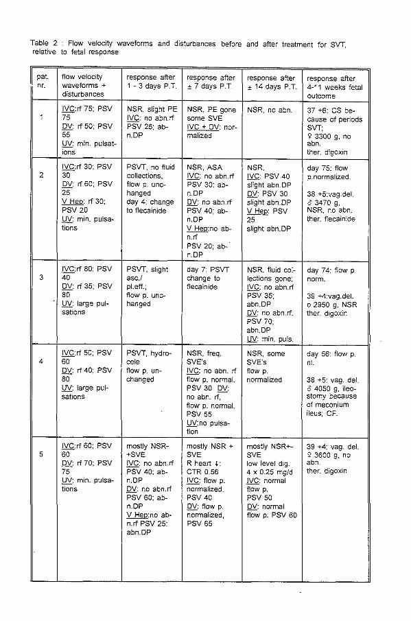
Table 2 Flow velocity waveforms and disturbances before and after treatment for SVT, relative to fetal response
pat. flow velocity response after response after response after response after nr. waveforms + 1 - 3 days P.T. ± 7 days P.T. ± 14 days P.T. 4-11 weeks fetal
disturbances outcome
IVC:rf 75; PSV NSR, slight PE NSR, PE gone NSR, no abn. 37 +6: cs be-1 75 IVC: no abn.rf some SVE cause of periods
OV: rf 50; PSV PSV 25; ab- IVC + DV: nor- SVT; 55 n.DP mallzed ~ 3300 g, no UV: min. pulsat- abn. ions ther. digoxin
IVC:rf 30; PSV PSVT, no fluid NSR,ASA NSR, day 75: flow 2 30 collections, IVC: no abn.rf IVC: PSV 40 p.normalized.
DV: rf 60; PSV flow p. unc- PSV 30; ab- slight abn.DP 25 hanged n.OP OV: PSV 30 38 +5:vag.del. V Hep: rf 30; day 4: change DV: no abn.rf slight abn.DP 0 3470 g, PSV 20 to flecainide PSV 40; ab- V Hep: PSV NSR, no abn. UV: min. pulsa- n.OP 25 ther. flecainide tions V Hep:no ab- slight abn.DP
n.rf PSV 20; ab-n.OP
IVC:rf 80; PSV PSVT, slight day 7: PSVT NSR, fluid col- day 74: flow p. 3 40 asc./ change to lections gone; norm.
OV: rf 35; PSV pl.eff.; flecainide DLQ: no abn.rf 80 flow p. unc- PSV 35; 39 +4:vag.del. UV: large pul- hanged abn.OP o 2950 g, NSR sations OV: no abn.rf, ther. digoxin
PSV 70; abn.OP UV: min. puis.
IVC:rf 50; PSV PSVT, hydro- NSR, freq. NSR, some day 56: flow p. 4 60 cele SVE's SVE's nl.
DV: rf 40; PSV flow p. un- IVC: no abn. rf flow p. 80 changed flow p. normal, normalized 38 +5: vag. del. UV: large pul- PSV 30 DV: o 4050 g, ileo-sations no abn. rf, stomy because
flow p. normal, of meconium PSV 55 ileus; CF. UV:no pulsa-tion
IVC:rf 60; PSV mostly NSR- mostly NSR + mostly NSR+- 39 +4: vag. del. 5 60 +SVE SVE SVE 2 3600 g, no
DV: rf 70; PSV IVC: no abn. rf R heart +; low level dig. abn. 75 PSV 40; ab- CTR 0.56 4 x 0.25 mg/d ther. digoxin UV: min. pulsa- n.OP .!YQ.: flow p. IVC: normal tions DV: no abn.rf normalized, flow p.
PSV 60; ab- PSV 40 PSV 50 n.OP DV: flow p. OV: normal V Hep:no ab- normalized, fiOW p. PSV 60 n.rf PSV 25; PSV 65 abn.DP

pat. nr.
6
7
8
flow velocity response after response after response after waveforms + 1 - 3 days P.T. ± 7 days P.T. ± 14 days P.T. disturbances
IVC: rf 40; PSV NSR, fluid col- NSR, fluid un- NSR, fluid < 50 lections un- changed; IVC: IVC: PSV 50 UV: min. pulsa- changed PSV 40 abn. DP tions high level flee abn. DP DV: PSV 65
2 x 100 mg/d DV: PSV 75 flow IVC: no abn.rf abn.DP p. normalized PSV 35; ab- V Hep: PSV V Hep: PSV n.DP 15 25 DV: no abn.rf abn.DP abn.DP PSV 65; ab- UV: min. puis. UV: min. puis. n.DP
IVC: rf 70; PSV periods NSR, NSR, fluid NSR, fluid col-40 fluid collecti- IVC: PSV 35 lections gone; DV: rf 60; PSV ons unchan- flow p. IVC: PSV 50 80 ged; IVC: rf normalized flow p. normal UV: min. pulsa- 20; PSV 40 DV: PSV 70 DV: PSV 80 tions abn.DP slight abn.DP flow p. normal
DV: no abn. rf V Hep: PSV PSV 40 ab- 35 n.DP flow p. normal V Hep:nl.flow p
IVC: rf 60; PSV SVT, fluid col- SVT+periods NSR, no fluid 70 lections gone, NSR collections DV: rf 25; PSV low level flee asc. gone IVC:rf 20; PSV 65 4 x 100 mg/d IVC: rf 15; 35 slight abn. UV: large pul- day 6: slight PSV DP sations asc. change to 30 abn.DP DV: PSV 45
digoxine DV: no abn.rf slight abn.DP PSV 80 v Hep:rf 15; abn.DP PSV 30 slight UV: min. puis. abn. DP
UV: no puis.
Abbreviations: fluid .J. = decrease of fluid collections abn.DP = abnormal diastolic flow pattern IVC = inferior vena cava
PE = pericard effusion pl.eff. = pleural effusion asc. = ascites
OV = ductus venosus UV = umbilical vein
rf = reverse flow {cm/s) P. T. = post treatment
response after 4-11 weeks;fetal outcome
day 31: fluid gone flow p. norm.-
37: vag. deL 9 2500 g, no abn. ther. digoxin
day 78: flow p. norm.
37 +2: vag. del. 2 4220 g , no abn. ther. flecainide
day 18: NSR+ periods SVT; asc. + pl.eff. IVC + DV + UV: flow p. idem
33 +5: cs 0 3000 g WPW; ther. di-goxin + prop-ranolol
V Hep = hepatic vein R heart + = decrease of right heart volume R heart t = increased right heart volume PSV = peak systolic veolocity (cm/s)
ASA = atriaL septum aneurysm nl. =normal
min. puis. = minimal pulsations SVE = supraventricular extrasystole SVT = supraventricular tachycardia flow p. = flow pattern vag. del. = vaginal delivery
ther. = therapy NSR == normal sinus rhythm freq. = frequent dig. = digoxine flee. = flecainide CF = cystic fibrosis CS = caesarean section

Chapter 5 Fetal variables and venous return l70
DISCUSSION
Combined two-dimensional ultrasonography and M-mode allow the most accurate
description of the type and severity of arrhythmia, despite one report on
diagnosis of fetal arrhythmias by Doppler ultrasound (Chan et al. 1990). Only a
few reports have appeared which documented venous velocity waveforms by
Doppler recording during this particular condition. Reed et al. (1990) described
the changes in inferior vena cava waveforms during different types of arrhythmia.
Our observations on inferior vena cava waveform modulation are in agreement
with their report. Especially in the fetuses with premature atrial contractions and
complete heart block the increase of the reverse flow component during altered
cardiac contractions is interesting. It reflects an overfilled right atrium contracting
when right ventriclar pressure is high or the ventricle is not relaxed. When the
atrial contraction occurs with the atrioventricular valves still closed during
ventricular systole, very high-velocity retrograde flow components are formed,
which are called "cannon a waves" by Reed et al.(1990). The same group also
reported on umbilical vein flow velocities in the same patients (Jndik et al. 1991).
They associated the high percentage reversed flow with the occurrence of
umbilical venous pulsations. No data exist on ductus venosus flow velocity
waveform modulation during arrhythmias except for one case of fetal tachycardia
described by Kiserud et al.(1991). The disturbed diastolic phase in the ductus
venosus waveform is the direct result of the different right atrial contraction
pattern. Normally without any retrograde flow component, the ductus venosus
flow velocity waveform during arrhythmia reflects the influence of an atrial
contraction of an overfilled heart chamber resulting in reversed flow.
To our knowledge this is the first report on Doppler assessment of venous inflow
before and after successful treatment of fetal supraventricular tachycardia. The
disappearance of the enormous reversed flow component in the fetal inferior
vena cava, ductus venosus and hepatic veins in case of a paroxysmal or
permanent normal sinus rhythm is remarkable. This significant change in blood
flow reflects the influence of the cardiac relaxation time on venous return. In
fetuses with tachycardia blood is pooled in the central venous system and the

Chapter 5 Fetal variables and venous return l71
right atrium, since the ventricles demonstrate shortened periods of filling during
the abnormally fast heart rhythm. In the diastolic phase of the cardiac cycle this
increased end-diastolic pressure results in a backward surge of right atrial blood
into the venous vessels. Inadequate forward flow will eventually lead, in most
untreated cases, to development of cardiac decompensation reflected by fetal
hydrops and asphyxia.
The normalization of flow velocity waveforms after the return of normal sinus
rhythm is also variable. In our study this is demonstrated by the interval from an
abnormal to a normal peak systolic/diastolic ratio in the inferior vena cava and
ductus venosus waveform. Successful anti-arrhythmic agents like digoxin and
flecainide may interrupt the abnormally fast heart rhythm, but there might still be
aberrant pathways or abnormal conductance which influence the contraction
force of the right atrium myocardium. We suggest that different factors play a role
in the recovery of the fetal circulatory function.
Almost no data exist about human fetal electro-physiology or myocardial
performance (Kleinman and Capel 1991). Knowledge has been acquired from
neonates and animals, but the variable response and tolerance of fetal
myocardium to various pharmaco-therapeutic regimes suggests different types of
mechanism which lead to SVT. The influence of gestational age and increasing
maturity may also play a role.
The role of the placenta as a barrier for drug transport into the fetal circulation
and as a compensating force for an abnormal venous pressure has not been
solved yet. The hydropic fetus, presumably due to hydrops of the placenta and
altered placental perfusion states, has considerably less absorption of maternally
administered medications than the non-hydropic fetus (Younis and Granat 1987;
Weiner et al. 1988; Gembruch et al. 1989; Hansmann et al. 1991).
The discussion about the presence and disappearance of fluid collections in
hydropic fetuses remains speculative. Ascites, pleural and pericardia! effusion
and skin edema have been diagnosed as signs of the fetal struggle with
abnormal heart rate. These symptoms are probably a better measure of fetal
cardiac response to the tachycardia then venous waveforms.

Chapter 5 Fetal variables and venous return l72
It can be concluded that during fetal arrhythmias venous return blood fiow is
altered. The diastolic phase of the cardiac cycle is mainly disturbed by the
change in right atrial filling patterns, reflected by increased retrograde fiow
components. After successful treatment of fetal tachycardia normalization of
blood fiow velocity waveforms is often slow, suggesting the variable response of
the fetal myocardium to periods of abnormal conductance.
SUMMARY
The objective of this study was to describe the changes in venous fiow velocity in
various cases of fetal arrhythmia and to evaluate these changes before and after
fetal therapy for tachycardia.
By means of Doppler ultrasonography a group of 22 fetuses was studied,
consisting of ten cases with supraventricular extrasystoles, eight fetuses with
supraventricular tachycardia, before and after successful treatment, and four
cases of bradycardia. Flow velocity waveform recordings were obtained from the
ductus venosus, inferior vena cava, hepatic veins and umbilical vein.
Peak systolic and diastolic velocity and their ratio were calculated. Presence of
reversed fiow and umbilical venous pulsations were noted.
All fetal arrhythmias were characterized by increased retrograde fiow components
and disturbed diastolic fiow patterns. In most cases umbilical venous pulsations
of varying amplitude were present. In the fetuses with tachycardia diastolic fiow
patterns remained disturbed after successful treatment, refiected by abnormal
peak systolic to peak diastolic ratios in the inferior vena cava and ductus venosus
waveform.
In conclusion, premature atrial contractions and complete heart block will
increase the reverse fiow component during the altered right atrial contractions,
refiecting an overfilled right atrium contracting when right ventriclar pressure is
high or the ventricle is not relaxed. The normalization of fiow velocity waveforms
after the return of normal sinus rhythm is in most cases slow suggesting the
variable response of the fetal myocardium to periods of abnormal conductance.

Chapter 5 Fetal variables and venous return 173
REFERENCES
Adeagbo ASO, Coceani F, Olley PM. The response of the lamb ductus venosus to prostaglandins
and inhibitors of prostaglandin and thromboxane synthesis. Circ Res 1982;51 :580-586.
Allan LD, Anderson RH, Sullivan ID et al. Evaluation of fetal arrhythmias by echocardiography.
Br Heart J 1983;50:240-245.
Brecher GA. Mechanism of venous flow under different degrees of aspiration.
Am J Physiol 1952;169:423-433.
Chacko AW, Reynolds SRM. Embryonic development in the human of the sphincter of the ductus
venosus. Anat Rec 1953; 115:151-173.
Chan FY, Woo SK, Ghosh A, Tang M, Lam C. Prenatal diagnosis of congenital fetal arrhythmias
by simultaneous pulsed Doppler velocimetry of the fetal abdominal aorta and inferior vena cava.
Obstet Gynecol 1990;76:200-204.
Chiba Y, Utsu M, Kanzaki T, Hasegawa T. Changes in venous flow and intra tracheal flow in fetal
breathing movements. Ultrasound Med Biol1985;11:43-49.
van Eyck J, Wladimiroff JW, Noordam MJ, Tonge HM, Prechtl HFR. The blood flow velocity
waveform in the fetal descending aorta; its relationship to fetal behavioural states in normal
pregnancy at 37-38 weeks of gestation. Early Hum Dev 1985;14:99-107.
van Eyck J, Wladimiroff JW, van den Wijngaard JAGW, Noordam MJ, Prechtl HFR. The blood
flow velocity waveform in the fetal internal carotid and umbilical artery; its relationship to fetal
behavioural states in normal pregnancy at 37 - 38 weeks of gestation. Br J Obstet Gynaecol
1987;94:736-741.
van Eyck J, Stewart PA, Wladimiroff JW. Human fetal foramen ovate flow waveforms relative to
behavioral states in normal term pregnancy. Am J Obstet Gynecol 1990;163:1239-1242.
van Eyck J, van der Mooren K, Wladimiroff JW. Ductus arteriosus flow velocity modulation by
fetal breathing movements as a measure of fetal lung development. Am J Obstet Gynecol
1990;163:558-566.

Chapter 5 Fetal variables and venous return 174
van Eyck J, Stewart PA, Wladimiroff JW. Human fetal foramen ovale flow velocity waveforms
relative to fetal breathing movements in normal term pregnancies.
Ultrasound Obstet Gynecol 1991;1:5-7.
Ferraz de Carvalho CA, Junqueira Rodrigues A. Beitrag zur funktionellen Anatom·le des Ductus
venosus im reifen menschlichen Fetus, mit besonderer BerOcksichtigung des Oberganges Ductus
venosus-Sinus umbilicalis. Anat Anz Bd 1975;137:207-220.
Gembruch U, Manz M, Bald R, Ruddel H, Redel DA, Schlebusch H, Nitsch J, Hansmann M.
Repeated intravascular treatment with amiodarone in a fetus with refractory supraventricular
tachycardia and hydrops feta!is. Am Heart J 1989;118:1335-1338.
Guyton AC. Phys·1cs of blood, blood flow and pressure: hemodynamics. In: Textbook of medical
physiology, Philadelphia: WB Saunders Company. 1981, pp 206-218.
Hansmann M, Gembruch U, Bald R, Manz M, Redel DA. Fetal tachyarrhythmias: transplacental
and direct treatment of the fetus - a report of 60 cases.
Ultrasound Obstet Gynecol 1991:1:162-170.
Holt JP. Collapse factor in the measurement of venous pressure: flow of fluid through collapsible
tubes. Am J Physiol 1941;134:292-299.
Huisman TWA, Stewart PA, Wladimiroff JW. Flow velocity waveforms in the fetal inferior vena
cava during the second half of normal pregnancy. Ultrasound Med Bioi 1991;17:679-682.
Huisman TWA, Stewart PA, Wladimiroff JW. Ductus venosus blood flow velocity waveforms in the
human fetus;a Doppler study. Ultrasound Med Bioi 1992;18:33-37.
lndik JH, Chen V, Reed KL Association of umbilical venous with inferior vena cava flow
velocities. Obstet Gynecol 1991;77:551-557.
Kircher BJ, Himelman RB, Schiller NB. Noninvasive estimation of right atrial pressure from the
inspiratory collapse of the inferior vena cava. Am J Cardiel 1990;66:493-496.
Kiserud T, Eik-Nes SH, B!aas HGK, Hellevik LR. Ultrasonographic velocimetry of the fetal ductus
venosus. Lancet 1991;338:1412-1414.
Chapter 5 Fetal variables and venous return l75
Kleinman CS, Capel JA, Weinstein EM, Santulli TV Jr., Hobbins JC. In utero diagnosis and
tyreatment of fetal supraventricular tachycardia. Sem Perinatal 1985;9:113-129.
Kleinman CS, Capel JA Electrophysiological principles and fetal antiarrhythmic therapy.
Ultrasound Obstet Gynecol 1991;1:286-297.
Kloosterman G. On intrauterine growth. lnt Gynecol Obstet 1970;8:895-912.
Mantell CO. Breathing movements in the human fetus. Am J Obstet Gynecol 1976;125:550-553.
Marsal K, Lindblad A, Lingman G, Eik-Nes SH. Blood flow in the fetal descending aorta; intrinsic
factors affecting fetal blood flow, i.e. fetal breathing movements and cardiac arrhythmia.
Ultrasound Med Bioi 1984;10:339-348.
Meyer W'N, lind J. The ductus venosus and the mechanism of its closure.
Arch Dis Childh 1966;41:597-605.
Mintz GS, Kotler MN, Parry WR, lskandrian AS, Kane SA Real-time inferior vena caval
ultrasonography: normal and abnormal findings and its use in assessing right-heart function.
Circulation 1981;64:1018-1025.
van der Mooren K, van Eyck J, Wladimiroff JW. Human fetal ductal flow velocity waveforms
relative to behavioural states in normal term pregnancy.
Am J Obstet Gynecol 1989;160:371-374.
Morin Ill FC. Prostaglandin E1 opens the ductus venosus in the newborn lamb.
Pediatr Res 1 987;21 :225-228.
Natale R. Maternal plasma glucose concentration and fetal breathing movements; a review.
Sem Perinatal 1980;4:287-293.
Natori H, Tamaki S, Kira S. Ultrasonographic evaluation of ventilatory effect on inferior vena caval
configuration. Am Rev Respir Dis 1979;120:421-427.
Nijhuis JG, Prechtl HFR, Martin CB jr., Bats RSGM. Are there behavioural states in the human
fetus? Early Hum Oev 1982;6:177-195.

Chapter 5 Fetal variables and venous return 176
Pearson AA, Sauter RW. Observations on the phrenic nerves and the ductus venosus in human
embryos and fetuses. Am J Obstet Gynecol 1971;110:560-565.
Reed KL, Appleton CP, Anderson CF, Shenker L, Sahn OJ. Doppler studies of vena cava flows in
human fetuses: insight into normal and abnormal cardiac physiology. Circulation 1990;81: 498-
505.
Richardson BS, Patrick JE, Abduljabbar H. Cerebral oxidative metabolism in the fetal lamb:
relationship to electrocortical state. Am J Obstet Gynecol 1985;153:426431.
Rizzo G, Ardu.ln·l D, Valens·lse H, Roman·lni C. Effects of behavioural states on cardiac output in
the healthy human fetus at 36-38 weeks of gestation. Early Hum Dev 1990;23:109-115.
Stewart PA Echocardiography in the human fetus. Thesis. Erasmus University Rotterdam, the
Netherlands. Promotor: Prof.J.W.Wiadimiroff.
Trudinger BJ. The umbilical circulation. Sem Perinatal 1987;11 :311-321.
Weiner CP, Thompson MIS. Direct treatment of fetal supraventricular tachycardia after failed
transplacental therapy. Am J Obstet Gynecol 1988;158:570-573.
Wexler L, Berge! DH, Gabe IT, Makin GS, Mills CJ. Velocity of blood flow in normal human venae
cavae. Circ Res 1968;23:349- 359.
Wladimiroff JW, Stewart PA Treatment of fetal cardiac arrhythmias.
Br J Hosp Med 1985;34:134-140.
Younis JS, Granat M. Insufficient transplacental digoxin transfer in severe hydrops fetalls. Am J
Obstet Gynecol 1987;157:1268-1269.

Chapter 5 Fetal variables and venous return l77

l78
Chapter 6
GENERAL CONCLUSIONS
In this thesis combined two-dimensional real-time and Doppler ultrasound was
used to study the fetal circulation in early and late pregnancy. In particular
venous inflow hemodynamics was investigated, because venous return is of great
importance to cardiac performance. Present transvaginal Doppler techniques
allow detailed information on fetal waveform characteristics and velocities as
early as 10 weeks of gestation. Transvaginal waveform recording was successful
up to approximately 14 weeks' gestation. Afterwards, waveform recording was
always carried out using the transabdominal approach because of increasing fetal
size.
Flow velocity waveforms obtained at various cardiovascular levels in late first and
early second trimester fetuses display an acceptable reproducibility in the
individual patient, except for the acceleration time, whereas all parameters depict
larger variabilities for between patient values. The possible clinical significance of
an early single flow velocity measurement will therefore be limited.
At 1 0-12 weeks, end-diastolic flow velocities were always absent in the fetal
descending aorta and umbilical artery, but were present in 58 % of the intra
cerebral artery waveforms, suggesting a relatively low cerebral vascular
resistance compared with that at the fetal trunk and umbilical placental level.
The pulsatility index at all three levels decreased significantly with advancing
gestational age, suggesting a reduction in fetal and umbilical placental vascular
resistance during the late first and early second trimester of normal pregnancy.
This may be due to the unplugging of the trophoblast at 13 to 14 weeks of
gestation. These Pulsatility Index changes are independent of the reduction in
FHR observed at this stage of early pregnancy.
A pulsatile flow pattern was observed in the ductus venosus and inferior vena
cava, whereas only the latter demonstrated a retrograde flow component during
right atrial contraction. The reduction in inferior vena cava flow reversal with

l79
advancing gestational age may be a result of raised ventricular compliance or
increased ventricular relaxation rate as well as reduction in afterload. The time
averaged fiow velocity in the ductus venosus is 2.7 and 3.2 times higher than
that in the inferior vena cava and umbilical vein in early pregnancy. The marked
difference in flow velocity between the ductus venosus and inferior vena cava
may result in a tendency not to mix and supports the assumption that two
channels of venous return exist inferior vena cava blood fiow will be channeled
to the tricuspid valve and ductus venosus blood flow will be mainly directed
towards the foramen ovale.
This is supported by our study on the anatomical relationships between the
venous vessels in the fetal hepatic region in the second half of pregnancy. The
abdominal inferior vena cava terminates in a funnel-like venous structure, a
subdiaphragmatic vestibulum, which contains the orifices of the hepatic veins as
well as the ductus venosus. It is suggested that information on blood flow
velocities in the individual vessels in the hepatic and subdiaphragmatic area
should be obtained more distally to the right atrium and not at the level of the
venous vestibulum.
In second and third trimester fetuses an acceptable within patient reproducibility
for venous waveform recordings was obtained. Venous Doppler waveforms,
however, should be interpreted with caution, since they are subject to variation
on the basis of variable sample placement and angle insonation or biological
variations in fetal hemodynamics.
The inferior vena cava flow velocity waveform displays a systolic and early
diastolic forward component with reversed flow during late diastole. Waveform
recording at the level of the inferior venous entrance into the right atrium provides
information on changes in venous return rather than changes in the inferior vena
cava per se.
The ductus venosus shows a pulsatile fiow pattern consisting of a systolic and
diastolic forward component without the late diastolic reverse component demon
strated in the inferior vena cava. Ductus venosus peak systolic velocities as high
as 40 - 80 cm/s were observed. A statistically significant increase in time
averaged velocity, peak systolic and peak diastolic velocity in the inferior vena

180
cava and ductus venosus with advancing gestational age was established. This
may be caused by increased volume flow, increased cardiac compliance and
stroke volume or reduced afterload. Age-related reduction in afterload may occur
in the human fetus as a result of the physiological decrease in placental vascular
resistance.
The influence on venous fiow velocity waveforms of various fetal intrinsic factors
such as fetal breathing movements, behavioural states and arrhythmias was
studied in late pregnancy. Flow velocity waveforms in the hepatic venous
vasculature close to the right atrium are markedly increased during fetal
breathing movements. These changes are independent of gestational age and
could be due to a raised pressure gradient between thorax and abdomen,
resulting in a reduction in venous vessel diameter and additional volume flow
being directed towards the right atrium. The significance of the inspiratory
collapse of the fetal inferior vena cava ("caval index") for the recognition of
elevated right atrial pressure in abnormal human fetal development needs further
investigation.
Flow velocity waveform recording distal to the partially collapsing venous vessel
wall merely demonstrated a reduction in fiow velocity. This may be· determined by
the increase in intra-abdominal pressure and the functional obstruction of the
collapsing vessel wall.
A fetal behavioural state dependent decrease of 30 % was observed in the
ductus venosus peak systolic, peak diastolic and time-averaged velocity,
suggesting a redistribution of umbilical venous blood through the ductus venosus
shunt during the passive sleep state as compared with the active sleep state in
the term fetus. The fetal behavioral state should be taken into account in future
studies on ductus venosus flow velocity waveforms in normal term pregnancies.
No behavioural state dependent changes were observed both in the proximal and
distal portion of the inferior vena cava. Possible modulation in relation to
behavioural states may, however, be obscured by considerable variation in fiow
velocities as demonstrated in the present study.

lBl
Finally, during fetal arrhythmias venous return blood fiow is altered. The diastolic
phase of the cardiac cycle is mainly disturbed by the change in right atrial filling
patterns, reflected by increased retrograde fiow components. After successful
treatment of fetal tachycardia normalization of blood fiow velocity waveforms is
often slow, suggesting a variable response of the fetal myocardium to periods of
abnormal conductance.

182

1S2
Summary
Chapter 1
In this introductory chapter it is pointed out that study of venous return in the
human fetus is dependent on non-invasive methods and variation by fetal intrinsic
variables_ The objectives of the present thesis are focused on Doppler
assessment of venous blood flow velocities in the human fetus relative to
gestational age, fetal heart rate and rhythm, fetal breathing movements and fetal
behavioural states.
Chapter 2
This chapter describes the acquisition of knowledge on fetal hemodynamics, in
particular embryological, anatomical and physiological factors that have influen
ced research on venous return_ It is pointed out that most information has been
acquired from animal experiments, so that a need for accurate data on the
human fetal circulation still remains. The non-invasive Doppler techniques
available for obtaining this information are discussed, comparing the significance
of both the transabdominal and transvaginal approach.
Chapter 3
In this chapter various aspects of fetal cardiovascular performance are discussed
using Doppler ultrasonography in women with late-first and early-second trimes
ter pregnancies_ A reproducibility study displays an acceptable biological variation
for individual flow velocity waveform parameters in the fetus_ Examination of
peripheral arteries demonstrates that the afterload conditions are characterized
by a high resistance at fetoplacental leveL This is reflected by the absence of
end-diastolic flow in flow velocity waveforms from the umbilical artery and fetal
descending aorta before 14 weeks of gestation_ At 12 to 13 weeks Doppler
184
velocity waveforms from the fetal intracerebral artery display an end-diastolic flow
component, which suggests a low resistance fetal cerebral circulation in the
presence of a high afterload resistance.
Data on atrioventricular flow velocity wavefonms at 10 to 14 weeks of gestation
suggest low compliance of the fetal ventricles reflected by late-diastolic filling
dominance.
This is also supported by the higher percentages of retrograde fiow in inferior
vena cava waveforms from first trimester fetuses as compared with the second
and third trimester. Moreover, study of preload parameters demonstrates that
time-averaged velocity in the inferior vena cava is markedly lower than in the
ductus venosus, supporting the assumption that two channels of venous return
exist side by side.
Chapter 4
This chapter begins with a detailed study of the anatomy of the fetal (peri) hepatic
vasculature. The presence of a funnel-like venous structure at venous inflow
level, named venous vestibulum, was established. This contains the orifices of
the hepatic veins, the inferior vena cava itself and the ductus venosus. The
anatomical complexity of all these veins may produce variable Doppler flow
velocity waveforms at venous infiow level. A reproducibility study demonstrates
acceptable within patient variance in inferior vena cava and ductus venosus fiow
recordings. In the following sub-chapters normal values for inferior vena cava and
ductus venosus fiow velocity parameters throughout the second half of pregnancy
are documented. A statistically significant increase in time-averaged and peak
systolic velocity with advancing gestation is observed.

185
Chapter 5
In this chapter the influence of various intrinsic factors is discussed in relation to
normal venous fiow velocity parameters. Variability in fiow measurements can .be
caused by (i) biological variance within the patient; (ii) fetal (breathing) move
ments; (iii) different fetal behavioural states and (iv) cardiac arrhythmia. Variation
in Doppler recording due to within patient variance components has been
extensively investigated in the reproducibility studies described in chapters 3.2
and 4.3.
Firstly, the role of fetal breathing movements was examined in second and third
trimester fetuses. Maternal glucose administration induced fetal breathing activity
and inferior vena cava, ductus venosus and hepatic vein fiow velocity waveforms
were documented. These waveforms, which were obtained close to the right
atrium, demonstrate a significant increase in peak systolic, diastolic and time
averaged velocity during fetal breathing movements. These changes are indepen
dent of gestational age and could be due to a raised pressure gradient between
thorax and abdomen, resulting in a reduction in venous vessel diameter and
additional volume fiow being directed towards the right atrium. The breathing
related reduction of the inferior vena cava vessel diameter or "inspiratory
collapse" had already been observed in the adult. The significance of this
collapse of the fetal inferior vena cava ( also called: "caval index") for the recog
nition of elevated right atrial pressure in abnormal human fetal development
needs further investigation. Flow velocity waveform recording distal to the
partially collapsing venous vessel wall merely demonstrates a reduction in fiow
velocity. This may be determined by the increase in intra-abdominal pressure and
the functional obstruction of the collapsing vessel wall.
Secondly, we focused on changes in venous return and venous blood fiow
velocities relative to different fetal behavioral states. During the passive sleep
state (FBS 1 F) a significant decrease in ductus venosus blood fiow velocity is
observed compared with the active sleep state (FBS 2F), while inferior vena cava
fiow parameters do not demonstrate any changes. It is suggested that a
redistribution of venous blood fiow takes place at the level of the ductus venosus

186
relative to the fetal behavioural state.
Finally, during cardiac arrhythmias marked changes in preload flow components
occur, in particular during the diastolic filling phase. The main alteration was the
appearance or increase of the retrograde flow component coincident with right
atrial contraction. These changes were also investigated in fetuses suffering from
supraventricular tachycardia, who were successfully treated with maternal
therapy of digoxine or flecanide. Normalization of the reversed flow component,
but in particular the diastolic flow characteristics, of ductus venosus and inferior
vena cava blood flow velocity waveforms is often slow, suggesting a variable
response of the fetal myocardium to periods of abnormal conductance.

l87
Samenvatting
Hoofdstuk 1
In dit inleidend hoofdstuk wordt gewezen op het feit dat onderzoek naar de
veneuze return in de humane foetus afhankelijk is van non-invasive methoden en
van variaties bepaald door foetale intrinsieke faktoren.
De doelstellingen van dit proefschrift concentreren zich op de evaluatie van
veneuze bloedstroom snelheden in de humane foetus door middel van Doppler
echografie in relatie tot de zwangerschapsduur. foetale hart frequentie en ritme,
foetale ademhalingsbewegingen en foetale gedragstoestanden.
Hoofdstuk 2
Dit hoofdstuk beschrijft de methoden ter verkrijging van kennis omtrent de foetale
hemodynamiek, in het bijzonder de embryologische, anatomische en fysiologi
sche faktoren die onderzoek naar de veneuze return hebben beinvloed. Er wordt
op gewezen dat de meeste informatie is vergaard in dierexperimentele proeven,
zodat er een noodzakelijkheid blijft bestaan voor accurate gegevens over de
humane foetale circulatie. De non-invasieve Doppler technieken die bekend zijn
om deze informatie te verkrijgen worden besproken, waarbij de plaats en
noodzakelijkheid van enerzijds de transabdominale en anderzijds de transvagina
le benadering worden vergeleken.
Hoofdstuk 3
In dit hoofdstuk worden de verschillende aspekten van foetaal cardiovasculair
functioneren met behulp van Doppler echografie beschreven bij vrouwen gedu
rende het eerste en vroeg tweede zwangerschapstrimester. Een reproduceer
baarheidsstudie demonstreert een acceptabele biologische variantie binnen de
veschillende bloedstroomsnelheids parameters in de individuele foetus.

l88
Onderzoek van de perifere arterien laat zien dat afterload kondities worden
gekenmerkt door een relatief hoge vasculaire weerstand op foeto-placentair
niveau. Dit wordt weerspiegeld in de afwezigheid van eind-diastolische fiow in het
bloedstroomsnelheidsprofiel van de arteria umbilicalis en de foetale aorta
descendens voor de zwangerschapsduur van 14 weken. In de zwangerschaps
periode van 12 - 13 weken vertoont Doppler echografie van de intracerebrale
arterie altijd de aanwezigheid van een eind-diastolische fiow component. hetgeen
een relatief lage vaatweerstand in de foetale cerebrale circulatie onder
omstandigheden van hoge afterload vaatweerstand suggereert.
Onderzoek naar de foetale cardiale functie bij 10 tot 14 weken zwangerschaps
duur suggereert een lage compliantie van de foetale ventrikels gekenmerkt door
dominantie van de laat-diastolische vullingsgraad. Dit wordt gesteund door het
hogere- percentage van retrograde fiow in de foetale vena cava inferior in het
eerste zwangerschapstrimester vergeleken met het tweede en derde trimester.
Bovendien heeft studie naar de "preload" parameters Iaten zien dat de gemiddel
de bloedstroom snelheid in de vena cava inferior beduidend lager ligt dan de
ductus venosus. Dit steunt de veronderstelling dat twee kanalen van veneuze
return naast elkaar bestaan.
Hoofdstuk 4
Dit hoofdstuk begin! met een gedetaileerde studie naar de anatomie van het
foetale (peri)hepatische bloedvatenstelsel. De aanwezigheid van een trechter
vormige veneuze structuur, veneus vestibulum genaamd, werd vastgelegd, welke
de openingen van de hepatische venen, de vena cava inferior zelf en de ductus
venosus bevat. De anatomische complexiteit van al deze venen zou mogelijk
variatie in de Doppler bloedstroom snelheidsprofielen registratie kunnen veroor
zaken. Een reproduceerbaarheidsstudie laat een acceptabele intrapatient variatie
in vena cava inferior en ductus venosus stroomsnelheidsmetingen zien. ln de
daaropvolgende subhoofdstukken worden de normaal waarden van de vena cava
inferior en ductus venosus stroomsnelheids parameters gedurende de tweede
helft van de zwangerschap gedocumenteerd. Een statistisch significante toename

189
in gemiddelde en piek systolische snelheden met toenemende zwangerschaps
duur wordt vastgesteld.
Hoofdstuk 5
In dit hoofdstuk wordt de invloed van verschillende intrinsieke faktoren onder
zocht in relatie tot normale veneuze stroomsnelheids parameters. Variabiliteit in
fiow metingen kunnen worden veroorzaakt door: (i) biologische variatie binnen de
patient; (ii) door foetale (ademhalings) bewegingen; (iii) verschillen in foetale
gedragstoestand en (iv) cardiale arritmieen. Variatie in Doppler registratie die te
danken is aan intrapatient variatie zijn reeds beschreven in hoofdstukken 3.2 en
4.3.
Ten eerste werd de rol van foetale ademhalingsbewegingen in het tweede en
derde zwangerschapstrimester onderzocht Maternale glucose toediening
induceerde foetale ademhalings aktiviteit. Bloedstroomsnelheidsprofielen van de
vena cava inferior, ductus venosus en hepatische venen werden vervolgens
vastgelegd. Deze profielen, die dicht bij he! rechter atrium werden verkregen,
vertonen een significante toename in piek systolische, diastolische en gemid
delde snelheid tijdens foetale ademhalingsbewegingen. Deze veranderingen zijn
onafihankelijk van de zwangerschapsduur en kunnen mogelijk worden verklaard
door een verhoogde drukgradient tussen thorax en abdomen, resulterend
enerzijds in een afname van de veneuze vaatwand diameter en anderzijds in een
additionele bloedstroom die in de richting van het rechter atrium wordt geleid.
Deze ademhalingsafihankelijke afname van de vena cava inferior vaat diameter of
"inspiratoire collaps" is reeds eerder beschreven bij de volwassene. De klinische
relevantie van deze collaps van de vena cava inferior vaatwand (ook genoemd:
"caval index") voor de diagnose van verhoogde rechter atriale druk tijdens
abnormale foetale ontwikkeling dient verder te worden onderzocht. Bloedstroom
snelheids metingen distaal van de gedeeltelijk collaberende veneuze vaatwanden
Iaten slechts een afname van de stroomsnelheid zien. Dit zou bepaald kunnen
worden door de gelijktijdige toename van de intra-abdominale druk en de

l90
functionele obstructie van de collaberende vaatwand.
Ten tweede was het onderzoek geconcentreerd op veranderingen in veneuze
bloedstroomsnelheden die gerelateerd zijn aan de verschillende foetale gedrags
toestanden. Tijdens de passieve slaaptoestand (FBS 1 F) wordt een significante
afname van de bloedstroomsnelheid in de ductus venosus vastgesteld,
vergeleken met de actieve slaaptoestand (FBS 2F), terwijl de vena cava inferior
flow parameters deze verandering niet vertonen. Dit suggereert dat een redistri
butie van veneuze bloedstroom plaats vindt op het niveau van de sinus umbilica
lis en de daaruit ontspringende ductus venosus, welke gerelateerd is aan de
foetale gedragstoestand.
Tenslotte vinden tijdens cardiale arritmieen opmerkelijke veranderingen plaats in
de zogenaamde "preload" stroomcomponenten, in het bijzonder gedurende de
diastolische vullingsfase. De meest in het oog springende verandering is verschij
ning of toename van de retrograde flow component tegelijkertijd met de contrac
tie van het rechter atrium. Deze veranderingen werden oak onderzocht bij
ongeborenen, die leden aan supraventriculaire tachycardie, en die via de moeder
succesvol behandeld waren met digoxine of flecainide. Normalisatie van de retro
grade flow, maar vooral de diastolische flow karakteristieken in de ductus
venosus en vena cava inferior is vaak langzaam, hetgeen een variabele respons
van het foetale myocard suggereert op perioden van abnormale prikkelgeleiding.

191

192
Dankwoord
Ten eerste gaat mijn oprechte dank uit naar mijn promotor, opleider en co-auteur
Prof.Dr.J.W. Wladimiroff, die mij, boven alles, heeft geleerd om op verantwoorde,
kritische wijze wetenschappelijk onderzoek te bedrijven dat leidt tot publiceerbaar
kwaliteitswerk.
De interessante en vriendelijke samenwerking met Prof.Dr.A.C. Gittenberger-de
Groot van het Anatomisch en Embryologisch lnstituut te Leiden uitte zich niet
aileen in een prachtige publicatie, maar ook in haar aanwezigheid in de promotie
commissie, waarvoor mijn hartelijke dank.
I consider it a great honor that Prof.S.H. Eik-Nes from Norway, one of the very
first pioneers in fetal ultrasound and venous Doppler research, participated in the
dissertation committee and express my gratitude for his willingness.
Voor zijn bereidheid om ook zitting te nemen in de promotiecommissie wil ik
graag Prof.Dr.J. Hess, kindercardioloog in het Sophia Kinderziekenhuis te
Rotterdam, bedanken.
De hoeveelheid support die ik mocht ontvangen van Patricia Stewart, vriendin,
collega, co-auteur, mentrix, paranimph etc.r, is niet te verwoorden, maar wordt
ontzettend gewaardeerd en nooit vergeten.
I~ de complexe en intrigerende wereld van de medische statistiek, inclusief zijn
sluipwegen plus valkuilen, werd duidelijkheid geschapen door Prof.R. van Strik,
maar vooral door co-auteur Theo Stijnen waarvoor mijn oprechte dank.
Mijn collega's van de prenatale diagnostie[<., Annette Reuss, Irene Groenenberg,
Titia Cohen, Roger Heydanus, Nicolette den Hollander, Marja Wessels, Dr.M.G.J.
Jahoda, Helen Brandenburg, Rik Quartero, Helga van der Elzen en Monique
Broekhuizen wil ik bedanken voor de professionele, maar tegelijkertijd gezellige
sfeer, waarmee zij deze afdeling een der besten der wereld maken.
Dank ook voor Jim van Eyck, die· in het begin verantwoordelijk was voor mijn
belangstelling voor de foetale circulatie, gedragstoestanden en prenatale shunts.
Helemaal in het begin leerde ik de basis echografie van Gerrie Bar, die ik
daarvoor nogmaals hartelijk dank.
Ook mijn co-auteurs Christoph Brezinka, Stefan van den Eijnde, Paula van

193
Splunder dienen apart vermeld te worden voor de bijdrage die ieder leverde voor
de tot standkoming van dit proefschrift.
De energieke hulp van vele assistenten van de polikliniek verloskunde bij de
recrutering van de patienten, hoofdzakelijk van kamer 3, werd enorm geappre
cieerd.
Piet Struijck ontwierp het computerprogramma, waarmee meer dan duizend
"waveforms" werden geanalyseerd en was vooral in het begin de enige die de
computer begreep en weer aan de praat kreeg.
Een aantal mensen van de Audio Visuele Dienst hebben, soms op zeer korte
termijn (lees: binnen een halve dag), mijn foto's, dia's en figuren op voortreffelijke
manier verzorgd. lneke, Gre, Madi, Frans, Frits en Kees, mijn hartelijke dank voor
de prettige samenwerking.
Gedurende de hele periode mocht ik telkens weer rekenen op secretariele
ondersteuning van Winnie, Yvonne, Eveline, Leonie, Wies en natuurlijk Sylvia.
Tenslotte ben ik de meer dan 500 patienten ontzettend dankbaar voor hun
enthousiaste medewerking aan aile onderzoeken, waardoor uiteindelijk dit
proefschrift tot stand is gekomen.

194
195
Curriculum vitae
De auteur van dit proefschrift werd op 5 april 1963 geboren te Amsterdam. Van
1975 tot 1981 werd de middelbare school bezocht, het Erasmiaans Gymnasium
te Rotterdam, alwaar het eindexamen Gymnasium 11 in 1981 werd behaald.
Door de numerus clausus werd in augustus 1981 met de geneeskunde studie
aangevangen aan het Rijks Universitair Centrum te Antwerpen (RUGA) in Belgie.
Na het behalen van het kandidaatsdiploma werd in 1985 gestart met de docte
raalfase aan het Universitair lnstituut Antwerpen (UIA). De medische studie werd
afgesloten met het verkrijgen van het docteraal diploma in februari 1988 (met
onderscheiding) en het artsdiploma op 30 juni 1989 (met onderscheiding) aan de
Universiteit van Antwerpen.
In april, mei en juni 1989 was de schrijver werkzaam als docent anatomie,
fysiologie en pathologie aan de verpleegopleiding "de Driemaster" in Rotterdam.
Van 1 november 1989 tot 31 mei 1993 werkte hij als arts in opleiding voor
onderzoeker (AIO) onder Ieiding van Prof.Dr. J.W. Wladimiroff binnen de vak
groep verloskunde/gynaecologie van de faculteit der geneeskunde aan de
Erasmus Universiteit Rotterdam (hoofd Prof.Dr.A.C. Drogendijk). In deze periode
werd tevens regelmatig routine obstetrisch en structureel echoscopisch onder
zoek verricht.
Vanaf 1 juni 1993 werkt de promovendus als arts-assistent-geneeskunde-niet-in
opleiding binnen de afdeling obstetrie/gynaecologie van het Academisch Zieken
huis Rotterdam - Dijkzigt (hoofd Prof.Dr.A.C.Drogendijk) en werd een opleidings
plaats obstetrie/gynaecologie verworven voor 1994 aan het Academisch Zieken
huis Groningen (hoofd Prof.Dr.J. Aalders).
Age, si quid agis


![Sri Lankan]. Bioi. 2017, 2 (2): 46-59 (2) - SLJOL](https://img.pdfslide.net/doc/110x75/620b18f9d42b3e06071b27f5/sri-lankan-bioi-2017-2-2-46-59-2-sljol.jpg)