Embed Size (px)
Citation preview
Dorsal Visual Pathway Changes in Patients withComitant ExtropiaXiaohe Yan1, Xiaoming Lin1*, Qifeng Wang2, Yuanchao Zhang2, Yingming Chen3, Shaojie Song1, Tianzi
Jiang2
1 State Key Laboratory of Ophthalmology, ZhongShan Ophthalmic Center, Sun Yat-sen University, Guangzhou, People’s Republic of China, 2 National Laboratory of
Pattern Recognition, Institute of Automation, Chinese Academy of Sciences, Beijing, People’s Republic of China, 3 Department of Radiology, First Affiliated Hospital, Sun
Yat-sen University, Guangzhou, People’s Republic of China
Abstract
Background: Strabismus is a disorder in which the eyes are misaligned. Persistent strabismus can lead to stereopsisimpairment. The effect of strabismus on human brain is not unclear. The present study is to investigate whether the brainwhite structures of comitant exotropia patients are impaired using combined T1-weighted imaging and diffusion tensorimaging (DTI).
Principal Findings: Thirteen patients with comitant strabismus and twelve controls underwent magnetic resonanceimaging (MRI) with acquisition of T1-weighted and diffusion tensor images. T1-weighted images were used to analyze thechange in volume of white matter using optimized voxel-based morphology (VBM) and diffusion tensor images were usedto detect the change in white matter fibers using voxel-based analysis of DTI in comitant extropia patients. VBM analysisshowed that in adult strabismus, white matter volumes were smaller in the right middle occipital gyrus, right occipital lobe/cuneus, right supramarginal gyrus, right cingulate gyrus, right frontal lobe/sub-gyral, right inferior temporal gyrus, leftparahippocampa gyrus, left cingulate gyrus, left occipital lobe/cuneus, left middle frontal gyrus, left inferior parietal lobule,and left postcentral gyrus, while no brain region with greater white matter volume was found. Voxel-based analysis of DTIshowed lower fractional anisotropy (FA) values in the right middle occipital gyrus and right supramarginal gyrus instrabismus patients, while brain region with increased FA value was found in the right inferior frontal gyrus.
Conclusion: By combining VBM and voxel-based analysis of DTI results, the study suggests that the dorsal visual pathwaywas abnormal or impaired in patients with comitant exotropia.
Citation: Yan X, Lin X, Wang Q, Zhang Y, Chen Y, et al. (2010) Dorsal Visual Pathway Changes in Patients with Comitant Extropia. PLoS ONE 5(6): e10931.doi:10.1371/journal.pone.0010931
Editor: Shaolin Yang, University of Illinois at Chicago, United States of America
Received November 21, 2009; Accepted May 10, 2010; Published June 3, 2010
Copyright: � 2010 Yan et al. This is an open-access article distributed under the terms of the Creative Commons Attribution License, which permits unrestricteduse, distribution, and reproduction in any medium, provided the original author and source are credited.
Funding: These studies were supported by grants 2007B031505002 and 2008B060600031 from the Research Foundation of Science and Technology PlanProject, Guangdong, People’s Republic of China. The funders had no role in study design, data collection and analysis, decision to publish, or preparation of themanuscript.
Competing Interests: The authors have declared that no competing interests exist.
* E-mail: [email protected]
Introduction
Comitant strabismus is a common form of strabismus, which
affects 1–4.2% of the population [1,2,3]. It is characterized by a
constant angle of deviation in different directions of gaze. In
clinical practice, comitant strabismus also presents with stereopsis
impairment, especially in those patients with onset in early
childhood, whose stereopsis is substantially impaired. Moreover,
stereopsis is still unrestored for many adults with early-onset and
long-standing strabismus though they underwent strabismus
surgery. These interesting phenomena may suggest that certain
regions in the brain, particularly the regions controlling stereopsis,
may be affected by early abnormal visual experience.
Previous studies have already suggested that brain plasticity
changes can be induced by early abnormal viusal experience, such
as strabismus. In primate experiments the number of binocular
neurons in the V1 region and the bilateral horizontal connections
between V1 areas are reduced by strabismus [4,5,6,7,8,9], while
those neurons responding to each eye are not affected [4,5,6,7].
Furthermore, optical imaging studies in cat have found that
strabismus can induce the segregation of ocular dominance
domains in area 17 [10], and modify the connection within area
18 and the connection from area 17 to area 18 [11]. In strabismus
cats, the neural deficits are not confined to the visual cortex, but
also in the thalamus [12,13].
Despite growing evidence for brain structure changes in
strabismus animals, few studies have focused on the impact of
strabismus on human brain. Chan et al. used voxel-based
morphometry (VBM) to analyze changes in gray and white matter
volume in 10 patients with comitant exotropia compared with 10
healthy volunteers [14]. They found that the gray matter of the
occipital eye field (OEF) and parietal eye field (PEF) was of smaller
volumes in the strabismus group, while the gray matter of the front
eye field (FEF), supplementary eye field (SEF), prefrontal cortex
(PFC) and some subcortical areas showed greater volumes in this
group. Chan and colleagues proposed that the brain of strabismus
patients had plastic changes, such that the oculomotor regions had
increased in volume in compensation for the atrophy of visual
PLoS ONE | www.plosone.org 1 June 2010 | Volume 5 | Issue 6 | e10931
cortex [14]. However, their study did not account for the potential
confounding effect of amblyopia, which is a developmental
problem in human brain and also accompanies with stereopsis
impairment [15]; in their study, most patients with strabismus
have amblyopia and some patients show very small deviation
angles. In addition, the magnetic field intensity in the study was as
low as 1T, in which spatial resolution was not as high as that in
high-field MR system because of lower signal/nosie ratio.
Therefore, the effect of comitant strabismus on human brain
remains undetermined.
T1-weighted imaging and diffusion tensor imaging (DTI) are
widely used in brain research, which are both noninvasive methods to
measure the brain structure in neurological disorders. The former
technique can be used to detect change in volume of gray and white
matter using VBM, which is based on voxel-by-voxel analysis [16].
The latter method can detect the integrity of white matter fibers
connectivity via analyzing the abnormality of fractional anisotropy
(FA), which is the main indicator reflecting directionality of water
diffusion in voxel-based analysis of DTI [17].
Here we used T1-weighted imaging and DTI to explore the
structure changes of brain plasticity in comitant strabismus
patients with normal corrected visual acuity. Our study aims to
investigate the changes in white matter structure of the strabismus
patients using optimized VBM and voxel-based analysis of DTI,
which can help to elucidate the effect of early abnormal experience
on the plasticity of human brain.
Methods
SubjectsThe study was approved by the Ethics Committee of
ZhangShan Ophthalmic Center, Sun Yat-sen University and
followed the tenets of the Declaration of Helsinki. All the
participants enrolled in the study signed informed consents and
received detailed eye examinations, including visual acuity, ocular
pressure, refraction, anterior segment anatomy, ophthalmoscopy,
binocular alignment, ocular motility, random-dot butterfly
stereogram and synoptophore. A total of 13 patients (6 female
and 7 male; average age 22.062.89 years) with comitant
exotropia and 12 normal volunteers (8 female and 4 male;
average age 23.1762.52 years) were enrolled in the study. The
mean age at strabismus diagnosis was 5.566.6 (range birth to 15
years) and the mean distance exodeviation was 79.7630.7 prism
dioptres (PD) (range 30 to 140). All patients had no stereopsis and
the normal subjects had good stereopsis as detected by random-
dot butterfly stereogram. The normal volunteers had no history
of strabismus. All the subjects had normal corrected visual acuity
for both eyes, right-handed, had no intermitent exotropia, other
ocular disease or surgery, neurological disorders, or brain
abnormality based on MRI scan. Detailed clinical data are
shown in Table 1.
Data acquisitionA Siemens Trio 3.0 T MR scanner was used to acquire T1-
weighted images and DTI images. The 3D magnetization-
prepared rapid gradient-echo imaging (MP-RAGE) sequence
was used for structural T1-weighted imaging in a sagital
orientation and spin-echo version of echo planar imaging (SE-
EPI) sequence for DTI imaging. Parameters were as follows:
Structural imaging: repetition time = 2000 ms; echo time =
2.6 ms; flip angle = 9u; acquisition matrix = 2566256; voxel
size = 16161 mm3; field of view = 2566224 mm2; bandwidth =
200 Hz/PX. The scanning time was about 5 min and a total of
192 images were obtained.
Diffuse tensor imaging: repetition time = 6300 ms; echo
time = 82 ms; acquisition matrix = 1286128; field of view =
2566256 mm2; voxel size = 26263 mm3; axial slices = 45; slice
thickness = 3 mm; bandwidth = 1502 Hz/PX. The diffusion sen-
sitizing gradients were applied along 12 nonlinear directions
(b = 1000 s/mm2), together with an acquisition without diffusion
weighting (b = 0 s/mm2), average time = 3. The scanning time was
about 6 min and a total of 39 images (1363) were obtained.
Data processingThe T1-weighted images were analyzed with optimized VBM
developed by Good et al [18]. Theses structural images were
processed with voxel-based morphometry toolbox (VBM5.1)
toolbox (http://dbm.neuro.uni-jena.de/vbm) implemented in the
Statistic Parametric Mapping 5 software package (SPM5, Well-
come Department of Cognitive Neurology, London, UK, http://
www.fil.ion.ucl.ac.uk/spm), which included normalization, seg-
mentation, modulation and smooth steps. The algorithm from
SPM5 and the extension of Hidden Markov Random Field
approach were used in the toolbox. The segmented white matter
images were modulated with Jacobian determinant to compensate
for volume changes during spatial normalization. Then the
modulated white matter images are smoothed with an 8 mm full
width at half maximization (FWHM) isotropic Gaussian kernel to
increase signal-to-noise ratio and improve the ability to detect
morphometric variations.
Diffuse tensor images were analyzed with voxel-based analysis
of DTI [19], and completed by Statistic Parametric Mapping 2
(SPM2, Wellcome Department of Cognitive Neurology, London,
UK) and FSL (FSL, Version 3.3; www.fmrib.ox.ac.uk/fsl)
software. The main steps were as follows: 1. Eddy current
correction was performed with FMRIB’s diffusion toolbox in FSL
software for the diffusion weighted images, which were then
transformed to corresponding b = 0 images through affine
registration. 2. Tensor reconstruction was performed based on
DTI, and tensor matrix was diagonalized to obtain eigenvalues l1,
l2 and l3, as well as corresponding eigenvectors, then the FA
value of each voxel was calculated according to the following
formula.
FA~
ffiffiffiffiffiffiffiffiffiffiffiffiffiffiffiffiffiffiffiffiffiffiffiffiffiffiffiffiffiffiffiffiffiffiffiffiffiffiffiffiffiffiffiffiffiffiffiffiffiffiffiffiffiffiffiffiffiffiffiffiffiffiffiffiffiffiffiffil1{l2ð Þ2z l1{l3ð Þ2z l2{l3ð Þ2
qffiffiffiffiffiffiffiffiffiffiffiffiffiffiffiffiffiffiffiffiffiffiffiffiffiffiffiffiffiffiffiffiffiffiffiffi2 l1
2zl22zl3
2� �q
3. b = 0 image of each subject was normalized to the Montreal
Neurological Institute (MNI) standard space using EPI template
with SPM2. Similarly, the relevant FA images were normalized to
Table 1. Clinical Characteristics.
Normal (12) Extropia (13) P value
Age(Year) 23.262.5 22.062.9 0.29a
Gender 4M/8F 7M/6F 0.30b
Age of Onset (Year) 5.566.6
Near Prism Exodeviation (PD) 63.3632.0
Distance Prism Exodeviation (PD) 79.7630.7
Stereopis (Seconds of arc) 40 none
aTwo sample t-test.bPearson Chi-square test.doi:10.1371/journal.pone.0010931.t001
Dorsal Pathway in Strabismus
PLoS ONE | www.plosone.org 2 June 2010 | Volume 5 | Issue 6 | e10931
MNI space, and their transformation parameters remained the
same as b0 images. As a result, the size of each voxel was
26262 mm3. 4. Space smoothing was conducted for each FA
image with 8 mm FWHM Gaussian kernel.
Statistical analysisIn the VBM method, two-sample t-tests were used to compare
the volume of each voxel between strabismus and normal control
groups using analysis of covariance, with age and sex as covariate
to control the effect of age and sex. The statistical difference was
defined when an uncorrected P,0.001 was obtained and the
cluster size was .20 voxels.
In the VBA method, two-sample t-tests were used to compare
the FA values between strabismus and normal control groups in
voxel-based manner using analysis of covariance, with age and sex
as covariate to control the effect of age and sex. The statistical
difference was defined when an uncorrected P,0.001 was
obtained and the cluster size was .30 voxels.
Results
Results of Optimized VBM analysisChanges in white matter volumes. Compared to the
normal control group, the strabismus group showed smaller
Table 2. Changes of white matter volumes in adult strabismus.
Brain regions Brodmann Cluster T MNI coordinate
area size score x y z (mm)
Strabismus,Normal Control
Right middle occipital gyrus 19, 37 28 4.06 49 271 5
Right occipital lobe/cuneus - 243 5.47 31 283 33
Right supramarginal gyrus 40 153 4.83 54 252 31
Right Cingulate Gyrus 31 151 4.10 241 243 29
Right frontal lobe/sub-gyral - 561 4.91 25 229 44
Right inferior temporal gyrus 20 153 5.39 54 223 225
Left parahippocampa gyrus - 28 4.72 236 228 224
Left cingulate gyrus 31 480 4.64 26 240 37
Left occipital lobe/cuneus - 72 4.44 243 289 9
Left middle frontal gyrus 46 23 4.18 10 42 30
Left inferior parietal lobule - 44 4.08 49 26 30
Left postcentral gyrus 3 95 5.33 2325 231 55
Left postcentral gyrus 1, 2, 4 155 7.64 247 220 49
White matter volumes were smaller in strabismus group than in the normal group, at the right middle occipital gyrus, right occipital lobe/cuneus, right supramarginal gyrus,right cingulate gyrus, right frontal lobe/sub-gyral, right inferior temporal gyrus, left parahippocampa gyrus, left cingulate gyrus, left occipital lobe/cuneus, left middle frontalgyrus, left inferior parietal lobule, and left postcentral gyrus. No brain region with greater white matter volume was found.doi:10.1371/journal.pone.0010931.t002
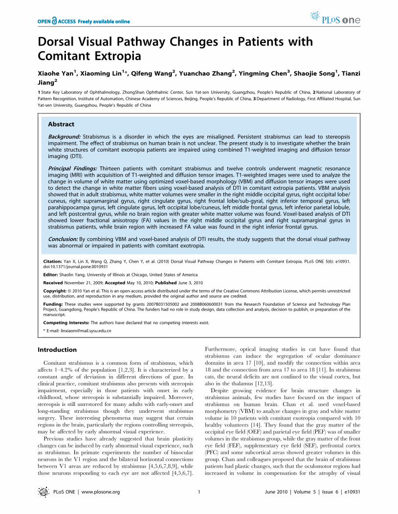
Figure 1. White matter regions with reduced volumes in adult strabismus. Strabismus patients showed smaller white matter in severalregions, including the right middle occipital gyrus (Z = +5), right occipital lobe/cuneus (Z = +33), right supramarginal gyrus (Z = +31), right cingulategyrus (Z = +29), right frontal lobe/sub-gyral (Z = +44), right inferior temporal gyrus (Z = 225), left parahippocampa gyrus (Z = 224), left cingulategyrus (Z = +37), left occipital lobe/cuneus (Z = +9), left middle frontal gyrus (Z = +30), left inferior parietal lobule (Z = 30), and left postcentral gyrus(Z = +55, Z = +49).doi:10.1371/journal.pone.0010931.g001
Dorsal Pathway in Strabismus
PLoS ONE | www.plosone.org 3 June 2010 | Volume 5 | Issue 6 | e10931
white matter volumes at the right middle occipital gyrus, right
occipital lobe/cuneus, right supramarginal gyrus, right cingulate
gyrus, right frontal lobe/sub-gyral, right inferior temporal gyrus,
left parahippocampa gyrus, left cingulate gyrus, left occipital lobe/
cuneus, left middle frontal gyrus, left inferior parietal lobule, and
left postcentral gyrus (P,0.001, uncorrected; see Table 2 and
Figure 1), while no brain region with greater white matter volume
was found at the right inferior temporal gyrus.
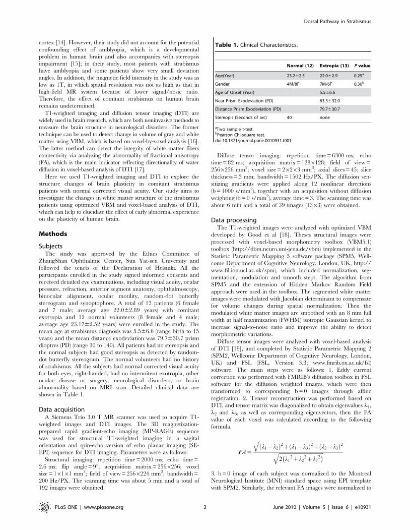
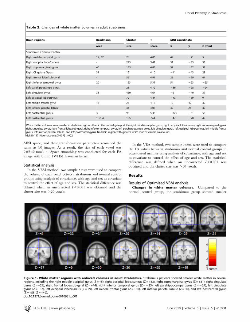
Results of voxel-based analysis of DTICompared to the normal control group, the FA values were
decreased in all the right hemisphere of the strabismus group,
including middle occipital gyrus and supramarginal gyrus
(P,0.001, uncorrected; see Table 3 and Figure 2). Right inferior
frontal gyrus with increased FA was found in the strabismus group
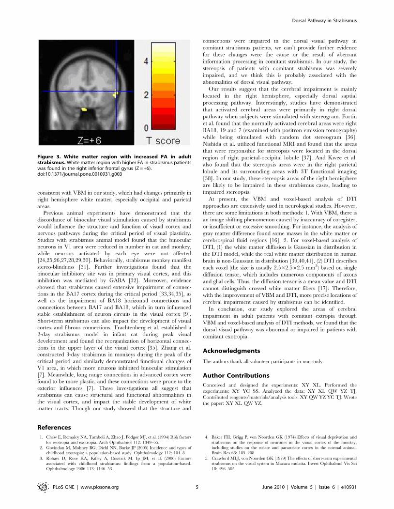
(P,0.001, uncorrected; see Table 3 and Figure 3).
Disscussion
In our study, VBM and voxel-based analysis of DTI showed
that there were structural abnormalities in occipital and parietal
areas, especially in the middle occipital gyrus and inferior parietal
lobule (supramarginal gyrus), which play important roles in the
dorsal visual pathway. Thus the major finding of the study is that
the dorsal visual pathway was abnormal in patients with comitant
exotropia. The dorsal visual pathway originates from V1 area,
passes through V2 and MT areas, and arrives at the inferior
parietal lobule. This pathway primarily takes part in the processing
of spatial position information and eye movement [20,21]. But we
don’t know whether the abnormalities in dorsal visual pathway is
the cause or result of the strabismus.
Our VBM results were partially consistent with the previous report
by Chan et al, that is, the occipital and parietal lobules showed
smaller volumes in strabismus patients. However, there were some
conflicting results, with our study showing that no regions with
greater volumes were found in strabismus patients, which may be
attributable to differences in participants, MRI scanner, magnetic
field intensity and the data analysis methods between the two studies.
The patients in our study were those with normal corrected visual
acuity and large deviation angles of exotropia, without stereopsis,
which can exclude the influence of confounding factors such as
amblyopia. The magnetic field intensity in our study was 3.0 T, which
has advantage over the 1T MRI intesity in Chan et al’s study, because
a high-field MR system provides higher signal/noise ratio, which
results in increased spatial resolution and better imaging quality
[22,23]. Moreover, we used optimized VBM which is superior to the
conventional VBM such as excluding the influence of non-cerebral
tissues and correcting the volume changes during normalization [18].
There is no prior voxel-based analysis of DTI study in comitant
strabismus, and the present study found that the FA value was
decreased in right middle occipital gyrus and right supramarginal
gyrus, indicating abnormalities of white matter fibers in these
areas. White matter region with higher FA in strabismus patients
was found in the right inferior frontal gyrus. The result was
Table 3. Locations of white matter regions with FA changes in adult strabismus.
Brain regions Brodmann Cluster T MNI coordinate
area size score x y z (mm)
Strabismus,Normal Control
Right middle occipital gyrus 19, 37 93 6.93 38 270 6
Right supramarginal gyrus 40 77 5.00 52 254 30
Left cingulate gyrus 31 26 4.05 24 236 38
Strabismus.Normal Control
Right inferior frontal gyrus 47 35 23.54 58 22 6
There are several white matter regions with significantly lower FA in adult strabismus than that in normal controls by voxel-based analysis of DTI. These regions includethe right middle occipital gyrus and right supramarginal gyrus. Region with increased FA in strabismus was found in the right inferior frontal gyrus.doi:10.1371/journal.pone.0010931.t003
Figure 2. Whiter matter regions with lower FA in adult strabismus. White matter regions with lower FA in strabismus patients, including rightmiddle occipital gyrus (Z = +6) and right supramarginal gyrus (Z = +30).doi:10.1371/journal.pone.0010931.g002
Dorsal Pathway in Strabismus
PLoS ONE | www.plosone.org 4 June 2010 | Volume 5 | Issue 6 | e10931
consistent with VBM in our study, which had changes primarily in
right hemisphere white matter, especially occipital and parietal
areas.
Previous animal experiments have demonstrated that the
discordance of binocular visual stimulation caused by strabismus
would influence the structure and function of visual cortex and
nervous pathways during the critical period of visual plasticity.
Studies with strabismus animal model found that the binocular
neurons in V1 area were reduced in number in cat and monkey,
while neurons activated by each eye were not affected
[24,25,26,27,28,29,30]. Behaviorally, strabismus monkey manifest
stereo-blindness [31]. Further investigations found that the
binocular inhibitory site was in primary visual cortex, and this
inhibition was mediated by GABA [32]. Moreover, evidence
showed that strabismus caused extensive impairment of connec-
tions in the BA17 cortex during the critical period [33,34,35], as
well as the impairment of BA18 horizontal connections and
connections between BA17 and BA18, which in turn influenced
stable establishment of neuron circuits in the visual cortex [9].
Short-term strabismus can also impact the development of visual
cortex and fibrous connections. Trachtenberg et al. established a
2-day strabismus model in infant cat during peak visual
development and found the reorganization of horizontal connec-
tions in the upper layer of the visual cortex [35]. Zhang et al.
constructed 3-day strabismus in monkeys during the peak of the
critical period and similarly demonstrated functional changes of
V1 area, in which more neurons inhibited binocular stimulation
[7]. Meanwhile, long range connections in advanced cortex were
found to be more plastic, and these connections were prone to the
exterior influences [7]. These investigations all suggest that
strabismus can cause structural and functional abnormalities in
the visual cortex, and impact the stable development of white
matter tracts. Though our study showed that the structure and
connections were impaired in the dorsal visual pathway in
comitant strabismus patients, we can’t provide further evidence
for these changes were the cause or the result of aberrant
information processing in comitant strabismus. In our study, the
stereopsis of patients with comitant strabismus was severely
impaired, and we think this is probably associated with the
abnomalities of dorsal visual pathway.
Our results suggest that the cerebral impairment is mainly
located in the right hemisphere, especially dorsal saptial
processing pathway. Interestingly, studies have demonstrated
that activated cerebral areas were primarily in right dorsal
pathway when subjects were stimulated with stereogram. Fortin
et al. found that the normally activated cerebral areas were right
BA18, 19 and 7 (examined with positron emission tomography)
while being stimulated with random dot stereogram [36].
Nishida et al. utilized functional MRI and found that the areas
that were responsible for stereopsis were located in the dorsal
region of right parietal-occipital lobule [37]. And Kwee et al.
also found that the stereopsis areas were in the right parietal
lobule and its surrounding areas with 3T functional imaging
[38]. In our study, these stereopsis areas of the right hemisphere
are likely to be impaired in these strabismus cases, leading to
impaired stereopsis.
At present, the VBM and voxel-based analysis of DTI
approaches are extensively used in neurological studies. However,
there are some limitations in both methods: 1. With VBM, there is
an image shifting phenomenon caused by inaccuracy of coregister,
or insufficient or excessive smoothing. For instance, the analysis of
gray matter difference found some masses in the white matter or
cerebrospinal fluid regions [16]. 2. For voxel-based analysis of
DTI, (1) the white matter diffusion is Gaussian in distribution in
the DTI model, while the real white matter distribution in human
brain is non-Gaussian in distribution [39,40,41]. (2) DTI describes
each voxel (the size is usually 2.562.562.5 mm3) based on single
diffusion tensor, which includes numerous components of axons
and glial cells. Thus, the diffusion tensor is a mean value and DTI
cannot distinguish crossed white matter fibers [17]. Therefore,
with the improvement of VBM and DTI, more precise locations of
cerebral impairment caused by strabismus can be identified.
In conclusion, our study explored the areas of cerebral
impairment in adult patients with comitant extropia through
VBM and voxel-based analysis of DTI methods, we found that the
dorsal visual pathway was abnormal or impaired in patients with
comitant exotropia.
Acknowledgments
The authors thank all volunteer participants in our study.
Author Contributions
Conceived and designed the experiments: XY XL. Performed the
experiments: XY YC SS. Analyzed the data: XY XL QW YZ TJ.
Contributed reagents/materials/analysis tools: XY QW YZ YC TJ. Wrote
the paper: XY XL QW YZ.
References
1. Chew E, Remaley NA, Tamboli A, Zhao J, Podgor MJ, et al. (1994) Risk factors
for esotropia and exotropia. Arch Ophthalmol 112: 1349–55.
2. Govindan M, Mohney BG, Diehl NN, Burke JP (2005) Incidence and types of
childhood exotropia: a population-based study. Ophthalmology 112: 104–8.
3. Robaei D, Rose KA, Kifley A, Cosstick M, Ip JM, et al. (2006) Factors
associated with childhood strabismus: findings from a population-based.
Ophthalmology 2006 113: 1146–53.
4. Baker FH, Grigg P, von Noorden GK (1974) Effects of visual deprivation and
strabismus on the response of neurones in the visual cortex of the monkey,
including studies on the striate and parastriate cortex in the normal animal.
Brain Res 66: 185–208.
5. Crawford MLJ, von Noorden GK (1979) The effects of short-term experimental
strabismus on the visual system in Macaca mulatta. Invest Ophthalmol Vis Sci
18: 496–505.
Figure 3. White matter region with increased FA in adultstrabismus. White matter region with higher FA in strabismus patientswas found in the right inferior frontal gyrus (Z = +6).doi:10.1371/journal.pone.0010931.g003
Dorsal Pathway in Strabismus
PLoS ONE | www.plosone.org 5 June 2010 | Volume 5 | Issue 6 | e10931
6. Crawford ML, Smith EL, 3rd, Harwerth RS, von Noorden GK (1984)
Stereoblind monkeys have few binocular neurons. Invest Ophthalmol Vis Sci 25:
779–81.
7. Zhang B, Bi H, Sakai E, Maruko I, Zheng J, et al. (2005) Rapid plasticity of
binocular connections in developing monkey visual cortex (V1). Proc Natl Acad
Sci U S A 102: 9026–31.
8. Kumagami T, Zhang B, Smith EL, III, Chino YM (2000) Effect of onset age of
strabismus on the binocular responses of neurons in the monkey visual cortex.
Invest Ophthalmol Vis Sci 41: 948–954.
9. Mori T, Matsuura K, Zhang B, Smith EL, 3rd, Chino YM (2002) Effects of the
duration of early strabismus on the binocular responses of neurons in the
monkey visual cortex (V1). Invest Ophthalmol Vis Sci 43: 1262–9.
10. Engelmann R, Crook JM, Lowel S (2002) Optical imaging of orientation and
ocular dominance maps in area 17 of cats with convergent strabismus. Vis
Neurosci 19: 39–49.
11. Schmidt KF, Lowel S (2008) Strabismus modifies intrinsic and inter-areal
connections in cat area 18. Neuroscience 152: 128–37.
12. Chino YM, Cheng H, Smith EL, 3rd, Garraghty PE, Roe AW, et al. (1994)
Early discordant binocular vision disrupts signal transfer in the lateral geniculate
nucleus. Proc Natl Acad Sci U S A 91: 6938–42.
13. Cheng H, Chino YM, Smith EL, 3rd, Hamamoto J, Yoshida K (1995) Transfer
characteristics of X LGN neurons in cats reared with early discordant binocular
vision. J Neurophysiol 74: 2558–72.
14. Chan ST, Tang KW, Lam KC, Chan LK, Mendola JD, et al. (2004)
Neuroanatomy of adult strabismus: a voxel-based morphometric analysis of
magnetic resonance structural scans. Neuroimage 22: 986–94.
15. McKee SP, Levi DM, Movshon JA (2003) The pattern of visual deficits in
amblyopia. J Vision 4: 380–405.
16. Busatto GF, Diniz BS, Zanetti MV (2008) Voxel-based morphometry in
Alzheimer’s disease. Expert Rev Neurother 8: 1691–702.
17. Assaf Y, Pasternak O (2008) Diffusion tensor imaging (DTI)-based white matter
mapping in brain research: a review. J Mol Neurosci 34: 51–61.
18. Good CD, Johnsrude IS, Ashburner J, Henson RN, Friston KJ, et al. (2001) A
voxel-based morphometric study of ageing in 465 normal adult human brains.
NeuroImage 14: 21–36.
19. Shu N, Li J, Li K, Yu C, Jiang T (2009) Abnormal diffusion of cerebral white
matter in early blindness. Hum Brain Mapp 30: 220–7.
20. Merigan WH, Maunsell JH (1993) How parallel are the primate visual
pathways? Annu Rev Neurosci 16: 369–402.
21. Tootell RBH, Hadjikhani NK, Mendola JD, Marrett S, Dale AM (1998) From
retinotopy to recognition: fMRI in human visual cortex. Trends Cogn Sci 2:
174–183.
22. Scarabino T, Nemore F, Giannatempo GM, Bertolino A, Di Salle F, et al. (2003)
3.0 T magnetic resonance in neuroradiology. Eur J Radiol 48: 154–64.
23. Alvarez-Linera J (2008) 3T MRI: advances in brain imaging. Eur J Radiol 67:
415–26.
24. Hubel DH, Wiesel TN (1965) Binocular interaction in striate cortex of kittens
reared with artificial squint. J. Neurophysiol 28: 1041–1059.25. Baker FH, Grigg P, von Noorden GK (1974) Effects of visual deprivation and
strabismus on the response of neurones in the visual cortex of the monkey,
including studies on the striate and parastriate cortex in the normal animal.Brain Res 66: 185–208.
26. Blakemore C (1976) Modification of visual function by early visual experience.Bull Schweiz Akad Med Wiss 32: 13–28.
27. Crawford MLJ, von Noorden GK (1979) The effects of short-term experimental
strabismus on the visual system in Macaca mulatta. Invest Ophthalmol Vis Sci18: 496–505.
28. Singer W, von Grunau M, Rauschecker J (1980) Functional amblyopia in kittenswith unilateral exotropia. I. Electrophysiological assessment. Exp Brain Res 40:
294–304.29. Crawford ML, Smith EL, 3rd, Harwerth RS, von Noorden GK (1984)
Stereoblind monkeys have few binocular neurons. Invest Ophthalmol Vis Sci 25:
779–81.30. Sengpiel F, Blakemore C, Kind PC, Harrad R (1994) Interocular suppression in
the visual cortex of strabismic cats. J. Neurosci 14: 6855–71.31. Crawford ML, von Noorden GK, Meharg LS, Rhodes JW, Harwerth RS, et al.
(1983) Binocular neurons and binocular function in monkeys and children.
Invest Ophthalmol Vis Sci 24: 491–5.32. Sengpiel F, Jirmann KU, Vorobyov V, Eysel UT (2006) Strabismic suppression
is mediated by inhibitory interactions in the primary visual cortex. Cereb Cortex16: 1750–8.
33. Lowel S, Singer W (1992) Selection of intrinsic horizontal connections in thevisual cortex by correlated neuronal activity. Science 255: 209–12.
34. Schmidt KE, Kim DS, Singer W, Bonhoeffer T, Lowel S (1997) Functional
specificity of long-range intrinsic and interhemispheric connections in the visualcortex of strabismic cats. J. Neurosci 17: 5480–92.
35. Trachtenberg JT, Stryker MP (2001) Rapid anatomical plasticity of horizontalconnections in the developing visual cortex. J. Neurosci 21: 3476–82.
36. Fortin A, Ptito A, Faubert J, Ptito M (2002) Cortical areas mediating stereopsis in
the human brain: a PET study. Neuroreport 13: 895–8.37. Nishida Y, Hayashi O, Iwami T, Kimura M, Kani K, et al. (2001) Stereopsis-
processing regions in the human parieto-occipital cortex. Neuroreport 12:2259–63.
38. Kwee IL, Fujii Y, Matsuzawa H, Nakada T (1999) Perceptual processing ofstereopsis in humans: high-field (3.0-tesla) functional MRI study. Neurology 53:
1599–1601.
39. Beaulieu C (2002) The basis of anisotropic water diffusion in the nervous system- a technical review. NMR Biomed 15: 435–55.
40. Assaf Y, Basser PJ (2005) Composite hindered and restricted model of diffusion(CHARMED) MR imaging of the human brain. Neuroimage 27: 48–58.
41. Jensen JH, Helpern JA, Ramani A, Lu H, Kaczynski K (2005) Diffusional
kurtosis imaging: the quantification of non-gaussian water diffusion by means ofmagnetic resonance imaging. Magn Reson Med 53: 1432–40.
Dorsal Pathway in Strabismus
PLoS ONE | www.plosone.org 6 June 2010 | Volume 5 | Issue 6 | e10931

![D V High [Dorsal] Low [Dorsal] No Dorsal Graded Dorsal Concentration Created by Mother Hierarchy of Gene Action in D/V Patterning Mesoderm Genes Neuroectoderm](https://img.pdfslide.net/doc/110x75/56649d3f5503460f94a18b80/d-v-high-dorsal-low-dorsal-no-dorsal-graded-dorsal-concentration-created.jpg)