Embed Size (px)
Citation preview

Dosimetry experience at IEO, Milan
MetroMRT 3rd Workshop
Clinical implementation of dosimetry for molecular
radiotherapyNPL 20-21 April 2015
Mahila Ferrari, Marta Cremonesi,
Francesca Botta

Marco Chinol
Stefano Papi
Francesca Botta
Marta Cremonesi
Laura Travaini
Guido Pedroli
Annalisa Rossi
Silvia Baio
Lisa Bodei
Marzia Colandrea
Paolo Baldassarre
Michele Calabrese
Sebastiano Croce
Angela Carollo
Alfio Cascio
Silvia Fracassi
Laura Gilardi
Paola Rocca
Gianni Bufi
Francesca Fassari
Daniele Paolucci
Valerio Massetti
Maurizio Fiorenza
Riccardo Mei
Domenico Militano
Andrea Vertua
Nuclear Medicine Division - IEO
Head physician: Chiara Grana

Our experience…..
MRT procedure Dosimetry data used for Dosimetry method
90Y- MoAbs (3 step)- 1995- Research- safety
Planar imagesMean doses
90Y- and 177Lu- PRRT- 1997-Research- kidney/red marrow safety
Planar imagesMean doses
High Activity Zevalin –2000
- Red marrow safety beforetransplantation-Safety
Planar imagesMean doses
IART (90Y-biotin) +EBRT in breast cancer – 2004
To combine MRT+EBRTPlanar images + 1 SPECTMean doses+DVH+ BED
SIRTEX 90Y - resinmicrospheres – 2007
To determine the activity to be injected
SPECT/CTDVH + BED etc….
NETTER-1 study (AAA) -dosimetry central lab -2014
To demonstrate thatdosimetry is notmandatory
Planar images + 1-2 SPECT/CTMean doses + kidneys BED +……work in progress

SIRT- how are dosimetric data used ?
Patients undergo dosimetry
Dosimetry is used to determine the 90Y activity to be injected in patient
The activity is established considering healthy liver (HL) EUBED < 40Gy
The actual activity injected relies on single patient “viability”, as resin
microspheres clug up vessels

SIRT - Dosimetric procedure
Radiopaque marker in CTs and SPECT for SPECT /CT fusionCT low dose for AC + CT with contrast medium
~ 74 MBq 99mTc-MAA to simulate therapy – 1 SPECT/CT (no biologial removal)
@ the end of interventional radiological procedure
manual delineation on contrast CT (Lesions - healthy liver)
120 projections (15 s each), 128x218,
OSEM, 8 iterations, 6 subsets, no filter
Scatter correction: energy window subtraction
Attenuation correction (GEHC Xeleris 3.1)
SPECT
Target volumes
Voxel dosimetry convolution (MATLAB ® support); S-voxel matrix (7x7x7) by MC
simulation (Penelope) to obtain for HL and lesions DVHs and
radiobiological parameters
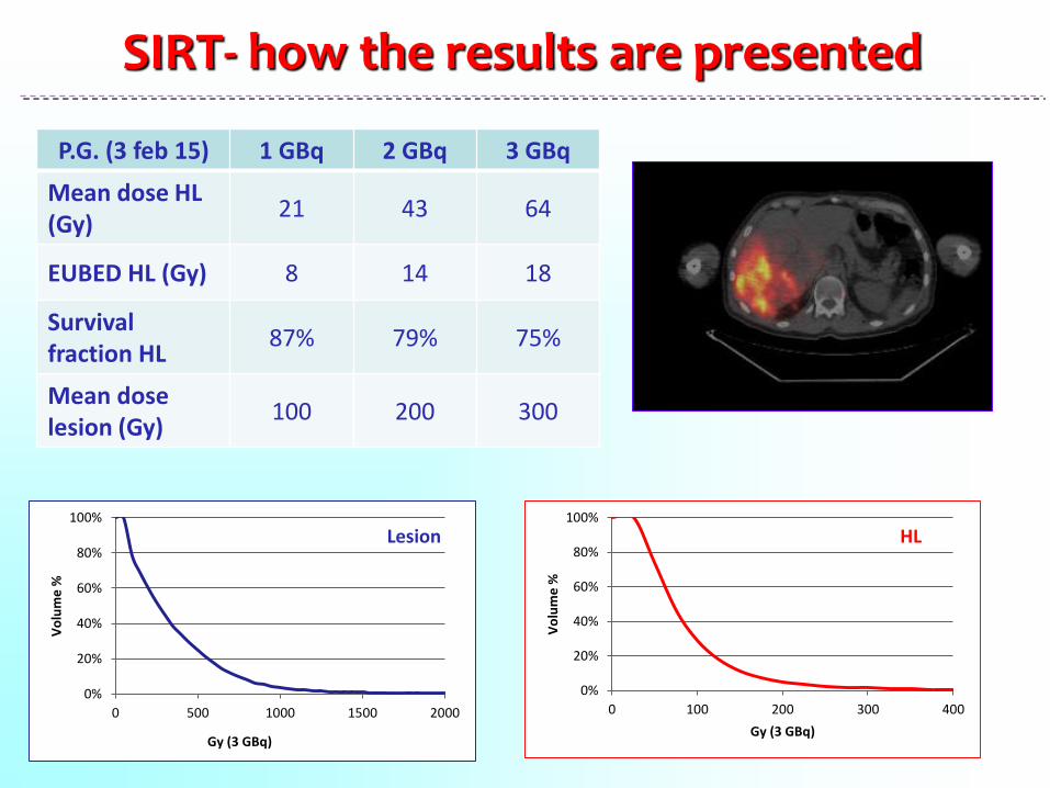
SIRT- how the results are presented
P.G. (3 feb 15) 1 GBq 2 GBq 3 GBq
Mean dose HL(Gy)
21 43 64
EUBED HL (Gy) 8 14 18
Survivalfraction HL
87% 79% 75%
Mean dose lesion (Gy)
100 200 300
0%
20%
40%
60%
80%
100%
0 100 200 300 400
Vo
lum
e %
Gy (3 GBq)
HL
0%
20%
40%
60%
80%
100%
0 500 1000 1500 2000
Vo
lum
e %
Gy (3 GBq)
Lesion

SIRT – How do calibrate the measurements?
We use relative calibration
SIRT – What type of accuracyvalidation measurements have you done?
NONE!

SIRT – Can you estimate the uncertainty in your dose measurements?
Accuracy influenced by:
Image quality isotope
actual resolution image reconstruction image corrections image noise…….
Activitymeasurement
in dose calibrator
Radiobiologicalparameters
…………….

Dose Calibrator
Geometry
Volume

For each volume and geometry anappropriate factor…..

Attention…. with an old calibrator….
The calibration factors were depending from geometry,
volume (not in a linear way) and …. RANGE OF ACTIVITIES!!!!
90Y: 0.6 GBq
90Y: 2.8 GBq
Volume ml
Cal
ibra
tio
nfa
cto
r

Attention ….. Spheres collapse……

About image quality/corrections……
Work in progress…….
QSPECT with 99mTc: impact of scatter
and attenuation corrections in the
prospective dosimetry for SIRTMassimiliano Pacilio, Marta Cremonesi, Carlo Chiesa, Mahila Ferrari,
Francesca Botta, Leda Lorenzon, Michael Ljungberg
To compare the patient dose images obtained from different methods ofreconstruction (with and without corrections) for simulated patients (SIMIND)and clinical cases, including different scatter correction, resolution and noiseof the images, relative and absolute calibration…..

0%
20%
40%
60%
80%
100%
120%
0 100 200 300 400
Vo
lum
e (
%)
Absorbed dose (Gy)
NAC NSC
NAC SC
AC NSC
AC SC
Pt 6_CG, 80 kg, lesion 100 g, healthy liver involved 825g , total
liver 1825 g
delta% NAC NSC AC NSC NAC SC
D mean -19% -7% -12%
D20 -22% -9% -14%
D70 -16% -5% -10%
D90 -7% 1% -8%
0%
20%
40%
60%
80%
100%
120%
0 10 20 30 40 50
Vo
lum
e (
%)
Absorbed dose (Gy)
NAC NSC
NAC SC
AC NSC
AC SC
delta% NAC NSC AC NSC NAC SC
D mean 8% 3% 5%
D20 19% 9% 12%
D90 910% 1067% -85%
lesion Healthy liver
Liver metastasis

SIRT - Do you think that the dosimetry youare doing could be incorporated in MRT
treatment planning on individual patientsand be used for personalised treatments?
YES!It’s easy and no time consuming
Useful for the management of the patient
Also local deposition can be considered without
compromising the results

177Lu-DOTATATE- how are dosimetric data used ?
AAA is conducting a dosimetry sub-study within the Phase III clinical trial,
with the primary objective to correlate whole body and organ radiation
dosimetry results with findings of the Erasmus Medical Center Phase I/II study
…...a planned treatment will be withheld if the resulting cumulative
bone marrow radiation dose exceeds 3.7 Gy, or if the cumulative
kidney radiation dose is determined to exceed 38 Gy of Biologically
Effective Dose (BED)
……. four treatments of 7.4 GBq of 177Lu-DOTA0-Tyr3-Octreotate
can be safely given without the need for individualized dosimetry assessments…….

177Lu-DOTATATE - Dosimetric procedure -1
Dosimetry based on:planar whole body imaging (WB; anterior and posterior views), in combination with transmission dataSPECT scans in the upper abdomen (to
include the kidneys, liver, and spleen) blood and urine analysis of radioactivity
calibration data: attenuation coefficient for gamma
camera calibration with a 177Lu reference source
for gamma counterSPECT of a phantom

177Lu-DOTATATE - Dosimetric procedure - 2
177Lu radioactivity in the organs (relative calibration): the conjugate-view techniqueis applied to ant and post images after background, scatter, attenuation, andphysical decay corrections
Counts are normalized at the first image, scanning the patient with 100% of theinjected activity subtracted by the percent of injected activity eliminated in theurine before the first image acquisition
The number of decays (NDs) per unit injected activity is calculated frommultiexponential fits to the time–activity curves
0 1 2 3 days
A(%
)
Absorbed doses to target organs arecalculated by entering the ND values for allthe source organs in the OLINDA/EXMsoftware and adjusting the doses reported bythe software for individual weight and organmasses
Kidney BED is also evaluated

177Lu-DOTATATE - how the results are presented

177Lu-DOTATATE – How do calibrate the measurements?
We use relative calibration, in WB planar dosimetry.
We are attempiting to perform 3D dosimetry with SPECT, but we have some
problems with absolute calibration and SPECTs alignment
177Lu-DOTATATE – What type of accuracyvalidation measurements have you done?
NONE!

177Lu-DOTATATE – Can you estimate the uncertainty in your dose measurements?
dose distribution
..... much more.... 2D/3D imaging
biokinetics
actual masses
biodistribution among cycles
Some key points for accuracy are:
Kidney models response evaluationradiobiological parameters.........

NDs were derived for 177Lu- & 90Y-peptides by: (i) trapezoidal+ physical-decay after experimental data (commercial software); (ii) trapezoidal+ biological-decay after the last 2 points; (iii) bi-exp fit; (iv) mono-exp fit.
4 and 64h notably impact the dose estimate
inappropriate model(i), overestimating ND(177Lu) up to 3-fold vs. (iii)
Model (ii) underestimates vs.(iii / iv)
differences biexp - monoexp: 8% (-50,+72)%.
Kinetic models strongly impact dose estimatesNDTAIL is major influencingBi-exp model better reflects the metabolic behaviour
biokinetics

dose rescaling factor based on patient specific mass of
kidneys or BW
0.9 (0.6-1.4)1.0 (0.8-1.3)
0
0.2
0.4
0.6
0.8
1.0
1.2
1.4
patients
resc
alin
g f
acto
r
Kidney mass
BW ratio
Kidneys: 200g, F, BW: 48 kg
Mono-kidney: 240gF, BW: 60 kg
For a group of 15 patients, comparing the actual mass of the kidneys vs. the standard values of 300 g (male) and 275 g (female) …
actual masses
0
100
200
300
400
500
600
kidney massses (g)
Up to 40% error

mass (g)
A% (24h)
dose (Gy)
Response
L Liver
200
4%
95
CR
L Pancreas
1150
25%
90
SD/MR
16-Jul-04 29-Aug-08, 3 yrs.5-Aug-05
Bodei, J Endocrinol Invest 2009,360-9
o Radiosensitivity (growth pattern, DNA
repair capacity)
o Tumour dimension
o Activity/dose distribution
average dose is not enough
o Non uniformity due to receptor density

Diffuse liver and bone mets from a pancreatic NETG2
basal MRI
final MRI
biodistribution can vary with cycles
final 177Lu-DOTATATE
Ant Post
1st 177Lu-DOTATATE
Ant Post
Uptake in tumour & organs can vary with cycles, especially in case of large burden

177Lu-DOTATATE - Do you think that the dosimetry you are doing could
be incorporated in MRT treatment planning on individual patients and be used for
personalised treatments?
-To avoid dosimetry just because apparently time consuming/expensive
- To perform “bad” dosimetry, i.e.:
- not collecting the essential data useful for future refinements
- not giving the method specifics for dosimetry
- To derive hasty conclusions without specifying how dosimetry wasmade
- To forget the concept of OPTIMIZATION
Whatshould not
be done
More than ever, dosimetry should be done, as accurate aspossible and providing, in any case, all the specifications, and refinements :
- revise and analyse possible ways to reduce inaccuracies
- focus efforts on the implementation of methods improvingdosimetry results
answers will be derived and the clinical results improved
Whatshould
be done
177Lu-DOTATATE - Do you think that the dosimetry you are doing could
be incorporated in MRT treatment planning on individual patients and be used for
personalised treatments?
Thank you!