Embed Size (px)
Citation preview
169
JARQ 50 (2), 169 - 173 (2016) http://www.jircas.affrc.go.jp
Histological Study of Bovine Nocardial EosinophilicGranuloma with Comparison of Splendore-Hoeppli Material
* Corresponding author: e-mail [email protected] 6 February 2015; accepted 3 August 2015.
Takeshi OYAMA1, Tomoko HIGASHI2, Yukiko TANIGUCHI3, Eiji HATA4,Shinichi HATAMA4, Yoshiharu ISHIKAWA4 and Koichi KADOTA4*1 Nemuro Livestock Hygiene Service Center (Betsukai, Hokkaido 086-0214, Japan)2 Shimane Livestock Hygiene Research Office (Izumo, Shimane 699-0822, Japan)3 Soya Livestock Hygiene Service Center (Hamatonbetsu, Hokkaido 098-5738, Japan)4 Hokkaido Research Station, National Institute of Animal Health, National Agriculture and Food Research Organization
(Sapporo, Hokkaido 062-0045, Japan)
AbstractNasal eosinophilic granuloma in two cases involving a 13-month-old Japanese Black heifer (case 1) and a two-year-old Japanese Black heifer (case 2) is described. The animals suffered from bilateral nasal obstruction, with the nasal cavities being occupied by large tumor-like masses. In case 1, the nasal mucous membrane was largely thickened due to severe eosinophilic infiltration; in case 2, there were many plasma cells and eosinophils. Epithelioid cells with bacterial organisms were observed in both animals. Extracellular eosinophilic deposits of Splendore-Hoeppli material, formed between intact filamentous bacteria and live epithelioid cells, appeared to be elongated spicules and resembled those in actinomycosis. In contrast, in a separate case of Trueperella pyogenes infection, neutrophils containing intact rod-shaped bacteria showed coagulation necrosis and fusion, and finally transformed into sulfur granules without projections on the surface. These differences suggest that at least two types of Splen-dore-Hoeppli phenomenon occur in bacterial infections.
Discipline: Animal healthAdditional key words: Allergy, cattle, Fite’s acid fast stain, immunohistochemistry
Introduction
The Splendore-Hoeppli phenomenon is seen by hema-toxylin and eosin (HE) staining as club-shaped or band-like eosinophilic structures surrounding microorganisms or foreign bodies (Hussein 2008, Schnadig 2010). Bacterial colonies with surrounding Splendore-Hoeppli material are designated sulfur granules, which may be observed in infec-tions of Actinomyces, Trueperella pyogenes, Actinobacillus, Staphylococcus aureus, and Pseudomonas aeruginosa in cattle (Kubo et al. 1980, 1981, 1982). Nocardial sulfur gran-ules are usually absent or inconspicuous (Husain 2012), but obvious in certain human cases (Rodig et al. 2001, Shimizu et al. 2001). Sulfur granules are absent, or at least have not been described in bovine nocardiosis with neutrophilic inflammation (Bawa et al. 2010, Pisoni et al. 2008, Saito et
al. 2009, Takahashi et al. 1999).Aspergillosis caused by several Aspergillus spp.
is histologically characterized by fungal hyphae and neutrophil infiltration, but epithelioid cell granuloma with eosinophil infiltration is formed in allergic bronchopulmo-nary aspergillosis in humans (Bosken et al. 1988). Allergic granuloma may occur in the skin and nasal cavity of cattle (Murakami et al. 2014, Riet-Correa et al. 1992, Shibahara et al. 2001). However, small amounts of allergens can cause severe inflammation in both humans and animals, implying difficulties in identifying its etiological agents. In a case of eosinophilic granuloma in the nasal vestibule of a Japanese Brown heifer, abundant Splendore-Hoeppli material sur-rounded by eosinophils was observed, and Corynebacte-rium ulcerans producing diphtheria toxin was isolated from the lesion (Murakami et al. 2014). Eosinophilic granuloma

170 JARQ 50 (2) 2016
T. Oyama et al.
caused by Nocardia sp. in the nasal cavities of a Holstein heifer has also been reported (Shibahara et al. 2001). Here, we describe the histology of two additional cases of bovine nocardial eosinophilic granuloma, with particular emphasis on the relation between Splendore-Hoeppli material and the inflammatory cells in certain bacterial infections.
Materials and methods
1. Animals and gross pathologyCase 1 was a 13-month-old Japanese Black heifer
that underwent examination due to dyspnea. A tumor-like elevation (8×3 cm in size) was detected on the mucosa of the right nasal cavity, but could not be surgically removed because of hemorrhage. Despite antibiotic and anti-inflam-matory treatments, the animal remained depressed and anorexic with labored breathing. Ten days later, steroid and antibiotic treatments were given based on the presence of many eosinophils in a biopsied tissue sample. The animal’s condition improved only marginally after the treatments, and both nasal cavities were narrowed seven days later. The animal was treated intermittently, but died five months after the initial examination. Case 2 was a two-year-old Japanese Black heifer with a three-month history of nasal discharge with blood. Despite antibiotic treatment, the condition alter-nated between better and worse. The animal was euthanized due to dyspnea, open mouth breathing, and nasal discharge with pseudomembranous material. In both cases, blood tests and culture for bacterial isolation were not conducted.
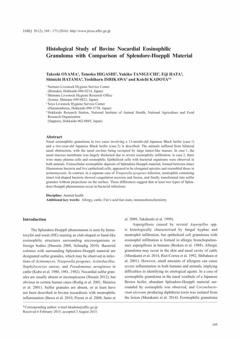
In case 1, necropsy revealed that both nasal cavities from the nostrils to the choana were occupied by tumor-like masses. The outer and cut surfaces of these masses were rough and yellowish white, with soft areas due to suppura-tion on the cut surfaces. The nasal septum became fragile at sites compressed by the masses. In case 2, greenish tumor-like masses with pus obstructed the nasal cavities (Fig. 1), and the left frontal bone was elevated.
2. Histological and immunohistochemical examina-tions
Formalin-fixed tissues were embedded in paraffin, sectioned at 4 μm, and stained with HE, Giemsa, and Gram (Twort’s and Nishioka’s methods) (Tanaka et al. 1997), and ordinary and modified Fite’s acid fast stain. Selected sections were dewaxed and labeled using the streptavidin-biotin-peroxidase complex (SAB) method. The primary reagent employed was a rabbit polyclonal antibody to Nocardia asteroides (1:2048; provided by Prof. Satoshi Murakami, Tokyo University of Agriculture, Kanagawa, Japan, and Dr. Mikio Inage, Hokubu Livestock Hygiene Service Center, Chiba, Japan). Subsequent procedures were performed using an immunoperoxidase labeling system (Nichirei, Tokyo, Japan).
For a comparison with the Splendore-Hoeppli phe-nomenon, tissues from a case of Trueperella pyogenes abscesses and mucoepidermoid carcinoma (located from the right parotid area to the angle of the right mandible in a 15-month-old Japanese Black heifer) were similarly treated
Fig. 1. Case 2Cross section of nasal region, viewed from behind, showing almost complete obstruction of the nasal meatuses by tumor-like masses.

171
Bovine Nocardial Eosinophilic Granuloma
and observed histologically as a control. In addition, stained sections from previously reported cases of nocardial eosino-philic granuloma (Shibahara et al. 2001) and actinomycosis (Murakami et al. 2014) were also observed as controls.
Results
Histological examination revealed that the tumor-like masses resulted from severe thickening of nasal mucosae in both cases. In case 1, the lamina propria was heav-ily infiltrated with eosinophils, admixed with macrophages and epithelioid cells. Plasma cells and mast cells were inconspicuous in most areas. Nasal gland cells were fre-quently filled with mucin. In case 2, the lamina propria was infiltrated with many plasma cells and eosinophils. Epithe-lioid cells were more readily recognized than in case 1, but mononuclear macrophages were not prominent. In places, eosinophilic abscesses surrounded by epithelioid cells were detected. There were more mast cells than in case 1, and in-traepithelial globule leukocytes were observed. Some nasal glands were composed exclusively of mucin-filled cells and highly dilated with large amounts of mucus.
In case 1, a few bacterial organisms were found in the cytoplasm of some epithelioid cells. Bacterial organ-isms with eosinophilic material were rarely seen and in close contact with the cell membrane of multinucleated epithelioid cells (Fig. 2 A). Similar findings were obtained in case 2. In addition, calcified Splendore-Hoeppli mate-rial was observed (Fig. 2 B), with many bacterial organisms being found within eosinophilic abscesses and surround-ing epithelioid cells. In both cases, the organisms were Gram-positive (Nishioka’s method) and partially acid-fast (Fite’s method) (Fig. 2 C), and stained positively with anti-
Nocardia antibody (Fig. 2 D). Unlike in case 1, immuno-positive products were observed even in the cytoplasm of epithelioid cells without bacterial organisms detected with Giemsa stain.
Similar bacteria and eosinophilic material as in cases 1 and 2 were ascertained in a previous case of nocardiosis (Fig. 2 E). In the case of actinomycosis, smaller bacterial colonies were surrounded by epithelioid cells, and radially projecting organisms were individually covered with eo-sinophilic material (Fig. 2 F). Splendore-Hoeppli material in the case of Trueperella infection (Fig. 2 G) appeared to be formed by a fusion of neutrophils containing intact bacteria and showing coagulation necrosis (Fig. 2 H).
Discussion
As in the previously reported case (Shibahara et al. 2001), eosinophil infiltration and epithelioid cells with filamentous bacterial organisms were observed in the present cases. As these organisms were positive with Gram (Nishioka’s method) (Tsutsumi 2000) and Fite’s acid fast stain, and included an anti-Nocardia antibody (Shibahara et al. 2001), Nocardia spp. were considered the causal agents of the eosinophilic granulomas. Similar host reactions are observed in human allergic bronchopulmonary aspergillosis, where fungal hyphae are located in the cytoplasm of multi-nucleated giant cells or within necrotic tissue (Bosken et al. 1988). Since this allergic disease occurs most commonly in patients with asthma or cystic fibrosis, especially those with coexisting atopy (Chetty 2003, Patterson & Strek 2010), the animals with nocardial eosinophilic granuloma were also considered to have an allergic constitution. Similar eosinophilic granulomas have been caused by C. ulcerans
Fig. 2 A. Case 1Intracytoplasmic and extracellular bacterial organ-isms are visible. Splendore-Hoeppli material is formed around the latter (arrow), and is adherent to multinuclear epithelioid cells (arrowheads). [Gi-emsa; Bar = 5 µm]
Fig. 2 B. Case 2Splendore-Hoeppli material is calcified and appears dendritic. Plasma cells and eosinophils are present in the surrounding tissue. [Giemsa; Bar = 5 µm]

172 JARQ 50 (2) 2016
T. Oyama et al.
(Murakami et al. 2014) and M. granulomatis (Riet-Correa et al. 1992), and further studies would find other organisms associated with bovine eosinophilic granuloma.
Splendore-Hoeppli material is said to consist of tissue debris, fibrin, and immunoglobulin (Hussein 2008, Johnson et al. 2010). In two cases of human conjunctival lesions exhibiting the Splendore-Hoeppli phenomenon, one revealed predominantly immunoglobulin deposition, whereas the other revealed primarily eosinophilic major basic protein (Read et al. 2005). Its composition varies and may be related to different factors, including the timing of biopsy and prior treatment (Read et al. 2005). In the current
and previous (Shibahara et al. 2001) Nocardia cases, small amounts of eosinophilic material were formed around single filamentous bacterial organisms, and located closely adjacent to the cell membrane of epithelioid cells. As has been demonstrated by electron microscopy (Kubo et al. 1980), each organism was encircled by Splendore-Hoeppli material in the Actinomyces case, and the material was in contact with epithelioid cells in smaller bacterial colonies. Such findings show that the bacteria and epithelioid cells play important roles at earlier stages of formation. On the other hand, in a case of Trueperella pyogenes infection, neutrophils containing intact bacteria became necrotic and
Fig. 2 C. Case 2Acid-fast bacteria in an eosinophilic abscess are indicated by arrows. [Fite’s method; Bar = 5 µm]
Fig. 2 D. Case 1Bacterial organisms positively stained with anti-Nocardia antibody are visible in the cytoplasm of epithelioid cells. [SAB; Bar = 5 µm]
Fig. 2 E. Control case of nocardiosisAs in Fig. 2A, Splendore-Hoeppli material is visible. The material is formed only adjacent to the cell membrane of a multinuclear epithelioid cell (ar-row), or between two epithelioid cells (arrowhead). [Giemsa; Bar = 5 µm]
Fig. 2 F. Control case of actinomycosisEach bacteria protruding from the colony is en-sheathed by eosinophilic material (arrow). [Giemsa; Bar = 5 µm]
Fig. 2 G. Control case of Trueperella infectionIntact bacteria are included within eosinophilic ma-terial, and their density and distribution are variable among sulfur granules. [Giemsa; Bar = 5 µm]
Fig. 2 H. Control case of Trueperella infectionNecrotic neutrophils with intact bacteria are visible (arrows). Splendore-Hoeppli material with intact bacteria (arrowheads) appears to be generated by a fusion of these necrotic cells. [Giemsa; Bar = 5 µm].

173
Bovine Nocardial Eosinophilic Granuloma
coalesced, and showed transition into sulfur granules. Like-wise, in a case of Corynebacterium ulcerans infection, cell death induced by cytotoxic diphtheria toxin was believed responsible for the formation of abundant Splendore-Hoep-pli material (Murakami et al. 2014). Elongated spicules of Splendore-Hoeppli material were not observed in these cases caused by toxigenic rod-shaped bacteria (Billington et al. 1997, Murakami et al. 2014), unlike in cases of branch-ing filamentous bacteria. Thus, at least two mechanisms are involved in forming the material, and its components may differ according to the bacterial species and immunoreac-tive cells present. Considering the degree of immunocom-petence and allergy in different hosts, and the fact that bacteria vary in such characteristics as shape, toxigenicity, and pathogenicity, the Splendore-Hoeppli material resulting from host-parasite interactions is probably heterogeneous.
In addition to a surgically removed mass, multiple raised nodules were detected in the case of Corynebacte-rium ulcerans, but not obstructive to breathing (Murakami et al. 2014). In spite of steroid or antibiotic treatment, the current nasal granulomas resulted in a poor prognosis or death. Because surgical excision or the continuous admin-istration of steroids and antibacterial drugs may be effective for treating smaller lesions, blood testing for eosinophilia (Shibahara et al. 2001) and cytological examination of nasal discharge or biopsied tissues should be conducted to ensure correct diagnosis in cases of nasal granulomas.
References
Bawa, B. et al. (2010) Bovine abortion associated with Nocardia farcinica. J. Vet. Diagn Invest., 22, 108-111.
Billington, S. J. et al. (1997) The Arcanobacterium (Actino-myces) pyogenes hemolysin, pyolysin, is a novel member of the thiol-activated cytolysin family. J. Bacteriol., 179, 6100-6106.
Bosken, C. H. et al. (1988) Pathologic features of allergic bronchopulmonary aspergillosis. Am. J. Surg. Pathol., 12, 216-222.
Chetty, A. (2003) Pathology of allergic bronchopulmonary aspergillosis. Front. Biosci., 8, e110-114.
Husain, A. N. (2012) Pulmonary infections. In Thoracic Pathol-ogy, Saunders, Philadelphia, 150-198.
Hussein, M. R. (2008) Mucocutaneous Splendore-Hoeppli phenomenon. J. Cutan. Pathol., 35, 979-988.
Johnson, M. M. 2010. Ear, nose, and throat infections. In Di-
agnostic Pathology of Infectious Disease, ed. Kradin, R. L., Saunders, Philadelphia, 99-123.
Kubo, M. et al. (1980) A histological and ultrastructural comparison of the sulfur granule of the actinomycosis and actinobacillosis. Natl. Inst. Anim. Health Q., 20, 53-59.
Kubo, M. et al. (1981) Morphology of sulfur granules produced by Pseudomonas aeruginosa in cows. Natl. Inst. Anim. Health Q., 21, 26-31.
Kubo, M. et al. (1982) Morphology of sulfur granules produced by Staphylococcus aureus and Corynebacterium pyogenes in cows. Natl. Inst. Anim. Health Q., 22, 130-137.
Murakami, K. et al. (2014) Eosinophilic granuloma with Splen-dore-Hoeppli material caused by toxigenic Corynebacterium ulcerans in a heifer. J. Vet. Med. Sci., 76, 931-935.
Patterson, K. & Strek, M. E. (2010) Allergic bronchopulmonary aspergillosis. Proc. Am. Thorac. Soc., 7, 237-244.
Pisoni, G. et al. (2008) Short communication: outbreak of Nocardia neocaledoniensis mastitis in an Italian dairy herd. J. Dairy Sci., 91, 136-139.
Read, R. W. et al. (2005) Splendore-Hoeppli phenomenon in the conjunctiva: immunohistochemical analysis. Am. J. Ophthal-mol., 140, 262-266.
Riet-Correa, F. et al. (1992) Bovine focal proliferative fibro-granulomatous panniculitis (lechiguana) associated with Pasteurella granulomatis. Vet. Pathol., 29, 93-103.
Rodig, S. J. et al. (2001) Splendore-Hoeppli phenomenon. Arch. Pathol. Lab. Med., 125, 1515-1516.
Saito, S. et al. (2009) Bovine stillbirth due to Nocardia farcinica. J. Vet. Med. Sci., 71, 1665-1668.
Schnadig, V. J. (2010) Cytopathology of infectious and inflam-matory diseases. In Diagnostic Pathology of Infectious Disease, ed. Kradin, R. L., Saunders, Philadelphia, 23-75.
Shibahara, T. et al. (2001) Bovine nasal eosinophilic, granuloma with blood eosinophilia caused by Nocardia species. Aust. Vet. J., 79, 363-365.
Shimizu, A. et al. (2001) Primary cutaneous nocardiosis due to Nocardia nova in a healthy woman. Br. J. Dermatol., 145, 154-156.
Takahashi, K. et al. (1999) Nasal nocardiosis in a calf. J. Vet. Med. Sci., 61, 421-423.
Tanaka, N. et al. (1997) The usability of a simple Gram stain procedure (Nishioka’s method). J. Jpn. Soc. Intensive Care Med., 4, 383 [In Japanese].
Tsutsumi, Y. (2000) Bacterial infections. In Atlas of Infectious Disease Pathology, Bunkodo, Tokyo, 3-106 [In Japanese].