Embed Size (px)
Citation preview

Journal of Pharmacognosy and PhytotherapyVolume 6 Number 1 January, 2014ISSN 2141-2502

ABOUT JPP The Journal of Pharmacognosy and Phytotherapy (JPP) is published monthly (one volume per year) by Academic Journals.
The Journal of Pharmacognosy and Phytotherapy (JPP) is an open access journal that provides rapid publication (monthly) of articles in all areas of the subject such as ethnobotany, phytochemistry, ethnopharmacology, zoopharmacognosy, medical anthropology etc.
The Journal welcomes the submission of manuscripts that meet the general criteria of significance and scientific excellence. Papers will be published shortly after acceptance. All articles published in JPP are peer-reviewed.
Submission of Manuscript Submit manuscripts as e-mail attachment to the Editorial Office at: [email protected]. A manuscript number will be mailed to the corresponding author shortly after submission.
The Journal of Pharmacognosy and Phytotherapy (JPP) will only accept manuscripts submitted as e-mail attachments. Please read the Instructions for Authors before submitting your manuscript. The manuscript files should be given the last name of the first author.
Editors Dr. (Mrs) Banasri Hazra Research Scientist (U.G.C.) Department of Pharmaceutical Technology Jadavpur University Calcutta - 700032 India Dr. Yuanxiong Deng Dept of Pharmaceutical Science School of Medicine Hunan Normal University Tongzipo Road 371, Changsha 410013, Hunan China Prof. Maha Aboul Ela Beirut Arab University, Faculty of Pharmacy, Beirut Campus Dr. S. RAJESWARA REDDY Assistant Professor, Division of Animal Biotechnology Department of Biotechnology, School of Herbal Studies and Naturo Sciences, Dravidian University, Kuppam – 517 425, A.P. India Dr. Mekhfi Hassane University Mohammed the First, Faculty of Sciences, Department of biology, Oujda, Morocco Morocco Dr. Ilkay Erdogan Orhan Faculty of Pharmacy, Gazi University, Ankara, Turkey Turkey Dr. Arun Kumar Tripathi Central Insttute of Medicinal and Aromatic Plants P.O. CIMAP, LUCKNOW-226015, India Dr. Wesley Lyeverton Correia Ribeiro Universidade Estadual do Ceará, Faculdade de Veterinária/Laboratório de Doenças Parasitárias Av. Paranjana, 1700 Itaperi - Fortaleza 60740-903, CE - Brazil
Dr. Maryam Sarwat C/O A.M. Khan, House No. 195 Dr. Yong-Jiang Xu Saw Swee Hock School of Public Health, National University of Singapore, Singapore. Prof. Dr. Adeolu Alex Adedapo Department of Veterinary Physiology, Biochemistry and Pharmacology University of Ibadan, Nigeria Dr. Joana S. Amaral Campus de Sta Apolónia, Ap. 1134, 5301-857 Bragança, Portugal Dr. Asad Ullah Khan Interdisciplinary Biotechnology UNIT Aligarh Muslim University, India Dr. Sunday Ene-ojo Atawodi Biochemistry Department Ahmadu Bello University Zaria, Nigeria Prof. Fukai Bao Department of Microbiology and Immunology, Kunming Medical College China Dr. Bhaskar C Behera Agharkar Research Institute Dept. of Secience &Technology, Plant Science Division India Prof. R. Balakrishna Bhat Walter Sisulu University Department of Botany Mthatha, South Africa Dr. Mohammad Nazrul Islam Bhuiyan BCSIR Laboratories; Chittagong cantonment; Chittagong-4220; Bangladesh

Dr. Baojun Bruce Xu Beijing Normal University-Hong Kong Baptist University United International College Zhuhai, Guangdong Province, China Dr. Hamad H. Issa Department of Physical Sciences, School of natural Sciences, The University of Dodoma, Tanzania Dr. Gagan Deep Department of Pharmaceutical Sciences School of Pharmacy, University of Colorado Denver, Colorado, USA Dr. Fengguo Xu Dept of Epidemiology and Public Health, Yong Loo Lin School of Medicine, National University of Singapore, Singapore Dr. Haitao Lv Medicine and Endocrinology, Albert Einstein College of Meidicine, Yeshiva University, USA Hassane MEKHFI University Mohammed the First, Faculty of Sciences, Department of biology, Laboratory of Physiology and Ethnopharmacology, Morocco Dr. Subhash C. Mandal Division of Pharmacognosy Pharmacognosy and Phytotherapy Research Laboratory, Department of Pharmaceutical Technology, Jadavpur University, India.
Dr. Adibe Maxwell Ogochukwu Clinical Pharmacy and Pharmacy Management, Faculty of Pharmaceutical Sciences, University of Nigeria, Nsukka Enugu state, Nigeria. Dr. Odukoya, Olukemi Abiodun Department of Pharmacognosy, Faculty of Pharmacy University of Lagos. Nigeria. Dr. Qinxue Richard Ding Medical Center at Stanford University, Palo Alto, USA Dr. Sulejman Redžic Faculty of Science of the University of Sarajevo 33-35 Zmaja od Bosne St., Sarajevo, Bosnia and Herzegovina Dr. Michal Tomczyk Medical University of Bialystok, Faculty of Pharmacy, Department of Pharmacognosy, Poland Dr. Ugur Çakilcioglu Firat University, Faculty of Science and Arts, Department of Biology, Elazig Turkey Prof. Samson Sibanda National University of Science and Technology Cnr Gwanda Road/Cecil Avenue, Ascot, Bulawayo, Zimbabwe

Instructions for Author
Electronic submission of manuscripts is strongly encouraged, provided that the text, tables, and figures are included in a single Microsoft Word file (preferably in Arial font). The cover letter should include the corresponding author's full address and telephone/fax numbers and should be in an e-mail message sent to the Editor, with the file, whose name should begin with the first author's surname, as an attachment. Article Types Three types of manuscripts may be submitted: Regular articles: These should describe new and carefully confirmed findings, and experimental procedures should be given in sufficient detail for others to verify the work. The length of a full paper should be the minimum required to describe and interpret the work clearly. Short Communications: A Short Communication is suitable for recording the results of complete small investigations or giving details of new models or hypotheses, innovative methods, techniques or apparatus. The style of main sections need not conform to that of full-length papers. Short communications are 2 to 4 printed pages (about 6 to 12 manuscript pages) in length. Reviews: Submissions of reviews and perspectives covering topics of current interest are welcome and encouraged. Reviews should be concise and no longer than 4-6 printed pages (about 12 to 18 manuscript pages). Reviews are also peer-reviewed. Review Process All manuscripts are reviewed by an editor and members of the Editorial Board or qualified outside reviewers. Authors cannot nominate reviewers. Only reviewers randomly selected from our database with specialization in the subject area will be contacted to evaluate the manuscripts. The process will be blind review. Decisions will be made as rapidly as possible, and the journal strives to return reviewers’ comments to authors as fast as possible. The editorial board will re-review manuscripts that are accepted pending revision. It is the goal of the JPP to publish manuscripts within weeks after submission.
Regular articles All portions of the manuscript must be typed double-spaced and all pages numbered starting from the title page. The Title should be a brief phrase describing the contents of the paper. The Title Page should include the authors' full names and affiliations, the name of the corresponding author along with phone, fax and E-mail information. Present addresses of authors should appear as a footnote. The Abstract should be informative and completely self-explanatory, briefly present the topic, state the scope of the experiments, indicate significant data, and point out major findings and conclusions. The Abstract should be 100 to 200 words in length. Complete sentences, active verbs, and the third person should be used, and the abstract should be written in the past tense. Standard nomenclature should be used and abbreviations should be avoided. No literature should be cited. Following the abstract, about 3 to 10 key words that will provide indexing references should be listed. A list of non-standard Abbreviations should be added. In general, non-standard abbreviations should be used only when the full term is very long and used often. Each abbreviation should be spelled out and introduced in parentheses the first time it is used in the text. Only recommended SI units should be used. Authors should use the solidus presentation (mg/ml). Standard abbreviations (such as ATP and DNA) need not be defined. The Introduction should provide a clear statement of the problem, the relevant literature on the subject, and the proposed approach or solution. It should be understandable to colleagues from a broad range of scientific disciplines. Materials and methods should be complete enough to allow experiments to be reproduced. However, only truly new procedures should be described in detail; previously published procedures should be cited, and important modifications of published procedures should be mentioned briefly. Capitalize trade names and include the manufacturer's name and address. Subheadings should be used. Methods in general use need not be described in detail.
Results should be presented with clarity and precision. The results should be written in the past tense when describing findings in the authors' experiments. Previously published findings should be written in the present tense. Results should be explained, but largely without referring to the literature. Discussion, speculation and detailed interpretation of data should not be included in the Results but should be put into the Discussion section.
The Discussion should interpret the findings in view of the results obtained in this and in past studies on this topic. State the conclusions in a few sentences at the end of the paper. The Results and Discussion sections can include subheadings, and when appropriate, both sections can be combined.
The Acknowledgments of people, grants, funds, etc should be brief. Tables should be kept to a minimum and be designed to be as simple as possible. Tables are to be typed double- spaced throughout, including headings and footnotes. Each table should be on a separate page, numbered consecutively in Arabic numerals and supplied with a heading and a legend. Tables should be self-explanatory without reference to the text. The details of the methods used in the experiments should preferably be described in the legend instead of in the text. The same data should not be presented in both table and graph form or repeated in the text. Figure legends should be typed in numerical order on a separate sheet. Graphics should be prepared using applications capable of generating high resolution GIF, TIFF, JPEG or Powerpoint before pasting in the Microsoft Word manuscript file. Tables should be prepared in Microsoft Word. Use Arabic numerals to designate figures and upper case letters for their parts (Figure 1). Begin each legend with a title and include sufficient description so that the figure is understandable without reading the text of the manuscript. Information given in legends should not be repeated in the text. References: In the text, a reference identified by means of an author‘s name should be followed by the date of the reference in parentheses. When there are more than two authors, only the first author‘s name should be mentioned, followed by ’et al‘. In the event that an author cited has had two or more works published during the same year, the reference, both in the text and in the reference list, should be identified by a lower case letter like ’a‘ and ’b‘ after the date to distinguish the works. Examples: Cole (2000), Steddy et al. (2003), (Kelebeni, 1983), (Bane and Jake, 1992), (Chege, 1998; Cohen, 1987a,b;Tristan, 1993,1995), (Kumasi et al., 2001)
References should be listed at the end of the paper in alphabetical order. Articles in preparation or articles submitted for publication, unpublished observations, personal communications, etc. should not be included in the reference list but should only be mentioned in the article text (e.g., A. Kingori, University of Nairobi, Kenya, personal communication). Journal names are abbreviated according to Chemical Abstracts. Authors are fully responsible for the accuracy of the references. Examples: Ansell J, Hirsh J, Poller L (2004). The pharmacology and management of the vitamin K antagonists: the Seventh ACCP Conference on Antithrombotic and Thrombolytic. Therapy. 126:204-233 Ansell JE, Buttaro ML, Thomas VO (1997). Consensus guidelines for coordinated outpatient oral anti coagulation therapy management. Ann. Pharmacother. 31:604-615 Charnley AK (1992). Mechanisms of fungal pathogenesis in insects with particular reference to locusts. In: Lomer CJ, Prior C (eds), Pharmaceutical Controls of Locusts and Grasshoppers: Proceedings of an international workshop held at Cotonou, Benin. Oxford: CAB International. pp 181-190. Jake OO (2002). Pharmaceutical Interactions between Striga hermonthica (Del.) Benth. and fluorescent rhizosphere bacteria Of Zea mays, L. and Sorghum bicolor L. Moench for Striga suicidal germination In Vigna unguiculata. PhD dissertation, Tehran University, Iran. Furmaga EM (1993). Pharmacist management of a hyperlipidemia clinic. Am. J. Hosp. Pharm. 50: 91-95 Short Communications Short Communications are limited to a maximum of two figures and one table. They should present a complete study that is more limited in scope than is found in full-length papers. The items of manuscript preparation listed above apply to Short Communications with the following differences: (1) Abstracts are limited to 100 words; (2) instead of a separate Materials and Methods section, experimental procedures may be incorporated into Figure Legends and Table footnotes; (3) Results and Discussion should be combined into a single section. Proofs and Reprints: Electronic proofs will be sent (e- mail attachment) to the corresponding author as a PDF file. Page proofs are considered to be the final version of the manuscript. With the exception of typographical or minor clerical errors, no changes will be made in the manuscript at the proof stage.
Fees and Charges: Authors are required to pay a $550 handling fee. Publication of an article in the Journal of Pharmacognosy and Phytotherapy (JPP) is not contingent upon the author's ability to pay the charges. Neither is acceptance to pay the handling fee a guarantee that the paper will be accepted for publication. Authors may still request (in advance) that the editorial office waive some of the handling fee under special circumstances. Copyright: © 2014, Academic Journals. All rights Reserved. In accessing this journal, you agree that you will access the contents for your own personal use but not for any commercial use. Any use and or copies of this Journal in whole or in part must include the customary bibliographic citation, including author attribution, date and article title.
Submission of a manuscript implies: that the work described has not been published before (except in the form of an abstract or as part of a published lecture, or thesis) that it is not under consideration for publication elsewhere; that if and when the manuscript is accepted for publication, the authors agree to automatic transfer of the copyright to the publisher. Disclaimer of Warranties In no event shall Academic Journals be liable for any special, incidental, indirect, or consequential damages of any kind arising out of or in connection with the use of the articles or other material derived from the JPP, whether or not advised of the possibility of damage, and on any theory of liability. This publication is provided "as is" without warranty of any kind, either expressed or implied, including, but not limited to, the implied warranties of merchantability, fitness for a particular purpose, or non-infringement. Descriptions of, or references to, products or publications does not imply endorsement of that product or publication. While every effort is made by Academic Journals to see that no inaccurate or misleading data, opinion or statements appear in this publication, they wish to make it clear that the data and opinions appearing in the articles and advertisements herein are the responsibility of the contributor or advertiser concerned. Academic Journals makes no warranty of any kind, either express or implied, regarding the quality, accuracy, availability, or validity of the data or information in this publication or of any other publication to which it may be linked.

Journal of Pharmacognosy and Phytotherapy
Table of Contents: Volume 6 Number 1 January 2014
ARTICLES
Research Articles Antitrypanosomal activity of Aristolochia ringens against Trypanosoma congolense infection in mice 1 Osho, I. B. and Lajide L. Antidiabetic potential of liquid-liquid partition fractions of ethanolic seed extract of Corchorus olitorious 4 Maxwell Osaronowen Egua, Emmanuel Udo Etuk, Shaibu Oricha Bello and Sanusi Wara Hassan

Vol. 6(1), pp. 1-3, January, 2014
DOI: 10.5897/JPP10.002
ISSN 2141-2502 ©2014 Academic Journals
http://www.academicjournals.org/JPP
Journal of Pharmacognosy and Phytotherapy
Full Length Research Paper
Antitrypanosomal activity of Aristolochia ringens against Trypanosoma congolense infection in mice
Osho, I. B.1* and Lajide L.2
1Department of Animal Production and Health, Federal University of Technology Akure, Nigeria.
2Department of Applied and industrial chemistry, Federal University of Technology Akure, Nigeria.
Accepted 3 December, 2013
The methanolic extract of Aristolochia ringens whole plant, commonly used in the traditional treatment of various diseases in humans and animal by some phytotherapist in Nigeria, was evaluated for anti-trypanosomal efficacy in mice infected with Trypanosoma congolense. Following three days dose intra-peritoneal administration, the extract produced anti-trypanosomal effect at the three dosage levels of the four tested that is, 433.2, 288.8 and 144.8 mg/kg body weight through the complete suppression or delay in parasite establishment. There was a reduction in the level of parasitaemia as well as enhanced survival of the infected mice, although the plant extract did not significantly (P < 0.05) increase the survival period of the mice compared to the negative control (infected untreated). The results suggest that the use of the extracts traditionally has a pharmacological basis. Key words: Aristolochia ringens, Trypanosoma congolense, anti-trypanosomal.
INTRODUCTION Animal trypanosomosis is one the major constraints to livestock production, particularly in the subhumid and to a lesser extent in the wetter parts of the semiarid zone of Africa (Osho, 2005). Extraction of bioactive compounds from medicinal plants permits the demonstration of their physiological activity. It also facilitates pharmacology studies leading to synthesis of a more potent drug with reduced toxicity (Ebana et al., 1991). Presently in the developing countries, synthetic drugs are not only expensive and inadequate for the treatment of diseases but are also often with adulterations and side effects (Shariff, 2001).
Aristolochia ringens is a plant that belongs to the family Aristolochiaceae (Watson and Dallwitz, 1992). The plant is commonly known in the south-western part of Nigeria as Akogun (Yoruba). It is an aromatic lianes, scamblers, climbing shrub or rhizomes. All species of Aristolochia have broad primary medullary rays. The herbal plant was known to contain alkaloids and aristolochic acids,
(Mabberley, 1993). In the present investigation the in vivo activity of the medicinal plant (A. ringens) which have some medicinal values among local herds men for the treatment of both human and animal diseases is therefore evaluated. MATERIALS AND METHODS Drying of samples and extraction of plant specimens The plant was purchased from the herbal sellers in an Akure
market, Ondo state, and was identified in the Department of Forestry and Wood Technology, Federal University of Technology, Akure. The plant was identified by the chief technologist of the forestry and wood technology department in The Federal University of Technology Akure, Dr ogunnika. The whole plant was air dried for two weeks and crushed into smaller particles using mortar and pestle and later pulverized into fine powder in a pulverizing machine (Thomas-Willey milling machine). Thereafter, 100 g of the pulverize-ed sample was measured and soaked in 300 to 500 ml of methanol
*Corresponding author. E-mail: [email protected].

2 J. Pharmacognosy Phytother. and left for 72 h after which it was filtered into a clean flask using muslin cloth and then filtered through Whatman No 42 (125 mm) filter paper. The extract obtained was then concentrated in vacuo using rotary evaporator until the entire methanol was removed. The methanolic extracts preparations were freeze-dried after concen-trated and stored in a freezer.
Inoculated parasite into mice
The Trypanosoma congolense inoculated was obtained from the National Institute of Trypanosomosis Research (NITR), Jos, Plateau State, Nigeria. The parasite was multiplied through animal passage.
Experimental animals
Blood was taken from the parasitised animals (the donor animal with parasite level at log 8.1 using "Rapid matching" method of Hebert and Lumsden (1976)). The donated blood was diluted in saline water to give log 6.9 before the mice weighing between 18.7 and 28.3 g were inoculated intraperitonially.
Experimental design
Thirty mice were weighed and grouped randomly into six experi-mental units of each unit, with five experimental animals per group. Therapy were given of the plant extract based on calculation in milligram per kilogram of body weight for three days.
Group 1: Infected untreated animals in this group did not receive any drug and served as (negative control). Group 2: Animals of group 6 were given the standard antitrypanosomal drug that is, Diminal® at 10 mg/kg intraperitoneally at manufacturer recommendation dosage for only one day .and this served as the positive control. Group 3: Infected and treated with 433.22 mg/kg/day. Group 4: Infected and treated with 288.8 mg/kg/day.
Group 5: Infected and treated with 144.8 mg/kg/day. Group 6: Infected and treated with 69.7 mg/kg. These doses were chosen for administration based on the calculated values of 75 to 12.5% of the obtained LD50 for the plant in a separate preliminary toxicity experiment.
Stages of the experiments
In vivo evaluation of antitrypanosomal activity The inoculated mice were left for two days for the parasite to get established, and after the appearance of parasitaemia in 48 h post inoculation attaining log value of 5.4, treatment commenced with the extracts at different dose levels .
Reconstitution and administration of extract
All extracts were freshly reconstituted in saline solution prepared with distilled water and administered at the various doses. Animals of group 2, 3, 4, 5 and 6 were administered the test extract consecutively for three days on the basis of calculated dose range levels derived from LD50 previously determined by intraperitoneal route. Mice were checked daily during and after the treatment to estimate the number of trypanosomes from blood at their tail in a wet blood film preparation.
Determination of parasitaemia After 24 h of administration of the extract, a drop of blood from tail of each mouse was taken for examination under microscope for determination of level of parasitaemia as described by Hebert and Lumsden (1976) to check for effect of the extract on the parasite. The absolute number of parasites per millilitre of blood was calculated as log using the rapid matching method for estimating the host’s parasitaemia according to Herbert and Lumsden (1976). Briefly, higher levels of infection, parasite levels was measured by matching microscopic fields of a wet blood film against charts and when fewer parasites were present, by counting the number of trypanosomes in 5, 10 or 20 such microscope fields.
Assesement of extract efficacy
For the assessment of the antitrypanosomal activity of the extracts, the level of parasitaemia (expressed as log of the absolute number of parasites per millilitre of blood) in treated animals was compared with that in the untreated control animals.
Statistical analysis
The therapeutic effects were assessed by subjecting the parasito-logical data of treated and control animals to one way analysis of variance (ANOVA) using statistical package for social sciences (SPSS) version 17 with Duncan multiple range test, and the P-values of < 0.05 considered as significant and those > 0.05 as
insignificant. RESULTS Effects of therapy of methalonic stem of A. ringens The effect of the efficacy of A. ringens on the level of parasitemia in mice infected with T. congonlense are shown on Table 1. The response on the first day post therapy showed that there was no significant difference (P > 0.05) in the effect observed on the mean level of parasitemia in all the extract levels of concentration and both positive and negative control. Visible effects were seen on the second day, where the positive control reduced (P < 0.05) the parasites in the mice. There was no significant difference (P > 0.05) in the reduction level of parasitemia of all levels of the extract concentration on the same day. However, on the third day post therapy the extract of concentrations of 433.2, 288.8 and 144.8 mg/kg were able to reduce the level of parasitemia and the pronounced effect of the cumulative therapy was seen. On the 4th day, when all grades of extract concentration further reduced, level of parasitemia in the infected mice showed no significant difference (P > 0.05) between the positive control and all three higher levels of the concentration of the extract used. A more noticeable effect was seen in the highest level of concentration of extract (433.2 mg/kg) as the cumulative plasma level completely reduced the parasites and appeared; the whole parasites were totally eliminated on the 4th day, however on the 5th day there was a recrudescence of

Osho and Lajide 3 Table 1. Effect of therapy of methanolic extract of stem bark of A. ringens in mice infected with T. congolense.
Treatment (mg/kg)
Level of parasitemia post therapy with extract (days)→
1 2 3 4 5 6 7
Negative control 5.40±0.00 5.58±0.07b 6.78±0.15
c 6.96±0.11
b 7.98±0.12
c 8.04±0.17
c 7.92±0.12
c
Positive control 5.40±0.03 3.42±1.40a 1.14±1.14
a 0.00±0.01
a 0.00±0.01
a 0.00±0.01
a 0.00±0.01
a
433.22 5.40±0.02 5.52±0.07b 2.22±1.36
a 0.00±0.01
a 1.08±1.08
a b 1.14±1.14
a 1.26±1.26
a
288.8 5.40±0.01 5.52±0.07b 3.24±1.32
a b 1.20±1.20
a 2.16
b±1.32
a 4.44±1.11
b 4.44±1.12
b
144.8 5.40±0.01 5.46±0.06b 3.42±1.40
a b 1.38±1.38
a 3.24±1.32
b 4.44±1.12
b 4.68±1.18
b
69.7 5.40±0.01 5.58±0.07b 5.70±0.30
bc 5.70±0.23
b 6.01±0.21
c 6.96±0.06
c 6.96±0.
26
b c
Values are log Mean ± standard error of mean (SEM) of 5 replicate determinations; Means with different superscript on the same vertical column
are significantly different (p<0.05) . * Level of parasitemia is expressed as log of the absolute number of parasite permillitre of blood.
Table 2. Post treatment survival period (days) of T. congolense infected mice treated graded
doses of methanolic stem bark extract of A. rigens.
Extract concentration (mg/kg)
Mean±S.D Minimum Maximum Range
69.70 13.40±1.75 8.00 18.00 10.00
144.80 12.00±0.86 9.00 14.00 5.00
288.80 14.20±2.65 8.00 24.00 16.00
433.22 16.40±2.04 9.00 21.00 12.00
Negative control 11.20±0.86 9.00 14.00 5.00
Mean Values ± standard Error of Mean (SEM) of 5 replicate determinations. Means with different superscripts on the same vertical column are significantly different (p<0.05).
parasitemia in some of the mice. The plant extract did not significantly increase the survival period of the mice compared to the negative control (Table 2). However some were able to stay longer than the negative control group. The maximum days obtained for the doses of 288.80 and 433.22 mg/kg were higher than the negative control.
DISCUSSION The demonstrable effect of the A. ringens on the level of parasitemia in the mice showed evidently from the results that the extract had antitrypanosomal activity in vivo at 433.288.8 and 144.8 (the dosage levels tested). Even at its crude status, the exhibited trypanocidal effect on the fourth day is an indication of high antitrypanosomal principle. This effect may be associated with level of some bioactive molecules (alkaloid) revealed as one the component. Hoet et al. (2003) reviewed that the quinoline alkaloids were from Cinchona bark (Rubiaceae). The observed action could also be attributed to some other phytochemical components present in the plant probably working in synergy. The relapse observed on day 5 for the highest dose which earlier caused the total clearance of the parasite might be due to reduced level of the extract in the plasma. The plant extract may also possess some valued antitrypanosomal principles which were responsible in prolonging some survival period of infected
mice.
Conclusion
A. rigens demonstrated antitrypanosomal action and requires further elucidation and exploratory investigation.
REFERENCES
Ebana RUB, Madunagu BE, Ekpe ED, Otung IN (1991). Microbiological exploitation of cardiac glycoside and alkaloids from Garcinia kola, Borreria ocymoides, Kola nitida and Citrus aurantifolia. J. Appl.
Biotechnol. 71:398-401. Herbert WJ, Lumsden WHR (1976). Trypanosoma brucei; A Rapid
Matching Method for Estimating the Host’s Parasiteamia. Exp.
Parasitol. 40:427-431. Hoet S, Oppereoes F, Brun R, Quetin-Lalercq J (2003). Natural
products active against African trypanosomes; a step towards
new drugs. Nat. Prod. Rep. 21:353-364. Mabberley DJ (1993). The plant book, Department of plant science;
university of Oxford .Press syndicate of the University of Cambridge.
Pp707. Osho IB (2005). Prevalence of trypanosomosis in small ruminants
managed under traditional system in Ondo and Ekiti States of
Nigeria. Appl. Trop. Agric. 10(2):91-94. Shariff ZU (2001). Modern Herbal Therapy for Common Ailments.
Nature Pharmacy Series (Volume 1), Spectrum Books Limited,
Ibadan, Nigeria in Association with Safari Books (Export) Limited, United Kingdom, pp. 9-84.
Watson L, Dallwitz MJ (1992). The Families of Flowering Plants:
Description, Illustrations Identification and Information Retrieval. The plant book (Revised Version). http:// biodiversity.uno.edu/delta.

Vol. 6(1), pp. 4-9, January, 2014
DOI: 10.5897/JPP2013.0294
ISSN 2141-2502 ©2014 Academic Journals
http://www.academicjournals.org/JPP
Journal of Pharmacognosy and Phytotherapy
Full Length Research Paper
Antidiabetic potential of liquid-liquid partition fractions of ethanolic seed extract of Corchorus olitorious
Maxwell Osaronowen Egua1*, Emmanuel Udo Etuk1, Shaibu Oricha Bello1 and Sanusi Wara Hassan2
1Department of Pharmacology, Usmanu Danfodio University, Sokoto, Nigeria. 4 Department of Biochemistry, Usmanu Danfodio University, Sokoto, Nigeria.
Accepted 3 December, 2013
The Corchorus olitorius seeds were pulverized (grounded) to powder. The powdered seed (200 g) was extracted with 500 ml of ethanol (99.9%) within a period of 24 h and the procedure repeated 3 times using the same powdered extract. Extraction and fractionation were carried out with some modification in the choice of primary solvent (water) and partitioning (separating) solvents (hexane, chloroform, ethyl acetate and butanol). The fractions obtained (hexane, chloroform, ethyl acetate, saturated butanol and last remaining aqueous) were tested for antidiabetic and phytochemical properties. Two doses were employed while testing in diabetic rats, 500 and 250 mg/kg body weight. Diabetes was induced by a single intraperitoneal injection of 150 mg/kg body wt alloxan (Sigma) in saline. Animals with a blood glucose level ≥ 150 mg/dl were considered diabetics. All the fractions had some bioactivity in alloxan induced diabetic rats. The activity being better with the 500 mg doses than the 250 mg. Statistical significance (p ˂ 0.05) in bioactivity (blood sugar change) was only seen with the aqueous fraction (1 h post treatment), chloroform fraction (1, 3 and 4 h post treatment) and the ethyl acetate fraction (2 and 3 h post treatment). The action of the seed extract can be attributed to phytochemical content of the extract. Of these flavanoids, alkaloids, saponins have been reported to have hypoglycaemic effect. Key words: Corchorus olitorius (CO), alloxan induced diabetic rat, fractionation, antidiabetic, phytochemical
INTRODUCTION The therapeutic cure for diabetes mellitus has remained elusive despite the discovery of an array of medications that can ameliorate the outcome of the disease (Holman, 2013). Plants have remained a veritable source for drug discovery the world over (Etuk, 2006). The leaves extract of Corchorus olitorius (CO) had been reported to possess hypoglycaemic effect (Abo et al., 2008) and high antibacterial activity (Adegoke and Adebayo, 2009). The crude ethanolic extract of the seed has been evaluated in our labouratory for antidiabetic properties in experimental animals (In Press). The current effort is aimed at fractionating the ethanolic seed extract of the plant and assessing the antidiabetic effect of each fraction in
alloxan induced diabetic rats. The outcome may stimulate the development of an antidiabetic drug from the plant extract.
The experimental model of a disease aids not only the understanding of the pathophysiology of the disease but also the development of drugs for its treatment (Etuk, 2010). Alloxan is a well known diabetogenic agent widely used to induce type II diabetes mellitus (DM) in animals (Viana et al., 2004). Alloxan causes selective necrosis of pancreatic islet β-cells producing different grades of the severity of DM by varying dose used. The simplistic argument made against the use of alloxan to induce type II DM is that alloxan produces β-cells damage thus
*Corresponding author. E-mail: [email protected].

leading to type I rather than type II DM. But studies showed that there are no differential response to hypoglycemic agents by alloxan and glucose loading hyperglycemic (with intact pancreatic cells) rats (Etuk, 2010). The best known drug induced DM is the alloxan induced, capable of inducing both type I and type II DM with proper dosage selection (Etuk, 2010). These suffice its use in this study.
The prevalence of diabetes for all age-groups worldwide was estimated to be 2.8% in 2000 and 4.4% in 2030. The total number of people with diabetes is projected to rise from 171 million in 2000 to 366 million in 2030. The prevalence of diabetes is higher in men than women, but there are more women with diabetes than men. The urban population in developing countries is projected to double between 2000 and 2030 (Sarah et al., 2004). In Africa, the prevalence of DM is estimated at about 2.4%, in Nigeria, at about 3.1% (Gill et al., 2009).
MATERIALS AND METHODS Laboratory animals
Male albino rats from the Biological Sciences Department of Usmanu Danfodiyo University (UDUS) were used for the study. The rats were housed in metal cages in the laboratory at temperature between 30 to 37°C; 12 h/12 h light/dark cycle and maintained with
free access to standard rat feeds and water, for 7 days before experimentation. 12 h before experimentation, food was withdrawn but water available ad libitum. Extraction and fractionation procedure
Extraction and fractionation were according to Gandhi et al. (2003) and Leila et al. (2007) with some modification in the choice of primary solvent (water) and partitioning (separating) solvents (hexane, chloroform, ethyl acetate and butanol). The powdered seed (200 g) was extracted with 500 ml of ethanol (99.9%) within a period of 24 h and the procedure repeated 3 times using the same powdered extract. The solvent was removed at 45°C under vacuum. The ethanol extract residue obtained was dissolved in water (500 ml) and exhaustively extracted by consecutive liquid/liquid partition with hexane (500 ml), chloroform (500 ml),
ethyl acetate (500 ml) and saturated butanol (500 ml) using a separating funnel (1000 ml). The hexane, chloroform, ethyl acetate, saturated butanol and last remaining aqueous fractions was evaporated to obtain fractions (Gandhi et al., 2003). The fractions obtained (hexane, chloroform, ethyl acetate, saturated butanol and last remaining aqueous) were tested to evaluate the antidiabetic and phytochemical properties.
Phytochemical analysis
The phytochemical constituents of the CO fractions were conducted using methods outlined by Odebiyi and Sofowora (1979). Antidiabetic studies
Induction of diabetes in rats Diabetes was induced by a single intraperitoneal injection of 150
Egua et al. 5 mg/kg body wt alloxan (Sigma) in saline. Animals with a blood glucose level ≥ 150 mg/dl were considered diabetic (Diniz et al., 2008).
The normal control was injected intraperitoneally with normal saline (2 ml/1 kg). A commercial available Glucometer (Accu Chek Active, Roche Diagnostics GmbH, D-68298 Germany) was used to determine blood glucose level in the animals (Glucose dye oxidoreductase mediator reaction method). Blood glucose was measured through tail tipping blood technique (Karl-Heinz et al., 2001). Hypoglycemic activity in alloxan induced diabetic rats
In this experiment, seven major groups of rats consisted of 5 alloxan induced diabetic rats each. A group without any form of treatment but 10% tween 20 in normal saline was used as diluents in the treatment groups (Gowthamarajan and Sachin, 2010). Another consisted of alloxan induced diabetic rats administered 0.2 mg/kg glibenclamide orally and groups 3 to 7 consisted of 2 dosage groups (250 and 500 mg/kg) each with 5 alloxan induced diabetic rats’ administerded the fractions (hexane, chloroform, ethyl acetate,
butanol and aqueous fractions). Glucose levels were measured just prior to and 1, 2 and 4 h after extract/drug administration (adm) (t = 0 min). Results were calculated as percentage decrease of the initial value (by the difference between the glucose level at time t = 0 min and at the respective hours) (Cunha et al., 2008). RESULTS Phytochemicals of fractions The phytochemicals isolated in the raw powdered seed were also seen in the ethanol extract, with exception of anthraquinone which was absent in the ethanol extract. All the fractions of the ethanolic seed extract were noticed to have volatile oil present. Also, all the fractions except the aqueous fraction contained alkaloids and cardiac glycosides. All the fractions, except the hexane fraction contained glycosides. The fractions were noticed to have phytochemicals in different combinations and proportions. While the aqueous fraction had the least, containing 3 (glycosides, volatile oil and balsams), hexane fraction contained 4 (alkaloids, tannins, cardiac glycosides and volatile oil), ethyl acetate had 5 (alkaloids, flavanoids, glycosides, cardiac glycoside and volatile oil), chloroform had 7 (alkaloids, tannins, flavanoids, glyco-sides, saponins, cardiac glycoside and volatile oil) and butanolic fraction had the highest 8 (alkaloids, tannins, flavanoids, glycosides, saponins, cardiac glycoside, volatile oil and balsams). All the fractions lacked steroids and anthraquinone present in the powdered seed (Table 1). Bioassay of fractions in alloxan induced diabetic rats The bioassay were carried out using two doses, 500 and 250 mg/kg and these showed the fractions all had some bioactivity in alloxan induced diabetic rats (Tables 2 to 4). The activity was better with the 500 mg doses than the

6 J. Pharmacognosy Phytother.
Table 1. Phytochemicals of fractions.
Parameter Powdered
Seed
Ethanol
extract
Hex
Frac
Chlo
Frac
Ethyl
Frac
But
Frac
Aqu
Frac
Alkaloids +++ +++ ++ ++ ++ ++ -
Tannins + ++ + ++ - ++ -
Flavonoids +++ +++ - +++ ++ +++ -
Glycosides ++ +++ - +++ + +++ ++
Saponins ++ + - ++ - ++ -
Saponin glycoside - - - - - - -
Steroids ++ +++ - - - - -
Cardiac glycoside ++ ++ +++ ++ ++ ++ -
Anthraquinone + - - - - - -
Volatile oil +++ +++ + ++ +++ + ++
Balsams + + - - - + +
+++ means high concentration; ++ means medium concentration and + means low concentration.
Table 2. Effect of CO on blood glucose level of alloxan-induced diabetic rats/ reduction% in blood sugar.
Treatment Dose
(mg/dl)
Blood glucose level (mg/dl) (%)
Pre-treatment 1 h 2 h 3 h 4 h
Control - 331.6±54.99 338.0±55.26 (-1.9) 329.4±56.19 (0.7) 319.0±5826 (3.8) 314.2±54.87 (5.2)
Glibenclamide 0.2 417.6±22.85 450.0±111.08 (-7.8) 403.2±112.0 (3.4) 339.2±116.45 (18.8) 285.2±112.71 (31.7)
Hexane fraction 500 413.2±116.78 339.0±132.14 (18) 306.2±127.94 (25.9) 330.8±147.03 (19.9) 322.6±140.96 (21.9)
250 365.6±149.61 348.8±131.63 (4.6) 318.8±121.64 (12.8) 324.4±120.30 (11.3) 325.6±127.64 (10.9)
Chloroform fraction 500 400.6±81.05 334.4±111.3 (16.5) 308.2±122.74 (23.1) 253.4±89.99 (36.7) 241.4±100.87 (39.7)
250 404.6±158.02 352.0±143.33 (13) 341.4±139.88 (15.6) 300.0±135.59 (25.9) 307.2±141.78 (24.1)
Ethyl acetate fraction 500 446.8±143.89 339.0±156.78 (24.1) 324.6±126.45 (27.4) 312.4±97.20 (30.1) 345.0±126.11 (22.8)
250 356.0±136.26 336.4±137.42 (5.5) 309.2±150.50 (13.1) 301.4±142.04 (15.3) 292.2±137.68 (17.9)
Butanol fraction 500 358.6±48.54 312.4±79.22 (12.9) 268.4±91.24 (25.2) 240.4±102.4 (33) 228.4±103.60 (36.3)
250 337.8±120.20 327.8±121.78 (3) 323.2±118.69 (4.3) 313.2±120.26 (7.3) 297.2±120.40 (12)

Egua et al. 7
Table 2. Contd.
Aqueous fraction 500 300.6±145.91 216.8±122.28 (27.9) 200.8±119.93 (33.2) 173.0±114.32 (42.4) 172.0±114.65 (42.8)
250 318.6±93.52 248.8±119.61 (21.9) 228.6±136.77 (28.2) 199.0±138.07 (37.5) 210.8±133.49 (33.8)
Values are mean ± SD (n=5). *Significant difference (p ˂ 0.05). (%) Reduction in blood sugar = untreated FBS - treated FBS/untreated FBS ×100.
Table 3. Blood sugar change due to treatment with CO fractions.
Treatment Dose
(mg/kg)
Blood sugar change
1 h 2 h 3 h 4 h
Control - -6.4±28.08 2.2±30.85 12.6±28.75 21.2±25.40
Glibenclamide 0.2 -32.8±23.18 14.4±36.95 78.4±45.46 132.4±67.96
Hexane fraction 500 74.2±63.11 107±49.95 82.4±64.12 90.6±55.98
250 16.8±51.46 46.8±40.81 41.2±33.85 40.0±30.27
Chloroform fraction 500 66.2±74.23 92.4±84.54 147.2±*62.27 159.2±*66.16
250 52.6±50.59# 63.2±70.71 104.6±127.53 97.4±146.28
Ethyl acetate fraction 500 107.8±94.39
# 122.6±68.99* 134.4±*60.95 101.8±77.8
250 19.6±33.78 46.8±32.82 54.6±28.6 63.8±45.35
Butanol fraction 500 46.2±39.02 90.2±51.39 119±69.58 130.2±72.33
250 10.0±9.90 14.6±4.67 24.6±9.26 40.6±31.33
Aqueous fraction 500 83.8±64.58 99.8±64.57 127.6±66.39 128.6±63.02
250 69.8±61.64##
90.0±82.90 119.6±91.47 107.8±85.78
Values are mean ± SD (n=5). *significant difference (p˂0.05) with respect to control. #significant difference (p˂0.05) with respect to
glibenclamide and ## p˂0.01.
250 mg. Statistical significance in bioactivity (blood sugar change) was only seen with the aqueous fraction (1 h post treatment), chloroform fraction (1, 3 and 4 h post treatment) and the ethyl acetate fraction (2 and 3 h post treatment) (Table
3). The calculated percentage reduction in blood sugar due to fractions (Table 4) showed that the aqueous fraction had the best bioactivity, followed by chloroform, butanol, ethyl acetate and hexane fractions in that order, respectively.
DISCUSSION In diabetic rats, the bioassay of fractions were carried out using two doses, 500 and 250 mg/kg and this showed the fractions all had some

8 J. Pharmacognosy Phytother.
Table 4. Calculated percentage reduction in blood sugar due to fractions.
Treatment Dose
(mg/kg)
% reduction in blood sugar
1 h 2 h 3 h 4 h
Control - -1.9 0.7 3.8 5.2
Glibenclamide 0.2 -7.7 3.4 18.8 31.7
Hexane fraction 500 18 25.0 19.9 21.9
250 4.6 12.8 11.3 10.9
Chloroform fraction 500 16.5 23.1 36.7 39.7
250 13.0 15.6 25.9 24.1
Ethyl acetate fraction 500 24.1 27.4 30.1 22.8
250 5.5 13.1 15.3 17.9
Butanol fraction 500 12.9 25.7 33.0 36.3
250 3.0 4.3 7.3 12.0
Aqueous fraction 500 27.9 33.2 42.4 42.8
250 21.9 28.2 37.5 33.8
% reduction in blood sugar=untreated FBS- treated FBS /untreated FBS ×100.
bioactivity in alloxan induced diabetic rats (Tables 2 to 4). The activity was better with the 500 mg doses than the 250 mg. Statistical significance (p ˂ 0.05) in bioactivity (blood sugar change) was only seen with the aqueous fraction (1 h post treatment), chloroform fraction (1, 3 and 4 h post treatment) and the ethyl acetate fraction (2 and 3 h post treatment) (Table 3). The Calculated percentage reduction in blood sugar due to fractions (Table 4) showed the aqueous fraction having the best bioactivity, followed by chloroform, butanol, ethyl acetate and hexane fractions in that order. Using the calculated percentage reduction in blood sugar (Table 4), in the 1st hour, all the fractions were noticed to have a better sugar control to glibenclamide in the following order; aqueous fraction, ethyl acetate, hexane, chloroform and butanol fractions. In the 2nd hour, the fractions had a better control to glibenclamide in this order; aqueous, ethyl acetate, hexane, butanol and chloroform. In the 3rd hour, the order was aqueous, chloroform, butanol, ethyl acetate, hexane and lastly glibenclamide. In the 4th hour, the order was aqueous, chloroform, butanol, glibenclamide, ethyl acetate and hexane. These findings suggested the different liquid-liquid partition fractions of the ethanolic seed extract of Corchorus olitorius had different efficacy, onset of action and period of action as an antidiabetic.
There are a number of other plants with acclaimed antidiabetic activity. Among these are Treculia africana and Bryophyllum pinnatum in the management of diabetes and heart disease (Ogbonnia et al., 2008), there
is also report that ethanol leaves extract of Cissampelos mucronata possess hypoglycemic activity instreptozocin induced diabetic rats. Gynostemma pentaphyllum Tea was found to improve insulin sensitivity in Type 2 diabetic patients (Huyen et al., 2013). Aqueous extract of Ganoderma lucidum has shown significant hypoglycemic effects in alloxan induced diabetic wistar rats (Mohammed et al., 2007). Aerial parts of Phyllanthus niruri have great potentials as anti-diabetic remedy (Nwanjo, 2007). Aqueous extract of Ficus religiosa bark possess significant anti diabetic activity (Rucha et al., 2010). Oral administration of Boerhaavia diffusa and Ocimum sanctum possess anti-hyperglycemic activity (Dwividendra et al., 2013).
Hypoglycemic activity of Fumaria parviflora in the treatment of diabetes has been validated (Fatemeh et al., 2013). The action of the liquid-liquid partition fractions of the seed extract can be attributed to phytochemical content of the extract. Of these phytochemicals, flavanoids (Taoying et al., 2009; Kaku et al., 2004), alkaloids (Day et al., 1990), saponins (George et al., 2002) have been reported to have hypoglycaemic effect. Several researchers have reported plant extracts (hypoglycaemic agents) with several combinations of phytochemicals to which the reported phytochemicals (Table 1) belong (Ahad et al., 2011; Ocho-Anin et al., 2010; Atangwho et al., 2009).
Adeneye and Adeyemi (2009) reported the phytoche-micals, alkaloids, flavonoids, tannins and glycosides of Hunteria umbellate to have hypoglycaemic effects in
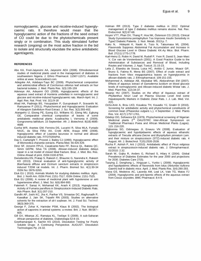
normoglycaemic, glucose and nicotine-induced hypergly-caemic rats. It therefore would mean that the hypoglycaemic action of the fractions of the seed extract of CO could be due to the phytotochemicals present singly or in combination. This study stimulated further research (ongoing) on the most active fraction in the bid to isolate and structurally elucidate the active antidiabetic agent/agents. REFERENCES Abo KA, Fred-Jaiyesimi AA, Jaiyesimi AEA (2008). Ethnobotannical
studies of medicinal plants used in the management of diabetes in southwestern Nigeria. J. Ethno Pharmacol. 11567-11571. Available online at www. Sciencedirect.com.
Adegoke AA, Adebayo-Tayo BC (2009). Phytochemical composition and antimicrobial effects of Corchorous olitorius leaf extracts on four
bacterial isolates. J. Med. Plants Res. 3(3):155-159
Adeneye AA, Adeyemi OO (2009). Hypoglycaemic effects of the aqueous seed extract of Hunteria umbellata in normoglycaemic and
glucose and nicotine-induced hyperglycaemic rats. March-April. Int. J.
Appl. Res. Nat. Prod. Vol. 2(1):9-18 Ahad HA, Padmaja BS, Yesupadam P, Guruprakash P, Sravanthi M,
Ramyasree P (2011). Phytochemical and Hypoglycaemic Evaluation of Alangium Salvifolium Root Extract. J. Sci. Res. 3(2):393-402.
Atangwho IJ, Ebong PE, Eyong EU, Williams IO, Eteng MU, Egbung GE. Comparative chemical composition of leaves of some
antidiabetic medicinal plants: Azadirachta I, Vernonia A (2009). Gongronema latifolium. 15 September. Afr. J. Biotechnol. 8(18):4685-
4689.
Cunha WR, Arantes GM, Ferreira DS, Lucarini R, Silva MLA, Furtado NAJC, da Silva Filho AA, Crotti AEM, Araujo ARB (2008). Hypoglycemic effect of Leandra lacunose in normal and alloxan
induced diabetic rats. FITOTERAPIA. Day C, Cartwright T, Provost J, Bailey CJ (1990). Hypoglycaemic effect
of Momordica charantia extracts. Planta Med. 56:426-429.
Diniz SF, Amorim FPLG, Cavalcante-Neto FF, Bocca QL, Batista QC, Simm GEPM, Silva TA (2008). Alloxan-induced diabetes delays repair in a rat model of closed tibial fracture. Braz. J. Med. Bio. Res.
Online Ahead of print. ISSN 0100-879X. Dwividendra KN, Pratap S, Rakesh C, Bhaumic G, Narendra K, Rakesh
KD (2013). Clinical evaluation of anti-hyperglycemic activity of Boerhaavia diffusa and Ocimum sanctum extracts in streptozocin
induced T2DM rat models. Int. J. Pharm. Biomed Sci. 4(1):30-34 ISSN No: 0976-5263.
Etuk EU ( 2010). Animals Models for studying diabetes mellitus. Agric. Biol. J. North Am. ISSN Print; 2151-7517, ISSN Online; 2151-7525.
Etuk EU (2006). A review of medicinal plant with hypotensive or anti
hypertensive effect. J. Med. Sci. 6(6):894-900. Fatemeh F, Sanaz H, Mohamad KK, Arash K (2013). Hypoglycemic
Activity of Fumaria parviflora in Streptozotocin-Induced Diabetic Rats.
Adv Pharm. Bull. 3(1):207-210. Gandhi AP, Joshi KC, Jha K, Parihar VS, Srivastav DC, Raghunadh P,
Kawalkar J, Jain SK, Tripathi RN (2003). Studies on alternative
solvents for the extraction of oil-I soybean. Int. J. Food Sci. Technol. 38(3):369-375.
George F, Zohar K, Harinder PSM, Klaus B (2002). The biological
action of saponins in animal systems: a review. Brit. J. Nutr. 88:587–605.
Gill GV, Mbanya JC, Ramaiya KL, Tesfaye S (2009). A sub-Saharan
African perspective of diabetes. Diabetologia 52:8-16. Gowthamarajan K, Sachin KS (2010). Dissolution Testing for Poorly
Soluble Drugs: A Continuing Perspective. AUGUST. Dissolution
Technologies Pp. 24-32.
Egua et al. 9 Holman RR (2013). Type 2 diabetes mellitus in 2012: Optimal
management of type 2 diabetes mellitus remains elusive. Nat. Rev. Endocrinol. 9(2):67-68.
Huyen VTT, Phan DV, Thang P, Hoa NK, Östenson CG (2013). Clinical Study Gynostemma pentaphyllum Tea Improves Insulin Sensitivity in Type2 Diabetic Patients. J. Nutr. Metab. 2013:p. 7.
Kaku N, Hideyuki K, Naoki A, Tozo N, Tatsumasa ML (2004). Flavonoids Suppress Abdominal Fat Accumulation and Increase in Blood Glucose Level in Obese Diabetic KK-Ay Mice. Biol. Pharm. Bull. 27(11):1775-1778.
Karl-Heinz D, Robin H, David M, Rudolf P, Yvon R, David S, Jean-Marc V, Cor van de Vorstenbosch (2001). A Good Practice Guide to the
Administration of Substances and Removal of Blood, Including Routes and Volumes. J. Appl. Toxicol. 21:15-23.
Leila Z, Eliandra de Sousa, Luisa HC, Anildo C Junior, Moacir GP,
Bruno S, Fatima R, Mena BS (2007). Effect of crude extract and fractions from Vitex megapotamica leaves on hyperglycemia in alloxan-diabetic rats. J. Ethnopharmacol. 109:151-155.
Mohammed A, Adelaiye AB, Abubakar MS, Abdurahman EM (2007). Effects of aqueous extract of Ganoderma lucidum on blood glucose
levels of normoglycemic and Alloxan-induced diabetic Wistar rats. J. Med. Plant Res. 1(2):34-37.
Nwanjo HU (2007). Studies on the effect of Aqueous extract of Phyllanthus Niruri Leaf on Plasma Glucose Level And some
Hepatospecific Markers in Diabetic Zistar Rats. I. J. Lab. Med. Vol. 2(2).
Ocho-Anin A, Brou LKD, Kouakou TH, Kouadio YJ, Gnakri D (2010).
Screening for antidiabetic activity and phytochemical constituents of common bean (Phaseolus vulgaris L.). 4 September. J. Med. Plants
Res. Vol. 4(17):1757-1761,
Odebiyi OO, Sofowora EA (1979). Phytochemical screening of Nigerian Medicinal plants 2
nd OAU/STRC Inter-African Symposium on
Traditional Pharmaco Poeia and African Medicinal Plants (Lagos).
115: 216-220. Ogbonnia SO, Odimegwu JI, Enwuru VN (2008). Evaluation of
hypoglycaemic and hypolipidaemic effects of aqueous ethanolic extracts of Treculia africana Decne and Bryophyllum pinnatum Lam.
and their mixture on streptozotocin (STZ)-induced diabetic rats. 4 August. Afr. J. Biotechnol. 7(15):2535-2539.
Rucha P, Ashish P, Arti J (2010). Antidiabetic effect of Ficus religiosa
extract in streptozotocin-induced diabetic rats. J. Ethnopharmacol. 01/2010: 2.32.
Sarah W, Gojka R, Anders G, Richard S, Hilary K (2004). Global Prevalence of Diabetes Estimates for the year 2000 and projections for 2030. Diabetes Care 27(5).
Taoying Z, Denghong L, Xingyuan L, Yunbo L (2009). Hypoglycemic and hypolipidemic effects of flavonoids from lotus (Nelumbo nuficera Gaertn) leaf in diabetic mice. April. J. Med. Plants Res. 3(4):290-293.
Viana GS, Medeiros AC, Lacerda AM, Leal LK, Vale TG, Matos FJ
(2004). Hypoglycemic and anti-lipemic effects of the aqueous extract from Cissus sicyoides. BMC Pharmacol. 8:4-9.

UPCOMING CONFERENCES
International Conference on Biological, Health and Environmental Sciences, London, UK, 19 Jan 2014
International Conference on Psychology, Psychiatry, Neurological, Behavioral and Cognitive Sciences, Barcelona, Spain, 27 Feb 2014

Conferences and Advert July 2014 17th World Congress of Basic and Clinical Pharmacology, Cape Town, South Africa, 13 Jul 2014
January 2014 International Conference on Medical, Biological and Pharmaceutical Sciences, London, UK, 19 Jan 2014

Journal of
Pharmacognosy and
Phytotherapy
Related Journals Published by Academic Journals
■ African Journal of Pharmacy and Pharmacology
■ Research in Pharmaceutical Biotechnology
■ Medical Practice and Reviews
■ Journal of Clinical Pathology and Forensic Medicine
■ Journal of Medicinal Plant Research
■ Journal of Drug Discovery and Development
■ Journal of Clinical Virology Research