Embed Size (px)
Citation preview
1
Title: Comparison of tetrazolium salts assays for evaluation of drug activity against 1
Leishmania spp. 2
Running title: Tetrazolium salts assay for leishmanicidal activities 3
Authors: Marine GINOUVES1, Bernard CARME1, 2, Pierre COUPPIE1, 3 and Ghislaine 4
PREVOT1# 5
Address: Université des Antilles et de la Guyane – Laboratoire d’ Epidémiologie des 6
parasitoses tropicales (EPAT) - UFR de médecine. Campus Saint-Denis 97306, 7
Cayenne, Guyane Française. 8
Affiliation: 1 Université des Antilles et de la Guyane, Laboratoire d’épidémiologie des 9
parasitoses tropicales EA 3593 – Labex CEBA UFR de médecine. Campus 10
Saint-Denis 97306, Cayenne, Guyane Française. 11
2 Laboratoire Hospitalo-universitaire de parasitologie et mycologie – centre 12
hospitalier de Cayenne, Guyane Française, Rue des Flamboyants, BP 6006, 97300 13
Cayenne, Guyane Française. 14
3 Institut Guyanais de Dermatologie Tropicale, Service de Dermatologie, Centre 15
Hospitalier de Cayenne, Rue des Flamboyants, BP 6006, 97300 Cayenne, Guyane 16
Française. 17
18
#Correspondent footnote: Ghislaine PREVOT [email protected] 19
20
JCM Accepts, published online ahead of print on 9 April 2014J. Clin. Microbiol. doi:10.1128/JCM.00201-14Copyright © 2014, American Society for Microbiology. All Rights Reserved.
on Decem
ber 30, 2019 by guesthttp://jcm
.asm.org/
Dow
nloaded from

2
ABSTRACT 21
In French Guiana, leishmaniasis is an essentially cutaneous infection. It constitutes a 22
major public health problem, with a real incidence of 0.2 to 0.3%. Leishmania guyanensis is the 23
causal species most frequently encountered in French Guiana. 24
The treatment of leishmaniasis is essentially drug-based, but the therapeutic compounds 25
available have major side effects (e.g. liver damage, diabetes) and must be administered 26
parenterally or are costly. The efficacy of some of these agents has declined due to the 27
emergence of resistance in certain strains of Leishmania. There is currently no vaccine against 28
leishmaniasis, and it is therefore both necessary and urgent to identify new compounds 29
effective against Leishmania. 30
The search for new drugs requires effective tests for evaluations of the leishmanicidal 31
activity of a particular molecule or extract. Microculture Tetrazolium Assays (MTAs) are 32
colorimetric tests based on the use of tetrazolium salts. We compared the efficacies of three 33
tetrazolium salts — MTT, XTT and WST-8 — for quantification of the promastigotes of 34
various species of Leishmania. We found that the capacity of Leishmania to metabolize a 35
tetrazolium salt depended on the salt used and the species of Leishmania. WST-8 was the 36
tetrazolium salt best metabolized by L. guyanensis and gave the best sensitivity. 37
38
INTRODUCTION 39
Leishmaniasis is a disease caused by a protozoan of the genus Leishmania, belonging to 40
the family Trypanosomatidae. The disease may take several forms — cutaneous, visceral or 41
mucocutaneous — depending on the species of Leishmania involved and the immune status of 42
the host. Leishmaniasis is a zoonosis affecting many mammals, including humans, and it is 43
transmitted by insect vectors: sandflies (Psychodidae: Phlebotominae). Leishmaniasis is a 44
public health problem in tropical countries, with more than two million new cases each year 45
(1). It is treated essentially with parasiticidal drugs. Several therapeutic compounds are 46
on Decem
ber 30, 2019 by guesthttp://jcm
.asm.org/
Dow
nloaded from

3
available (e.g. antimony derivatives and pentamidine), but they may have major adverse effects 47
(such as liver damage and diabetes), and may require parenteral administration or are too costly 48
to be readily available in certain countries. The oldest of the compounds used are antimony 49
derivatives, which were released onto the market in the 1930s but have since decreased in 50
efficacy, due to the emergence of resistance in certain strains of Leishmania infantum and 51
Leishmania donovani (2, 3). The other most widely used compounds are pentamidine and 52
amphotericin B. In the first decade of this century, a new compound, miltefosine, was released 53
onto the market. It can be administered orally, a key advantage, but its use is not authorized in 54
all countries. 55
There is currently no vaccine against leishmaniasis. It is therefore both necessary and 56
urgent to identify new compounds active against Leishmania. The search for such compounds 57
requires an in vitro test for the reliable evaluation of their effects on the viability and 58
proliferation of Leishmania. 59
Cell counts can be carried out by simple and readily accessible techniques, such as direct 60
counting under the microscope, or by much more sophisticated and costly techniques, such as 61
flow cytometry. The reliability of a test depends on both the method used and the cell type 62
used, or even the stages or species considered, which must be defined in advance. Leishmania 63
can be cultured in their mobile promastigote form, or as amastigotes, either resident within 64
macrophages or axenic. Infected macrophages more closely resemble the situation in humans, 65
but the use of promastigotes is more widespread in tests of the activity of molecules or complex 66
extracts, due to the ease with which these cells can be cultured in vitro. 67
Direct counting under the microscope, which is still used in some laboratories, is a 68
particularly straightforward technique. However, the mobility of the promastigotes and the 69
presence of rosettes may complicate the task and, although the cells can be immobilized with 70
glutaraldehyde, this method is not suitable for the simultaneous counting of cells in multiple 71
on Decem
ber 30, 2019 by guesthttp://jcm
.asm.org/
Dow
nloaded from
4
samples, the cells counted include dead cells and the bias resulting from the presence of 72
rosettes remains. This fastidious and time-consuming technique is not very suitable for precise 73
evaluations of leishmanicidal activity. Methods based on the incorporation into DNA of 74
radiolabeled markers, such as tritiated uracil ([5-3H] uracil ; (4)) or tritiated thymidine ([3H] 75
thymidine ; (5)) are rapid and simple and facilitate large-scale analyses of samples. The 76
assimilation of this radioactive isotope into the DNA during its synthesis makes it possible to 77
follow cell proliferation, as there is a linear correlation between the radiation emitted and cell 78
multiplication. The major disadvantages of this technique are the need to manipulate 79
radioactive compounds, for which accreditation is required, and the production of radioactive 80
waste, the elimination of which remains problematic. In addition, the incorporation of these 81
radioactive isotopes into cells during division may lead to DNA damage, cell cycle arrest and 82
cell apoptosis, with erroneous results. The replacement of radioactive isotopes with stable 83
isotopes is possible, but requires a mass spectrophotometer (6), an item of equipment not 84
available in all laboratories. Flow cytometry (7, 8) can provide a quantification of the number 85
of cells that is so precise that it is possible to determine the absolute number of cells per unit 86
volume of culture, for cell densities from 100 cells/ml to 1 x 10⁶ cells/ml, in a highly 87
reproducible manner (9). However, the excessively high cost of this technique in terms of 88
materials and reagents constitutes a major barrier to the widespread adoption of this technique. 89
Other similar but less cumbersome devices that are practical and simple to use, such as 90
automatic portable or bench cell counters, are accessible, but consumables costs remain 91
excessive. 92
As attested by many publications, the technique of choice remains colorimetric 93
determination, which is cheap, effective, simple and rapid. A wide selection of reagents is 94
available, including resazurin-3H-phenoxazin-3-one 10-oxide (Alamar blue®), a blue molecule 95
that is reduced by the dehydrogenase activity of mitochondria to give rise to a pink molecule, 96
resorufin (7-hydroxy-3H-phenoxazin-3-one), which can be quantified by spectrophotometry. 97
on Decem
ber 30, 2019 by guesthttp://jcm
.asm.org/
Dow
nloaded from
5
This technique provides results similar to those obtained by tritiated thymidine incorporation 98
(10). However, it has been reported (11) that the reduction of resazurin by the mitochondrial 99
enzymes of Leishmania is much slower than that observed in other cell types (Trypanosoma, 100
mammalian cells). Indeed, Leishmania must be cultured at a temperature of about 27°C, 101
whereas Trypanosoma and mammalian cells can be cultured at 37°C, which is the optimal 102
temperature for resazurin reduction. The reaction rate is thus much lower in Leishmania, 103
resulting in much lower absorbance values for similar incubation times (11). 104
Microculture tetrazolium assays (MTAs) are colorimetric tests based on the use of 105
tetrazolium salts, including 3-(4,5-dimethylthiazol-2-yl)-2,5-diphenyltetrazolium bromide 106
(MTT), 3-(4,5-dimethylthiazol-2-yl)-5-(3-carboxymethoxyphenyl)-2(4-sulfonyl)-2H-tetrazo-107
lium (MTS), 2,3-bis-(2-methoxy-4-nitro-5-sulfophenyl)-2H-tetrazolium-5-carboxanilide 108
(XTT), (2-(4-iodophenyl)-3-(4-nitrophenyl)-5-(2,4-disulfophenyl)-2H-tetrazolium (WST-1), 2-109
(4-iodophenyl)-3-(2,4-dinitrophenyl)-5-(2,4-disulfophenyl)-2H-tetrazolium (WST-3) and 2-(2-110
methoxy-4-nitrophenyl)-3-(4-nitrophenyl)-5-(2,4-disulfophenyl)-2H-tetrazolium (WST-8), ... 111
These methods are based on the cleavage of a tetrazolium salt by a mitochondrial enzyme, 112
succinate dehydrogenase, leading to the formation of a colored product, formazan (12), which 113
can be quantified by spectrophotometry. In practice, most of these tetrazolium salts have been 114
found to be of variable efficacy for the quantification of Leishmania (13-17). 115
In this study, we compared the efficacy of three tests based on tetrazolium salts – MTT, 116
XTT and WST-8 – for quantifying promastigotes of different strains of Leishmania. Then, the 117
most suitable salt was used to evaluate the reliability of the test : in the presence of an ethanolic 118
plant extract, Lantana camara and a drug used as first-line treatment of cutaneous 119
leishmaniasis in French Guiana, pentamidine. 120
121
on Decem
ber 30, 2019 by guesthttp://jcm
.asm.org/
Dow
nloaded from
6
MATERIALS AND METHODS 122
Parasites and cultures 123
Tests were carried out on promastigotes from several strains of Leishmania: eight New-124
World strains of Leishmania, including two reference strains (Leishmania (Viannia) guyanensis 125
MHOM/GF/97/LBC6 (LG-R) and Leishmania (Leishmania) amazonensis 126
MPRO/BR/72/M1845 (LA-R)) and six strains isolated from patients in French Guiana (L. (V.) 127
guyanensis 12.01.13-2008 (LG-1), L. (V) guyanensis 04.04.13-2068 (LG-2), L. (V) guyanensis 128
01.03.13-2014 (LG-3), L. (V.) braziliensis 09.10.12-2031 (LB-1), L. (V.) braziliensis 24.05.12-129
2068 (LB-2), L. (L.) amazonensis 10.10.12-2048 (LA-1), together with one Old-World strain 130
(Leishmania (L.) donovani MHOM/IN/96/THAK72 (LD-R)). The reference strains were 131
obtained from the national reference center for leishmaniasis in Montpellier, France, and the 132
other strains were kindly supplied by the parasitology and mycology laboratory of Cayenne 133
University Hospital, French Guiana, with strict respect for patient anonymity. 134
The reference strains, which were stored in liquid nitrogen, were thawed and then 135
cultured in 3 ml of RPMI 1640 (GIBCO®) containing L-glutamine, 20 mM HEPES and Phenol 136
Red and supplemented with 20% inactivated calf fetal serum (CFS; GIBCO®), 50 IU/ml 137
penicillin (Invitrogen®), 0.05 mg/ml streptomycin (Invitrogen®) and nonessential amino acids 138
(GIBCO®) at 26°C. 139
The medium was changed when the cells reached stationary phase: the cultures were 140
centrifuged for 5 minutes at 514 x g and the parasites were resuspended in 10 ml of RPMI 1640 141
medium (GIBCO®) containing glutamine but devoid of Phenol Red, to limit background noise 142
(18), and supplemented with 10% inactivated CFS (GIBCO®), 50 IU/ml penicillin 143
(Invitrogen®), 0.05 mg/ml streptomycin (Invitrogen®) and nonessential amino acids (GIBCO®). 144
This medium is referred to as RPMIØRP medium. The cells were cultured at 26°C until they 145
reached the exponential growth phase. 146
on Decem
ber 30, 2019 by guesthttp://jcm
.asm.org/
Dow
nloaded from
7
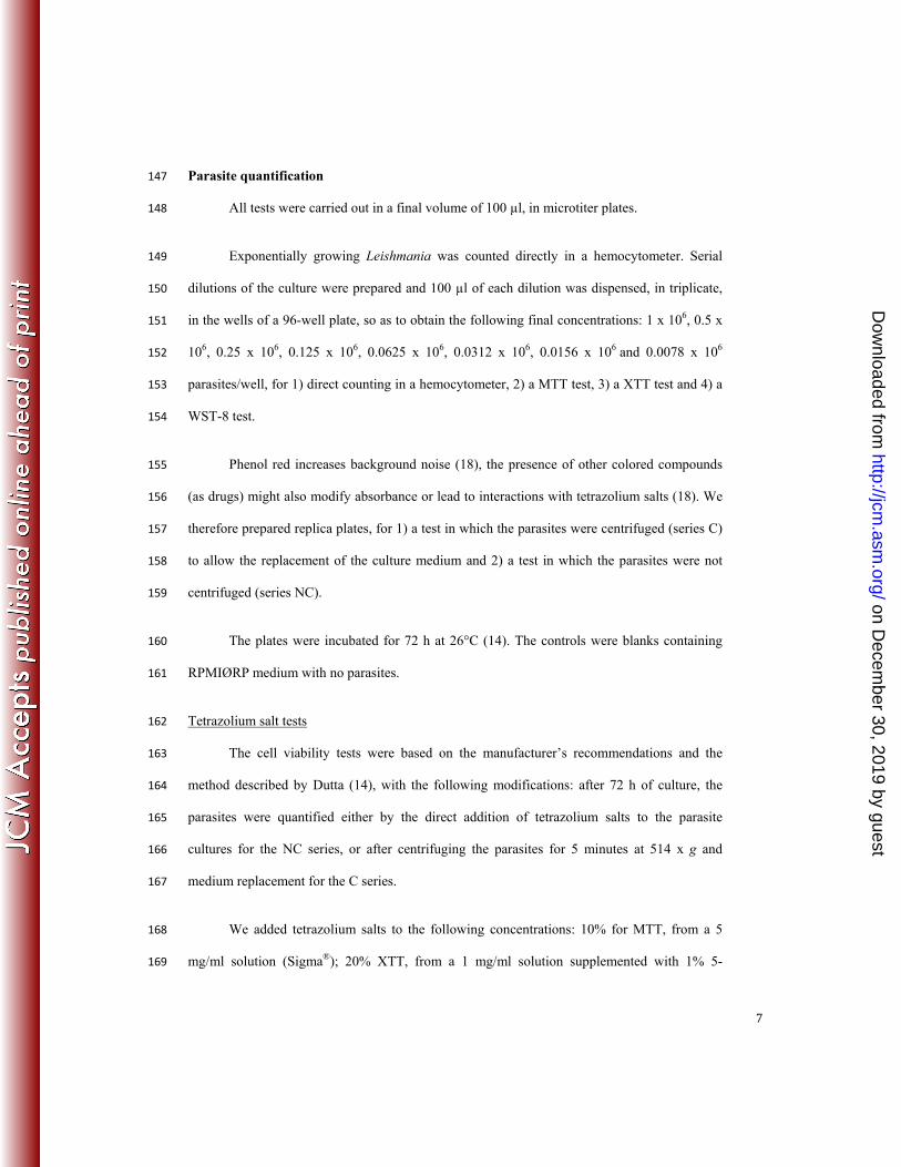
Parasite quantification 147
All tests were carried out in a final volume of 100 µl, in microtiter plates. 148
Exponentially growing Leishmania was counted directly in a hemocytometer. Serial 149
dilutions of the culture were prepared and 100 µl of each dilution was dispensed, in triplicate, 150
in the wells of a 96-well plate, so as to obtain the following final concentrations: 1 x 10⁶, 0.5 x 151
106, 0.25 x 10⁶, 0.125 x 10⁶, 0.0625 x 10⁶, 0.0312 x 10⁶, 0.0156 x 10⁶ and 0.0078 x 10⁶ 152
parasites/well, for 1) direct counting in a hemocytometer, 2) a MTT test, 3) a XTT test and 4) a 153
WST-8 test. 154
Phenol red increases background noise (18), the presence of other colored compounds 155
(as drugs) might also modify absorbance or lead to interactions with tetrazolium salts (18). We 156
therefore prepared replica plates, for 1) a test in which the parasites were centrifuged (series C) 157
to allow the replacement of the culture medium and 2) a test in which the parasites were not 158
centrifuged (series NC). 159
The plates were incubated for 72 h at 26°C (14). The controls were blanks containing 160
RPMIØRP medium with no parasites. 161
Tetrazolium salt tests 162
The cell viability tests were based on the manufacturer’s recommendations and the 163
method described by Dutta (14), with the following modifications: after 72 h of culture, the 164
parasites were quantified either by the direct addition of tetrazolium salts to the parasite 165
cultures for the NC series, or after centrifuging the parasites for 5 minutes at 514 x g and 166
medium replacement for the C series. 167
We added tetrazolium salts to the following concentrations: 10% for MTT, from a 5 168
mg/ml solution (Sigma®); 20% XTT, from a 1 mg/ml solution supplemented with 1% 5-169
on Decem
ber 30, 2019 by guesthttp://jcm
.asm.org/
Dow
nloaded from
8
methylphenazinium methyl sulfate (PMS) (Sigma®); or 10% WST-8, supplemented with 1-170
methoxy-PMS (Cell Counting Kit-8 – Sigma®). 171
The plates were incubated for 3 h at 26°C. We then added 100 µl of dimethyl sulfoxide 172
(DMSO) to the wells containing MTT and the plates were returned to the 26°C incubator for 1 173
hour. 174
We determined absorbance at 570 nm for the MTT tests, and at 450 nm for the XTT and 175
WST-8 tests, with a Tristar LB941 spectrophotometer (Berthold Technologies®). We 176
eliminated the background noise by subtracting the value of the blank (no parasites and no 177
tetrazolium salts) from the values obtained in the tests. 178
Counting in a hemocytometer 179
The cells were counted directly, with or without prior centrifugation, in a 180
hemocytometer. Counts were carried out in parallel for the three tetrazolium salt tests. 181
Sensitivity tests 182
The sensitivity tests were carried out on L. guyanensis isolate LG-2, obtained from a 183
patient. When the parasites reached the exponential growth phase, we added 90 µl of culture, 184
containing 106 parasites, to each well. We dispensed 10 µl of various dilutions of pentamidine 185
(Sigma®) or of an extract in ethanol (70:30) of Lantana camara leaves into the test wells, so as 186
to obtain final concentrations of 0.125, 0.0625, 0.0312, 0.015, 0.0078 and 0.0039 µg/ml 187
pentamidine, or 2000, 1000, 500, 250, 125 and 62.5 µg/ml Lantana camara leaf extract. We 188
added 10 µl of RPMIØRP to the control wells. The tests were carried out in triplicate. 189
The blanks for the tests consisted of 90 µl of RPMIØRP medium and 10 µl of the 190
various concentrations of pentamidine or plant extract. The blanks for the controls consisted of 191
100 µl of RPMIØRP medium. 192
on Decem
ber 30, 2019 by guesthttp://jcm
.asm.org/
Dow
nloaded from
9
After incubation for 48 h at 26°C, we added 10% WST-8 supplemented with 1-193
methoxy-PMS to each well, including the blanks. The plate was incubated for a further 24 h. 194
The results were read, by spectrophotometry at 450 nm, after 72 h in total of incubation in the 195
presence of the drug or plant extract. 196
The absorbance values from the blanks were subtracted to the corresponding test 197
absorbance, and the percentage inhibition was calculated as follows: 198
199
x 100 200
The IC50 was then calculated with GraphPad Prism6 software. 201
Statistical analysis 202
We calculated the standard deviation (SD) for each point, with Excel (ECARTYPEP). 203
The r coefficient of correlation was determined with Excel to assess correlation between 204
parasite number and OD. 205
206
RESULTS 207
The various colorimetric tests based on MTT, XTT and WST-8 were compared on the 208
reference strain L. guyanensis LG-R. 209
Centrifugation impact on quantification tests 210
Direct counts of parasites were obtained in parallel, with a hemocytometer, to validate the 211
colorimetric results. Parasite quantification tests were carried out at different cell densities, with 212
and without centrifugation, to evaluate the potential impact of centrifugation on the results 213
obtained (Fig. 1). 214
on Decem
ber 30, 2019 by guesthttp://jcm
.asm.org/
Dow
nloaded from
10
215
Comparison of the 3 tetrazolium salts tests 216
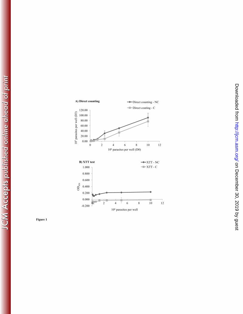
Direct counting of the parasites in a hemocytometer after 72 h of culture generated a linear 217
growth curve (Fig. 1A), with a proportional relationship between the number of parasites used 218
to inoculate the medium on D0 and the number of parasites counted on D3. However, this 219
linear relationship was observed at parasite densities exceeding 1.25 x 10⁵ parasites/well. We 220
consider this density to be the threshold of detection for direct counting. Centrifugation did not 221
seem to affect the results obtained. Indeed, the number of parasites was smaller in series C, but 222
remained proportional to that in series NC. Centrifugation led to a reproducible loss of cells, 223
with a coefficient of about 2.5, in all wells. 224
An increase in the population by a factor of 10 after 72 h of culture was noted. We can 225
therefore consider the number of parasites to be 10 times higher on D3 than on D0, and these 226
values can be used to interpret the results of colorimetric tests. 227
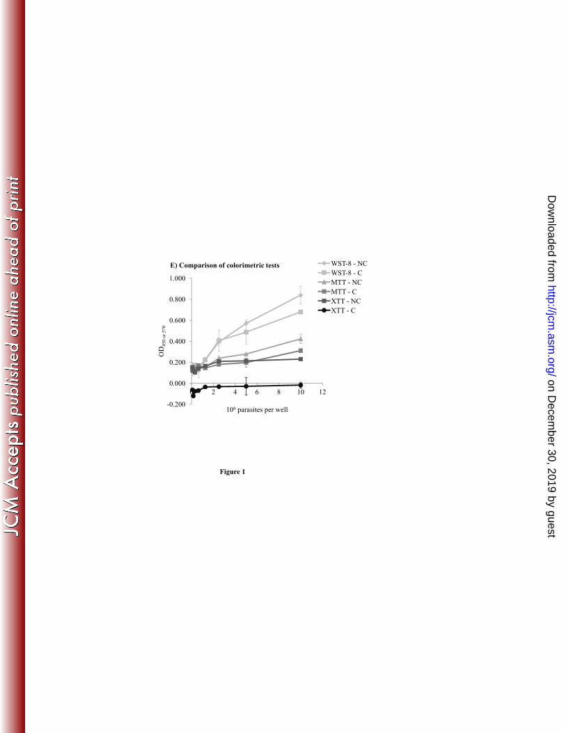
Parasite quantification by colorimetric tests based on XTT (Fig. 1B) gave very low, or 228
even negative absorbance values, ranging from -0.121 to -0.015 for series C and from 0.105 to 229
0.230 for series NC. The values obtained in this test were too low to demonstrate any real 230
differences in the numbers of cells present. 231
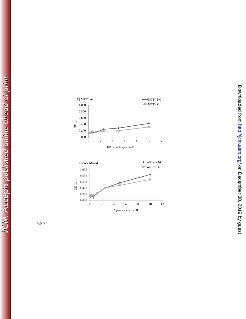
By contrast, parasite quantification with the MTT test (Fig. 1C) gave higher absorbance 232
values, ranging from 0.118 to 0.423 for series NC and from 0.129 to 0.311 for series C. The 233
curves for the C and NC series were almost parallel and the numbers of parasites before and 234
after centrifugation may therefore be considered proportional. 235
The threshold of detection for parasites was 2.5 x 10⁶ parasites/well, the curve 236
increasing after this point. The standard deviations for MTT tests were relatively high (data not 237
shown). 238
on Decem
ber 30, 2019 by guesthttp://jcm
.asm.org/
Dow
nloaded from

11
Quantification in the WST-8 test (Fig. 1D) gave absorbance values well above those 239
obtained in the other two colorimetric tests (MTT and XTT), with values ranging from 0.177 to 240
0.839 for series NC and from 0.143 to 0.679 for series C. The threshold for parasite detection 241
was about 0.625 x 10⁷ parasites/well. 242
For series NC, each of the coefficients of correlation (r) was calculated. Thus, direct 243
counting yielded an r of 0.99, the MTT test had an r of 0.98, the WST-8 test had an r of 0.98 244
and the XTT test had an r of 0.82. 245
Metabolisation of tetrazolium salts by L.guyanensis and L. amazonensis 246
We compared the metabolism of the various tetrazolium salts and the efficacy of these tests, by 247
omitting the 72 h incubation after cell inoculation and the centrifugation step. The tetrazolium 248
salts were added to the cultures directly after inoculation. 249
We evaluated the metabolism of the three tetrazolium salts after four hours of 250
incubation, with the reference strains L. guyanensis LG-R and L. amazonensis LA-R, at three 251
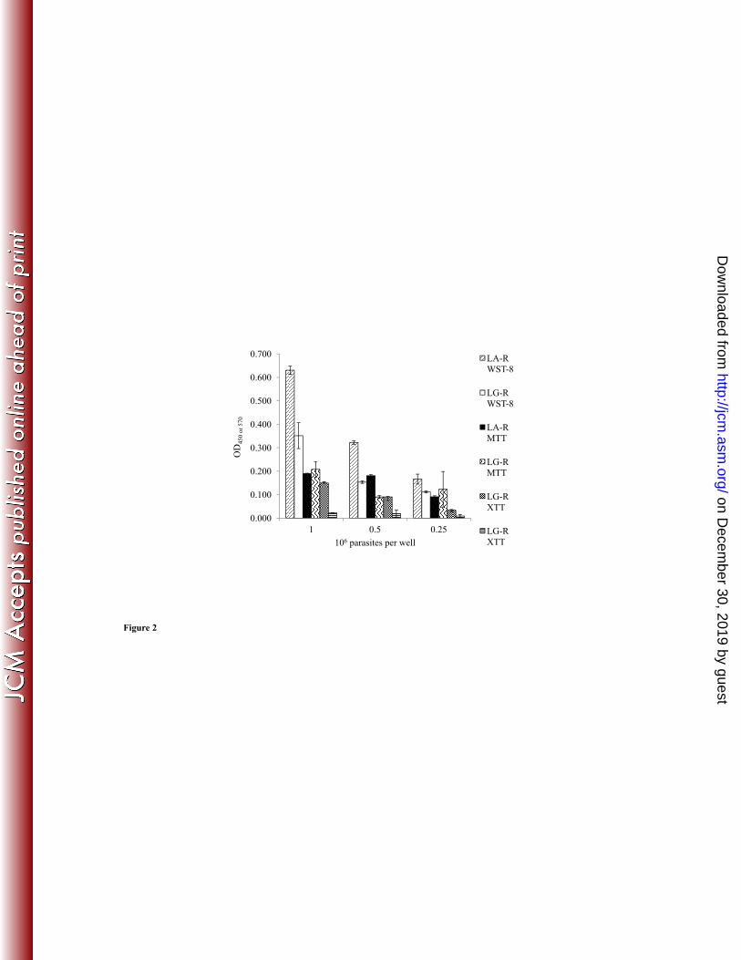
concentrations: 1 x 10⁶, 5 x 10⁵ and 2.5 x 10⁵ parasites/well. The results are presented as a 252
histogram, in Figure 2. The absorbance values obtained in the MTT tests were very similar for 253
the three densities of L. amazonensis LA-R (r = 0.80) tested, whereas those for L. guyanensis 254
LG-R (r = 0.82) decreased with decreasing cell density. 255
For the XTT tests, the absorbance results obtained were consistent with the number of 256
parasites for L. amazonensis LA-R (r = 0.98), whereas, for L. guyanensis LG-R (r = 0.89), the 257
absorbance values obtained were very low, or zero. 258
The absorbance values obtained in WST-8 tests were higher than those for the other 259
tests, particularly for L. amazonensis LA-R, which had an absorbance value of 0.630, versus 260
0.351 for the L. guyanensis LG-R strain, for a density of 1 x 10⁶ parasites/well. The absorbance 261
on Decem
ber 30, 2019 by guesthttp://jcm
.asm.org/
Dow
nloaded from
12
values decreased proportionally with the number of cells for the L. amazonensis LA-R strain (r 262
= 1) and L. guyanensis LG-R (r = 0.98). 263
The WST-8 test was the colorimetric test with the best coefficient of correlation (r), the 264
best sensitivity (Fig. 1D) and the best tetrazolium salt metabolism (Fig. 2). 265
Metabolisation of WST-8 by Leishmania spp. 266
We assessed the efficacy of the WST-8 test for quantifying the various strains of Leishmania, 267
using strains L. guyanensis LG-R, L. guyanensis LG-1, L. amazonensis LA-R, L. amazonensis 268
LA-1, L. braziliensis LB-1, L. braziliensis LB-2, and L. donovani LD-R, at densities of 1 x 10⁶, 269
5 x 10⁵ and 2.5 x 10⁵parasites/well (Fig. 3). Absorbance depended on the strain tested, with 270
values ranging from 0.606 for L. braziliensis LB-2 to 1.05 for L. amazonensis LA-R, for 1 x 271
106 parasites/well. Some strains from different species gave similar absorbance values. For 272
example, L. amazonensis LA-R (A=0.738) and L. braziliensis LB-1 (A=0.723) for 5 x 273
105 parasites/well, and L. guyanensis LG-R (A=0.993) and L. donovani LD-R (A=0.992) for 1 274
x 106 parasites/well. 275
Optimal incubation time for the WST-8 quantification test 276
We determined the optimal incubation time for the WST-8 quantification test, with the patient 277
isolate L. guyanensis LG-3, in the presence of various concentrations of amphotericin B (Fig. 278
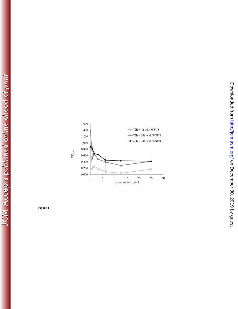
4). Incubation for four hours with WST-8 was not sufficient to obtain consistent results (Fig. 4) 279
and yielded very low absorbance values due to cells death. Incubation with WST-8 for longer 280
periods was therefore required, with 24 h identified as the most functional incubation time. 281
However, incubation for 24 h in the presence of WST-8, after 72 h of cell culture in the 282
presence of amphotericin B, gave a similar curve to incubation for four hours in the presence of 283
WST-8, and resulted in a total incubation time of 96 h rather than 72 h. For the performance of 284
the test over a period of 72 h, we recommend adding the WST-8 after 48 h of incubation with 285
the drug or plant extract, and then reading absorbance 24 h later. 286
on Decem
ber 30, 2019 by guesthttp://jcm
.asm.org/
Dow
nloaded from
13
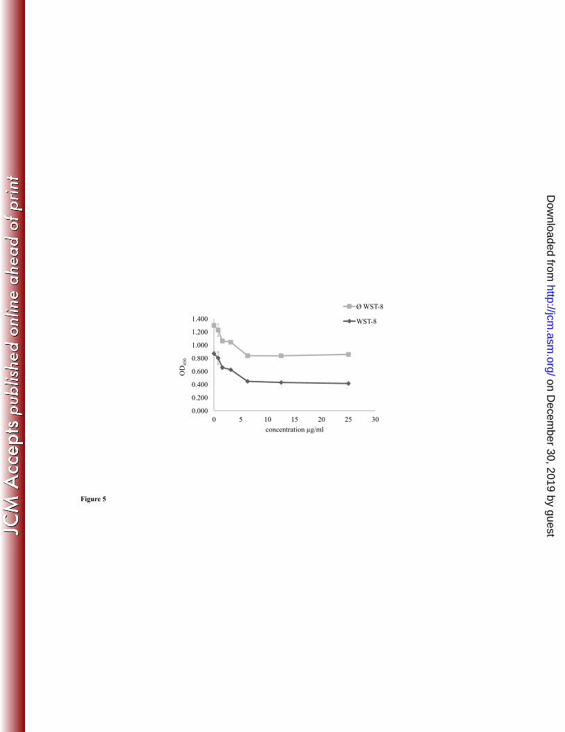
Interaction between amphotericin B and WST-8 287
We checked that the compounds tested did not interact with WST-8. Figure 5 shows the results 288
obtained in the presence of amphotericin B. Similar curves were obtained for the absorbance 289
measured in the presence or absence of WST-8. 290
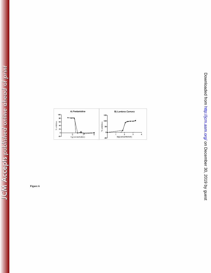
Sensitivity test of plant extracts and pentamidine 291
The sensitivity of the patient isolate L. guyanensis LG-2 to various antimicrobial compounds 292
was assessed as a function of incubation time determined above. The results obtained in the 293
presence of pentamidine or an extract of Lantana camara leaves in ethanol are shown in 294
Figures 6A and 6B, respectively. The IC50 values obtained were 0.018 µg/ml for pentamidine 295
and 86.81 µg/ml for Lantana camara leaf extract. Ethanol didn’t affect parasite growth (data 296
not shown). 297
298
DISCUSSION 299
Parasite quantification is a crucial step in proliferation and cytotoxicity tests. Simple, 300
rapid and effective tests are required, allowing the analysis of multiple samples. 301
Many cell quantification techniques are available, including colorimetric tests, in 302
particular. In this study, we compared three tetrazolium salt tests: those based on MTT, XTT 303
and WST-8. We chose to include the MTT test on the basis of its reputation, this test being the 304
most widely used in cell quantification studies (12, 19, 20). XTT is also widely used, as it 305
palliates some of the deficiencies reported for the MTT test (21-23), and WST-8 is one of the 306
most recently synthesized tetrazolium salts (24). 307
We found that the sensitivity of the MTT test was quite low for Leishmania guyanensis, 308
indicating that this species probably metabolizes this compound poorly, if at all. This test also 309
on Decem
ber 30, 2019 by guesthttp://jcm
.asm.org/
Dow
nloaded from

14
displayed a lack of reproducibility, as already reported by Wan et al. (25), due to the 310
insolubility of formazan, the product formed. The MTT test therefore requires a solubilization 311
step, to dissolve the product in an organic solvent, such as DMSO (26). Unfortunately, this 312
dissolution is not always complete and may generate biases, also, the presence of bubbles 313
generated by this additional step interfere with absorbance readings. In this study, we obtained 314
standard deviations of more than 0.2 in some tests (data not shown). In addition, the quality of 315
the correlation with the results obtained by tritiated thymidine counting remains variable (13, 316
27). 317
XTT (21) and MTS (28) tests were developed to overcome these problems of solubility. 318
Indeed, these salts are reduced to soluble derivatives of formazan, and their reduction is 319
accelerated by the addition of an electron-coupling agent, Phenazine MethoSulfate (PMS; 320
(29)), thereby increasing test sensitivity. However, although the efficacy of these tests has been 321
demonstrated in several studies (17, 27) for evaluations of the sensitivity of Leishmania to 322
drugs or plant extracts (30), we found that XTT tests displayed a lack of sensitivity with L. 323
guyanensis strains. Furthermore, the presence of PMS may lead to the formation of crystals in 324
the culture medium, modifying the absorbance of the product formed and resulting in erroneous 325
findings (21). Menadione, another electron-coupling agent, also promotes the rapid reduction of 326
XTT (31) in many cell lines, and its use also decreases background noise. However, crystals are 327
also observed in the presence of this electron-coupling agent, albeit in smaller numbers than for 328
PMS (21). 329
The WST-8 test gave highly satisfactory results in our study, with an acceptable 330
sensitivity and a proportional relationship between the number of parasites and absorbance. 331
WST-8 is, in fact, an improved version of WST-3, which is itself an improved version of WST-332
1: it combines the high stability of WST-1 with the high sensitivity of WST-3 (24). WST-3 is 333
less stable than WST-1, due to the presence of two nitro (-NO2) groups on the same benzene 334
on Decem
ber 30, 2019 by guesthttp://jcm
.asm.org/
Dow
nloaded from

15
ring. However, it is a much more sensitive agent than WST-1 for tests of viability. WST-8 was 335
synthesized to improve the stability of this molecule, whilst conserving its sensitivity. It was 336
produced by transferring one of the nitro groups of WST-3 onto another benzene ring (24). The 337
electron-coupling agent used with WST-8, 1-methoxy-PMS, is also an improved form of PMS 338
that is more effective and insensitive to the photochemical deterioration observed with PMS 339
(32). 340
The results presented in this study are consistent with those obtained by Ishiyama et al. 341
and Tominaga et al., whose work on human and rabbit cells showed the absorbance values 342
obtained to be proportional to the number of cells, highlighting the greater sensitivity of the 343
WST-8 test over those based on the other tetrazolium salts tested, WST-1, XTT and MTS (24, 344
33). 345
Many studies have highlighted the efficacy of MTA tests, but this efficacy varies as a 346
function of 1) the type of tetrazolium salt used (33); in our study XTT was less well 347
metabolized than WST-8; 2) the species of Leishmania tested (15); L. amazonensis was, in this 348
study, the species that best metabolized most of the tetrazolium salts tested, whereas L. 349
guyanensis metabolized these salts poorly; 3) the strains studied, the mitochondria of which 350
may have different enzymatic activities (15, 21). Indeed, our observations show that, for a 351
density of 1 x 10⁶ parasites/well, the absorbance value obtained with L. braziliensis LB-2 was 352
0.730, whereas that obtained with L. braziliensis LB-1 was 1.096. 353
The sensitivity tests with patient isolate L. guyanensis LG-2 incubated with pentamidine 354
or extract of Lantana camara leaves, carried out with WST-8, provided satisfactory results, 355
with IC50 values of 0.018 µg/ml for pentamidine and 86.81 µg/ml for Lantana camara extract. 356
The results obtained were consistent with microscopy observations carried out after 72 h of 357
culture in the presence of pentamidine. 358
on Decem
ber 30, 2019 by guesthttp://jcm
.asm.org/
Dow
nloaded from
16
Certain substances present in drugs or plant extracts may interfere with tetrazolium salts 359
through chemical effects on cellular respiration processes (34). In such cases, centrifugation 360
can be used to decrease this phenomenon (cell washing step). We therefore evaluated the 361
impact of a centrifugation step on cell quantification. We found that the centrifugation step did 362
not cause a bias (constant cell loss). However, in the presence of a drug or plant extract, the 363
results obtained after centrifugation (data not shown), were not consistent for XTT and MTT. 364
These results may reflect interactions between these tetrazolium salts and the leishmanicidal 365
compounds tested. By contrast, with WST-8, the results obtained were good enough to 366
eliminate the parasite centrifugation step. Indeed, WST-8 did not seem to interact with the 367
drugs or plant extracts, because the results obtained were proportional in both the presence and 368
absence of WST-8, with and without centrifugation. A control would nevertheless be required 369
to check for the absence of interference with all new products tested. 370
In conclusion, this study confirms that the efficacy of tetrazolium salt reduction depends 371
on the strain of Leishmania tested and the tetrazolium salt used. L. guyanensis accounts for 372
90% of the strains isolated from patients with cutaneous leishmaniasis in French Guiana. A test 373
suitable for evaluating the level of susceptibility to drugs of the strains of Leishmania 374
responsible for these cases of leishmaniasis is therefore required. We show here that the most 375
widely used tests, the XTT and MTT tests, are not suitable for studies of the susceptibility of L. 376
guyanensis. WST-8 is the tetrazolium salt best metabolized by L. guyanensis, resulting in better 377
sensitivity. It also displays satisfactory levels of efficacy in tests of susceptibility to drugs or 378
plant extracts. Thus, this process is ideal for quantification of cells, either to assess cell 379
sensitivity to drugs, or to screen new antimicrobial compounds. For this, we suggest performing 380
the tests with WST-8 according to the following protocol : drug or compound is added to cells 381
from exponential phase. The addition of WST-8 is advised after 48 h of incubation and the 382
absorbance is read in a spectrophotometer at 450 nm after 24 h. However, if the cells 383
on Decem
ber 30, 2019 by guesthttp://jcm
.asm.org/
Dow
nloaded from

17
metabolize the tetrazolium salt well, addition of WST-8 may be performed after 72 h of 384
incubation and reading, after 4 h of incubation (Fig. 7). 385
This WST-8 assay is now applied in our lab for preliminary screening of field isolates for 386
resistance to drugs usually used to treat leishmaniasis and for screening of new antileishmanial 387
agents. Thus, the WST-8 assay is a valuable tool to assess viability and proliferation of 388
Leishmania promastigotes or for a variety of other cell types. 389
390
ACKNOWLEDGEMENTS 391
This work was supported by the University of the French West Indies and French Guiana and 392
the Ministère Français de l'Enseignement Supérieur et de la Recherche Scientifique. 393
This work has benefited from an "Investissement d’Avenir" grant managed by Agence 394
Nationale de la Recherche (CEBA, ref. ANR-10-LABX-25-01). 395
This work was supported by the Conseil Régional de la Guyane and the European Union 396
(FEDER- Presage N° 31454). 397
REFERENCES 398
1. WHO. 2010. Control of the leishmaniases. WHO Technical Report Series. 949:. 399
2. Faraut-Gambarelli, F., R. Piarroux, M. Deniau, B. Giusiano, P. Marty, G. Michel, B. 400
Faugère, and H. Dumon. 1997. In vitro and in vivo resistance of Leishmania infantum to 401
meglumine antimoniate: a study of 37 strains collected from patients with visceral 402
leishmaniasis. Antimicrobial Agents and Chemotherapy. 41:827-830. 403
3. Lira, R., S. Sundar, A. Makharia, R. Kenney, A. Gam, E. Saraiva, and D. Sacks. 1999. 404
Evidence that the high incidence of treatment failures in Indian kala-azar is due to the 405
on Decem
ber 30, 2019 by guesthttp://jcm
.asm.org/
Dow
nloaded from

18
emergence of antimony-resistant strains of Leishmania donovani. J. Infect. Dis. 180:564-567. 406
doi: 10.1086/314896. 407
4. Berman, J. D., and J. V. Gallalee. 1985. Semiautomated assessment of in vitro activity of 408
potential antileishmanial drugs. Antimicrob. Agents Chemother. 28:723-726. 409
5. Sharief, A. H., E. A. Gasim Khalil, T. G. Theander, A. Kharazmi, S. A. Omer, and M. 410
E. Ibrahim. 2006. Leishmania donovani: An in vitro study of antimony-resistant amphotericin 411
B-sensitive isolates. Exp. Parasitol. 114:247-252. doi: 10.1016/j.exppara.2006.03.016. 412
6. Hu, V. W., G. E. Black, A. Torres-Duarte, and F. P. Abramson. 2002. 3H-thymidine is a 413
defective tool with which to measure rates of DNA synthesis. The FASEB Journal. . doi: 414
10.1096/fj.02-0142fje. 415
7. Di Giorgio, C., O. Ridoux, F. Delmas, N. Azas, M. Gasquet, and P. Timon-David. 2000. 416
Flow cytometric detection of Leishmania parasites in human monocyte-derived macrophages: 417
application to antileishmanial-drug testing. Antimicrob. Agents Chemother. 44:3074-3078. 418
8. Kamau, S. W., R. Nunez, and F. Grimm. 2001. Flow cytometry analysis of the effect of 419
allopurinol and the dinitroaniline compound (Chloralin) on the viability and proliferation of 420
Leishmania infantum promastigotes. BMC Pharmacol. 1:1. 421
9. Ross, D. D., C. C. Joneckis, J. V. Ordóñez, A. M. Sisk, R. K. Wu, A. W. Hamburger, 422
and R. E. Nora. 1989. Estimation of Cell Survival by Flow Cytometric Quantification of 423
Fluorescein Diacetate/Propidium Iodide Viable Cell Number. Cancer Research. 49:3776-3782. 424
10. Gogal, R. M.,Jr, S. A. Ahmed, and C. T. Larsen. 1997. Analysis of avian lymphocyte 425
proliferation by a new, simple, nonradioactive assay (lympho-pro). Avian Dis. 41:714-725. 426
on Decem
ber 30, 2019 by guesthttp://jcm
.asm.org/
Dow
nloaded from

19
11. Mikus, J., and D. Steverding. 2000. A simple colorimetric method to screen drug 427
cytotoxicity against Leishmania using the dye Alamar Blue. Parasitol. Int. 48:265-269. 428
12. Mosmann, T. 1983. Rapid colorimetric assay for cellular growth and survival: application 429
to proliferation and cytotoxicity assays. J. Immunol. Methods. 65:55-63. 430
13. Berg, K., L. Zhai, M. Chen, A. Kharazmi, and T. C. Owen. 1994. The use of a water-431
soluble formazan complex to quantitate the cell number and mitochondrial function of 432
Leishmania major promastigotes. Parasitol. Res. 80:235-239. 433
14. Dutta, A., S. Bandyopadhyay, C. Mandal, and M. Chatterjee. 2005. Development of a 434
modified MTT assay for screening antimonial resistant field isolates of Indian visceral 435
leishmaniasis. Parasitol. Int. 54:119-122. doi: 10.1016/j.parint.2005.01.001. 436
15. Ganguly, S., S. Bandyopadhyay, A. Sarkar, and M. Chatterjee. 2006. Development of a 437
semi-automated colorimetric assay for screening anti-leishmanial agents. J. Microbiol. 438
Methods. 66:79-86. doi: 10.1016/j.mimet.2005.10.011. 439
16. Sereno, D., and J. L. Lemesre. 1997. Use of an enzymatic micromethod to quantify 440
amastigote stage of Leishmania amazonensis in vitro. Parasitol. Res. 83:401-403. 441
17. Williams, C., O. A. Espinosa, H. Montenegro, L. Cubilla, T. L. Capson, E. Ortega-442
BarrÃa, and L. I. Romero. 2003. Hydrosoluble formazan XTT: its application to natural 443
products drug discovery for Leishmania. J. Microbiol. Methods. 55:813-816. 444
18. Denizot, F., and R. Lang. 1986. Rapid colorimetric assay for cell growth and survival: 445
Modifications to the tetrazolium dye procedure giving improved sensitivity and reliability. J. 446
Immunol. Methods. 89:271-277. doi: 10.1016/0022-1759(86)90368-6. 447
on Decem
ber 30, 2019 by guesthttp://jcm
.asm.org/
Dow
nloaded from
20
19. Liu, Y., D. A. Peterson, H. Kimura, and D. Schubert. 1997. Mechanism of Cellular 3-448
(4,5-Dimethylthiazol-2-yl)-2,5-Diphenyltetrazolium Bromide (MTT) Reduction. J. Neurochem. 449
69:581-593. doi: 10.1046/j.1471-4159.1997.69020581.x. 450
20. Carmichael, J., W. G. DeGraff, A. F. Gazdar, J. D. Minna, and J. B. Mitchell. 1987. 451
Evaluation of a Tetrazolium-based Semiautomated Colorimetric Assay: Assessment of 452
Radiosensitivity. Cancer Research. 47:943-946. 453
21. Scudiero, D. A., R. H. Shoemaker, K. D. Paull, A. Monks, S. Tierney, T. H. Nofziger, 454
M. J. Currens, D. Seniff, and M. R. Boyd. 1988. Evaluation of a Soluble 455
Tetrazolium/Formazan Assay for Cell Growth and Drug Sensitivity in Culture Using Human 456
and Other Tumor Cell Lines. Cancer Research. 48:4827-4833. 457
22. Roehm, N. W., G. H. Rodgers, S. M. Hatfield, and A. L. Glasebrook. 1991. An 458
improved colorimetric assay for cell proliferation and viability utilizing the tetrazolium salt 459
XTT. J. Immunol. Methods. 142:257-265. doi: 10.1016/0022-1759(91)90114-U. 460
23. Meshulam, T., S. M. Levitz, L. Christin, and R. D. Diamond. 1995. A Simplified New 461
Assay for Assessment of Fungal Cell Damage with the Tetrazolium Dye, (2,3)-bis-(2-Methoxy-462
4-Nitro-5-Sulphenyl)-(2H)-Tetrazolium-5-Carboxanilide (XTT). Journal of Infectious 463
Diseases. 172:1153-1156. doi: 10.1093/infdis/172.4.1153. 464
24. Ishiyama, M., Y. Miyazono, K. Sasamoto, Y. Ohkura, and K. Ueno. 1997. A highly 465
water-soluble disulfonated tetrazolium salt as a chromogenic indicator for NADH as well as 466
cell viability. Talanta. 44:1299-1305. doi: 10.1016/S0039-9140(97)00017-9. 467
25. Wan, H., R. Williams, P. Doherty, and D. F. Williams. 1994. A study of the 468
reproducibility of the MTT test. J. Mater. Sci. Mater. Med. 5:154-159. doi: 469
10.1007/BF00053336. 470
on Decem
ber 30, 2019 by guesthttp://jcm
.asm.org/
Dow
nloaded from

21
26. Marshall, N., C. Goodwin, and S. Holt. 1995. A critical assessment of the use of 471
microculture tetrazolium assays to measure cell growth and function. Growth Regulation. 5:69-472
84. 473
27. Wang, L., J. Sun, M. Horvat, N. Koutalistras, B. Johnston, and A. G. Ross Sheil. 1996. 474
Evaluation of MTS, XTT, MTT and3HTdR incorporation for assessing hepatocyte density, 475
viability and proliferation. Methods in Cell Science. 18:249-255. doi: 10.1007/BF00132890. 476
28. Cory, A. H., T. C. Owen, J. A. Barltrop, and J. G. Cory. 1991. Use of an aqueous 477
soluble tetrazolium/formazan assay for cell growth assays in culture. Cancer Commun. 3:207-478
212. 479
29. Buttke, T. M., J. A. McCubrey, and T. C. Owen. 1993. Use of an aqueous soluble 480
tetrazolium/formazan assay to measure viability and proliferation of lymphokine-dependent 481
cell lines. J. Immunol. Methods. 157:233-240. doi: 10.1016/0022-1759(93)90092-L. 482
30. Cole, S. P. C. 1986. Rapid chemosensitivity testing of human lung tumor cells using the 483
MTT assay. Cancer Chemother. Pharmacol. 17:259-263. doi: 10.1007/BF00256695. 484
31. Singh, U., S. Akhtar, A. Mishra, and D. Sarkar. 2011. A novel screening method based 485
on menadione mediated rapid reduction of tetrazolium salt for testing of anti-mycobacterial 486
agents. J. Microbiol. Methods. 84:202-207. doi: 10.1016/j.mimet.2010.11.013. 487
32. Hisada, R., and T. Yagi. 1977. 1-Methoxy-5-methylphenazinium methyl sulfate. A 488
photochemically stable electron mediator between NADH and various electron acceptors. J. 489
Biochem. 82:1469-1473. 490
33. Tominaga, H., M. Ishiyama, F. Ohseto, K. Sasamoto, T. Hamamoto, K. Suzuki, and 491
M. Watanabe. 1999. A water-soluble tetrazolium salt useful for colorimetric cell viability 492
assay. - Anal. Commun. - 47. 493
on Decem
ber 30, 2019 by guesthttp://jcm
.asm.org/
Dow
nloaded from
22
34. Pearse, A. G. E. 1972. Principles of oxidoreductase histochemistry. In: I lisio 494
chemistry. Theoretical and Applied, 3rd ed., Chap. 20. Edinburgh: Churchill Livingston. 495
496
497
Figure legends 498
499
Figure 1: Quantification of L. guyanensis LEM 3319 parasites seeded at various concentrations 500
(0, 0.0078 x 10⁶, 0.015 x 10⁶, 0.0312 x 10⁶, 0.0625 x 10⁶, 0.125 x 10⁶, 0.25 x 10⁶, 0.5 x 10⁶, 1 x 501
106 parasites/well), after 72 h of incubation, by direct counting in a hemocytometer (A), and 502
colorimetric tests with XTT (B), MTT (C) and WST-8 (D). For each test, two series were 503
carried out, one in which the parasites were centrifuged (series C), and another in which the 504
parasites were not centrifuged (series NC). The results of the various colorimetric tests with 505
tetrazolium salts were then compared (E). Absorbance was measured at 570 nm for MTT tests, 506
and at 450 nm for XTT and WST-8 tests. 507
508
Figure 2: Comparison of the metabolism of different tetrazolium salts (XTT, MTT and WST-509
8) by the LG-R and LA-R strains, at densities of 1 x 10⁶, 0.5 x 10⁶, 0.25 x 10⁶ parasites/well. 510
511
Figure 3: Comparison of the metabolism of WST-8 by strains LG-R, LG-1, LA-R, LA-1, LB-512
1, LB-2 and LD-R, at densities of 1 x 10⁶, 0.5 x 10⁶, 0.25 x 10⁶ parasites/well. 513
514
on Decem
ber 30, 2019 by guesthttp://jcm
.asm.org/
Dow
nloaded from
23
Figure 4: Optimization of the incubation time of parasites in the presence of WST-8. LG-3 515
parasites (1 x 10⁶ parasites/well) were placed in contact with various concentrations of 516
amphotericin B for 48 h or 72 h and were then incubated for 4 or 24 h in the presence of WST-517
8. 518
519
Figure 5: Demonstration of the absence of interaction between WST-8 and amphotericin B. 520
We placed 1 x 10⁶ parasites/well of LG-3 in contact with various concentrations of 521
amphotericin B. The test blanks consisted of 90 µl of RPMIØRP and 10 µl of amphotericin B 522
at various concentrations, with (WST-8) or without (Ø WST-8) the addition of WST-8. The 523
absorbance values of the test blanks were subtracted from the test absorbance values. 524
525
Figure 6: Percentage inhibition of patient isolate LG-2 in the WST-8 test in the presence of 526
pentamidine (A) at concentrations of 0.125, 0.0625, 0.0312, 0.015, 0.0078, 0.0039 and 0 µg/ml 527
and of Lantana camara leaf extract in ethanol (B) at concentrations of 2000, 1000, 500, 250, 528
125, 62.5 and 0 µg/ml, for a parasite density of 1 x 10⁶ cells/well. 529
530
Figure 7: Protocol recommended for quantification of cells by WST-8 test according to their 531
metabolism. 532
533
on Decem
ber 30, 2019 by guesthttp://jcm
.asm.org/
Dow
nloaded from
A) Direct counting Direct counting - NC
Direct couting C
60.00
80.00
100.00
120.00
per w
ell (
D3)
Direct couting - C
0.00
20.00
40.00
0 2 4 6 8 10 12105
para
site
s p
1 000
B) XTT test XTT - NCXTT C
106 parasites per well (D0)
0 400
0.600
0.800
1.000
D45
0
XTT - C
-0 200
0.000
0.200
0.400
0 2 4 6 8 10 12
OD
0.200106 parasites per well
Figure 1
on Decem
ber 30, 2019 by guesthttp://jcm
.asm.org/
Dow
nloaded from
1.000
C) MTT test MTT - NCMTT - C
0.400
0.600
0.800
OD
570
0.000
0.200
0 2 4 6 8 10 12
106 parasites per well
1 000
D) WST-8 test WST-8 - NCWST-8 - C
0.400
0.600
0.800
1.000
OD
450
0.000
0.200
0 2 4 6 8 10 12
O
106 it ll106 parasites per well
Figure 1
on Decem
ber 30, 2019 by guesthttp://jcm
.asm.org/
Dow
nloaded from
0 800
1.000
E) Comparison of colorimetric tests WST-8 - NCWST-8 - CMTT - NCMTT - C
0.400
0.600
0.800
50 o
r 570
XTT - NCXTT - C
0.000
0.200
0.400
OD
45
-0.200
0 2 4 6 8 10 12
106 parasites per well
Figure 1
on Decem
ber 30, 2019 by guesthttp://jcm
.asm.org/
Dow
nloaded from
0.600
0.700 LA-R WST-8
0 300
0.400
0.500
D45
0 or
570
LG-R WST-8
LA-R MTT
0.100
0.200
0.300
OD
LG-R MTT
LG-R XTT
0.0001 0.5 0.25
106 parasites per well
XTT
LG-R XTT
Figure 2gu e
on Decem
ber 30, 2019 by guesthttp://jcm
.asm.org/
Dow
nloaded from

1 400
1.000
1.200
1.400LA-R
LB-1
0.400
0.600
0.800
OD
450
LG-R
LD-R
LA 1
0.000
0.200
1 0.5 0.25106 parasites per well
LA-1
LG-1
LB-210 parasites per well
Figure 3
on Decem
ber 30, 2019 by guesthttp://jcm
.asm.org/
Dow
nloaded from
1.600
1.000
1.200
1.400 72h + 4h with WST-8
72h + 24h with WST-8
48h + 24h with WST-8
0.400
0.600
0.800
OD
450
0.000
0.200
0 5 10 15 20 25 30concentration µg/mlµg
Figure 4
on Decem
ber 30, 2019 by guesthttp://jcm
.asm.org/
Dow
nloaded from
1 000
1.200
1.400
Ø WST-8
WST-8
0.400
0.600
0.800
1.000
OD
450
0.000
0.200
0 5 10 15 20 25 30concentration µg/ml
Figure 5
on Decem
ber 30, 2019 by guesthttp://jcm
.asm.org/
Dow
nloaded from