Embed Size (px)
Citation preview
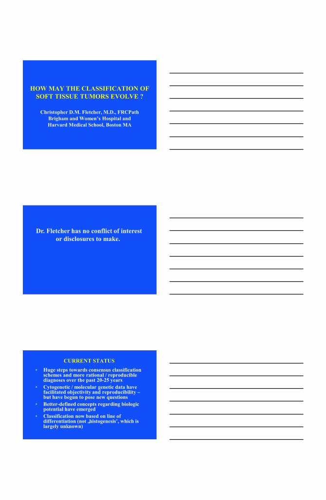
HOW MAY THE CLASSIFICATION OF SOFT TISSUE TUMORS EVOLVE ?
Christopher D.M. Fletcher, M.D., FRCPath
Brigham and Women‟s Hospital and Harvard Medical School, Boston MA
Dr. Fletcher has no conflict of interest or disclosures to make.
CURRENT STATUS • Huge steps towards consensus classification
schemes and more rational / reproducible diagnoses over the past 20-25 years
• Cytogenetic / molecular genetic data have facilitated objectivity and reproducibility – but have begun to pose new questions
• Better-defined concepts regarding biologic potential have emerged
• Classification now based on line of differentiation (not „histogenesis‟, which is largely unknown)
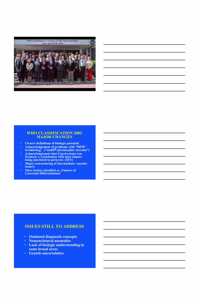
WHO CLASSIFICATION 2002 MAJOR CHANGES
• Clearer definitions of biologic potential • Acknowledgement of problems with “MFH”
terminology (“undiffd pleomorphic sarcoma”) • Acknowledgement that h‟pericytoma was
formerly a wastebasket with most tumors being unrelated to pericytes (SFT)
• Major restructuring of intermediate vascular tumors
• More lesions classified as „Tumors of Uncertain Differentiation‟
ISSUES STILL TO ADDRESS
• Outdated diagnostic concepts • Nomenclatural anomalies • Lack of biologic understanding in
some broad areas • Genetic uncertainties
OUTDATED DIAGNOSTIC CONCEPTS
• “Malignant fibrous histiocytoma” • “Haemangiopericytoma” • “Fibrosarcoma” (at least in adults)
Challenges posed by major change Power of existing literature across multiple disciplines
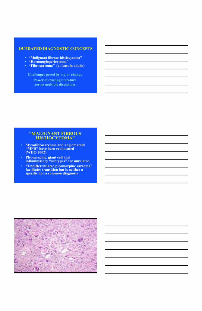
“MALIGNANT FIBROUS HISTIOCYTOMA”
• Myxofibrosarcoma and angiomatoid “MFH” have been reallocated (WHO 2002)
• Pleomorphic, giant cell and inflammatory “subtypes” are unrelated
• “Undifferentiated pleomorphic sarcoma” facilitates transition but is neither a specific nor a common diagnosis
PLEOMORPHIC SARCOMAS APPROX RISK OF METASTASIS AT 5 YRS
Dedifferentiated liposarcoma 15-20% High grade myxofibrosarcoma 30-35% Pleomorphic liposarcoma 40-50% Pleomorphic leiomyosarcoma 60-70% Pleomorphic rhabdomyosarcoma 80-90%
• Not an „entity‟ – but synonymous with undifferentiated pleomorphic sarcoma
• Diagnosis of exclusion • Accounts for no more than 5% of adult
sarcomas • Subclassification of pleomorphic sarcomas has
clinical relevance (myogenic is bad…) • MFH terminology should now disappear
(WHO 2013), but clinicians may need to understand why
PLEOMORPHIC „MFH‟ KEY POINTS
• No continuing rationale for maintaining the term, other than “clinical convenience”
• No good definition for fibrohistiocytic differentiation
• Need to begin to acknowledge existence of undifferentiated or unclassified sarcomas as a routine clinical problem
• Create category of undifferentiated sarcomas with criteria for inclusion/exclusion
„MALIGNANT FIBROUS HISTIOCYTOMA‟
WHAT TO DO NEXT ?
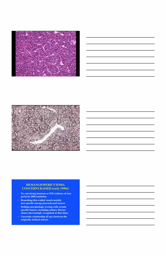
HEMANGIOPERICYTOMA CONCERNS RAISED (early 1990s)
• No convincing immuno or EM evidence of true pericytic differentiation
• Branching thin-walled vessels notably non-specific among mesenchymal tumors
• Striking morphologic overlap with certain specific tumors, including solitary fibrous tumor (increasingly recognised at that time)
• Uncertain relationship (if any) between the originally defined subsets
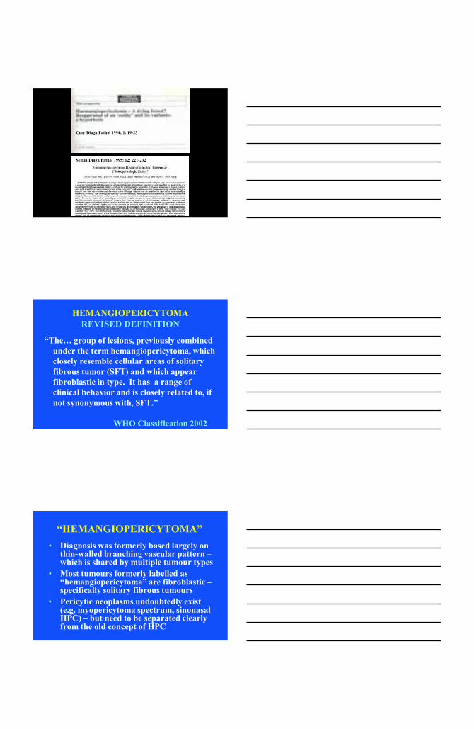
Curr Diagn Pathol 1994; 1: 19-23
Semin Diagn Pathol 1995; 12: 221-232
HEMANGIOPERICYTOMA REVISED DEFINITION
“The… group of lesions, previously combined under the term hemangiopericytoma, which closely resemble cellular areas of solitary fibrous tumor (SFT) and which appear fibroblastic in type. It has a range of clinical behavior and is closely related to, if not synonymous with, SFT.”
WHO Classification 2002
“HEMANGIOPERICYTOMA” • Diagnosis was formerly based largely on
thin-walled branching vascular pattern – which is shared by multiple tumour types
• Most tumours formerly labelled as “hemangiopericytoma” are fibroblastic – specifically solitary fibrous tumours
• Pericytic neoplasms undoubtedly exist (e.g. myopericytoma spectrum, sinonasal HPC) – but need to be separated clearly from the old concept of HPC
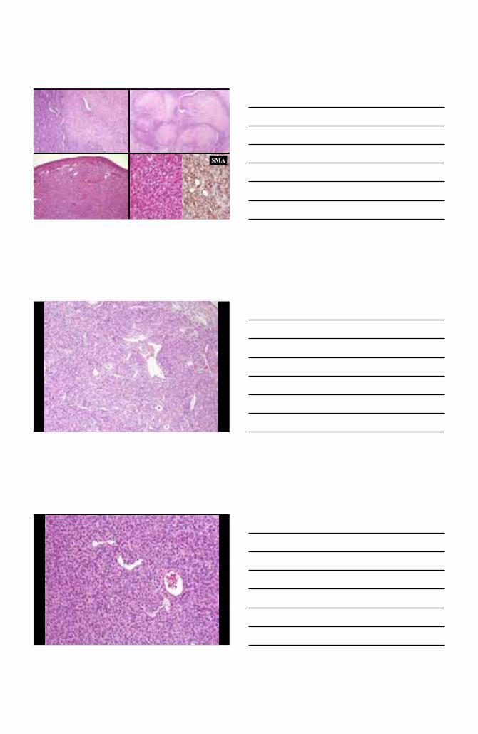
SMA
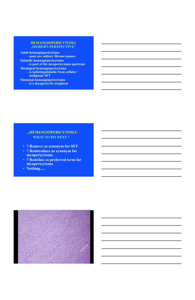
HEMANGIOPERICYTOMA „MODERN PERSPECTIVE‟
Adult hemangiopericytoma - most are solitary fibrous tumors Infantile hemangiopericytoma - is part of the myopericytoma spectrum Meningeal hemangiopericytoma - is indistinguishable from cellular / malignant SFT Sinonasal hemangiopericytoma - is a myopericytic neoplasm
• ? Remove as synonym for SFT • ? Reintroduce as synonym for
myopericytoma • ? Redefine as preferred term for
myopericytoma • Nothing….
„HEMANGIOPERICYTOMA‟ WHAT TO DO NEXT ?
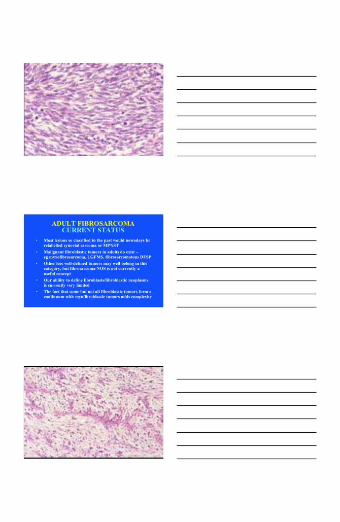
ADULT FIBROSARCOMA CURRENT STATUS
• Most lesions so classified in the past would nowadays be relabelled synovial sarcoma or MPNST
• Malignant fibroblastic tumors in adults do exist – eg myxofibrosarcoma, LGFMS, fibrosarcomatous DFSP
• Other less well-defined tumors may well belong in this category, but fibrosarcoma NOS is not currently a useful concept
• Our ability to define fibroblasts/fibroblastic neoplasms is currently very limited
• The fact that some but not all fibroblastic tumors form a continuum with myofibroblastic tumors adds complexity
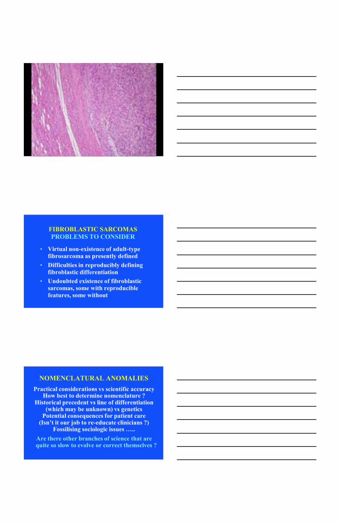
FIBROBLASTIC SARCOMAS PROBLEMS TO CONSIDER
• Virtual non-existence of adult-type fibrosarcoma as presently defined
• Difficulties in reproducibly defining fibroblastic differentiation
• Undoubted existence of fibroblastic sarcomas, some with reproducible features, some without
NOMENCLATURAL ANOMALIES Practical considerations vs scientific accuracy
How best to determine nomenclature ? Historical precedent vs line of differentiation
(which may be unknown) vs genetics Potential consequences for patient care
(Isn‟t it our job to re-educate clinicians ?) Fossilising sociologic issues …..
Are there other branches of science that are quite so slow to evolve or correct themselves ?
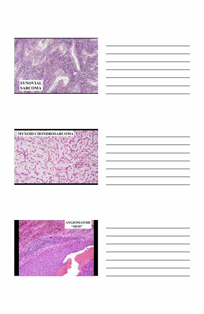
SYNOVIAL SARCOMA
MYXOID CHONDROSARCOMA
ANGIOMATOID “MFH”
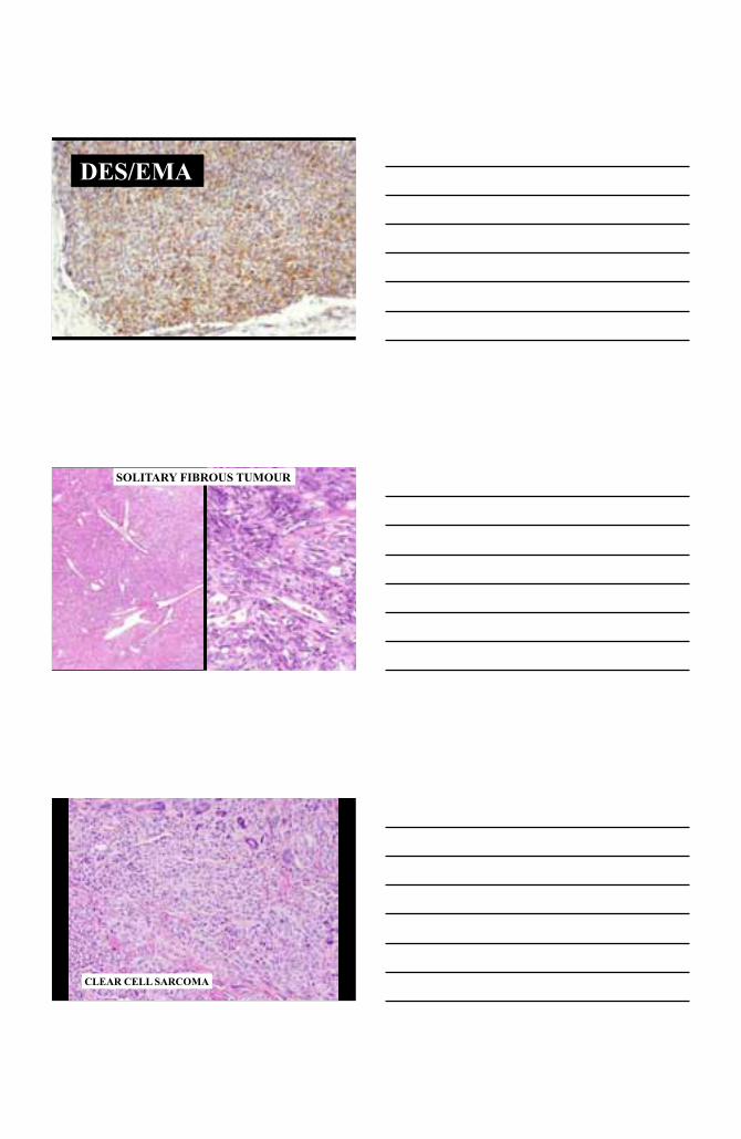
DES/EMA
SOLITARY FIBROUS TUMOUR
CLEAR CELL SARCOMA
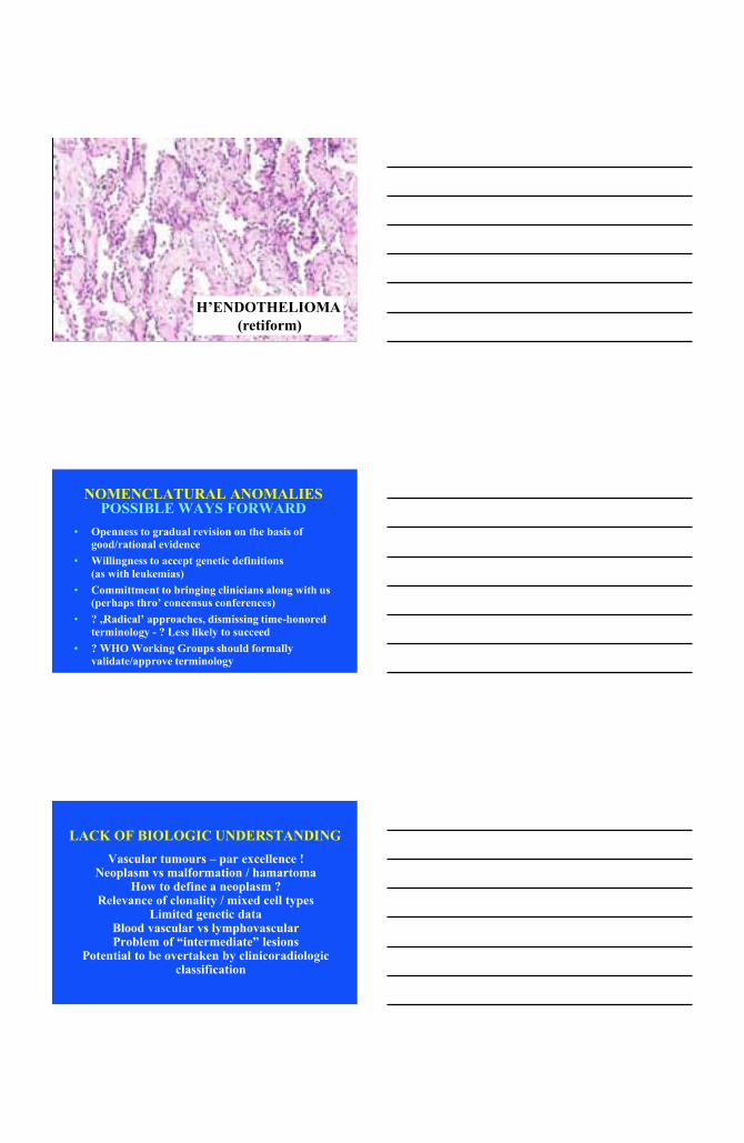
H‟ENDOTHELIOMA (retiform)
NOMENCLATURAL ANOMALIES POSSIBLE WAYS FORWARD
• Openness to gradual revision on the basis of good/rational evidence
• Willingness to accept genetic definitions (as with leukemias)
• Committment to bringing clinicians along with us (perhaps thro‟ concensus conferences)
• ? „Radical‟ approaches, dismissing time-honored terminology - ? Less likely to succeed
• ? WHO Working Groups should formally validate/approve terminology
LACK OF BIOLOGIC UNDERSTANDING Vascular tumours – par excellence !
Neoplasm vs malformation / hamartoma How to define a neoplasm ?
Relevance of clonality / mixed cell types Limited genetic data
Blood vascular vs lymphovascular Problem of “intermediate” lesions
Potential to be overtaken by clinicoradiologic classification
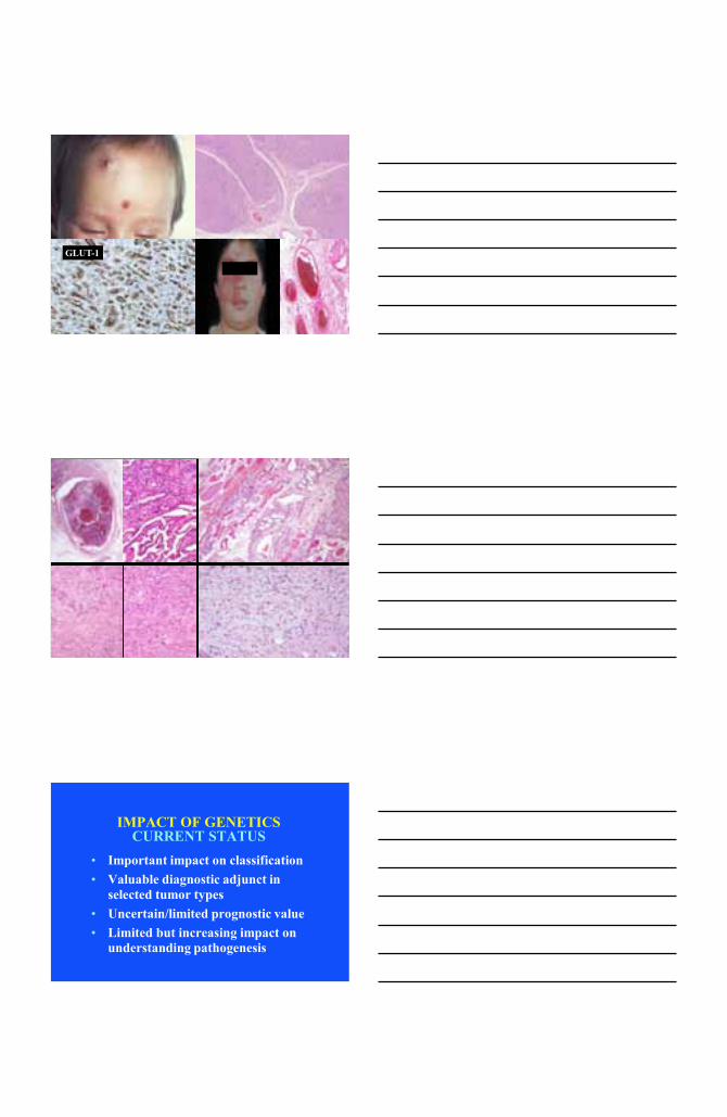
GLUT-1 XXXX
IMPACT OF GENETICS CURRENT STATUS
• Important impact on classification • Valuable diagnostic adjunct in
selected tumor types • Uncertain/limited prognostic value • Limited but increasing impact on
understanding pathogenesis
CYTOGENETIC ABERRATIONS IN SOFT TISSUE SARCOMAS
Tumor type Cytogenetic changes Gene fusion Ewing‟s sarcoma/primitive t(11;22)(q24;q12) FLI-1-EWSR1 neuroectodermal tumor t(21;22)(q22;q12) ERG-EWSR1 t(7;22)(p22;q12) ETV1-EWSR1 t(17;22)(q12;q12) EIAF-EWSR1 t(2;22)(q33;q12) FEV-EWSR1 t(16;21)(p11;q22) FUS-ERG Alveolar rhabdomyosarcoma t(2;13)(q35;q14) PAX3-FOXO1A t(1;13)(p36;q14) PAX7-FOXO1A Myxoid/round cell liposarcoma t(12:16)(q13;q11) DDIT3-FUS t(12;22)(q13;q11-12) DDIT3-EWSR1 Desmoplastic small round cell tumor t(11;22)(p13;q12) WT1-EWSR1 Synovial sarcoma t(X;18)(p11.2;q11.2) SSX1-SS18 SSX2-SS18 Clear cell sarcoma/ t(12;22)(q13;q12) ATF-1-EWSR1 so-called angiomatoid „MFH‟ t(2;22)(q33;q12) CREB1-EWSR1 Extraskeletal myxoid t(9;22)(q22;q12) NR4A3-EWSR1 chondrosarcoma t(9;17)(q22;q11) NR4A3-TAF15 Dermatofibrosarcoma protuberans/ t(17;22)(q22;q13) PDGFB-COL1A1 giant cell fibroblastoma Infantile fibrosarcoma t(12;15)(p13;q25) ETV6-NTRK3 Alveolar soft part sarcoma t(X;17)(p11;q25) ASPL-TFE3 Low grade fibromyxoid sarcoma t(7;16)(q33;p11) FUS-CREB3L2 t(11;16)(p13;p11) FUS-CREB3L1 Myxoinflammatory fibrobl. sarcoma t(1;10)(p22;q24) TGFBR3-MGEA5
RECENTLY IDENTIFIED SPECIFIC CYTOGENETIC / MOLECULAR GENETIC ABERRATIONS IN SOFT TISSUE TUMORS
Myoepithelial tumors EWSR1 and various fusion partners Nodular fasciitis t(17;22)(p13;q12.3) USP6-MYH9 Mesenchymal chondrosarc t(8;8)(q21.1;q13.3) HEY1-NCOA2 Epithelioid h‟endothelioma t(1;3)(p36.3;q25) WWTR1-CAMTA1 Pseudomyogenic hemangioendothelioma t(7;19)(q22;q13) ??? Soft tissue angiofibroma t(5;8)(p15;q13) AHRR-NCOA2 Undiffd (Ewing-like) sarcoma t(4;19)(q35;q13.1) CIC-DUX4 t(4;10)(q35;q26) CIC-DUX4 Ossifying fibromyxoid tumor Rearrangement of PHF1 at 6p21 Solitary fibrous tumor inv12 (q13;q13) NAB2-STAT6
More to come.....
IMPACT OF GENETICS POSSIBLE INFLUENCE ON
NOMENCLATURE AND / OR CLASSIFICATION ?
• DFSP Giant cell fibroblastoma • Spindle cell lipoma Mammary-type myofibroblastoma Cellular angiofibroma Just „related‟ ? Or variants of
a single „entity‟ ?
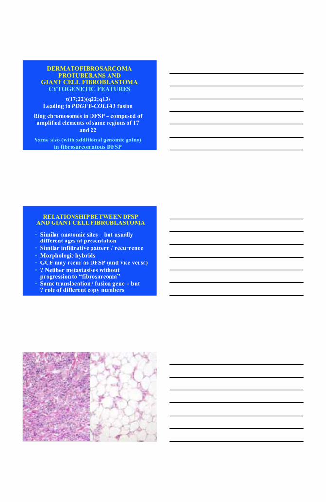
DERMATOFIBROSARCOMA PROTUBERANS AND
GIANT CELL FIBROBLASTOMA CYTOGENETIC FEATURES
t(17;22)(q22;q13) Leading to PDGFB-COL1A1 fusion
Ring chromosomes in DFSP – composed of amplified elements of same regions of 17
and 22 Same also (with additional genomic gains)
in fibrosarcomatous DFSP
• Similar anatomic sites – but usually different ages at presentation
• Similar infiltrative pattern / recurrence • Morphologic hybrids • GCF may recur as DFSP (and vice versa) • ? Neither metastasises without
progression to “fibrosarcoma” • Same translocation / fusion gene - but
? role of different copy numbers
RELATIONSHIP BETWEEN DFSP AND GIANT CELL FIBROBLASTOMA
• Generally different anatomic sites - does this influence the phenotype ? • Morphologic overlap with subtle
differences • Immunophenotypic differences • Same rearrangement/loss of 13q14 • All benign/rarely recur • Cellular angiofibroma may perhaps have
potential for progression
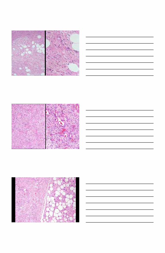
RELATIONSHIP BETWEEN SPINDLE CELL LIPOMA,
MAMMARY-TYPE MYOFIBROBLASTOMA & CELLULAR ANGIOFIBROMA
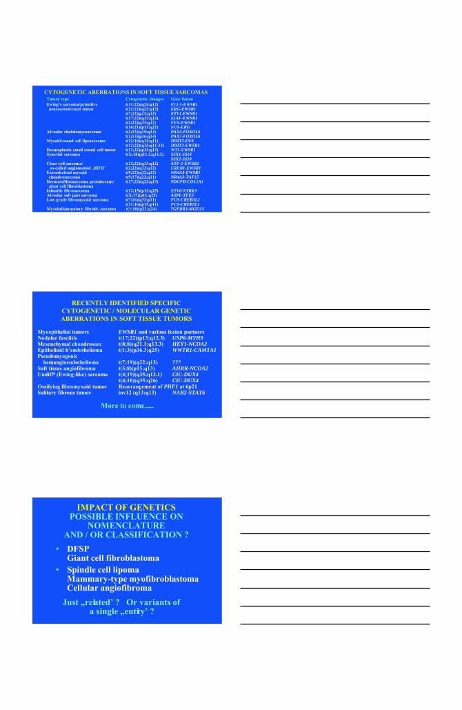
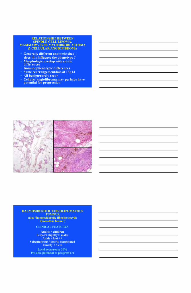
HAEMOSIDEROTIC FIBROLIPOMATOUS TUMOUR
(aka “haemosiderotic fibrohistiocytic lipomatous lesion”)
Adults > children Females slightly > males
Ankle / foot ++ Subcutaneous / poorly marginated
Usually < 5 cm Local recurrence 30%
Possible potential to progress (?)
CLINICAL FEATURES
Female aged 46 with lesion on dorsum of foot – 2 different components
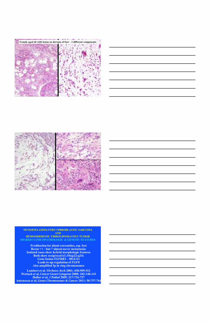
MYXOINFLAMMATORY FIBROBLASTIC SARCOMA AND
HEMOSIDEROTIC FIBROLIPOMATOUS TUMOR SHARED CLINICOPATHOLOGIC & GENETIC FEATURES
Predilection for distal extremities, esp. feet Recur ++ - but ? almost never metastasise
Isolated cases show hybrid morphologic features Both show reciprocal t(1;10)(p22;q24)
Gene fusion TGFBR3 – MGEA5 Leads to up-regulation of FGF8
Also amplified 3p in ring chromosomes Lambert et al, Virchows Arch 2001; 438:509-512
Wettach et al, Cancer Genet Cytogenet 2008; 182:140-143 Hallor et al, J Pathol 2009; 217:716-727
Antonescu et al, Genes Chromosomes & Cancer 2011; 50:757-764
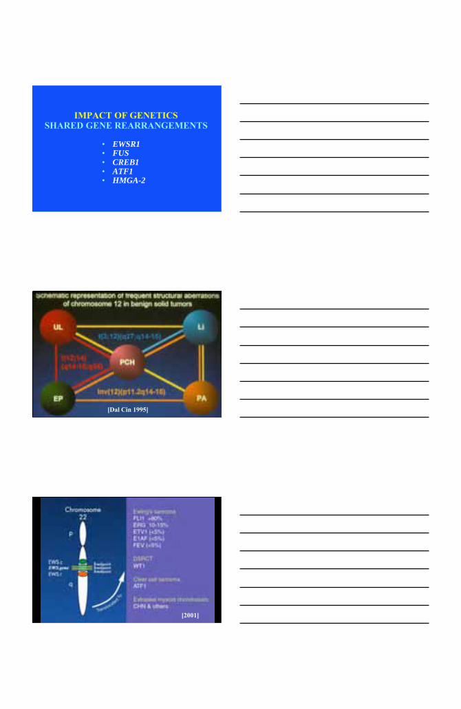
IMPACT OF GENETICS SHARED GENE REARRANGEMENTS
• EWSR1 • FUS • CREB1 • ATF1 • HMGA-2
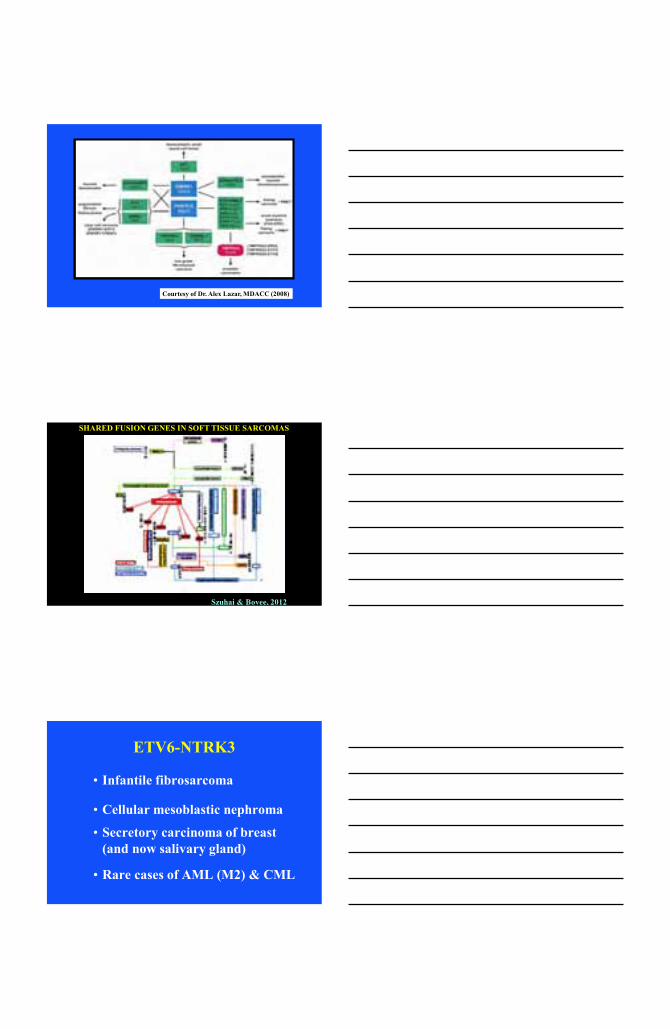
[Dal Cin 1995]
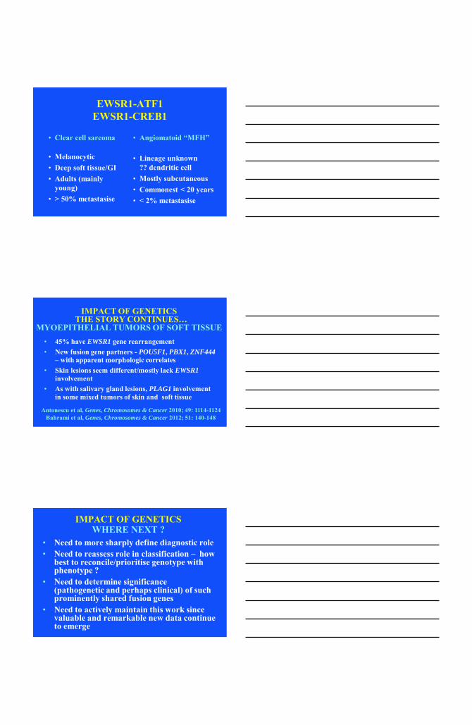
[2001]
Courtesy of Dr. Alex Lazar, MDACC (2008)
SHARED FUSION GENES IN SOFT TISSUE SARCOMAS
Szuhai & Bovee, 2012
ETV6-NTRK3
• Infantile fibrosarcoma
• Cellular mesoblastic nephroma
• Secretory carcinoma of breast (and now salivary gland)
• Rare cases of AML (M2) & CML
EWSR1-ATF1 EWSR1-CREB1
• Clear cell sarcoma
• Melanocytic • Deep soft tissue/GI • Adults (mainly
young) • > 50% metastasise
• Angiomatoid “MFH”
• Lineage unknown ?? dendritic cell
• Mostly subcutaneous • Commonest < 20 years • < 2% metastasise
IMPACT OF GENETICS THE STORY CONTINUES…
MYOEPITHELIAL TUMORS OF SOFT TISSUE • 45% have EWSR1 gene rearrangement • New fusion gene partners - POU5F1, PBX1, ZNF444
– with apparent morphologic correlates • Skin lesions seem different/mostly lack EWSR1
involvement • As with salivary gland lesions, PLAG1 involvement
in some mixed tumors of skin and soft tissue
Antonescu et al, Genes, Chromosomes & Cancer 2010; 49: 1114-1124 Bahrami et al, Genes, Chromosomes & Cancer 2012; 51: 140-148
IMPACT OF GENETICS WHERE NEXT ?
• Need to more sharply define diagnostic role • Need to reassess role in classification – how
best to reconcile/prioritise genotype with phenotype ?
• Need to determine significance (pathogenetic and perhaps clinical) of such prominently shared fusion genes
• Need to actively maintain this work since valuable and remarkable new data continue to emerge
WHO CLASSIFICATION 2012/2013 MAJOR CHANGES
• Introduction of undifferentiated / unclassified sarcomas
• Disappearance of malignant fibrohistiocytic tumors
• Disappearance of hemangiopericytoma • Introduction of some „new‟ entities • Inclusion of GIST and nerve sheath
tumors in soft tissue volume
WHO CLASSIFICATION 2012/2013 „NEW ENTITIES‟
• Pseudomyogenic hemangioendothelioma • Hybrid nerve sheath tumors • Acral fibromyxoma • Hemosiderotic fibrolipomatous tumor • Phosphaturic mesenchymal tumor
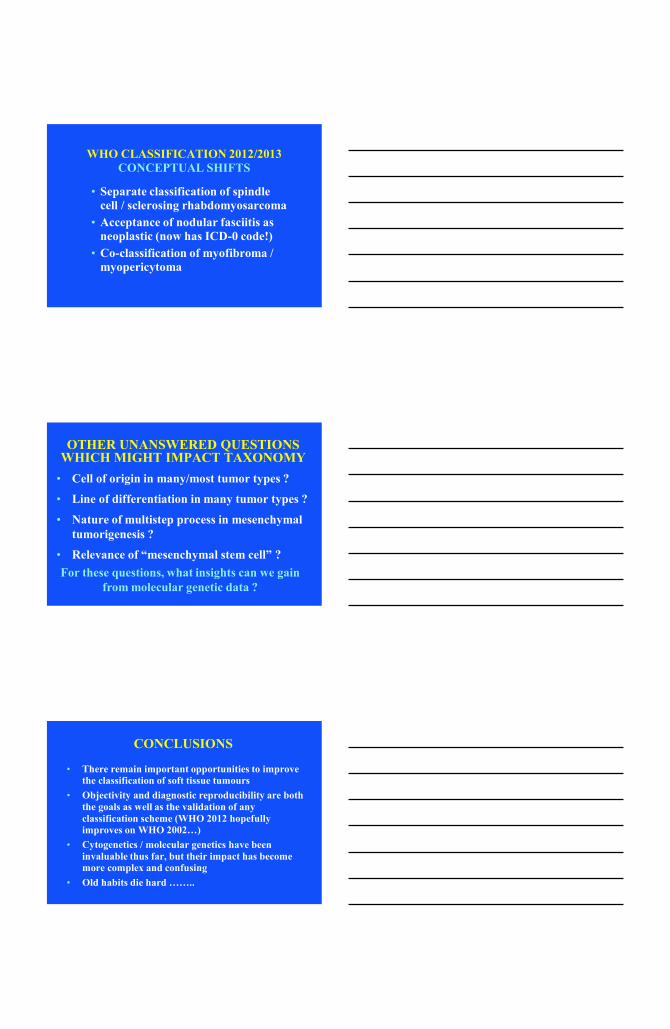
WHO CLASSIFICATION 2012/2013 CONCEPTUAL SHIFTS
• Separate classification of spindle cell / sclerosing rhabdomyosarcoma
• Acceptance of nodular fasciitis as neoplastic (now has ICD-0 code!)
• Co-classification of myofibroma / myopericytoma
OTHER UNANSWERED QUESTIONS WHICH MIGHT IMPACT TAXONOMY
• Cell of origin in many/most tumor types ?
• Line of differentiation in many tumor types ?
• Nature of multistep process in mesenchymal tumorigenesis ?
• Relevance of “mesenchymal stem cell” ?
For these questions, what insights can we gain from molecular genetic data ?
CONCLUSIONS
• There remain important opportunities to improve the classification of soft tissue tumours
• Objectivity and diagnostic reproducibility are both the goals as well as the validation of any classification scheme (WHO 2012 hopefully improves on WHO 2002…)
• Cytogenetics / molecular genetics have been invaluable thus far, but their impact has become more complex and confusing
• Old habits die hard ……..