Embed Size (px)
Citation preview

Dr Mahboube Daneshvar
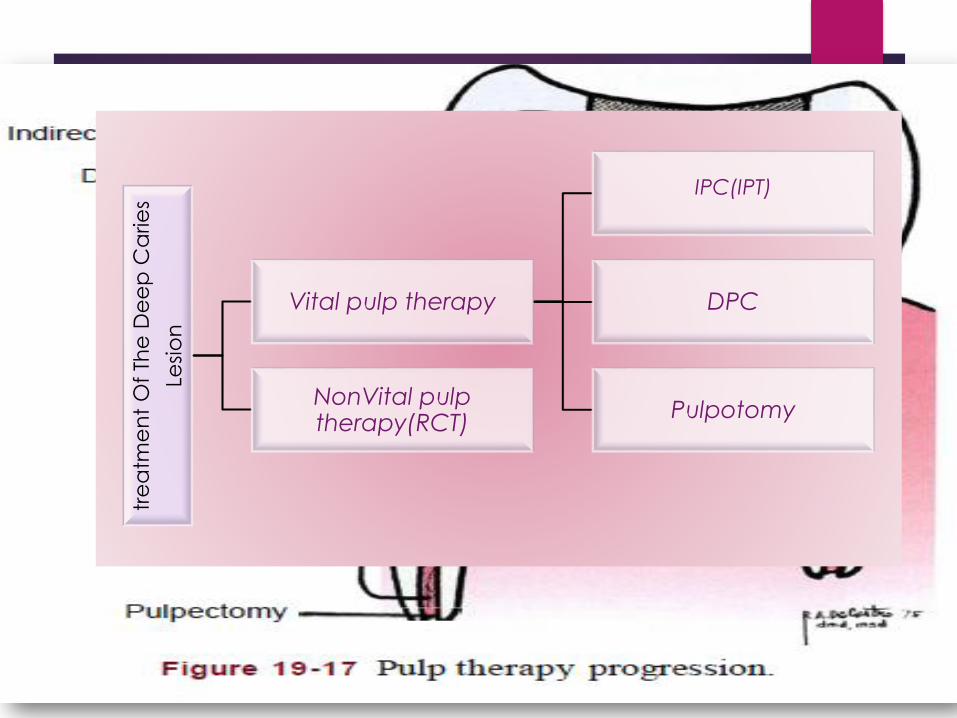
tre
atm
en
t O
f Th
e D
ee
p C
arie
s
Lesi
on
Vital pulp therapy
IPC(IPT)
DPC
PulpotomyNonVital pulp therapy(RCT)
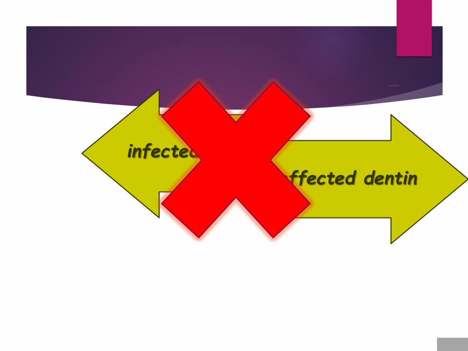
infected dentin
affected dentin
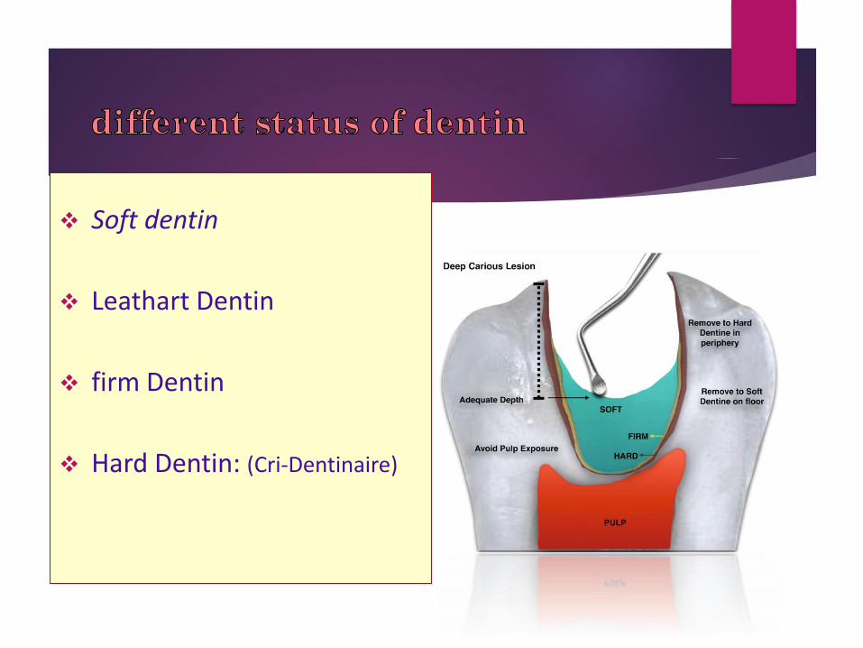
Soft dentin
Leathart Dentin
firm Dentin
Hard Dentin: (Cri-Dentinaire)

remove carious tissue
Nonselective removal to hard dentin (complete excavation or complete caries removal) : overtreatment and is no longer advocated (ICCC).
Selective removal to firm dentin:in shallow or moderately
cavitated dentinal lesions (radiographically extending less than the
pulpal third or quarter of dentin).
Selective removal to soft dentin: in deep cavitated lesions (radiographically extending into the pulpal third or quarter of dentin).
Stepwise removal:

Indirect Pulp Treatment
75% of teeth with deep caries actually have clinical pulp exposures.
90% of asymptomatic teeth with deep caries can be successfully treated by IPC.
Indication:
Teeth with deep caries that are free of symptoms of painful pulpitis
removing the gross caries but allowing sufficient caries to remain over the pulphorn to avoid exposure of the pulp.
The walls of the cavity are extended to sound tooth structure to preventmicroleakage
Large round bur no.6 or 8
Spoon excavator
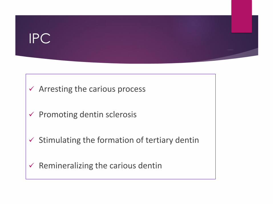
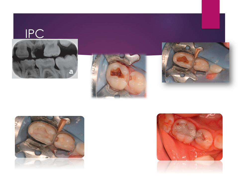
IPC
Arresting the carious process
Promoting dentin sclerosis
Stimulating the formation of tertiary dentin
Remineralizing the carious dentin
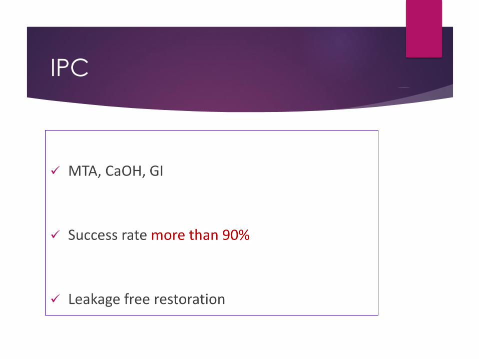
IPC
MTA, CaOH, GI
Success rate more than 90%
Leakage free restoration


Carisolv
A chemomechanical approach to caries excavation
A gel: three amino acids and a low concentration of sodium hypochlorite
with specially designed hand instruments.
sound and carious dentin are clinically separated
only carious dentin is removed
a more conservative preparation.
the time needed to complete the procedure
IPC
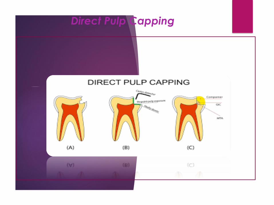
DPC
Indication:
healthy pulp has been inadvertently exposed during an operative procedure.
The tooth must be asymptomatic,
the exposure site must be pinpoint in diameter and free of oral contaminants.
with sterile instruments in clean conditions.
Use of the rubber dam

DPC
calcium hydroxide medicament
to stimulate dentin formation
“heal” the wound and maintain
vitality of the pulp
DPC of a carious pulp exposure in
primary tooth :is not recommended
immature permanent teeth : can be used with success
Direct Pulp Capping
DPC
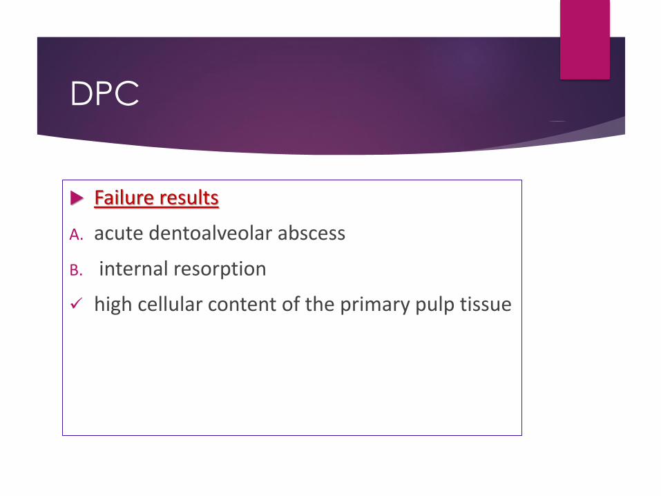
Failure results
A. acute dentoalveolar abscess
B. internal resorption
high cellular content of the primary pulp tissue
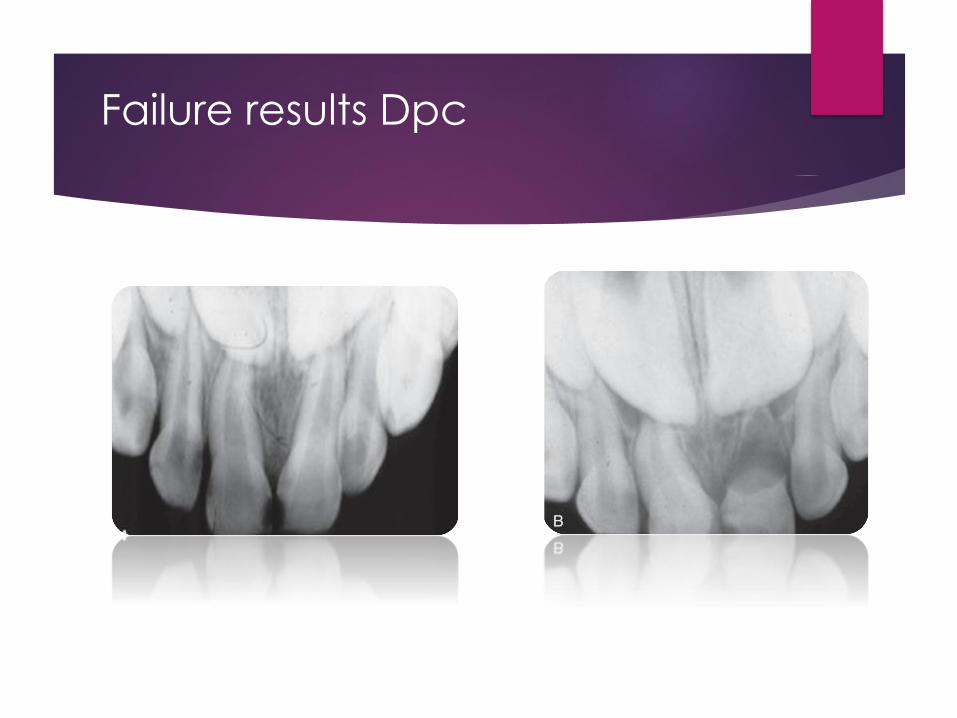
Failure results Dpc
DPC
DPC in primary teeth still be viewed with
reservation, only recommended for exposed
pulp in older children, 1 or 2 year before
normal exfoliation
Pulpotomy
Pulpotomy
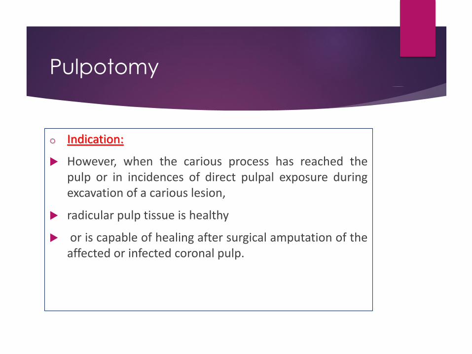
o Indication:
However, when the carious process has reached thepulp or in incidences of direct pulpal exposure duringexcavation of a carious lesion,
radicular pulp tissue is healthy
or is capable of healing after surgical amputation of theaffected or infected coronal pulp.
Pulpotomy
Contraindications : Swelling (of pulpal origin)
Fistula
Pathologic mobility
Pathologic external root resorption
Internal root resorption
Periapical or interradicular radiolucency
Pulp calcification
Excessive bleeding from amputated radicular stumps
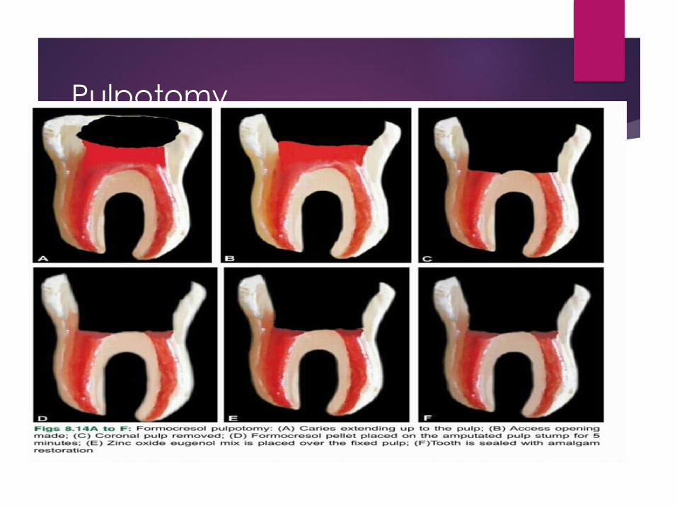
Pulpotomy Technique
Anesthesia
Caries removal all around the cavity walls
Axial wall the final wall for caries removal
The least amount of infected dentin when the exposure
happens
Roof of pulp chamber removed by dental 330 bur water
cooled high speed joining the pulp horns

Pulpotomy Technique
Coronal pulp removal by sharp excavator or slowly revolving large round bur
Pulpal floor perforation
No tags of tissue
Cotton pellets over amputation sites for a few minutes
Hemostasis or minor bleeding
Buckley’s solution 5 min or 1 min (no contact with gingival tissues)
Dark brown or dark red
Pulp chamber dried with new pellets
A thick paste of ZOE
SSC

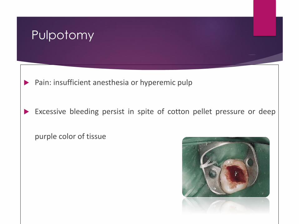
Pulpotomy
Pain: insufficient anesthesia or hyperemic pulp
Excessive bleeding persist in spite of cotton pellet pressure or deep
purple color of tissue

Ideal Dressing for Radicular Pulp
Bactericidal
Harmless to pulp and surrounding tissues
Promote healing of radicular pulp
No interfere with physiologic root resorption
unfortunately the “ideal” pulp dressing material has not yet been identified.
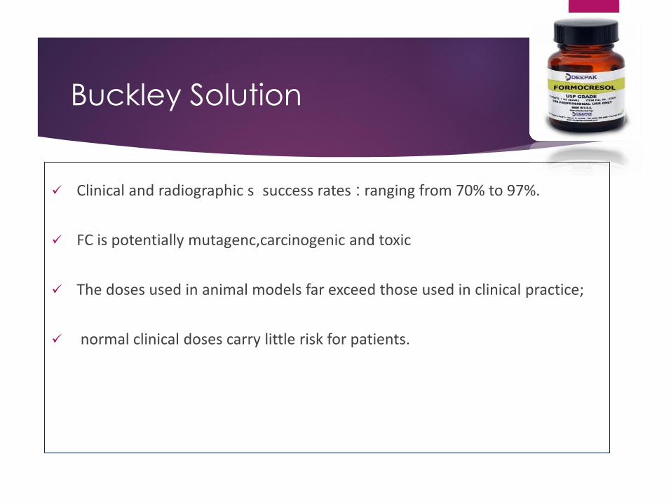
Buckley Solution
Clinical and radiographic s success rates : ranging from 70% to 97%.
FC is potentially mutagenc,carcinogenic and toxic
The doses used in animal models far exceed those used in clinical practice;
normal clinical doses carry little risk for patients.
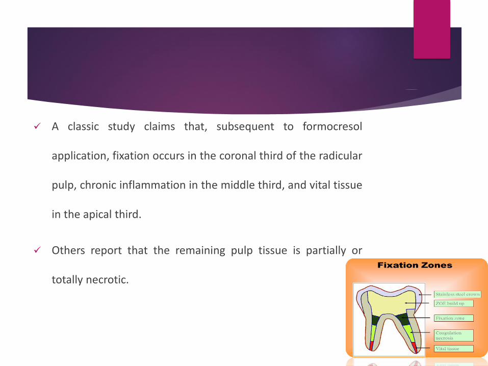
A classic study claims that, subsequent to formocresol
application, fixation occurs in the coronal third of the radicular
pulp, chronic inflammation in the middle third, and vital tissue
in the apical third.
Others report that the remaining pulp tissue is partially or
totally necrotic.

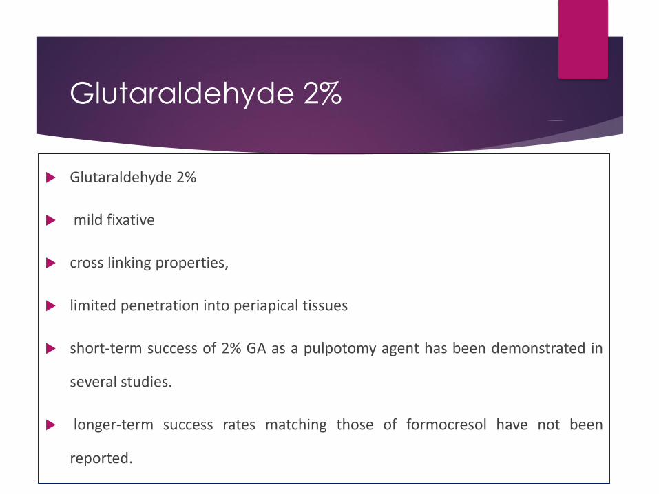
Glutaraldehyde 2%
Glutaraldehyde 2%
mild fixative
cross linking properties,
limited penetration into periapical tissues
short-term success of 2% GA as a pulpotomy agent has been demonstrated in
several studies.
longer-term success rates matching those of formocresol have not been
reported.
NaOCL 5%
studies:
clinical and radiographic success rates for NaOCl pulpotomies are comparable to FS and formocresol pulpotomies.
further studies with longer observation periods are needed
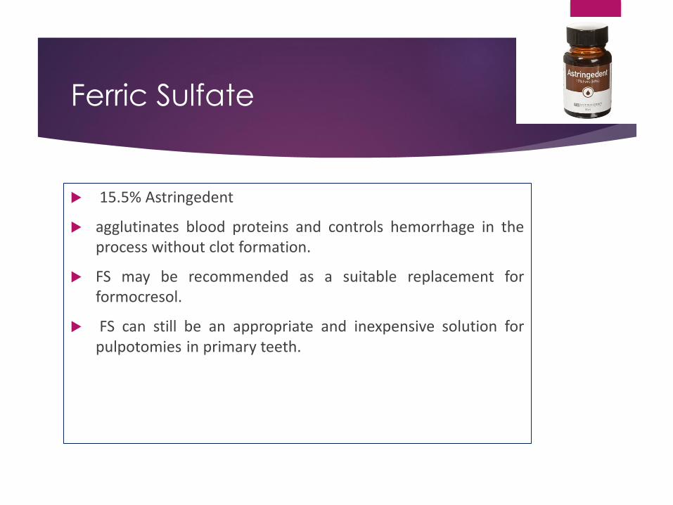
Ferric Sulfate
15.5% Astringedent
agglutinates blood proteins and controls hemorrhage in theprocess without clot formation.
FS may be recommended as a suitable replacement forformocresol.
FS can still be an appropriate and inexpensive solution forpulpotomies in primary teeth.

MTA
Portland cement, dicalcium silicate, tricalcium silicate, tricalcium aluminate,
gypsum, and tetracalcium aluminoferrite; bismuth oxide (opaque)
many positive properties such as
excellent biocompatibility,
alkaline pH
radiopacity
high sealing capacity
ability to induce the formation of dentin, cement, and bone.
MTA
MTA: Gray or White
White MTA no tetracalcium aluminoferrite
Dentin bridge formation significantly better with gray MTA compared to white MTA
The gray MTA had 100% radiographic success, and the white had a 93% success rate.
The negative attributes include:
1. difficulty of handling
2. the exceptional cost.
Portland Cement :
no bismuth ion and presence of potassium ion
compared PC, MTA : Both materials have comparable antibacterial activityand almost identical properties macroscopically, microscopically
compared the success rates of PC, MTA, formocresol in primary molarpulpotomies and found similar clinical and radiographic effectiveness after 24months.
before routine clinical use of PC can be recommended, further studies withlarge samples and long follow-up assessments are needed.
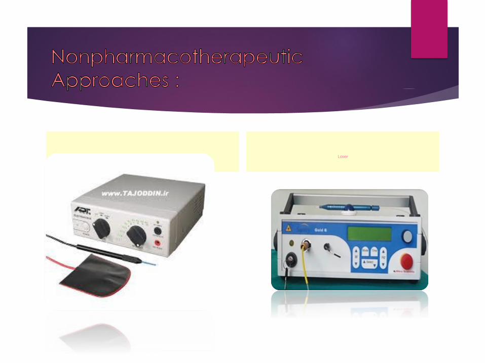
Electrocautery Laser
Summary
alternatives to formocresol as a pulp dressing in primary tooth
pulpotomies has yet to reveal an ideal agent or technique.
formocresol (either in a 1/5 or full strength), FS, or MTA can be used as
capping agents in primary tooth pulpotomies.

Internal Resorption
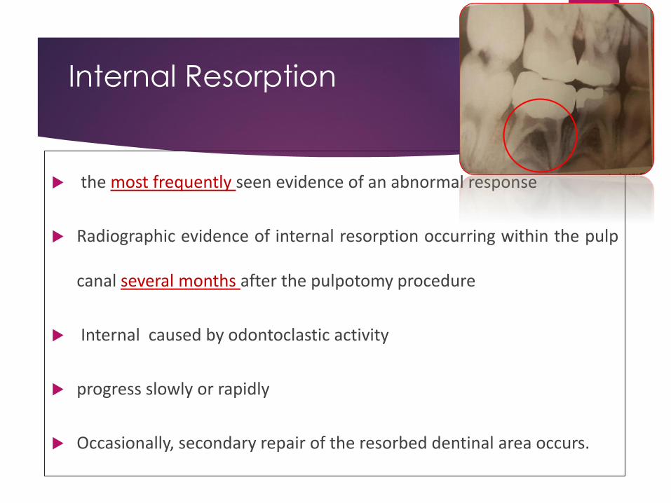
the most frequently seen evidence of an abnormal response
Radiographic evidence of internal resorption occurring within the pulp
canal several months after the pulpotomy procedure
Internal caused by odontoclastic activity
progress slowly or rapidly
Occasionally, secondary repair of the resorbed dentinal area occurs.
Internal Resorption
o that with a true carious exposure of the pulp, an inflammatory process will bepresent to some degree.
abnormal pulp tissue may be allowed to remain.
If the inflammation extended to the entrance of the pulp canal, odontoclasts may havebeen attracted to the area;
tooth histologically : small bays of resorption would be evident.
This condition may exist at the time of pulp therapy,
although there is no way to detect it.
The only indication would be the clinical evidence of a hyperemic pulp.
o internal resorption even though the pulp is normal at the time of treatment :
Inflammatory cells drawn to the area as a result of the placement of an irritatingcapping material might well attract odontoclastic cells and initiate internal resorption.
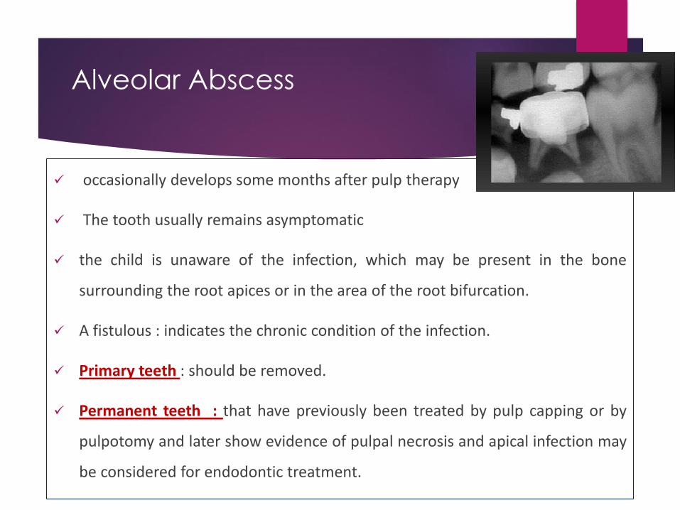
Alveolar Abscess
occasionally develops some months after pulp therapy
The tooth usually remains asymptomatic
the child is unaware of the infection, which may be present in the bone
surrounding the root apices or in the area of the root bifurcation.
A fistulous : indicates the chronic condition of the infection.
Primary teeth : should be removed.
Permanent teeth : that have previously been treated by pulp capping or by
pulpotomy and later show evidence of pulpal necrosis and apical infection may
be considered for endodontic treatment.

Early Exfolation
loosen and exfoliate (or require extraction) :
It is believed that such a condition results from low-grade, chronic,
asymptomatic, localized infection.
abnormal and incomplete root resorption patterns of the affected
teeth are also observed.

Over-Retention
tendency for primary teeth undergoing successful pulpotomies orpulpectomies to be over-retained.
result of interfering with the normal eruption of permanent teeth and adversely
affecting the developing occlusion.
Extraction of the primary tooth is usually sufficient.
normal physiologic exfoliation is delayed by the bulky amount of cement
contained in the pulp chamber.
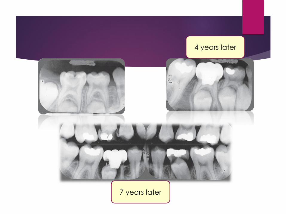
4 years later
7 years later


![Untitled-2 [] · Dr. Akshay Dr. Gade Dr. pratiksha Pat" Dr. Suresh Dr. AmitRajput Bensley Dr. Shefali Karkhanis Dr. Patil Dr. S Mulay Dr. Kamini Lakhiani Dr. Shah Dr. Jaydeep Rev](https://img.pdfslide.net/doc/110x75/5adee08b7f8b9a8f298c298a/untitled-2-akshay-dr-gade-dr-pratiksha-pat-dr-suresh-dr-amitrajput-bensley.jpg)