Embed Size (px)
Citation preview
Dr Rosline Hassan
Haematology Department,
School of Medical Sciences,
Universiti Sains Malaysia,
Kelantan
THE FIRST ASEAN FEDERATION OF HAEMATOLOGY AND THE VIIITH MALAYSIAN
NATIONAL HAEMATOLOGY SCIENTIFIC MEETING
ABO blood group was discovered by Karl Landsteiner in 1900
1970’s : Biochemical basis was elucidated carbohydrate
structure of glycoproteins was worked out
1990 : ABO gene was determined
Existence of a character in two or more variant forms in a population and the least common form
present is more than 1% of individuals[1].
Eg: blood group has a frequency of more than 1% and less than 99%,
it is polymorphic.
[1] Kendrew J. (Ed.) The encyclopedia of
molecular biology. Oxford1994. BlackweI1 Science.
1- Insight about RBC antigens and antibodies
2-Implication in management of transfusion
medicine
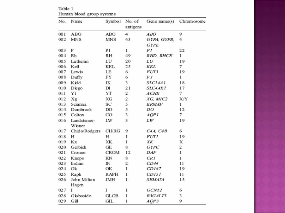
To date nearly 300 blood groups phenotypes identify from an almost 30 blood group system
The most common cause of blood group polymorphismmissense mutation nucleotide change encoding substitution of one amino acid for
another.
Gene deletion.
Deletion of a whole gene only applies to
the D polymorphism of the Rh system
Homozygosity : deletion of the whole
region of GYPB accounts for : S-s-U-
phenotype
Single nucleotide deletion.
Deletion of single nucleotide : shift in
reading-frame for the common O alleles
and A2 allele of the ABO system
Sequence duplication plus nonsense mutation :
inactive RHD gene (RHDΨ),
Intergenic recombination between closely-
linked genes, : hybrid genes
MNS system :GYP(B-A-B) gene responsible for
the GP.Mur phenotype in the Far East.
Rh systems include RHD-CE-Ds produces no D
and is polymorphic in Africans
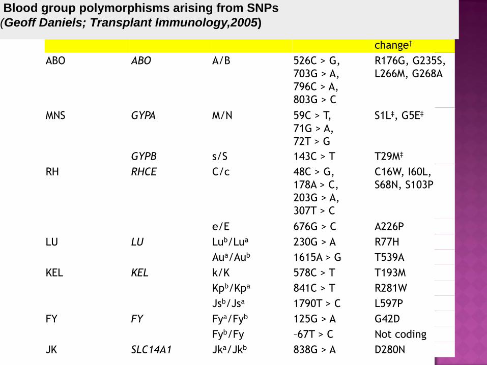
System Gene Polymorphism SNP Amino acid
change†
ABO ABO A/B 526C > G,
703G > A,
796C > A,
803G > C
R176G, G235S,
L266M, G268A
MNS GYPA M/N 59C > T,
71G > A,
72T > G
S1L‡, G5E‡
GYPB s/S 143C > T T29M‡
RH RHCE C/c 48C > G,
178A > C,
203G > A,
307T > C
C16W, I60L,
S68N, S103P
e/E 676G > C A226P
LU LU Lub/Lua 230G > A R77H
Aua/Aub 1615A > G T539A
KEL KEL k/K 578C > T T193M
Kpb/Kpa 841C > T R281W
Jsb/Jsa 1790T > C L597P
FY FY Fya/Fyb 125G > A G42D
Fyb/Fy –67T > C Not coding
JK SLC14A1 Jka/Jkb 838G > A D280N
Blood group polymorphisms arising from SNPs
(Geoff Daniels; Transplant Immunology,2005)
H antigen is an essential precursor to the
ABO blood group antigens.
H locus located on chromosome 19.
contains 3 exons and encodes a
fucosyltransferase that produces the H
Ag.
ABO locus is located on chromosome 9
7 exons & encodes glycosyltransferase
three alleleic forms: A, B, and O.
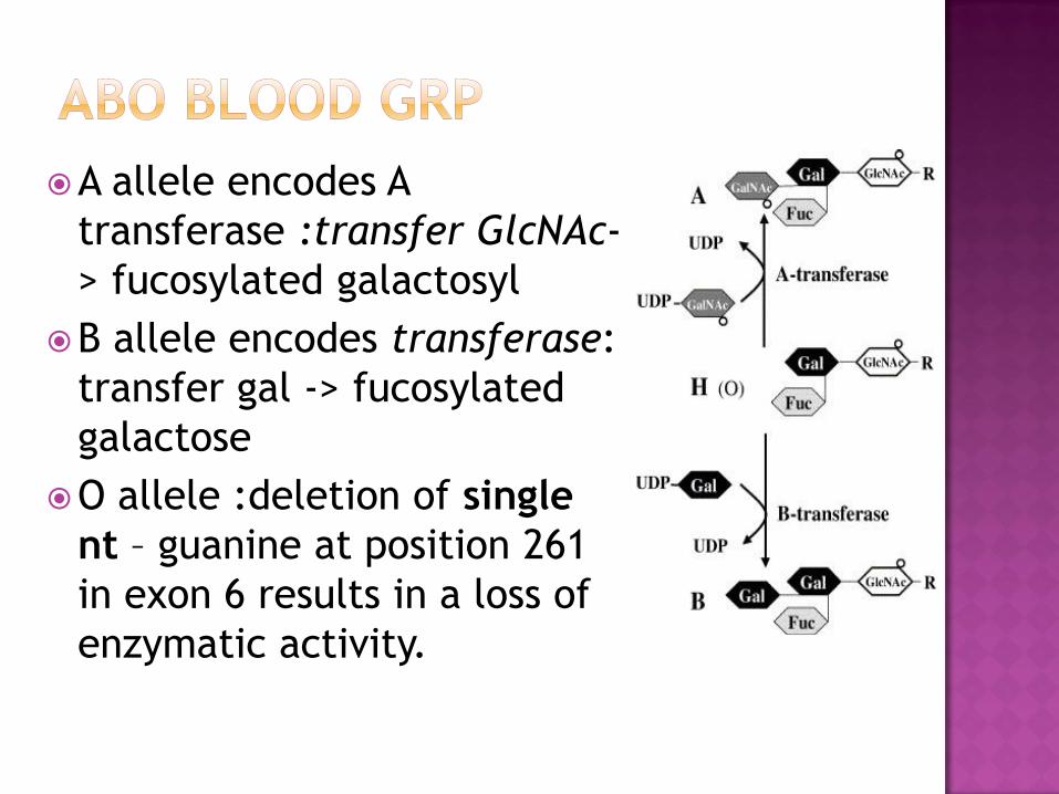
A allele encodes A
transferase :transfer GlcNAc-
> fucosylated galactosyl
B allele encodes transferase:
transfer gal -> fucosylated
galactose
O allele :deletion of single
nt – guanine at position 261
in exon 6 results in a loss of
enzymatic activity.
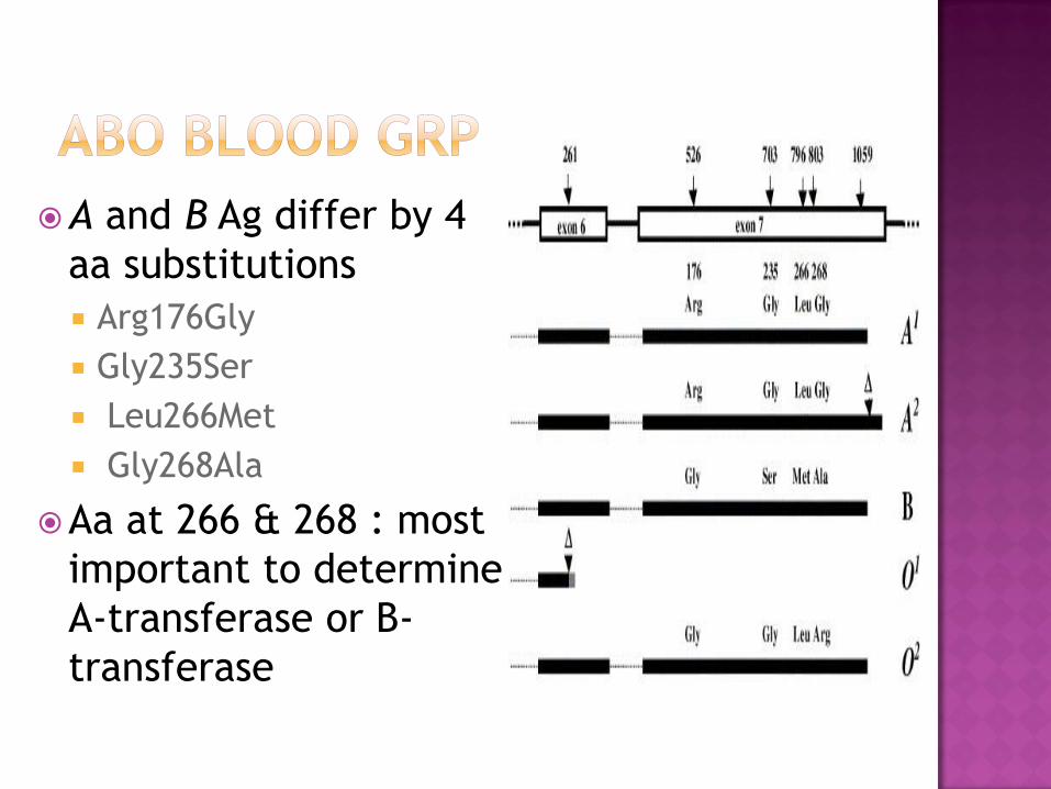
A and B Ag differ by 4
aa substitutions
Arg176Gly
Gly235Ser
Leu266Met
Gly268Ala
Aa at 266 & 268 : most
important to determine
A-transferase or B-
transferase
Seltsam A et al (2003). Blood 102 (8): 3035
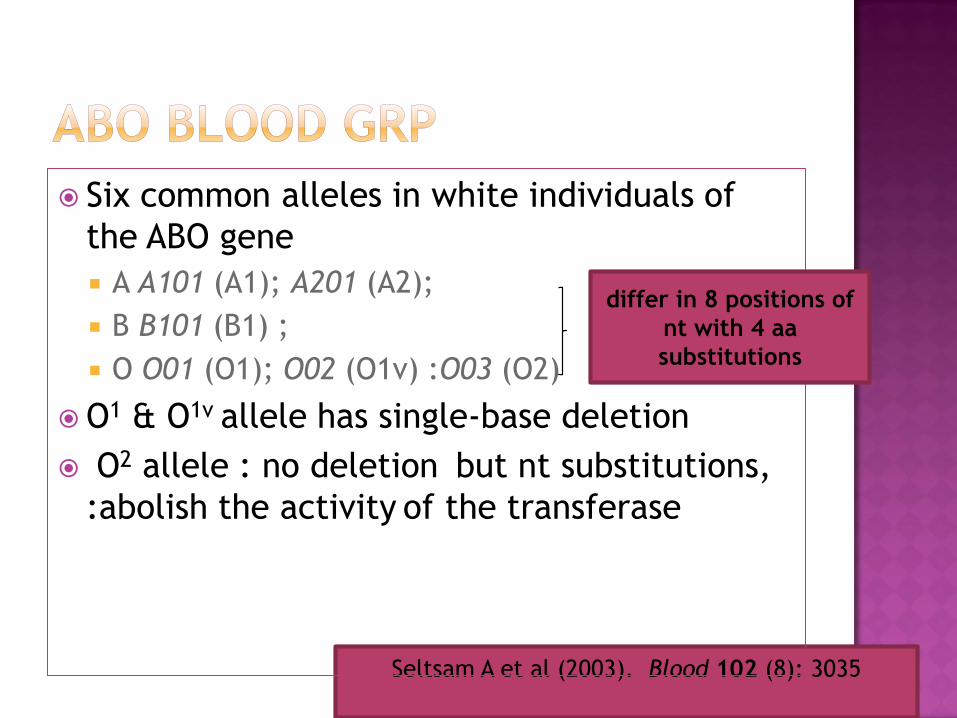
Six common alleles in white individuals of
the ABO gene
A A101 (A1); A201 (A2);
B B101 (B1) ;
O O01 (O1); O02 (O1v) :O03 (O2)
O1 & O1v allele has single-base deletion
O2 allele : no deletion but nt substitutions,
:abolish the activity of the transferase
differ in 8 positions of
nt with 4 aa
substitutions
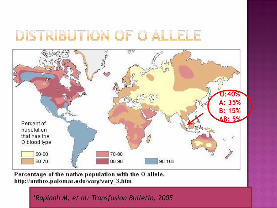
O:40%
A: 35%
B: 15%
AB: 5%
*Rapiaah M, et al; Transfusion Bulletin, 2005
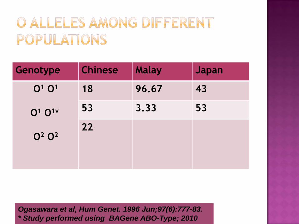
Genotype Chinese Malay Japan
O1 O1
O1 O1v
O2 O2
18 96.67 43
53 3.33 53
22
Ogasawara et al, Hum Genet. 1996 Jun;97(6):777-83.
* Study performed using BAGene ABO-Type; 2010
P. Han et at showed incidence of HDN due
to ABO incompatibility In Singapore was
3.7% of all group O mothers
Correlate with
low distribution of grp 0 among Asian
pop
Homogenous grp 0 alelle
P. Han et al: J.of Paed and Child Health; 2008
Great importance for transfusion
medicine
High immunogenicity
Rh system are encoded by two genes, RHD
and RHCE.
These genes located on chromosome 1
Both have high level of homology with
93.8% identity
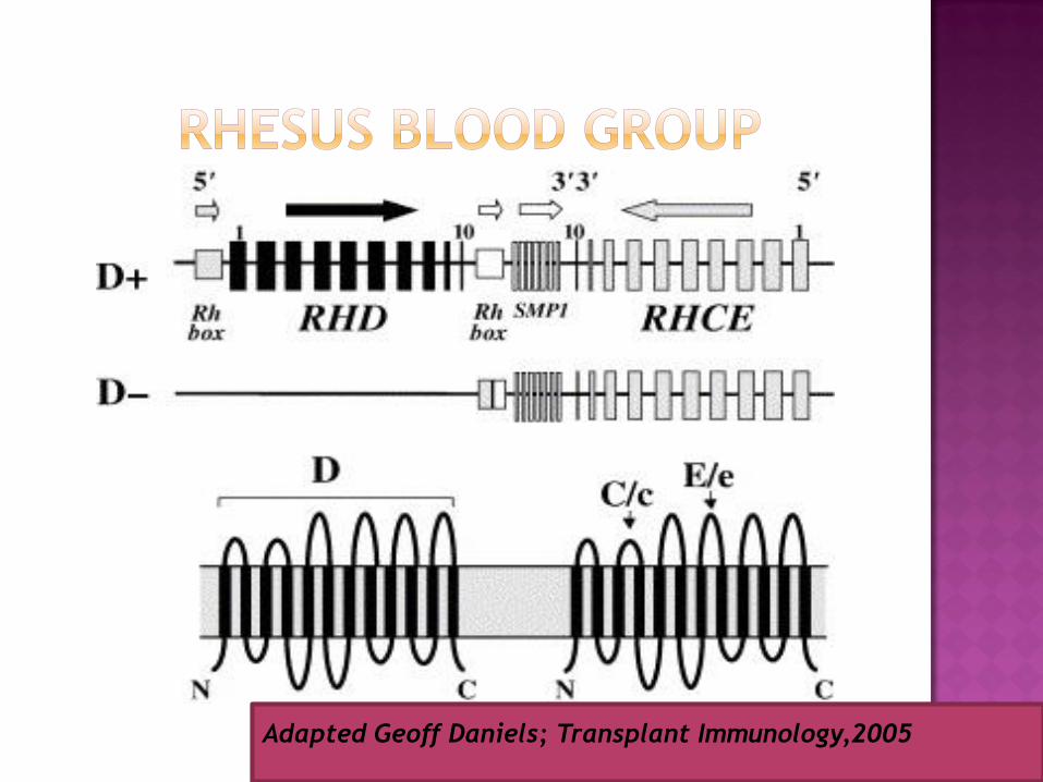
Adapted Geoff Daniels; Transplant Immunology,2005
D antigen comprises several different
antigenic epitopes.
It is classified into 6 distinct categories (DII to
DVII, DI being obsolete)
Characterization of partial D is performed by
differential reactivity with monoclonal anti-D
antibodies
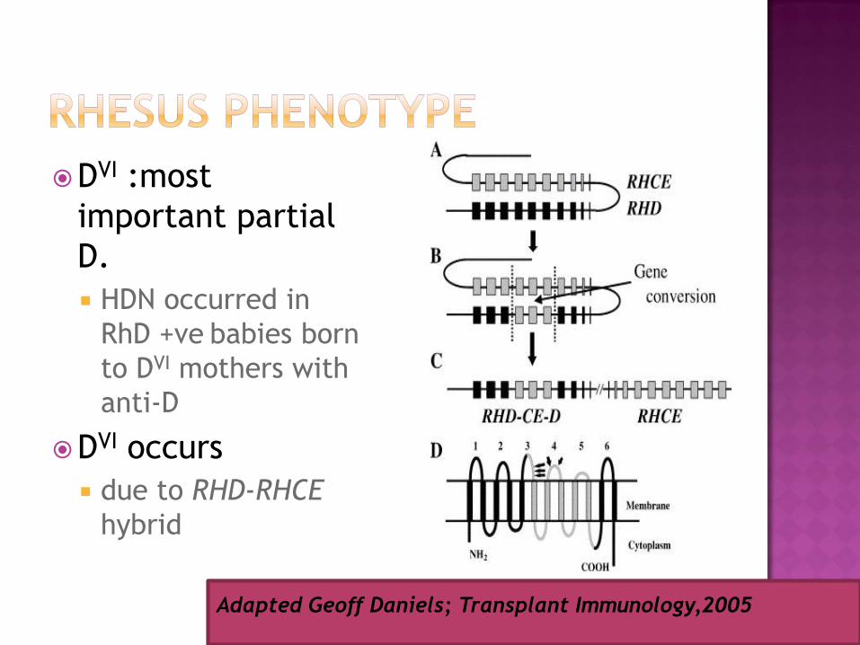
DVI :most
important partial
D.
HDN occurred in
RhD +ve babies born
to DVI mothers with
anti-D
DVI occurs
due to RHD-RHCE
hybrid
Adapted Geoff Daniels; Transplant Immunology,2005
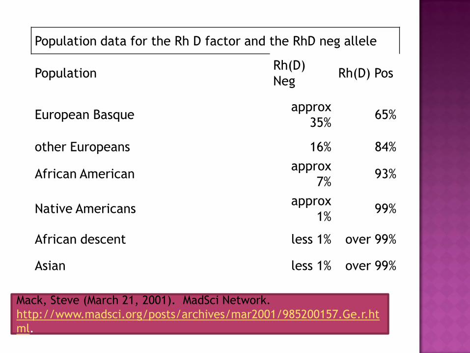
Population data for the Rh D factor and the RhD neg allele
PopulationRh(D)
NegRh(D) Pos
European Basque approx
35%65%
other Europeans 16% 84%
African Americanapprox
7%93%
Native Americansapprox
1%99%
African descent less 1% over 99%
Asian less 1% over 99%
Mack, Steve (March 21, 2001). MadSci Network.
http://www.madsci.org/posts/archives/mar2001/985200157.Ge.r.ht
ml.
Rh neg haplotypes in Africans & Asian :
1. RHD deletion & normal RHCE
2. RHD pseudogene, RHDΨ.
RHD gene duplication: premature stop codon
3. RHD-CE-D, a hybrid gene
Exons from RHD, plus exons from RHCE, followed
by exons from RHD.
hybrid gene produces no D Ag, but prob produce
abnormal C Ag.
Geoff Daniels; Transplant Immunology,2005)
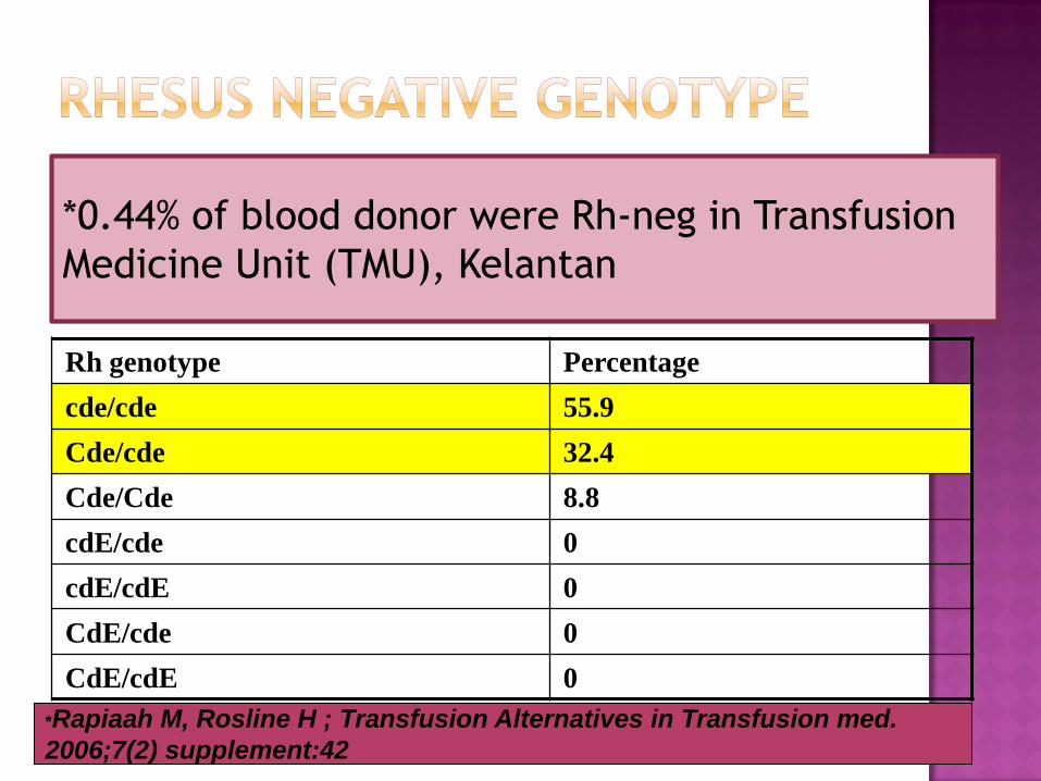
Rh genotype Percentage
cde/cde 55.9
Cde/cde 32.4
Cde/Cde 8.8
cdE/cde 0
cdE/cdE 0
CdE/cde 0
CdE/cdE 0
*0.44% of blood donor were Rh-neg in Transfusion
Medicine Unit (TMU), Kelantan
*Rapiaah M, Rosline H ; Transfusion Alternatives in Transfusion med.
2006;7(2) supplement:42
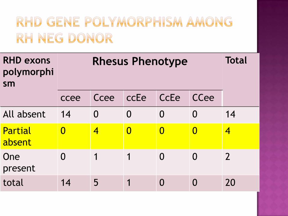
RHD exons
polymorphi
sm
Rhesus Phenotype Total
ccee Ccee ccEe CcEe CCee
All absent 14 0 0 0 0 14
Partial
absent
0 4 0 0 0 4
One
present
0 1 1 0 0 2
total 14 5 1 0 0 20
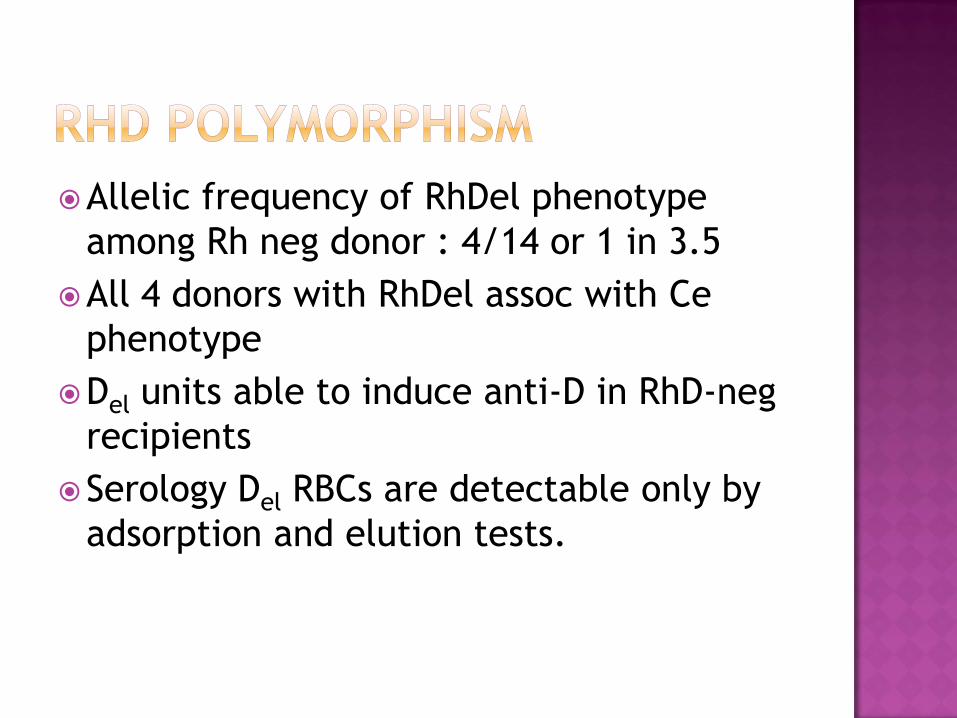
Allelic frequency of RhDel phenotype
among Rh neg donor : 4/14 or 1 in 3.5
All 4 donors with RhDel assoc with Ce
phenotype
Del units able to induce anti-D in RhD-neg
recipients
Serology Del RBCs are detectable only by
adsorption and elution tests.
Transfusion of RhD-Positive Blood in “Asia Type”
DEL RecipientsThe RhD status of transfusion recipients and donors is routinely
matched for red-cell transfusion. This worldwide practice is due to the
potent immunogenicity of RhD. In East Asians, the frequency of RhD-
negative status is only about 0.3%, which sharply limits the supply of
RhD-negative blood. However, approximately 30% of RhD-negative
persons carry an RhD variant, termed "Asia type" DEL.1
Beginning in 2008, my colleagues and I organized a collaborative
group of 10 laboratories, located in 10 cities in northern, central, and
southern China
.
. Shao, N Engl J Med 362(5):472-473 February 4, 2010
Antibody-based technology has been the
basis for blood group typing
Current expansion in molecular
knowledge of RBC and platelet has made
a progression in the laboratory aspect of
Transfusion Medicine
Polymorphism of blood group in a population
Patient with AIHA or positive DAT
Recently transfused patient
Rare blood group phenotypes or
discrepancies in blood group testings
Prenatal testing
Investigate ABO and Rhesus HDN
Determine fetal bld grp & rhesus
Determine RHD zygosity for fathers
Rhesus antigen is highly polymorphic eg
Asian
Required further type Rhesus negative
donors and recipients
Safe transfusion can be assured
To identify RHDel
Shao et al,2010 found RHD gene–intact
but antigen D–alleles in the Ce
haplotype and highly associated with
the RHD 1227A allele.
Determine paternal zygosity & gene
expression
HDN :
homozygous for the gene, all children
Rh +ve
father with deletion in the RHD gene or
has inactive RHD gene require a
monitoring of the pregnancy
neonatal alloimmune thrombocytopenia
Fetal status is determined by testing fetal DNA for HPA-1a/1b from cells obtained by amniocentesis or
Testing fetal-derived DNA present inmaternal plasma at > 5 weeks gestation
If fetus antigen is negative,
mother and fetus need not undergo invasive, costly monitoring or receiveimmune-modulating agents.
Not indicated for routine use of DNA-
based to determine variants of D
especially in area with low prevalence
Extensive pretransfusion matching of
donor blood for patients with diseases
that have a high risk of alloimmunization
sickle cell anemia
thalassemia
Presence of donor RBCs makes typing
inaccurate
DNA-based methods overcome these
limitations
regions of genes common to all alleles
are targeted
minor amounts of donor DNA
outcompeted by patient DNA
Accurate typing in massive transfusions
with non–leukocyte-reduced blood
DNA isolated from a buccal swab
Another indication of DNA arrays
genetic screening to establish
susceptibility to common diseases

DNA-based blood group typing is to
complement conventional ABO typing and
Ab screening but not as independent test
Replacement of Ag & Ab by conventional
methods for pretransfusion testing with
molecular methods is not straightforward.
Majority of transfusion does not require
cross matching beyond ABO and D type
Identifying blood group polymorphism in a
population is the basis for future planning
in the application DNA technology in
transfusion medicine
It is highly recommended to do further
typing for Rhesus negative donor to
detect RHDel which is highly prevalent in
our population