Embed Size (px)
Citation preview

Introduction to histology
Dr. Samah Kotb2015
Histology Techniques CLS 322

Objectives to learn histology
To understand what is Histology.
To understand the importance of Histology in medical
field.
To recognize the normal microscopic features of all
organs of human body.

Introduction to histology
Definition of histology:
Histology is the study of the tissues of the body and
how these tissues are arranged to constitute organs.

Importance of histology in clinical field
Histology is the science that tells about the normal
microscopic feature of all the cells and tissues in the
body.
Histology is the step before going to understand the
pathology of diseased tissues.

Histological techniques or procedures
To study the microscopic features of cells and tissues, the
tissues are processed through various stages to be
viewed under microscope, all these steps are called
HISTOLOGICAL TECNIQUES OR PROCEDURES.

• In the past manual methods were used; but histotechnology
has now advanced to a paramedical science of automation
and advanced techniques to help the pathologist in
diagnosis.
• Histotechnologist should not only know what to do; but
should also know the principle and the theory of what he or
she is doing.
Histotechnology

• To Demonstrate such structures; the tissue must be
prepared in such a way to allow microscopically
observation and study of these different structures.
• This preparation includes the preservation of tissue
materials, processing of tissue block, cutting and staining
of thin sections.

1.Fixation
2.Dehydration
3.Embedding
4.Cutting
5.Staining
Techniques These techniques are :
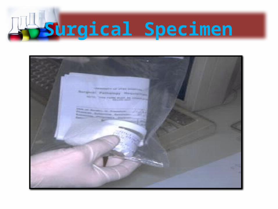
Surgical Specimen

Fixation

Taking Samples

Dehydration and Clearing

Embedding

Sectioning
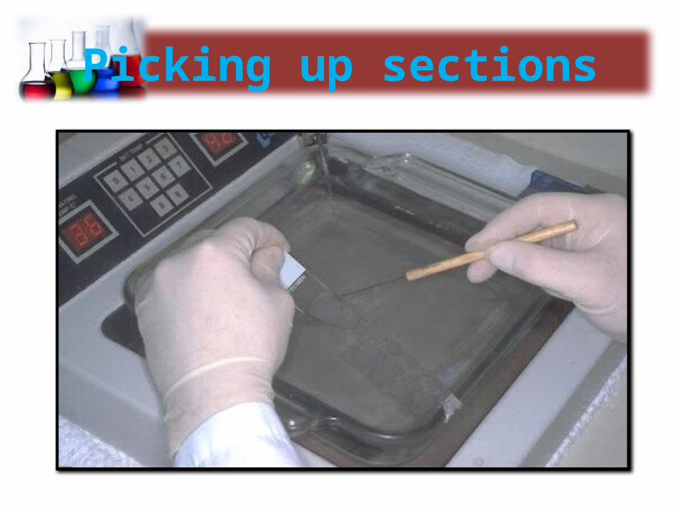
Picking up sections

Microscope slide preparation

Staining

Cover Slipping

Reporting


PREPARATION OF TISSUES FOR STUDY
Biological tissues must undergo a series of treatments
to be observed with light and electron microscopes.
The process begins by stabilization of the tissue with
chemical fixatives.
Next, the tissue is made rigid to allow sectioning.
Finally, it is stained to provide contrast for
visualization in the microscope.

1.Fixation
Techniques
Preserves cellular structure and maintains the
distribution of organelles.
Formaldehyde the most commonly used chemical
fixatives.

2.Dehydration
Techniques
The water is first extracted from the fragments by
bathing them successively in a graded series of
mixtures of ethanol and water, usually from 70% to
100% ethanol (dehydration).

Techniques
The liquid form of the embedding compound, for
example, paraffin wax, replaces the intermediate
solvent.
The liquid embedding medium is allowed to
solidify, thereby providing rigidity to the tissue for
sectioning.
3.Embedding

Techniques
The hard blocks containing the
tissues are then placed in an
instrument called a microtome
and are sliced by the microtome's
steel or glass blade into sections
1 to 10 micrometers thick.
4.Cutting

Techniques
To be studied microscopically sections must typically
be stained or dyed because most tissues are colorless.
Methods of staining tissues have therefore been
devised that not only make the various tissue
components clear but also permit distinctions to be
made between them.
5.Staining

There are various stains used to make histology slides, commonly used are the following:
STAIN TISSUES
Haematoxyline Stains nucleus of cell (blue)
Eosin Stains cytoplasm of cell (pink)
Masson’s trichome
Stains connective tissue
Wright’s stain Stains blood cells
Periodic acid Schiff(PAS)
Stains basement membrane


BASIC HUMAN BODY TISSUES
Human body tissues are classified into four basic types.
1.Epithelial tissue 2.Connective tissue
3.Muscular tissue 4.Nervous tissue

BASIC HUMAN BODY TISSUES
1.Epithelium:
Covers body surfaces, lines organs and cavities.

BASIC HUMAN BODY TISSUES
2. Connective tissue: The different types of connective tissue are
responsible for providing and maintaining the form of organs throughout the body.
provide a matrix that connects and binds other tissues and cells in organs

BASIC HUMAN BODY TISSUES
3.MUSCULAR TISSUE:
Histological, there are three types of muscular tissues, all
responsible for different kind of movements.

BASIC HUMAN BODY TISSUES
4.NERVOUS TISSUE:
It is the main component of the nervous system - the
brain, spinal cord, and branching peripheral nerves.
It is composed of neurons and neuroglia cells.

Thank you