Embed Size (px)
Citation preview

Myocarditis and PericarditisDr .Ali. M Somily
Prof . Hanan A. HabibCardiovascular block
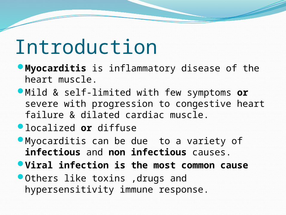
IntroductionMyocarditis is inflammatory disease of the
heart muscle.Mild & self-limited with few symptoms or severe
with progression to congestive heart failure & dilated cardiac muscle.
localized or diffuseMyocarditis can be due to a variety of
infectious and non infectious causes.Viral infection is the most common cause Others like toxins ,drugs and hypersensitivity
immune response.


Myocarditis
Epidemiology ,Etiology and Risk Factors Epidemiology : no accurate estimate of
incidence as many cases are mild & brief and diagnosis is not made.
Coxsackie virus B is the most common cause of myocarditis
Other virus like Coxsackie virus A, Echoviruses, Adenoviruses ,Influenza, EBV, Rubella, Varicella, Mumps, Rabies, Hepatitis viruses and HIV.
Bacterial causes include Corynebacterium diptheriae, Syphilis ,Lyme disease or as a complication of bacterial endocarditis.
Parasitic cause includes Chagas diseases, Trichinella spiralis, Taxoplasma gondii and Echinococcus.
Others organisms includes Rickettsiae, Fungi, Chlamydia, enteric pathogens, Legionella and Tuberculosis.
Giant cell myocarditis due Thymoma, SLE (systemic lupus erythromatosis ) or Thyrotoxicosis.

Infectious NoninfectiousViruses 1. Coxsackie B2. HIV
Systemic Diseases 1. SLE2. Sarcoidosis3. Vasculities(Wegener’s
disease)4. Celiac disease
Bacterial 1. Corynebacterium diphtheriae (diphtheria)
Neoplastic infiltration
Protozoan 1. Trypanosoma cruzi (Chagas disease)
Drugs & Toxins 1. Ethanol2. Cocaine3. Radiation4. Chemotherapeutic
agents - Doxorubicin
Spirochete1. Borrelia burgdorferi ( Lyme
disease)
Clinical PresentationHighly variable ; days to weeks after onset
of acute febrile illness or with heart failure without any known antecedent symptoms .
Fever, headache, muscle aches, diarrhea, sore throat and rashes similar to any viral infection
Chest pain, arrhythmias or sweating , fatigue and may present with congestive heart failure.

Differential DiagnosisAcute MyocarditisVasculitisCardiomyopathy ( due to drugs or radiation)

DiagnosisWBCs, ESR, Troponin and CK-MB usually
elevated ECG (nonspecific ST-T changes and conduction delays are
common)
Blood cultures Viral serology and other specific test for Lyme
disease, diphtheria and Chagas disease may be indicated on a case by case basis.
Chest X-rays : show cardiomegallyRadiology : MRI and Echocardiogram Heart muscle biopsy

ECG of normal heart

Endomyocardial DiagnosisPathologic exam may reveal lymphocytic
inflammatory response with necrosis, but this is not sensitive because of the patchy areas of distribution.
“Dallas” criteria for histopathologic diagnosis
“Giant cells” may be seen.

Giant cells-Myocarditis

ManagementOften supportive;
Restricted physical activity in heart failure.Specific antimicrobial therapy is indicated
when an infecting agent is identified.Treatment of heart failure arrhythmiaOther drugs indicated in special situations like
anticoagulant, NSAID (nonsteroidal antiinflammatory
drugs) , steroid or immunosuppressive immunomodulatory agents.
Heart transplant

ManagementMost cases of viral myocarditis are self
limited.One third of the patients are left with lifelong
complications, ranging from mild conduction defects to severe heart failure.
Patient should be followed regularly every 1-3 months.
Sudden death may be the presentation of myocarditis in about 10% of cases.
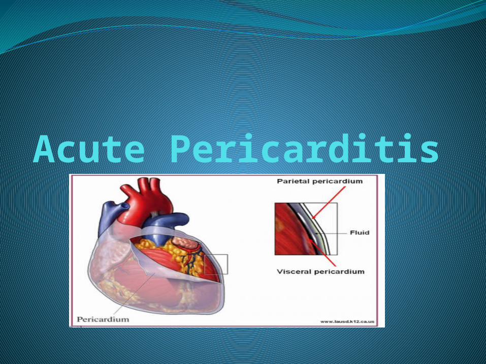
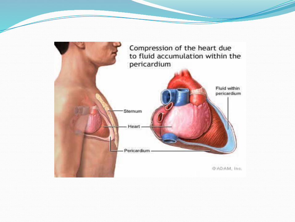
Acute Pericarditis

PericarditisPericarditis is an inflammation of the
pericardium usually of infectious etiology ( viral, bacterial, fungal or parasitic)
Viral Pericarditis:Coxsackievirus A and B, Echovirus are the
most common causes.Other viruses includes Herpes viruses,
Hepatitis B , Mumps, Influenza, Adenovirus ,Varicella and HIV.

PathophysiologyContiguous spread
lungs, pleura, mediastinal lymph nodes, myocardium, aorta, esophagus, liver.
Hematogenous spreadsepticemia, toxins, neoplasm, metabolic
Lymphangetic spreadTraumatic or irradiation

PathophysiologyInflammation provokes a fibrinous exudate
with or without serous effusionThe normal transparent and glistening
pericardium is turned into a dull, opaque, and “sandy” sac
Can cause pericardial scarring with adhesions and fibrosis.

Bacterial pericarditis usually a complication of pulmonary infections (e.g. pneumonia ,empyema):
S. pneumonia, M. tuberculosis, S. aureus, H. influenzae, K. pneumoniae & Legionella.
HIV patients may develop pericardial effusions (M.tuberculosis , M.avium complex).
Disseminated fungal infection (Histoplasma, Coccidioides)
Parasitic infections (disseminated toxoplasmosis, contagious spread of Entamoeba histolytica )are rare causes.

Types of PericarditisCaseous Pericarditis commonly
tuberculous in origin.Serious Pericarditis due to autoimmune
diseases (rheumatoid arthritis, SLE).Fibrous Pericarditis a chronic pericarditis
usually caused by suppurative, caseous, or encased in a thick layer of scar tissue.

Types of Effusive FluidSerous
Transudative - heart failureSuppurative
Pyogenic infection with cellular debris and large number of leukocytes
HemorrhagicOccurs with any type of pericarditisEspecially with infections and malignancies
Serosanguinous
9/98 medslides.com 22
Constrictive Pericarditis
IdiopathicRadiotherapyCardiac surgeryConnective tissue disordersDialysisBacterial infection
23

Clinical presentationPatients with pericarditis will present with
sudden plueritic chest pain, fever, dyspnea and a friction rub.
Patient with tuberculous pericarditis has insidious onset of symptoms.
On examination exaggerated pulses , paradoxus JVP and tachycardia.
As the pericardial pressure increases, palpitations , presyncope or syncope may occur.
Tuberculous PericarditisIncidence of pericarditis in patients with
pulmonary TB ranges from 1 – 8 %Physical findings: fever, pericardial friction
rub, hepatomegalyTuberculin skin test usually positiveFluid smear for AFB often negativePericardial biopsy more definitive
9/98 medslides.com 25
Acute PericarditisDifferential DiagnosisAcute myocardial infarctionPulmonary embolismPneumoniaAortic dissection
DiagnosisECG will show ST elevation, PR depression and T-wave
inversion may occur later.Blood cultureLeukocytosis and an elevated ESR are typical Other routine testing : urea and creatinine.Tuberculin skin test is usually positive in tuberculous
Pericarditis.Chest x-ray may show enlarged cardiac shadow or
calcified pericardium and CT scan show pericardial thickening >5mm.
Pericardial fluid or pericardial biopsy specimens for fungi, antinuclear antibody tests and Histoplasmosis complement fixation in endemic area.

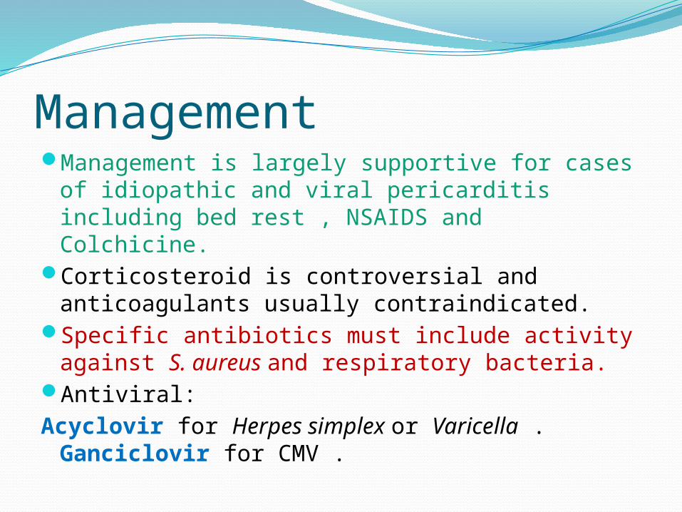
ManagementManagement is largely supportive for cases
of idiopathic and viral pericarditis including bed rest , NSAIDS and Colchicine.
Corticosteroid is controversial and anticoagulants usually contraindicated.
Specific antibiotics must include activity against S. aureus and respiratory bacteria.
Antiviral:Acyclovir for Herpes simplex or Varicella .
Ganciclovir for CMV .
ManagementPericardiocentesis to relief tamponade.Patients who recovered should be observed
for recurrence.Symptoms due to viral pericarditis usually
subsided within one month.
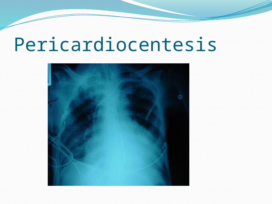
Pericardiocentesis