Embed Size (px)
Citation preview
ANNUAL REPORT 2008DRCMR
DANISH RESEARCH CENTRE FOR MAGNETIC RESONANCE

2
DRC
MR
Annu
al R
epor
t 20
08Co
nten
ts
CONTENTSIntroduction 3 DRCMR at a Glance 4 Dansk Resumé 5 Organisation and Staff 6
Collaborations 7 Clinical Imaging 8 Basic Research 10 Clinical Research 20 Preclinical
Research 27 Other Activities 30 Publications 32 Acknowledgements 34

3
DRC
MR
Annu
al R
epor
t 20
08In
trod
ucti
on
INTRODUCTION
This report summarizes the aims and organization of the Danish Research Centre for Magnetic Resonance (DRCMR), also known as the Department of Magnetic Resonance, at Hvidovre Hospital and describes the accomplishments of the DRCMR staff during 2008.
The main aim of the DRCMR is to advance the use of magnetic resonance as a clinical and investigative tool in biomedical science. During the recent years, the department has been reorganized. The reorganization continued in 2008 with the appointment of Hartwig R. Siebner as consultant (overlæge) with responsibility for research activities, a position which has been consolidated by an appointment as professor in functional brain mapping in the beginning of 2009. Reorganization of the research section will continue in the coming year. In the research section, the activities connected to the “Center for integrated molecular brain imaging” (Cimbi) established in 2006 and spon-sored by the Lundbeck Foundation have been a major focus area.
In 2008 public funding was obtained for replacement of the department’s two oldest scanners for clinical use, both from 1994 with two new scanners to be installed in the coming year. With the new scanners the department will have two 3 tesla and a 1.5 tesla scanner for clinical use.
Also the experimental research has received funding for upgrading and new equipment. The Centre has thus been able to establish facilities for hyperpolarization of carbon-13 labeled substances and a major upgrade of the small bore 4.7 tesla MR-scanner has been ordered.
The DRCMR is well prepared to meet new challenges in the coming years. DRCMR will continue towards further strengthening the department as one of the most dynamic, flexible and innovative MR clinical and research units in this part of Europe.
Finally, I would like to express our gratitude towards the foundations and institutions whose support over the years has enabled the Centre to achieve and maintain its front-line position in MR research.
Olaf B. PaulsonHead of the DRCMR
This report is published by
Danish Research Centre for Magnetic ResonanceCopenhagen University Hospital, HvidovreKettegård Allé 302650 HvidovreDenmark
tel: (+45) 36 32 28 85fax: (+45) 36 47 03 02http://www.drcmr.dk
Editing & LayoutKaram Sidaros
© Copyright 2008-2009

4
DRC
MR
Annu
al R
epor
t 20
08D
RCM
R at
a G
lanc
e
A unique strength of the Danish Research Centre for Magnetic Resonance (DRCMR) is the multi-disciplinary nature of its activities. The Centre is home to an active clinical department providing a full range of diagnostic MRI services. Patient referrals come from a broad range of sources, including Hvidovre Hospital as well as other hospitals in Copenhagen and other parts of Denmark. The clinical services of the department are performed alongside the investigative imaging, providing valu-able integration between primary clinicians and clinical researchers.
Distinguishing the DRCMR from other academic radio-logy settings in Denmark is the juxtaposition within the Centre of a vigorous basic research program with the patient-oriented activities of the department. This ensures the highest level of scientific support for the Centre’s biomedical mission, and places it at the forefront of MR method development. Through inter-action with research partners in the Copenhagen Brain Research Center and elsewhere, the DRCMR also par-ticipates in groundbreaking research in neurology, neuroinformatics, neuropharmacology, neuropsychiatry, and cognitive science.
Imaging facilities
The Centre has three Siemens whole-body clinical scan-ners. A Magnetom Trio (3 tesla) MR-scanner was installed in 2002 after a generous donation from the Simon Spies Foundation. The two other clinical scanners 1.5 and 1.0 tesla were installed in 1994 and will in 2009 be replaced by a Siemens 3 tesla Verio scanner and a Siemens 1.5 tesla Avanto scanner. All three clinical scanners are located in area 340A of the hospital, where there also are facilities for clinical work and conferences.
In addition, the Centre has an experimental Varian 4.7 tesla MR-scanner, suitable for MR studies in small ani-mals. The experimental scanner is located in area 340B where there are also facilities for data analysis and other research activities. A major upgrade of the exper-imental animal scanner will take place in the beginning of 2009 where the gradient coil will be replaced and a new console will be installed increasing the imaging speed of the scanner significantly. This 4.7 tesla system is the only modern MR scanner in Copenhagen for stud-ies of small experimental animals. The pre-clinical group is involved in a set of promising new studies of cerebral connectivity using fixed brains scanned with a very high resolution for fibre-tracking. These studies are important in defining and extending the limits of new MR methods and illustrate the advantages of com-bined high-field human and animal imaging facilities (and the scientists who use them) in one site. Thanks to a generous donation by the Simon Spies Foun-dation, a carbon-13 hyperpolarizer was installed in 2008 which opens up for a whole new area of research. Only a very limited number of MR-centres in Europe have access to this technology for in vivo research. Using this
instrument it is possible to hyperpolarize substances labelled with carbon-13 thus increasing their MR signal more than 10.000 times. By injecting such hyperpolar-ized substances intravenously it is possible to follow their conversion in metabolic processes non-invasively and in real time by MR spectroscopy.
Clinically orientated activities
The Centre is a provider of local and national radio-logical services in response to physician referrals. The department’s radiological expertise is also in demand as a reading and MR coordination site for several large clinical trials. An essential component of these trials is image analysis, and the Centre continues to make considerable effort and progress in extending the “configurable” analysis pipeline with new meth-ods. MR images acquired using sequences designed to obtain differing morphological, physiological or func-tional information are entered into the ‘pipeline’ and automatically analyzed using a wide range of methods including alignment, intensity correction and segmen-tation. In the last year, continuing development of this pipeline has narrowed the gap between traditional radiological practices and the informatics approaches of the future.
Organisation of Departmental Research
The research organisation in the Centre is currently being restructured and the new organisation is expected to be functional during the coming year. The organisa-tion will thus shift from a methodologically oriented organisation to an organization with more focus on all the research areas covered by the researchers in the department. This will give a better balance between methodological and applied research and strengthen the research profile of the Centre. Furthermore, there will be an increased focus on career development for the Centre’s senior researchers. The leaders of the indi-vidual research groups meet regularly in the Research Coordination Group (RCG).
2008 and the future
In the research one of the most exciting developments of the year was the department’s role in the continu-ous work with the Center for Integrated Molecular Brain Imaging (Cimbi), established in 2006 and funded by the Lundbeck Foundation. The Cimbi group is led by Profes-sor Gitte Moos Knudsen of the Neurobiology Research Unit at Rigshospitalet and included contributions from principal investigators at the Faculty of Pharmaceutical Sciences, University of Copenhagen (led by Professor Mikael Begtrup), the Technical University of Denmark (led by Professor Lars Kai Hansen) as well as the DRCMR (led by Professors Olaf B. Paulson and Terry Jernigan). The research in Cimbi focuses on the neural bases of personality dimensions that predispose individuals to affective and substance use disorders, with special emphasis on the serotonergic neurotransmitter system.
DRCMR AT A GLANCE
Denne rapport giver et indblik i målene, visionerne og organisationen af MR-afdelingen på Hvidovre Hospital og beskriver afdelingens aktiviteter i 2008. Én af afdelingens styrker er netop tværfagligheden af aktiviteterne, der spænder fra et aktivt klinisk miljø med en lang række diagnostiske MR-tjenester til et omfattende forskningsprogram, der dækker klinisk MR såvel som basal forskning. Centret blev grundlagt efter en stor donation fra Simon Spies i 1984 og allerede fra starten var der lagt lige vægt på såvel forskning som kliniske anvendel-ser. I 2002 sikrede Simon Spies Fonden med donationen af landets første højfeltsskanner, at afdelingen er forblevet i front. Afdelingen råder over tre humane MR-skannere. De to ældste er fra 1994 og bliver i 2009 udskiftet med to nye skannere med feltstyrke på 3 og 1,5 tesla. Derudover råder afdelingen over en 4,7 tesla dyreeksperimentel skanner, som får en større opgradering i begyndelsen af 2009.
Dette har sikret international anerkendelse i form af blandt andet projektstøtte fra EU, samarbejde med udenlandske forskningsinstitutioner, omfattende publikationsaktivitet i internationale tidsskrifter og udvælgelsen af afdelingen til MR-evalueringscenter ved inter-nationale medicinafprøvninger.
Året 2008 blev et år med fortsatte organisatoriske ændringer på MR afdelingen. I slutningen af 2006 og begyndelsen af 2007 fik afdelingens kliniske sektion i stort omfang nyt personale og har siden gennemgået en væsentlig reorganisation. I forskningssektionen er fokus i sti-gende grad blevet rettet mod aktiviteter knyttet til ”Center for integrated molecular brain imaging” (Cimbi), som blev etableret i 2006 med støtte af Lundbeckfonden. En reorganise-ring i forskningssektionen er ligeledes fortsat med ansættelse af overlæge Hartwig R. Siebner i efteråret 2008 (udnævnt til professor i begyndelsen af 2009). Reorganiseringen i forsknings-sektionen forventes at fortsætte i det kommende år. Disse nyskabelser vil få stor betydning for afdelingens fortsatte virke inden for klinik og forskning. Afdelingen forventer store udford-ringer i de kommende år og er vel rustet til at møde disse og til at sikre afdelingen som et af de førende kliniske og forsknings MR-centre i denne del af Europa.
DANSK RESUMÉ
Both PET and MRI are employed in studies of human subjects, and these are complemented with relevant studies using animal models. Advanced informatics techniques, new tracer compounds, and novel sero-tonergic challenge paradigms are also being developed within the Centre. The work also involves collaborating laboratories in Europe and the US.
The 3 tesla whole body system provides a demanding environment where researchers continue to invest sig-nificant effort developing new powerful imaging and spectroscopy methods. The high quality of morphologi-cal and functional images obtained at 3 tesla ensures that the system will continue to have an important future in the department’s research activities. As men-tioned earlier, new scanners will be installed at the end of 2009.
The accomplishments of the year, described within this report, illustrate the depth and breadth of exper-tise within the department. The interaction between radiologists, clinicians, psychologists, physicists and engineers together with other scientists from different disciplines within both the department and collaborat-ing centres continues to create a rich multi-disciplinary environment to pursue MR research and apply it to clini-cal problems.
With the anticipated new clinical and research initia-tives over the next year, the department is confident that it will continue to make significant clinical and sci-entific contributions and remain at the forefront of MR research at an international level.

6
DRC
MR
Annu
al R
epor
t 20
08O
rgan
isat
ion
and
Staf
f
Department Chair
Olaf B. Paulson
Senior Staff, Clinical
Marianne Dalsgaard, Head TechnologistPer Åkeson, MD, DMSc, Senior Physician and Clinical ChiefErland Magnussen, MD, Senior PhysicianMichel Nemery, MD, Senior Physician
Senior Staff, Research
William Baaré, PhD, PsychologistDaniela Balslev, PhD, MDMark Schram Christensen, PhD, Engineer Tim B. Dyrby, PhD, EngineerEllen Garde, PhD, MDLars G. Hanson, PhD, Physicist Pernille Iversen, PhD, BiophysicistTerry L. Jernigan, Professor, PhD, PsychologistJulian Macoveanu, PhD, EngineerKristoffer Madsen, PhD, EngineerPeter Magnusson, PhD, Physicist Maurice Ptito, Visiting Professor, PhD, DMScThomas Ramsøy, PhD, PsychologistPoul Ring, MSc, Engineer Karam Sidaros, PhD, EngineerHartwig Siebner, Professor, MDLise Vejby Søgaard, PhD, PhysicistXingchen Wu, PhD, MD
Junior Staff, Clinical
Camilla Gøbel Madsen, MD
Junior Staff, Research
PhD studentsBrian Villumsen Broberg, MSc (pharm.) Sadia Asghar Butt, MSc, BiochemistAnne-Marie Dogonowski, MDTorsten Dorniok, MSc, PhysicistBjørn Ebdrup, MDSofie Gelskov, MSc, BiologistMartin Vestergaard Hansen, PsychologistMette Hauge Lauritzen, MSc, Human BiologistFrederik Hengstenberg, MDDavid Alberg Holm, MSc, EngineerBettina Hornbøll, MSc, BiologistAstrid Lou, MDHenrik Lundell, MSc, EngineerKathrine Skak Madsen, MSc, BiologistRobin de Nijs, MSc, PDEng, Medical PhysicistCharlotte Ryberg, MSc, Biologist
Annette Sidaros, MDArnold Skimminge, MSc, PhysicistJon Wegener, MSc, Life Sciences and Chemistry
Junior ResearchersJens Bundgaard, MSc, Physicist Matthew Liptrot, MSc, EngineerHenrik Lund, MSc, BiologistMartin Skov, MA, Nordic Languages and Literature
Research Assistants and StudentsAyna Baladi Nejad, MScSajjad Chughtai
Technologists
Yalda Ansari, RadiographerMarion Berge, RadiographerSiri Eggum, RadiographerSascha Gude, Laboratory TechnicianSussi Larsen, Research TechnicianJane Næsby, RadiographerPia Olsen, RadiographerJesper Rohde, Radiographer Hanne Schmidt, RadiographerAnn-Sofi Sjöqvist, RadiographerKristin Storli, Radiographer
Secretarial and Administrative Staff
Nana Siggard-AndersenLena Bech Joan HustedTina Bo Kyong Skov Ina Tech
Cleaning Assistants
Ruth KielstrupElsebeth Nielsen
Conscientious Objectors
Theis Groth
Visiting Staff
Börje Bjelke, DMSc, MD Daniel Robert Chebat, PsychologistAnnika R. Langkilde, PhD, MDJakob Marstrand, PhD, MDIsabelle Matteau, PsychologistEline Bruun Ofei, Psychology studentTrine Stavngaard, PhD, MDZhi Wang, PhD, MD
ORGANISATION AND STAFF

7
DRC
MR
Annu
al R
epor
t 20
08Co
llabo
rati
ons
COLLABORATIONS
National CollaborationsCenter for Integrated Molecular Brain Imaging (Cimbi), Rigs-
hospitaletCenter of Functionally Integrative Neuroscience, University
of Aarhus Copenhagen Biocentre Department of Physics, the Technical University of Den-
markDepartment of Informatics and Mathematical Modelling,
the Technical University of DenmarkDepartment of Exercise and Sport Sciences, University of
CopenhagenDepartment of Neuroscience and Pharmacology, Panum
Institute, University of CopenhagenCopenhagen Muscle Research Centre, CMRCLearning Lab Denmark, University of AarhusDepartment of Psychology, University of CopenhagenCenter for Marketing Communication, Copenhagen Busi-
ness School PhaseOne Trials A/SThe Niels Bohr Institute, University of Copenhagen
Copenhagen University Hospitals
Departments at Hvidovre HospitalDepartment of NeurorehabilitationDepartment of Orthopedic SurgeryDepartment of PaediatricsDepartment of RadiologyDepartment of Respiratory Medicine
Departments at RigshospitaletThe Bartholin InstituteClinic for Spinal Cord InjuriesDanish Multiple Sclerosis CenterDepartment of Clinical Physiology and Nuclear MedicineDepartment of Infectious DiseasesDepartment of NeurophysiologyDepartment of NeurosurgeryDepartment of OphthalmologyDepartment of PsychiatryDepartment of RadiologyDiagnostic Centre, Section 3731Finsen LaboratoryThe Memory Disorders Research UnitThe Neurobiology Research Unit
Departments at other Copenhagen University HospitalsChild and Adolescent Psychiatric Centre F, Bispebjerg
HospitalDepartment of Psychiatry, Glostrup HospitalResearch Laboratory for Stereology and Neuroscience,
Bispebjerg Hospital
International CollaborationsAcademic Unit of Radiology, University of Sheffield, Shef-
field, United KingdomBuffalo Neuroimaging Analysis Center, New York State Uni-
versity at BuffaloCentre for Medical Image Computing, Dept. of Computer
Science, University College London, United KingdomDepartment of Clinical Neurosciences, Cambridge Univer-
sity, United Kingdom
Department of Interventional and Diagnostic Radiology, Johannes Gutenberg-University Medical School, Mainz, Germany
Department of Medical Radiation Physics, Lund University, Lund, Sweden
Department of Neurology, Sahlgrenska University Hospital, Göteborg University, Göteborg, Sweden
Department of Physics, Johannes Gutenberg-University Medical School, Mainz, Germany
Department of Physiology, Ecole d’Optometrie, University of Montreal, Canada
Department of Radiation Physics, Lund University Hospital, Sweden
Department of Radiology, University Hospital MAS, Lund University, Sweden
Department of Radiology, MMRRCC B1 Stellar-Chance Labo-ratories, University of Pennsylvania, USA
GE HealthcareGRSNC, Physiology Department, University of Montreal,
CanadaImage Science & Biomedical Engineering, University of
Manchester, United KingdomLaboratory of Cognitive Imaging, University of California,
San Diego, USAMedical Imaging Research Institute GmbH, HeidelbergMRC Cognition and Brain Sciences Unit, University of Cam-
bridge, United KingdomMRDAC, University Hospital of Freiburg, GermanyNeuroImageNord Imaging Center, Universities of Hamburg,
Kiel and Lübeck, GermanyNeuroscience and Psychiatry Department, The University
of Manchester, United KingdomSchool of Psychology, University of Birmingham, United
KingdomSchool of Physiotherapy and Exercise Science, Griffith Uni-
versity, Queensland, AustraliaSection for Pulmonology, Department of Internal Medicine,
University Hospital MAS, Lund University, Sweden
International Multi-Centre Research Collaborations
The DiMI Project: An international network of excellence for the advancement of diagnostic molecular imaging (DiMI).
The EU project: Leukoaraiosis and Disability in the elderly (LADIS)
EU 6th framework Program; Research and Training Net-work (RTN), Marie Curie Actions: Polarized Helium Lung Imaging Network (PHeLINet).
Collaboration in Clinical TrialsThe DRCMR participates in national and international clinical phase I-III drug trials in collaboration with The Danish Multiple Sclerosis Center and The Memory Disorders Research Group at Rigshospitalet and with the PhaseOne Trial Center at Hvidovre Hospital. The pharmaceutical com-panies involved in our 2008 and current trials are Biogen Idec Denmark, BioMS Technology Corp., F. Hoffmann-La Roche Ltd, GE Healthcare Ltd, Genmab A/S, Genzyme Europe, GlaxoSmithKline, Merck Serono International / EMD Serono Inc., Novartis Healthcare A/S, Sanofi-Aventis, Shire and Zymenex A/S.

8
DRC
MR
Annu
al R
epor
t 20
08Cl
inic
al Im
agin
g
CLINICAL IMAGING
Since the major reorganisation of the clinical section of the DRCMR during 2006 and the consolidation phase during 2007 the organisation ha stabilized. The patient throughput has levelled out to around 4000 examina-tions per year. The majority of the examinations are referrals from Hvidovre Hospital, although about half of them are referred from other hospitals or specialists inside or outside the Copenhagen area. During 2008 two new MR-scanners have been ordered and are planned to be installed during 2009. These new scanners, a 3 Tesla and a 1.5 Tesla scanner, will replace two older scanners from 1994.
Investigations of neurological diseases, e.g. suspicion of stroke, multiple sclerosis, intracranial tumours, intracranial haemorrhage, dementia and epilepsy are an important part of daily clinical radiology and still dominate the panorama of examinations together with spinal examinations. Diffusion tensor imaging has been introduced as a clinical tool although still not used extensively. Investi-gations of orthopaedic cases, e.g. intraarticular diseases such as meniscal tears or osteoarthritis, extraarticular diseases such as tendinitis and soft tissue diseases as well as soft tissue tumours have continued to increase in numbers. The clinical section has also continued to help the department of radiology at Hvidovre Hospital during vacation and sickness, also concerning abdomi-nal cases.
The clinical section is represented in the ‘EPI-KIR’ group, a group responsible for national epilepsy patient management that selects patients suitable for surgi-cal intervention and is responsible for postoperative patient management. Consequently, many patients with epilepsy have been imaged for the presence of structural brain lesions causing seizures. Many of the patients with epilepsy were investigated with a special protocol. Patients are received from all over Denmark for these examinations.
Patients with suspected intracranial vascular diseases such as arteriovenous malformations and aneurysms are regularly referred to the department for investi-gation with MRI and MR angiography. MR imaging and angiography are performed both without and with contrast agents. The use of the 3T scanner for these examinations has further improved the results due to the very high resolution that can be achieved and has become routine. An example of a very large vascular malformation is shown in the figure. Infectious diseases like encephalitis of different origins, isolated affections of one cranial nerve or central nerv-ous system tuberculosis and different other more or less seldom infections have also continued to increase in numbers. An example is shown in the second figure where a left-sided inflammation of the facial nerve is
Transversal T2-weighted scan of the brain of a 44 year old person showing a very large right-sided arterio-venous vascular malformation.
9
DRC
MR
Annu
al R
epor
t 20
08Cl
inic
al Im
agin
g
seen (arrow) which was consistent with a Bell’s paraly-sis, a reversible inflammation of the facial nerve.
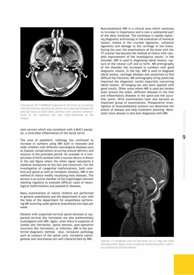
The area of paediatric radiology has continued to increase in numbers using MRI both in neonates and older children with different neurological diseases such as hypoxic complications occurring around delivery and seizures in the postnatal period. An example of a com-plication of birth assisted with a suction device is shown in the last figure where the white signal represents a subdural hematoma of the falx and tentorium. For the investigation of congenital malformations, both cere-bral and spinal as well as metabolic diseases, MRI is the method of choice readily visualizing most diseases. The section is an active member of the Copenhagen network meeting regularly to evaluate difficult cases of neuro-logical malformations and paediatric diseases.
Many examinations of mainly children are performed in general anaesthesia and the department is now with the help of the department for anaesthesia perform-ing MR-scanning under general anaesthesia two days per week.
Patients with suspected cervical spinal stenoses or sus-pected cervical disc herniation are also preferentially investigated with MRI. Again, when there is suspicion of lumbar disc herniation, spinal stenosis, post-operative recurrent disc herniation, or infection, MRI is the pre-ferred diagnostic method. Also, intradural pathology such as tumours of the spinal cord, intradural menin-geomas and neurinomas are well characterised by MRI.
Sagittal T1-weighted scan of the brain of a 7 day old child showing white signal from a subdural hematoma after a deliv-ery assisted by suction device.
Musculoskeletal MRI is a clinical area which continues to increase in importance and is now a substantial part of the daily workload. The technique is rapidly replac-ing diagnostic arthroscopy in the evaluation of meniscal lesions, lesions in the cruciate ligaments, collateral ligaments and damage to the cartilage in the knees. During the year the examinations of the knee with the 3T scanner has become the method of choice with valu-able improvement of the investigative results. In the shoulder, MRI is used in diagnosing labral lesions, rup-ture of the rotator cuff and so forth. MR-arthrography of the shoulder has increased in numbers with good diagnostic results. In the hip, MRI is used to diagnose labral lesions, cartilage diseases and sometimes to find difficult hip fractures. MR-arthrography of hip joints has improved the diagnostic results especially concerning labral lesions. 3D-imaging has also been applied with good results. Other areas where MRI is used are tendon tears around the ankle, different diseases in the foot and inflammatory diseases in the spine and the sacro-iliac joints. Wrist examinations have also become an important group of examinations. Preoperative inves-tigation of musculoskeletal tumours can determine the extent of disease and help treatment planning. Meta-static bone disease is also best diagnosed with MRI.
Transversal 3D T1 MPRAGE sequence of the brain of a grownup with Gd-contrast showing the facial nerve passing through the petrosal bone on both sides (arrows) with contrast enhance-ment on the inflamed left side (right-hand-side of the image).

10
DRC
MR
Annu
al R
epor
t 20
08Ba
sic
Rese
arch
As a major MR research facility, the DRCMR uses cut-ting-edge methodologies to advance MR methods and techniques on an international level for the benefit of patients. Basic research is a key component in this. Unlike research targeted at immediate application, basic research is the foundation for future clinical application or applied research, and it is therefore a key focus area within the DRCMR. It includes• developing and evaluating new methods which can
provide quicker, more accurate and more robust results. Example areas are development of measure-ment methods, hardware (e.g. monitoring systems), software and analysis methods (statistical calcula-tions, optimization, mathematical modeling, etc).
• research that leads to a better biological understand-ing of the healthy human body and the interaction between biology and MRI methods. Example areas include research in brain function and how physi-ological factors are reflected in MR images.
The basic research at the Centre can be divided into six categories:1. MR physics and methodology2. Development of novel post-processing strategies
and experimental design (MR informatics)3. Mapping of the cognitive functions in the brain
(brain mapping)4. Investigation of the neural bases of personality
dimensions with special emphasis on the serotoner-gic neurotransmitter system (Cimbi)
5. Studying the maturation of the brain in children6. Studying the neural mechanisms of decision makingThe activities of the Centre within each of these cat-egories are described in the following.
MR Physics and Methodology
MR measurements involve complicated patterns of radio wave transmission, magnetic field changes and signal sampling. The measurement schemes programmed for the scanner are called sequences. Although numer-ous clinical MR sequences are provided with the MR scanners by the scanner manufacturers, there are a variety of research projects within the Centre that rely on sequences that are either written in-house or are modified versions of provided sequences. The Centre therefore has research agreements with Siemens and Varian that give researchers access to the source code of the manufacturers’ sequences. This is necessary to modify and optimize MR methods.
Education in MR physics and methodology is a focus area at the DRCMR since a basic knowledge of MR techniques is required to interpret MR measurements correctly. A well-attended and free annual course open for exter-nal participants has been offered for years. In 2008 it was extended with an application part coordinated by Arnold Skimminge. The acquisition part of the course was as usual coordinated by Lars G. Hanson, whose widely used course notes are available on the DRCMR
BASIC RESEARCH
homepage together with educational software used internationally.
Teaching of MRI is challenging, since the many MR meth-ods involve quite complicated manipulations of atomic nuclei using radio waves and gradient pulses. The com-plexity of these matters is reflected in the fact that several Nobel prizes were awarded for the invention of basic MR methods. These can nevertheless be under-stood by most, when explained properly. For a number of MR educators, it has been a source of frustration that most books on MRI actually do not explain basic MR well. Quantum mechanics is often presented as being necessary whereas MR is actually fully understandable in terms of classical mechanics that is an intuitive set of physical laws describing everyday phenomena. In contrast, quantum mechanics offers a more complete description of certain phenomena, but it has coun-ter-intuitive aspects and is incomprehensible to most people, including many authors of books on MRI who, nevertheless, try to describe MR in terms of quantum mechanics. Typically, quantum mechanics adds noth-ing but confusion to descriptions of MRI. This has been known by some MR physicists for decades, but it is not recognized widely. In 2008, Lars G. Hanson lost patience and published a paper called “Is quantum mechanics necessary for understanding magnetic resonance?” arguing that many introductory books contain myths and
Scene from an animation available on the DRCMR website (http://www.drcmr.dk/MR) demonstrating how resonant radio waves can rotate the magnetic moments of a collection of nuclei oriented almost randomly. Figures like this repre-sent better alternatives to typical counter-intuitive figures inspired by quantum mechanics appearing in MR text books (more correct from both quantum and classical viewpoints). This was pointed out in a DRCMR paper published in 2008 that spurred considerable positive interest.
11
DRC
MR
Annu
al R
epor
t 20
08Ba
sic
Rese
arch
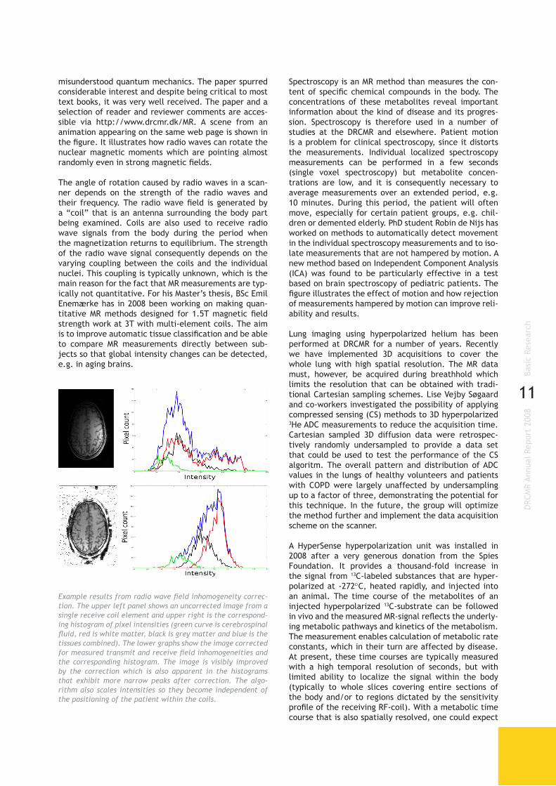
misunderstood quantum mechanics. The paper spurred considerable interest and despite being critical to most text books, it was very well received. The paper and a selection of reader and reviewer comments are acces-sible via http://www.drcmr.dk/MR. A scene from an animation appearing on the same web page is shown in the figure. It illustrates how radio waves can rotate the nuclear magnetic moments which are pointing almost randomly even in strong magnetic fields.
The angle of rotation caused by radio waves in a scan-ner depends on the strength of the radio waves and their frequency. The radio wave field is generated by a “coil” that is an antenna surrounding the body part being examined. Coils are also used to receive radio wave signals from the body during the period when the magnetization returns to equilibrium. The strength of the radio wave signal consequently depends on the varying coupling between the coils and the individual nuclei. This coupling is typically unknown, which is the main reason for the fact that MR measurements are typ-ically not quantitative. For his Master’s thesis, BSc Emil Enemærke has in 2008 been working on making quan-titative MR methods designed for 1.5T magnetic field strength work at 3T with multi-element coils. The aim is to improve automatic tissue classification and be able to compare MR measurements directly between sub-jects so that global intensity changes can be detected, e.g. in aging brains.
Spectroscopy is an MR method than measures the con-tent of specific chemical compounds in the body. The concentrations of these metabolites reveal important information about the kind of disease and its progres-sion. Spectroscopy is therefore used in a number of studies at the DRCMR and elsewhere. Patient motion is a problem for clinical spectroscopy, since it distorts the measurements. Individual localized spectroscopy measurements can be performed in a few seconds (single voxel spectroscopy) but metabolite concen-trations are low, and it is consequently necessary to average measurements over an extended period, e.g. 10 minutes. During this period, the patient will often move, especially for certain patient groups, e.g. chil-dren or demented elderly. PhD student Robin de Nijs has worked on methods to automatically detect movement in the individual spectroscopy measurements and to iso-late measurements that are not hampered by motion. A new method based on Independent Component Analysis (ICA) was found to be particularly effective in a test based on brain spectroscopy of pediatric patients. The figure illustrates the effect of motion and how rejection of measurements hampered by motion can improve reli-ability and results.
Lung imaging using hyperpolarized helium has been performed at DRCMR for a number of years. Recently we have implemented 3D acquisitions to cover the whole lung with high spatial resolution. The MR data must, however, be acquired during breathhold which limits the resolution that can be obtained with tradi-tional Cartesian sampling schemes. Lise Vejby Søgaard and co-workers investigated the possibility of applying compressed sensing (CS) methods to 3D hyperpolarized 3He ADC measurements to reduce the acquisition time. Cartesian sampled 3D diffusion data were retrospec-tively randomly undersampled to provide a data set that could be used to test the performance of the CS algoritm. The overall pattern and distribution of ADC values in the lungs of healthy volunteers and patients with COPD were largely unaffected by undersampling up to a factor of three, demonstrating the potential for this technique. In the future, the group will optimize the method further and implement the data acquisition scheme on the scanner.
A HyperSense hyperpolarization unit was installed in 2008 after a very generous donation from the Spies Foundation. It provides a thousand-fold increase in the signal from 13C-labeled substances that are hyper-polarized at -272°C, heated rapidly, and injected into an animal. The time course of the metabolites of an injected hyperpolarized 13C-substrate can be followed in vivo and the measured MR-signal reflects the underly-ing metabolic pathways and kinetics of the metabolism. The measurement enables calculation of metabolic rate constants, which in their turn are affected by disease. At present, these time courses are typically measured with a high temporal resolution of seconds, but with limited ability to localize the signal within the body (typically to whole slices covering entire sections of the body and/or to regions dictated by the sensitivity profile of the receiving RF-coil). With a metabolic time course that is also spatially resolved, one could expect
Example results from radio wave field inhomogeneity correc-tion. The upper left panel shows an uncorrected image from a single receive coil element and upper right is the correspond-ing histogram of pixel intensities (green curve is cerebrospinal fluid, red is white matter, black is grey matter and blue is the tissues combined). The lower graphs show the image corrected for measured transmit and receive field inhomogeneities and the corresponding histogram. The image is visibly improved by the correction which is also apparent in the histograms that exhibit more narrow peaks after correction. The algo-rithm also scales intensities so they become independent of the positioning of the patient within the coils.

12
DRC
MR
Annu
al R
epor
t 20
08Ba
sic
Rese
arch
to be able to detect and discriminate differences in the kinetics of the metabolism and the metabolic conver-sion rates between sub-regions of an organ of interest. Methodological MR research was therefore initiated during 2008 by Peter Magnusson and co-workers with the aim to develop and optimize sequences for metabolic imaging of hyperpolarized substances at the pre-clinical MR-scanner, and to further explore the possibilities to increase the spatial and temporal resolution. A spec-troscopic imaging sequence (CSI) was implemented on the pre-clinical MR-scanner, along with reconstruc-tion, which was optimized for spectroscopic imaging of hyperpolarized 13C-labelled pyruvate. To facilitate high SNR, the sequence employs a spiral trajectory in order for the central parts of k-space to be sampled first while the signal has the highest amplitude. This spiral k-space trajectory also limits the risk of large jumps in signal between adjacent points in k-space and thereby limits the risk for ringing effects in the MR-image. In order to reduce the acquisition time, the sequence was designed to sample only an elliptical part of k-space excluding the corners. This sequence was further modi-fied to a second version, without spatial encoding, for slice-selective time series measurements with high temporal resolution. Both versions were adapted and used for the two applications of hyperpolarized 13C of cardiac ischemia and breast cancer as described in the Preclinical research section of this report. A multi-echo version of a fast spin-echo sequence (RARE) was imple-mented for rapid metabolic imaging which is the first step towards increasing both the spatial and the tempo-ral resolution beyond what is currently achievable with the CSI-sequence on the pre-clinical system.
Diffusion imaging is a special MRI technique that offers a unique probe into the microstructure of brain tissue, and with the addition of tractography, allows the visu-alisation of the brain’s internal connectivity. Today most clinical research projects at DRCMR incorporate diffusion MRI to probe changes in brain connectivity and WM microstructure which are consequences of, for example, normal maturation in school children, or of diseases such as MS, dementia or traumatic injuries. The enormous potential of in vivo diffusion MRI drives improvements of complex methods, and motivates development of novel approaches. However, the need for method verification and validation also increases, and basic research into the ways in which diffusion MRI can be adjusted to reflect specific microstructure, such as cellular organization including cell size and density, become more important.
It was in this vein that Tim Dyrby defended his PhD in 2008, entitled “Modelling Brain Tissue using Magnetic Resonance Imaging”, financially supported by the Velux Foundation, and performed as a collaboration between the Technical University of Denmark, the Department of Neurology at Righospitalet and DRCMR. As part of the research project he investigated the validity of proba-bilistic tractography methods typically used in clinical research projects, employing dual in vivo non-inva-sive tracers injected into pig brains as a gold-standard against which to reference. The validation study under-lined the potential of tractography methods, but also
The top panel shows spectra from 48 repeated spectroscopy acquisitions of an 8 ml voxel located in the brain of a pre-term infant. Metabolite signals appear as vertical stripes. If motion was absent, all rows in the image should be similar but there is significant movement, particularly around acqui-sition number 33. The middle panel shows how Independent Component Analysis (ICA) can group data into similar spectra (components). ICA reveals in a fully automated way that the spectra changed at acquisition number 33 and that the head did not return to the original position. The bottom panel pro-vides a comparison between different strategies to correct for motion. The choline and creatine peaks at 3.0 and 3.2 ppm are shifted due to motion, and considerable variation is seen depending on the analysis method employed. The novel method based on ICA was found to consistently improve spec-tral quality the most.

13
DRC
MR
Annu
al R
epor
t 20
08Ba
sic
Rese
arch
demonstrated pitfalls in the detection, i.e., “false positive” and “false negative” fibre tracts as well as potential termination problems in the smaller deep-GM structures. The published study has already been highly cited, and the results widely used in educa-tional courses at international meetings, e.g. ISMRM in Toronto, 2008, and Human Brain Mapping in Melbourne, 2008. As part of the validation study, Tim Dyrby and co-workers also developed a postmortem imaging pipeline, implemented on a high-field experimental MR scanner, to generate high-quality diffusion MRI datasets with high SNR and image resolution, and importantly, without the usual artifacts characteristic of in vivo scans, such as physiological noise and sequence related distortions, which can substantially degrade the image quality.
After his PhD, Tim Dyrby was appointed at DRCMR as Research Principle for the diffusion MRI group (leading and coordinating role) to continue his basic research in diffusion MRI method development, and with an impor-tant remit to further the links between state of the art diffusion research and clinical studies.
In 2008, Matthew Liptrot started working on a method development project within the diffusion MRI group aimed at addressing the very common problem of path-length dependency (PLD) in tractography data. PLD is simply the reduction in the ability of a streamline - a calculated trajectory through a diffusion data space - to find its way from a seed to a target region. This implies that long-range connections are difficult to find, and as the level of difficulty varies depending on the sub-ject, it means that comparing tracking results between different subjects becomes extremely problematic. As such, this is one of the major hurdles preventing more widespread use of tractography in the clinical environ-ment. A new framework for performing such tracking is therefore being developed, ICE-T (Iterative Confi-dence Enhancement for Tractography), which is hoped will overcome the PLD problem and for the first time allow the exciting results offered by tractography to be directly compared between subjects.
Henrik Lundell is a PhD student at DRCMR and Depart-ment of Exercise and Sport Sciences at the University of Copenhagen, working mainly with diffusion MRI of the spinal cord. As one part of his project, he is adapting methods for diffusion tensor imaging (DTI) for use in patients with spinal cord injuries. Due to the demand-ing imaging conditions around the spinal cord, care has to be taken to reduce artifacts from cardiac pulse, res-piration and susceptibility induced distortions. During development of the method, and to achieve additional anatomical information and a golden standard refer-ence, postmortem scans are also being performed on fixed tissue using the high-field preclinical scanner at the department. The diffusion imaging results will provide a measure of cervical spinal cord integrity and will be compared with clinical and electrophysiological data. The applicability of the method to clinical use will then be assessed.
MR Informatics
Many studies running at the DRCMR utilise images of different modalities and with differing contrasts. In order to perform tasks upon this multitude of images, including registration, normalisation, preprocess-ing and analysis in an efficient and reproducible manner, a common framework for these tasks is cru-cial. The major task of implementing a standardised environment for all studies based on the popular SPM5 software package was initiated by PhD-student Arnold Skimminge and co-workers. In particular recent developments such as physiological noise modelling for fMRI studies and methods for DTI data processing have been implemented into the standard workflow of ongoing studies, and hence these methods can now be easily incorporated into the analysis of MR data. Neuroimaging often includes data from several modali-ties, e.g. structural MRI, functional MRI (fMRI), and PET as well as “metadata” such as age and gender. However, most analysis methods are modality-spe-cific (considering only one modality at a time). Peter Mondrup, concluded his DTU MSc project entitled “Mul-timodal analysis of the serotonergic neurotransmitter system” during 2008, where the focus was on integra-tion of multiple modalities into a single analysis. Such multimodal analysis performed on voxel-level can be used to asses more complex questions in neuroscience. Kristoffer H. Madsen concluded his PhD-thesis entitled “Modelling Strategies for Functional Magnetic Resonance Imaging” during 2008 with focus mainly on unsupervised analysis of fMRI data. In particular this includes factor analysis type decompositions, independent component analysis and the extensions to higher order decompo-sitions such as the canonical decomposition / parallel factor analysis model. The work has focused mainly on extending these simple linear decompositions in order to provide more realistic models for the analysis of fMRI signals. In particular extension of these models to include shifts and imposing several constraints including temporal/spatial smoothness, sparseness and non-nega-tivity have been developed together with Morten Mørup from the Technical University of Denmark. These meth-ods can help in more accurately identifying activated regions in fMRI data as well as reduction of nuisance effects by explicitly modelling of these effects. The decomposition algorithms have been developed spe-cifically with computational efficiency in mind making them suitable for large scale data sets such as multi-subject fMRI data.
Brain Mapping
An extremely useful aspect of MRI is its ability to meas-ure functional activation of the brain based on the magnetic properties of oxy- and deoxy-haemoglobin. This technique of functional MRI (fMRI) is widely used to make functional maps of the brain. This is one of the key research areas at the DRCMR and a variety of projects rely on this technique to achieve a better understand-ing of brain function.

14
DRC
MR
Annu
al R
epor
t 20
08Ba
sic
Rese
arch
Together with Michael J. Grey from the Department of Exercise and Sport Sciences and Department of Neuroscience and Pharmachology at the University of Copenhagen, Mark Schram Christensen conducted a study in which they investigated the role of proprio-ceptive feedback in functional electrical stimulation (FES). FES is a technique used in rehabilitation typically after stroke. FES helps patients to perform movements by applying current stimulation to the muscles, which then are moved automatically. Therapeutic functional electrical stimulation (TFES) uses patterned electrical stimulation of muscle synergies (i.e. FES) in conjunc-tion with voluntary motor drive. Previously, studies have demonstrated that cortical areas responsible for sensory-motor integration are active during TFES, in particular secondary somatosensory cortex (SII). In the present study Michael Grey and Mark Schram Chris-tensen investigated if peripheral sensory feedback is required for the cortical activation patterns revealed during movements performed with TFES. To investigate this question they used ischemic nerve block (INB) to block transmission of peripheral afferents while sub-jects performed finger movements with or without electrical stimulation during fMRI. They tested the hypothesis that cortical activation will decrease during INB in regions responsible for sensory motor integration during the TFES and FES conditions. They found a signif-icant interaction between the presence and absence of proprioceptive feedback and voluntary movements and FES. The study showed SII activation was reduced during
INB but only significantly for the FES condition and that peripheral ischemia reduced sensory motor cortex acti-vation for the FES condition but only slightly for TFES. The study may provide insight into the underlying physi-ology for the clinical benefits previously reported for therapeutically applied FES. The work was presented at the Society for Neuroscience’s annual meeting in Wash-ington DC USA.
Eye position helps locate visual targets relative to one’s own body and modulates the distribution of attention in visual space. While in the monkey, proprioceptive eye position signals have been recorded in the soma-tosensory cortex, in humans, no brain site has yet been associated with eye position. Daniela Balslev and her colleague from Birmingham, Chris Miall, aimed to disrupt the proprioceptive representation of the right eye in the left somatosensory cortex, presumably located near the representation of the right hand, using repetitive transcranial magnetic stimulation (rTMS). Head-fixed subjects reported their perceived visual straight-ahead position using both left and right eye monocular vision, before and after 15 min of 1 Hz rTMS. rTMS over the left somatosensory but not over the left motor cortex shifted the perceived visual straight ahead to the left, whereas non-visual detection of body midline was unchanged for rTMS over either brain area. These results can be explained by the underestimation of the angle of gaze of the right eye when fixating the target. To link this effect more tightly to an altered ocular
Bayesian detection of nuisance effects in fMRI time series: The 4 panels show the spatial distribution of complexity (model order) of each of the 4 nuisance signal types (low frequency drift – top left, motion – top right, cardiac cycle – bottom left ,respiration –bottom right). Effects of the highpass-filter are widely distributed over the brain whereas motion is most prominent at the edges of the brain. At the large arteries of the brain such as the Circle of Willis and the Medial Cerebral Artery the detected model com-plexity is highest as it is to be expected. Respiratory effects are present at the edges of the brain as well as in the ventricles.

15
DRC
MR
Annu
al R
epor
t 20
08Ba
sic
Rese
arch
proprioception, we applied a passive deviation to the right eye before the visual straight-ahead task. Passive eye displacement modulated the shift in the perceived straight ahead induced by somatosensory rTMS, without affecting the perceived straight ahead at baseline or after motor cortex rTMS. Daniela Balslev and Chris Miall concluded that the anterior parietal cortex in humans encodes eye position and that this signal has a proprio-ceptive component.
As a part of his PhD project, Henrik Lundell is, together with Dorothy Barthélemy and Jens Bo Nielsen, com-paring cortical activity for patients with spinal cord injuries and normal controls. The aim of the study is to investigate the role of cortical plasticity after spinal cord injuries and to understand the role of subcortical networks in regeneration of locomotion after rehabili-tation.
Cimbi
The Lundbeck Foundation Center for Integrated Molecu-lar Brain Imaging (Cimbi) was founded in 2006 with the DRCMR being a main participant. The research in Cimbi focuses on the neural bases of personality dimensions that predispose individuals to affective and substance use disorders, with special emphasis on the seroton-ergic neurotransmitter system. Both PET and MRI are employed in studies of human subjects, and these are complemented with relevant studies using animal models. Advanced informatics techniques, new tracer
compounds, and novel serotonergic challenge para-digms are also being developed within the Centre.
The role of the DRCMR in Cimbi is twofold: Terry Jerni-gan leads a project focusing on the relation between personality, biochemistry and brain structure while Olaf B. Paulson heads a group focusing on functional brain imaging under serotonergic challenges.
One of the important issues addressed in Cimbi is the influence on human behaviour of genes that affect the brain serotonin system. Previous brain imaging research suggests that one of the ways that these genes may act is through their influence on the structure of the brain, perhaps during the process of brain development. That is, genetic polymorphisms may influence the size of certain cell populations in the brain, or the numbers of connections that are established, or preserved, between specific brain structures. This may lead to dif-ferences in brain morphology. Structural neuroimaging methods continue to improve in sensitivity and anatom-ical resolution, and it is now possible to examine brain morphology, and even the physical connections between brain areas in remarkable detail. The major aim of this
Areas showing a significant interaction between movement type, i.e. functional electrical stimulation and voluntary movement, and presence or absence of proprioceptive feedback.
CimbiCenter for integrated molecular brain imaging

16
DRC
MR
Annu
al R
epor
t 20
08Ba
sic
Rese
arch
project is to apply these new structural neuroimaging approaches to volunteer subjects, so that differences in anatomy can be linked to genetic variability on the one hand, and to personality traits, cognitive functions, and other functional parameters on the other. Thus it may be possible to determine to what extent genetic influences on serotonin function and behaviour may be mediated by their effects on the brain’s anatomical structure.
Senior researcher William Baaré and PhD-student Kath-rine Skak Madsen are working on the morphological Cimbi project. Jens Bundgaard left the team in the beginning of 2008 and in October 2008 the team was strengthened with Pernille Iversen who has a PhD in Bioinformatics. Pernille extended and improved the Cimbi related MR database and is especially involved in cortical thickness/shape analyses, automated struc-tural segmentation and multivariate statistical image analyses.
In the third year of Cimbi the project team continued to improve the integration between the activities of the current project with those of other projects in Cimbi.
Considerable attention has been given to the integra-tion of the anatomical information provided with the serotonin receptor imaging. Several data sets were generated for further (voxel-wise) analyses including datasets for assessing anatomical and serotonin receptor binding asymmetries. Together with Cimbi investigators we continued our work on implementing and improv-ing image analysis procedures on the Cimbi computing cluster at the Danish Technical University (see figures). The project also continued to contribute actively to the efforts to design and implement a centralized database for Cimbi observations. This initiative is increasingly important as Cimbi investigators begin to perform the cross-project analyses that will take advantage of syn-ergy within the centre.
Last year we, together with our collaborators, observed healthy individuals who have a twin afflicted with major depression (i.e. high risk subjects) had reduced hip-pocampal volumes. This “marker” of depression may either be associated with the genetic predisposition for depression, or to environmental factors that are shared by affected and healthy twins growing up in the same environment, but it is unlikely to be entirely due to the depressive illness itself or to the treatments for depres-sion, neither of which afflicted these healthy co-twins of depressed individuals. Recent preliminary findings suggest that the observed hippocampal volume reduc-tions might be associated with specific polymorphisms of the serotonin transporter gene (5HTTLPR), which have been related to affective and anxiety disorders, neurot-icism and reduced hippocampal volume. In agreement with a recent study in patients with major depression
Coronal (upper left), sagittal (upper right), and axial (lower left) slices depicting the mean FA skeleton (in red) overlaid on a mean FA image. The cingulum is depicted in blue (note that only part of the cingulum can be visualized in 2D). In TBSS all subjects’ FA images are aligned into a common target space using high dimensional nonlinear registration. Next, a cross-subject mean FA image is created and thinned to create a mean FA skeleton, representing the centres of all tracts common to the group. Each subject’s aligned FA image is then projected onto the mean skeleton by locating the highest local FA value in the direction perpen-dicular to the skeleton tracts and assigning this value to the skeleton.
DARTEL (“Diffeomorphic Anatomical Registration Through Exponentiated Lie algebra”) performs high dimensional non-linear inter subject registration allowing voxel-wise group as well as region of interest analyses. (Left) Top row shows a rendering of the aver-age GM map of 112 subjects. The bottom row shows an axial, coronal and sagittal slice through the average of the 112 warped T1 weighted structural images. The clear definition of gyri and subcortical structures indicate that DARTEL successfully diminished inter-individual anatomical variation. (Right) Each box depicts axial GM and CSF slices of two subjects before (top row) and after (bottom row) warping.

17
DRC
MR
Annu
al R
epor
t 20
08Ba
sic
Rese
arch
we observed that healthy individuals at risk of major depression and homozygous for the long variant of the 5HTTLPR polymorphism had smaller right hippocampal volumes as compared to healthy high-risk individuals with a short variant and low risk healthy individuals.
We made significant progress in analyzing diffusion weighted imaging data, which allows assessing white matter microstructure integrity. Specifically we inves-tigated the cingulum, which is the major fibre tract connecting the cingulate gyrus to medial temporal brain structures such as the amygdala and hippocampus. These brain structures are known to play an important role in emotional processes. Using Tract-based Spatial Statistics (TBSS, see figure) to allow group comparisons of major fibre tracts in the brain, we observed that a shift in laterality of fractional anisotropy (FA; a measure of white mater integrity) of the cingulum, adjusted for age and gender, was linked to higher trait neuroticism, a known risk factor for affective disorders, in a cohort of healthy subjects participating in the Cimbi project.
The aim of the functional studies is to investigate the relationship between the cerebral activation responses and the serotonergic system using fMRI. The work is coordinated by post-doc Julian Macoveanu and involves two PhD-students, Bettina Hornbøll and Jon Wegener. Jon has finished his PhD during the year and moved on to new challenges. The research focuses heavily on a set of behavioural constructs that have been linked to serotonergic function: self-discipline, vulnerability (and trait anxiety), and decision making. Activation paradigms for use with fMRI were designed to evoke, or probe, each of these behavioural dimensions, and the first results show they reliably produce activity in the fronto-limbic regions known to have high con-centrations of 5-HT1A, 5-HT2A and 5-HT4 receptors (see figures). To elucidate the role of serotonergic function in these mental processes, brain activation patterns are being studied under challenge of the serotonergic system by different types of drug interventions. Addi-tionally, the activation responses will be correlated to
the receptor density and to genetic polymorphism of serotonin receptors and transporters, data provided by other Cimbi groups. In collaboration with another group lead by Lars Kessing at the psychiatry department at Rigshospitalet we have initiated a new functional MRI project where we used the same activation paradigms in a different group of subjects. The subjects recruited by Lars Kessings’s group have a high risk of developing depression, a condition linked to an imbalance in the serotonergic system.
Brain Maturation
The brain maturation study (“Hjernens Udvikling hos Børn
og Unge“: HUBU) in school-aged children was started in 2007. This project is led by Professor Terry Jernigan and represents collabora-
tion between the DRCMR and Learning Lab Denmark of
the Danish School of Education, Aarhus University. Also partnering
in the project are three local schools, their pupils, and their families. The project involves two annual exami-nations with psychological and academic tests as well as structural brain imaging. Recent research has shown that biological maturation of brain tissues continues throughout childhood in the form of changes in corti-cal morphology, and particularly, in changing structure of the brain’s fibre tracts. What is less well-known is the degree of individual variability in the timing and pattern of these changes, and whether such variability has relevance for the developing mental functions of the child. These questions are the main focus of this study.
Ninety children, ranging in age from 7 to13 years, were studied at their baseline visits, and many completed their third visit to the department. The fourth assess-
Decreased activity in orbito-frontal cortex caused by blockade of serot-onin receptors during an emotional face processing paradigm.
Statistical map showing correlations during two conditions of a gambling task, assumed risk (red) and winning (green).

18
DRC
MR
Annu
al R
epor
t 20
08Ba
sic
Rese
arch
ment will be completed in the beginning of 2009. The original research protocol has been extended to include the assessment of personality, alcohol and drug use, as well as social and emotional functioning. With respect to the latter we among other things implemented an emotional go/no-go task.
Two manuscripts were written on measures of fibre tract organization in specific neural systems and the relation-ship with behavioural inhibition and working memory, respectively (see figure). Both the capacity to successfully inhibit a “primed” response and the ability to keep in mind and use an accurate spatial model of the environment are func-tions that continue to develop substantially over the school-aged years. For both of these functions, specific neural systems have been implicated in adults. In the DRCMR study, measures of organization of fibre tracts in these systems were significantly correlated with the behavioural measures, and the analyses suggested that these associations were relatively specific to the organ-ization of the implicated neural systems.
Though these are exciting results, the challenge is to interpret them correctly. Children may vary in the phase of maturation in the brain networks subserving response inhibition and working memory, and this vari-ability may mediate these associations. This is plausible since both fibre tract maturation and these functions continue to develop during this age range. Alternatively,
the associations could be mediated by stable individual differences reflecting underlying neural system connec-tivity. Longitudinal observations currently continuing in the DRCMR are needed to help distinguish between these, and other, explanations.Finally, preliminary results suggest that changes in grey matter (reduction) and white matter (increase) over time can already be observed over a 6 months period.
Decision Neuroscience
The Decision Neuroscience Research Group (DNRG) was initiated in early 2008 with the collaboration between DRCMR and Professor Flemming Hansen at the Copenhagen Business School, and was formalised by the employment of Thomas Z. Ramsøy as a post-doc researcher at the CBS. Understanding the brain basis of preferences and decisions may generally improve models of human behaviour, and may influence concep-tions and actions towards less than optimal decisions, including financial decisions, gambling, political behav-iour and consumer behaviour.
The DNRG soon gained momentum and at the end of 2008 had approximately 7 researchers directly or indirectly connected to the group. Members of the group include people with divergent backgrounds, including economy, biology, psychology and literature. By the end of 2008, DNRG members included Jon S. Wegener (DRCMR/CBS),
TBSS skeleton in yellow and in red the right inferior frontal gyrus (IFG) overlaid on a FA map. B) Response inhibition improves significantly (e.g. lower stop signal task - SST - reaction times: SSRT) with higher right IFG FA. The partial regression plot shows the variation in SSRT explained by right IFG FA, adjusted for age and whole skeleton FA.
Axial (A) and Sagittal (B) views of significant grey matter volume decrease (Dark blue - Light blue scale) and white matter volume increase (Red - Yellow scale) in typically-developing children over a 6 month period. Numbers in the colour bar represent T- values (range 2.35 - 6).

19
DRC
MR
Annu
al R
epor
t 20
08Ba
sic
Rese
arch
Martin Skov (DRCMR), Sofie Gelskov (DRCMR/CBS), Lars Bech Christensen (CBS) and Jesper Clement (CBS).
The primary aim of this group is to study the neural underpinnings of preference formation and decision making in many different contexts. Projects initiated in 2008 include the neural bases of branding and memory, aesthetics and design, creativity, gambling, politics, and socio-behavioural economics.
The study of brands and brand mascots implies a study of how the brain stores culturally specific information. Brands and mascots are items that, within a particu-lar culture, represent a wider range of associations. Thus, one may contend that brands tap into the neural processes underlying general memory functions. In this study, the aim is to study whether brand mascots and brands are processed in different ways than generic fig-ures or well-known cartoon figures. Preliminary results suggest that, compared to generic figures, brand mas-cots increase the activation of areas such as the medial temporal lobe (MTL) and dorsolateral prefrontal cortex (PfC). This may suggest that the higher number of asso-ciations for brands mascots evoke stronger activations of memory-related activation.
Intertemporal choice involves a trade-off between get-ting a lower reward soon, and getting a higher reward after a longer delay. In our study, the aim was to study how the delay, independent of reward and subjec-tive value, is processed during intertemporal choices. Imaging results suggest that a network involving the temporo-parietal junction and anterior temporal lobe are decreasingly activated as a function of delay. This suggests that a network known in previous studies to underlie our ability to model the intentions of others may also be involved when modelling the intentions of our future selves.
Preliminary results demonstrating increased prefrontal acti-vations when a subject is looking at brand mascots compared to unknown (but visually similar) figures.
Group level fMRI results from inverse parametric modulation of time delay irre-spective of choice (n=19, p(FDR)<0.05). rTPJ is right temporo-parietal junction. rPT is right posterior temporal cortex. rAT is right anterior temporal cortex. lAT is left anterior temporal cortex, and lTPJ is left temporo-parietal junction.

20
DRC
MR
Annu
al R
epor
t 20
08Cl
inic
al R
esea
rch
Neuropsychiatric Disorders
Major depressive and bipolar disorder (MDD; BPD), and schizophrenia are severe psychiatric illnesses affect-ing respectively 4-8%, 1.3-1.6%, and 1% of the general population. Although the aetiologies of these diseases are unknown, genetic factors as well as environmen-tal factors are involved. Heritability estimates for MDD range between 31% and 66%. The heritability of BPD is approximately 70% and that of schizophrenia is around 80%. The underlying pathophysiology of the disorders is largely unknown. However, post-mortem and func-tional and structural in vivo neuroimaging studies have provided accumulating evidence for the presence of functional and structural abnormalities in the brains of patients with affective disorders and schizophrenia as compared to healthy controls. Indeed, in vivo imaging studies have been pivotal for our understanding of schizophrenia as a brain disease. Studies of the prodro-mal and early stages of a disease such as for example in first-episode (drug-naïve) schizophrenia patients are important as they control, to a large extent, for effects of factors such as long-term hospitalization, medication treatment and disease chronicity.
The MR investigations predominantly address the follow-ing questions: (a) which brain abnormalities are present before onset of a disorder? (b) Which abnormalities are related to an increased (genetic) risk to develop a disorder? (c) Which abnormalities are present at ill-ness onset? (d) Which abnormalities emerge during the course of the illness? (e) Which abnormalities progress in the first years of the illness? (f) How are these abnor-malities and changes related to cognitive functions, pharmaceutical treatment, behavioral symptoms, and social and medical history? (g) Which abnormalities and changes are predictive of treatment response and clini-cal outcome?
MR techniques used in the different projects include the following: structural MRI including T1, proton den-sity and T2-weighted, FLAIR and diffusion weighted imaging (DWI). DWI allows investigating white matter microstructure. Additionally, fMRI is used to investigate brain function while subjects are engaged in different cognitive tasks such as verbal working memory (in drug-naïve first episode schizophrenia patients), reward and emotional tasks (in the AGENDA project, see below).
Patients and healthy controls are recruited and clinically evaluated by the psychiatry departments at the Copen-hagen University Hospitals of Rigshospitalet (Affective disorders: Prof. Dr. Lars Kessing and Maj Vinberg, PhD), and Bispebjerg (Schizophrenia: Katrine Pagsberg. PhD) and the Center for Neuropsychiatric Schizophrenia Research (CNSR) at Glostrup Hospital (Schizophrenia: Professor Birte Glenthøj).
There are several ongoing projects. From the DRCMR Hartwig Siebner, William Baaré, and Arnold Skimminge are involved in all projects while Bettina Hornbøll and Julian Macoveanu, and Ayna Nejad, Bjørn Ebdrup (CNSR), and Trine Borg Hammer (CNSR), are involved in the affective disorders and schizophrenia projects respectively.
Projects in affective disorders include the UBFAL (“Udløsende og beskyttende faktorer ved affektiv lidelse”) project in which healthy mono– and dizygotic twins (age > 18 years) with a high and a low risk of
CLINICAL RESEARCH
Risk effect for hippocampal volume adjusted for age, gender and years of education
Voxel-wise non-parametric statistic results showing areas were patients (Ptall) had smaller caudate nucleus GM volumes than healthy controls (HC: yellow), areas where patients without a history of substance abuse (Pt non-ab) had smaller volumes than HC (purple), and the overlap of the two con-trasts (orange). Displayed voxels survived a FDR-correction (p< 0.05) using a small volume correction restricted to the caudate nucleus. Results are projected on sagittal slices of the average of all DARTEL warped MPRAGE images. From top to bottom row the images are respectively 13, 18, and 23 mm from the mid sagittal plane. Right hemisphere images are mir-rored and shown in the right column. Left hemisphere images are presented in the left column.

21
DRC
MR
Annu
al R
epor
t 20
08Cl
inic
al R
esea
rch
developing affective disorder are investigated and the AGENDA (“Associationer mellem genpolymorfier, endofænotyper for depression og antidepressiv behan-dling”) project in which 40 healthy subjects with at least one parent diagnosed with an affective disorder participate. A paper on grey matter changes in twins at risk for affective disorders was resubmitted in the end of 2008. People at risk tended to have reduced hip-pocampal volumes as compared to healthy controls (see figure). The data acquisition in the AGENDA will be con-cluded in the beginning of 2009.
Psychiatrist Bjørn Ebdrup is clinical responsible and PhD student on the project “Structural and functional brain changes in drug-naïve first-episode schizophrenia patients: relation to cognitive function and anti-psy-chotic medication”. Patients and controls were assessed longitudinally with a 6 month interval, in which patients were treated with clinical doses of quetiapine. A man-uscript on the structural MRI baseline assessments reporting reduced hippocampal and caudate volumes in patients as compared to healthy controls is in prepa-ration. Ayna Nejad is in the process of analyzing the verbal working memory baseline fMRI data. Patients as compared to controls showed among other things higher activity in the dorsolateral prefrontal lobe while failing to “turn off” non task related brain regions.
Other projects in schizophrenia include: “Structural and functional brain abnormalities in early onset first-episode schizophrenia” and “First episode psychotic children and adolescents: a 5 year follow-up study of brain structure and function” (Katrine Pagsberg); and a 5-10 year follow-up study of schizophrenia patients: “Skizofreni: Sygdomsprocessens kliniske, psykofysiolo-giske og neurobiologiske manifestationer” (Trine Bjørg Hammer, the last subjects for this study will be scanned in the beginning of 2009).
Brain Aging and Neurodegenerative Disorders
The Centre is the site of several studies of normal aging and the neurodegenerative disorders that afflict the elderly; and is a participating site in a broader multi-site investigation by European Union collaborators entitled, “Leukoaraiosis and Disability in the Elderly” (LADIS). The latter is an ongoing structural MRI study of the known changes that occur with aging in the white matter of the brain. The objective is to better describe the predictors and consequences of these changes. Eld-erly volunteers were scanned at entry into the study and a 3-year follow-up scan has now been completed. These measures are correlated with extensive neu-robehavioural assessments. In 2008 Ellen Garde, who initiated the LADIS study and defended her PhD-thesis from the DRCMR in 2000, and since has worked at the National Hospital of Neurology and Neurosurgery in
Boxplot of hippocampal volumes in the three subject groups. Volumes are corrected for age, gender and ICV. In the box-and-whisker plot, the central box represents the values from the lower to upper quartile. The transverse line in the box repre-sents the median corrected volume. The vertical line extends from the minimum to the maximum value, excluding outside values. Outside values are defined as values smaller or larger than the lower quartile minus 1.5 times the inter quartile range and are displayed as separate points (o). No outliers were identified.
a) Contrast showing the group-by-WM load interaction effect, significant at p=0.05, FDR cor-rected. b) the interaction effect contrast masked by the control group’s WM activation: red areas denote brain regions where activ-ity is increased significantly more from low to high WM load in schiz-ophrenia patients as compared to controls. Blue areas are brain regions in which schizophrenia patients show a failure of attenu-ated deactivation with increasing WM load.

22
DRC
MR
Annu
al R
epor
t 20
08Cl
inic
al R
esea
rch
London, replaced Egill Rostrup as senior DRCMR investi-gator involved with the LADIS studies.
Several DRCMR subprojects have developed from the LADIS initiative, all involving the development of advanced methods for automated measurement of abnormalities in cerebral white matter. Tim Dyrby who completed his PhD ‘Modelling brain tissue using Mag-netic Resonance Imaging’ also contributed to the LADIS study. He developed and validated a tissue segmenta-tion method based on an optimised artificial neural network to produce probabilistic maps of age-related white matter changes, the grey and white matter and cerebrospinal fluid (see figure). The automatic segmen-tation method was applied to MR scans of 362 subjects from the 11 centers in the LADIS study and demon-strated high consistency between subjects and centers but also discrepancy between semi-manual and neural network segmentation. Combining information from MPRAGE, T2 and the FLAIR image modalities signifi-cantly improved cross-center generalisability compared to neural networks using the FLAIR image only. Expert knowledge not available to the neural networks was a minor source of discrepancy, while variation in MR scan quality constituted the largest source of error. Due to the promising results of the segmentation method we
are in the progress of implementing the method in other relevant clinical studies such as in multiple sclerosis.
PhD-student Charlotte Ryberg is also related to the LADIS study focusing on the relationship between the corpus callosum, which is the major cerebral white matter structure carrying most interhemispheric connections, and age related white matter changes. An automated method to recognise and quantify the volume of corpus callosum has been developed in collaboration with the group of Professor Rasmus Larsen at the Technical Uni-versity of Denmark. The full cross-sectional dataset of 578 subjects has been analysed, and a significant cor-relation between the area of the rostrum and splenium regions of the corpus callosum and age-related white matter changes load in most brain regions demon-strated. Two papers were published in 2008 based on the work from this study.In order to assess the spatial distribution of age-related white matter changes Egill Rostrup developed a voxel-based approach in which lesion probability was mapped as a function of clinical risk factors. The method was applied on 605 subjects from the LADIS study and, interestingly, the distribution pattern seems to depend on risk factors involved suggesting regionally different pathogenesis in the white matter.
In 2008, Thomas Z. Ramsøy completed his PhD on the role of healthy aging on the functions of the brain. Using both structural and functional MRI methods, complemented by neuropsychological tests, the main focus was on the medial temporal lobe (MTL) structures including the amygdala, hippocampus and rhinal corti-ces. In particular, the PhD work focused on the role of age on MTL processing during episodic memory, where it was found that increasing age was associated with a general loss in the brain’s specialisation for different kinds of information, such as object and position infor-mation. These observed age-related alterations were most pronounced during encoding and retrieval of such information, and that the MTL region was particularly affected for object information. Further studies of the effects of aging on MTL function are now focusing on other cognitive and emotional aspects of MTL function. In addition, this project has led to the development of novel solutions to structural and functional imaging of this region, and a protocol for drawing Regions of Inter-est of MTL structures. These advances are currently being applied on the study of the effects of recreational use of ecstasy and hallucinogens on the structures and functions of both the MTL and the whole brain.
Multiple Sclerosis
The DRCMR has a long history of doing MRI research on multiple sclerosis (MS), and a major part of this research is performed in collaboration with external groups and thus has an extensive multidisciplinary input ranging from neuroimmunology and neurophysiology to neuropsychology.
MS is an inflammatory demyelinating and neurodegen-erative disease of the central nervous system (CNS). It is the leading cause of nontraumatic neurological dis-ability among young adults. About 85% of MS patients
A result of the tissue segmentation method based on opti-mised artificial neural network to produce probabilistic maps of white matter changes (WMC), the grey and white matter and cerebrospinal fluid in a subject with substantial age-related white matter changes. Re-sampled FLAIR image (A, B), semi-manual re-sampled WMC (C, D), and probability map of WMC using a F2 neural network (E, F). Automatic segmen-tation was found to be more conservative in detecting WMC compared to manual (arrow).

23
DRC
MR
Annu
al R
epor
t 20
08Cl
inic
al R
esea
rch
initially experience a relapsing-remitting clinical course (RRMS) with transient symptoms followed by a secondary progressive course (SPMS) characterized by gradual progression of disability. In the majority of cases patients show episodes of neurological dysfunc-tion (relapse, attack) separated by partial or complete recovery. MRI is a valuable tool for the diagnosis of MS and for monitoring the disease evolution. However, the correlation is limited between conventional MRI (T1, T2, and FLAIR) measures and clinical findings. There are several reasons for this mismatch: (a) the low patho-logical specificity of conventional MRI and the inability of conventional MRI to quantify the extent of damage in the so-called normal-appearing white matter (NAWM), (b) A relative mismatch between whole brain meas-ures of lesion burden by MRI and the fact that clinical measures of disability are relatively pathway and region specific and (c) the severity of clinical manifestations of MS does not result simply from the extent of tissue destruction, but rather represents a complex balance between tissue damage, tissue repair, cortical discon-nection and reorganization. In general, the MS research projects at DRCMR aim towards improving the specificity and sensitivity of MR imaging to detect the heteroge-neous pathological changes and cerebral adaptation in MS. In addition, we also use MRI as a tool to evaluate the treatment effects of different immunomodula-tory agents. The projects in this area are carried out by Henrik Lund, PhD-student Anne-Marie Dogonowski and senior researcher Xingchen Wu, who has extensive experience within the areas of both clinical and experi-mental MS research.
The pathological mechanisms of MS are investigated quantitatively by applying different MR techniques to two groups of MS patients. From these studies Henrik Lund and his collaborators hope to learn important details on the breakdown of myelin sheaths as well as of the blood-brain-barrier. The participating patients in one of the groups were newly diagnosed at entry and have been scanned three times – just before start of treatment, after 3 months and again after 6 months. This longitudinal study gives us an excellent opportu-nity to monitor the effects of different treatments. The patients in the other group have had MS for at least half a year and are volunteering in a cross-sectional study. For both groups the outcomes are correlated to a vast range of immunological and neurological measures col-
lected by our collaborators at Copenhagen University Hospital, Rigshospitalet.
The different MR-techniques all aim at the exploration of structural changes caused by the pathology of MS. For example, applying so-called q-space analysis to our diffusion data, it is possible to acquire structural information on the various biological barriers and com-partments. One limitation with traditional diffusion tensor imaging (DTI) is that the calculated diffusion coefficients are not expected to depend on the diffu-sion weighting or diffusion time. This is correct only in perfectly homogenous media and not in vivo where the diffusion of water is hindered by tissue structures. Normal DTI analysis gives one (apparent) diffusion coef-ficient (ADC) based on the assumption that the sample behaves as a perfect Gaussian distribution. However, an increased ADC does not tell us whether the viscosity is lowered or whether the sample has fewer barriers. The q-space analysis, which is based on several diffu-sion weightings provides a size distribution of the tissue compartments and thus potentially gives clinically rel-evant information on the breakdown of the structures. This technique will be used to analyse the water diffu-sion orthogonal to the fibres since an alteration in this diffusion is hypothesized to reflect a direct immunologi-cal breakdown of the myelin and/or axons. Additionally, anterograde (Wallerian) and terminal axonal degenera-tion as a response to focal lesions possibly gives rise to more diffuse changes. Hence, the approach is expected to provide information on diffuse as well as focal pathol-ogies and we aim at showing that q-space imaging can provide clinically relevant information in MS at scan-ning times suitable for the clinic. The data analyses and interpretation are performed in collaboration with Finn Sellebjerg, Rigshospitalet and Lars G. Hanson, DRCMR.
In addition, methods are currently being implemented to gain insight into the breakdown of the blood-brain-barrier. After contrast injection, focal enhancing lesions appear hyperintense on T1-weighted scans because con-trast agent accumulating in the tissue that surrounds a broken blood-brain-barrier increases the MR signal. It is hypothesized that a subtle breakdown of the bar-rier in regions that do not appear as focal enhancing lesions still gives rise to a measurable change in the signal intensity. This diffuse increase in signal intensity is measured quantitatively and compared to brain tissue of healthy subjects.
Functional MRI (fMRI) can give information on brain plas-ticity following MS-related structural injury, with the potential to limit the clinical manifestations of the dis-ease. Studies of cerebral activation in MS patients have successfully been carried out using the blood oxygen level dependent (BOLD) fMRI technique, with which metabolic and haemodynamic consequences of brain activity can be dynamically followed. Furthermore, it is now believed that processing of a functional task by the brain can only be performed through interaction of seg-regated regions within a complex network. Functional connectivity MRI (fcMRI) is a new method of assessing cerebral connectivity by mapping regions with synchro-nous slow fluctuations in cerebral blood oxygenation
Colour-coded FA map from a patient with relapsing-remit-ting multiple sclerosis. Bright colours indicate that water diffuses primarily in one direc-tion (nerve fibre bundles) while black indicates isotropic diffu-sion. The colours indicate the direction of the nerve fibures. Red is from left to right. Green is from front to back. Blue is from head to foot.

24
DRC
MR
Annu
al R
epor
t 20
08Cl
inic
al R
esea
rch
and flow. Assessing functional cerebral connectivity of resting state BOLD appears to be an informative way to determine the clinical impact of the overall diffuse and focal injury in MS. These techniques have recently been refined by our Centre by increasing the signal-to-noise ratio and improving data analysis. Anne-Marie Dog-onowski, Xingchen Wu and collaborators investigate the recovery mechanism of MS patients by evaluating the cerebral activation and functional cerebral connectiv-ity in MS patients during acute relapse and after clinical recovery. In order to explore the correlation between cerebral connectivity and clinical disability, a cross-sectional study of a group of MS patients with varying degrees of disability and a group of healthy controls are investigated by using the same method.
Diffusion-weighted imaging (DWI), which can quantify the extent of microstructure changes within and outside conventional MRI visible MS lesions. There is increasing evidence showing that DWI is sensitive to detect subtle changes in the NAWM, and the fibre tracking capabilities of DWI are well suited for evaluation of fibre pathway changes associated with MS lesions. These two groups of MS patients included in fcMRI studies are also investi-gated using whole brain DWI in 61 directions. In addition detection of immunological markers from blood sam-ples and clinical examinations is carried out at the same time points with MRI by collaborators at the Danish MS Center, Rigshospitalet. Patients are still being recruited for both projects.
MR spectroscopy (MRS) can provide information about neuronal loss or dysfunction by measuring N-acetyl aspartate (NAA), a suitable marker for neuronal integ-rity. Cognitive dysfunction can be seen in 50% of MS patients, and a substantial number of studies suggest that cognitive dysfunction is related to the overall dis-ease burden of the brain. A group of patients with early RRMS have been investigated using multi-slice echo planar spectroscopic imaging (EPSI) and a large battery of cognitive measures. The study showed that the cog-nitive performances of the patients were correlated to subtle pathological changes in the CNS. Rather intrigu-ingly, we showed that subtle changes in the so-called normal appearing white matter were partly responsible for the highly disabling deterioration of the cognitive function. This project has been carried out by Henrik K. Mathiesen, Lars G. Hanson, and in close collabora-tion with Per Soelberg Sørensen and Agnete Jønsson, both at Rigshospitalet. A 7 years follow up study of the same group of patients are undertaken by Xingchen Wu and Lars G. Hanson, DRCMR and Morten Blinkenberg and Agnete Jønsson, Rigshospitalet.
Traumatic Brain Injury
Traumatic brain injury (TBI), predominantly caused by motor vehicle accidents, is the leading cause of death and long-term morbidity among younger age groups in Western countries. In survivors of severe TBI the final outcome is both highly variable, ranging from almost full recovery to persistent vegetative state, and extremely difficult to predict, especially in cases of prolonged unconsciousness. Several different types of lesions can occur in TBI, but diffuse primary and sec-
ondary lesion types are thought to be major prognostic determinants. However, these diffuse lesions are highly underestimated by conventional imaging. Advanced quantitative MR techniques, such as diffusion tensor imaging (DTI), have the potential to improve detection of these important lesions and provide useful clinical tools for outcome prediction.
A PhD project on TBI is headed by Annette Sidaros who submitted her thesis in December 2008. The project is carried out in collaboration between the DRCMR and the Department of Neurorehabilitation, Brain Injury Unit, at Hvidovre Hospital. In this prospective longitu-dinal study, adult patients with severe TBI have been scanned at mean 8 weeks and 1 year post-injury. Healthy controls were scanned for comparison. In addition to conventional MRI sequences the project applied DTI and spectroscopic imaging. Clinical outcome of patients was evaluated at 1 year post-trauma.
Thirtyone patients have been included and scanned in the late subacute phase; of these 25 completed the 1-year follow-up scan. DTI data were published in Brain during 2008 and data-analyses focusing on the brain atrophy that occurs during the 1-year follow-up period has been completed and accepted for publication in NeuroImage during 2008. Advanced morphological tech-niques were used to assess both global and regional atrophy. At the first scan time-point, it was found that brain volume of patients was 8.4% smaller than that of the matched controls. Furthermore, during the scan interval, patients exhibited continued atrophy with percent brain volume change (%BVC) averaging -4.0%. Interestingly, %BVC improved prediction of long-term functional status over and above what could be pre-dicted using functional status at the first scan time point. Tensor-based morphometry, used for the assess-ment of regional atrophy, showed that atrophy was most pronounced in areas susceptible to traumatic axonal injury. The study indicates that the long-term atrophy is attributable to consequences of traumatic axonal injury. Despite progressive atrophy, remarkable clinical improvement occurred in most patients. The results of this project might provide important diagnostic, prognostic and pathophysiological informa-tion useful in the clinical management of brain-injured patients.
Neonatal Brain Maturation
Infants born prematurely are at risk of brain injury and neurodevelopmental deficits in later life. The pathogen-esis of brain lesions is still controversial but apparently both infection in pregnancy and perinatal ischemia influence the development of white matter damage. In an ongoing collaboration with the department of Pae-diatrics, a study headed by Maria J. Miranda aims to demonstrate an association between infection in preg-nancy and white matter damage in the immature brain at term-equivalent age. The study aimed at including 200 premature infants born at either Hvidovre Hospital or Rigshospitalet at a gestational age (GA) less than 33 weeks. The placenta was histologically and microbio-logically examined by a pathologist, while blood from the umbilical cord was examined for bacterial endotox-

25
DRC
MR
Annu
al R
epor
t 20
08Cl
inic
al R
esea
rch
ins and several inflammatory cytokines. These data are compared with the number and extent of brain lesions and lactate accumulation found in MR scans performed at term-equivalent age. MR single voxel spectroscopy (MR-SVS) data from the 100 infants scanned to date (analyzed by Robin de Nijs), revealed other important findings in the metabolic pattern of the brain of premature infants at term-equiv-alent-age as compared with healthy term-born controls. Choline/Creatine ratios were significantly different between the groups. The decreased Choline-levels might indicate either earlier or slower myelination in preterm infants (not seen on MRI), at least in some areas of the brain, even in preterm infants at term without brain lesions. The developed methods and results of this study have been accepted for publication.Another modality that was used to examine the pre-term infants is Diffusion Tensor Imaging (DTI) which can be used to study white matter microstructure as well white matter tracts. Comparing diffusion measures in the preterm infants with term-born controls revealed no differences between the groups where white matter structures of interest were traced manually. However, comparing the whole brain statistically with voxel based morphometry (VBM), an advanced development of white matter regions in occipital white matter was found. This is in contrast to current belief, where a delayed white matter development is expected due to the detrimental effects of prematurity. These data have been published during 2008 in Neuroimage. The DTI study was carried out by Maria J. Miranda in collaboration with Peter Born (a previous DRCMR researcher), Egill Rostrup, Lars G. Hanson and Terry Jernigan, DRCMR, as well as Zoltan Nagy, Karolinska Institute, Sweden and Monica Gimenez, Barcelona University, Spain.
Visual Plasticity
There is a strong tradition for investigating the visual system with fMRI at DRCMR. Several projects investigat-ing human vision with fMRI are currently ongoing. In a project that was recently initiated PhD student Astrid Rosenstrand Lou and co-workers investigate struc-tural as well functional MRI data to assess plasticity in the adult human visual cortex. The main focus of this project is on the anatomical and functional adaptabil-ity of the central nervous system throughout the entire human adult life-span. The project involves several experiments designed in order to investigate how the brain reacts to acute changes in visual input.Visiting Professor Maurice Ptito is heading a research program on congenital blindness supported by the Danish
Medical Research Council and the Harland Sanders Foundation in Canada. The research involves functional investigations of plasticity in congenital blindness using several different types of sensory stimulation including stimulation of the tongue by the so-called tongue dis-play unit (TDU). The TDU is able to project “images” on the tongue by pulsed electrical stimulation thereby enabling the possibility of delivering tactile information about visual scenes to blind subjects.
Pulmonary Function
Imaging of the lungs poses a number of difficulties with respect to traditional MRI. Large susceptibility differ-ences at the air-tissue interfaces cause the MR signal to decay very rapidly and, in addition, the proton density of lung tissue is low compared to other tissues. On con-ventional MR images the lungs appear as dark regions and only by using very sophisticated and advanced MR methods can some information about the lung structure and function be extracted from the images. Another MR approach is based on imaging an inhaled hyperpolarized gas. DRCMR has been active in this field for a number of years now, starting with the very successful EU funded PHIL (Polarized Helium for Lung Imaging) project.
The partners from the PHIL project and a few additional organisations formed the research network PHeLINet that obtained funding from the EU 6th Framework Pro-gramme as a Marie Curie Research Training Network. The PHeLINet network commenced in 2007 and supports one foreign Early Stage Researcher employed at the DRCMR. Torsten Dorniok from Germany was recruited for this position and is working as a PhD-student. In 2008 the network arranged two training schools to provide the recruited fellows in PHeLINet with the best possible background for their studies in the field of hyperpolar-ized helium lung imaging.
Lise Vejby Søgaard and Peter Magnusson are locally responsible for MR lung imaging at the DRCMR. The hyperpolarized 3He gas for the studies at the DRCMR is produced by the PHeLINet partner at the Physics Department at Johannes Gutenberg-University in Mainz, Germany and shipped to Copenhagen as air freight.
In 2007 a new clinical study on COPD (chronic obstruc-tive pulmonary disease) patients was initiated together with the Department of Cardiology and Respiratory Medicine. Thirty patients and 10 healthy smokers were recruited among the participants in the ECLIPSE study, a non-drug study aiming at identifying relevant markers of the progression of COPD disease. The selected sub-
Hyperpolarized 3He lung ADC maps in a healthy volunteer. The colorscale spans values from 0 to 1.0 cm2/s. The left image is acquired after inspi-ration to total lung capacity, the right shows the corresponding slice acquired after expiration to residual lung volume. The ADC correlates to alveolar size and is therefore useful to detect emphysema and could potentially be used to monitor progression of emphysema. By combin-ing inspiration and expiration measurements it is expected that additional information can be obtained.

26
DRC
MR
Annu
al R
epor
t 20
08Cl
inic
al R
esea
rch
jects are MR scanned 3 times with one year intervals in order to evaluate whether hyperpolarized 3He MRI can be used to monitor disease evolution. PhD-student Fre-derik Hengstenberg is responsible for this clinical study where 29 patients and 9 healthy smokers completed their first hyperpolarized helium MR scan in 2008.The MR protocol includes morphological imaging provid-ing information about the ventilation distribution and diffusion imaging that has been shown to correlate with the alveolar sizes in the lung. It has been suggested that the lung Apparent Diffusion Coefficient (ADC) values can be used as a sensitive marker for the progression of emphysema. The ADC is measured both in inspiration and expiration using a 3D sequence to provide full lung coverage at high resolution.
Clinical Trials
In 2007 a remarkable clinical trial was initiated at Hvidovre Hospital including the DRCMR: A potential treatment for the rare child disease Metachromatic Leukodystrophy (MLD) is currently being tested. In its late infantile form, MLD is a genetic disease that leads to demyelination of the brain. The children develop normally until approximately two years of age. Severe disabilities accumulate subsequently due to lack of an enzyme needed for sustained brain myelination. This horrible disease is normally fatal before age 10, but is luckily very rare: It affects only 1 out of 40,000 children. A Danish company Zymenex (now owned by the pharmaceutical company Shire) developed a way to produce the missing enzyme and initiated a clinical trial of treatment based on injection of the enzyme. In advance, it was far from clear that this would be suf-ficient for stopping the disease as the enzyme needs to move from the blood stream into the site of action to have any effect. Also, not all injected substances are tolerated by the body. The immune system can for example do away with the drug before it has any effect. The prospects, however, were sufficiently promising to warrant a try and for the drug to be sold to the phar-maceutical company Shire. A company PhaseOne Trials that is located at and partly owned by Hvidovre Hospi-
tal, was hired to conduct a clinical trial. Due to the low prevalence of the disease, this task is far from trivial: With two weeks interval, children from all over Europe that participate in the trial come to Hvidovre Hospital with their parents to get the treatment. This is neces-sary to ensure that the drug is used as intended and to monitor if there are effects (good or bad). Every half year, the stay is extended over two days that involve very extensive testing, including two hours of MR-scan-ning under anaesthesia. Making this possible is obviously a challenge for the children, parents, the involved departments, the individual healthcare workers/sci-entists, companies and the study coordinators. It had not been possible without giving the project priority or without everybody demonstrating much more flexibility than normally possible in a busy environment.
The children were followed through 2008 by radiogra-phers Siri Eggum and Ann-Sofi Sjøqvist who conducted scans based on a protocol setup by Lars G. Hanson in collaboration with collaborators in Tübingen, Germany. It included diffusion tensor measurements, single voxel spectroscopy and spectroscopic imaging (short and long echo times). The practical aspects and the analysis have been discussed mainly with Christine i Dali from Rigshospitalet who is the clinical investigator and the main paediatrician involved in all parts of the study. Such a relatively large patient group (thirteen patients) was never examined longitudinally with MRI before (every half year). In particular, spectroscopic imaging was never reported for these rare patients despite the fact that this methodology can possibly offer the ear-liest sign of disease onset and provide unique insight into the progression of the disease which is important for monitoring of treatment. The spectroscopic imaging sequence developed by Lars G. Hanson offers rapid metabolic maps over the entire cerebrum. Clear corre-lations were found between measures of the metabolite N-acetyl-aspartate (NAA) present in living neurons, and between the clinical performance of the children. Based on the encouraging results, spectroscopy is likely to play an important role in a planned follow-up study of the drug.

27
DRC
MR
Annu
al R
epor
t 20
08Pr
eclin
ical
Res
earc
h
Working with animal models, the preclinical research group studies anatomy, physiology and disease pro-gression at a more fundamental level. The studies are mostly focused on brain disease and cancer. A 4.7T Varian MR-scanner designed for small animal imaging and spectroscopy research is used for studying mice and rats, often in longitudinal studies where the same animal is imaged several times. The majority of the pre-clinical work is undertaken in collaboration with other research groups within the Copenhagen area, providing the opportunity for exciting multi-disciplinary projects to be performed with contributions from researchers with different scientific expertise and experience.
In 2008 exciting new possibilities opened with the installation of a HyperSense from Oxford Instruments. Using this instrument it is possible to hyperpolarize sub-stances labelled with carbon-13 (and other nuclei such as nitrogen-15) thus increasing their MR signal more than 10.000 times. By injecting such hyperpolarized substances intravenously it is possible to follow their conversion in metabolic processes non-invasively and in real time by MR spectroscopy. The first substance that has been used at DRCMR is pyruvate which is of particu-lar interest because it is converted to acetyl-CoA, which enters the Krebs cycle and is one of the main fuels for energy production from the mitochondria in our cells. The conversion of pyruvate to acetyl-CoA gives the by-product carbon dioxide which is in rapid equilibrium with bicarbonate. The bicarbonate production is easily measured over time and is an indicator of the mito-chondrial activity or for example in ischemia a measure of mitochondrial vitality. Other products of the normal pyruvate metabolism are the amino acid alanine and the carboxylic acid lactate. All the metabolic products appear at different chemical shifts in the carbon MR spectra, and by acquiring spectra as a function of time it is possible to model the rate constants for the indi-vidual metabolic conversions. Alternatively one can use
chemical shift imaging to map the spatial distribution of the different metabolites and thereby spatially locate differences in activity such as low activity in infarcts or high activity in cancer cells. The first animal models that this new technique has been applied to at DRCMR are cancer in a mouse model and cardiac ischemia in a rat model.
The transgenic mouse model of breast cancer, express-ing the oncoprotein polyoma middle T antigen (PymT) in the mammary epithelium, resembles human breast cancers in many ways. The transgenic PymT mice form spontaneous tumours and are well-suited to study cancer progression and treatment and for characterization of metabolism by MR based methods. A study aiming at characterizing the tumour perfusion in the mammary cancer mouse model was performed in 2008. This study showed that T1-weighted contrast enhanced MRI can be used to estimate perfusion and leakage parameters. Furthermore, the different tumour stages could be dis-tinguished based on permeability, plasma volume and distribution volume. All showed an initial increase peak-ing at the adenoma stage followed by decrease towards the late carcinoma stage. This indicates maturation of the microvasculature and/or development of necrosis. All tumours were well perfused and displayed a short mean transit time (MTT) - the time it takes the contrast agent to traverse the vasculature. The high perfusion of the spontaneous transgenic tumours makes this model an excellent choice for studies using hyperpolarized pyruvate and for delivery of treatment agents to the tissue. The first experiments using hyperpolarized pyruvate have been used to optimize the experimental proce-dures. Time-series experiments were used to determine the optimal time for start of the chemical shift imaging scan after pyruvate injection. The time is a crucial point as the hyperpolarisation has a decay time con-stant of approximately 20-30 seconds in vivo, and the acquisition has to be done as quickly as possible while the lactate metabolite signal is at its maximum. Our first CSI results from PymT mice show that we are able to measure metabolism in the tumour and adjacent leg muscle. Lactate levels were high in tumour and signif-icantly lower in the leg muscle. In contrast high and lower alanine signal levels were observed in the muscle and tumour tissue, respectively. The high lactate level in the tumour reflects elevated glycolysis and the char-acteristic anaerobic metabolism for tumour tissue. The first results confirm that our setup can be used to study the pattern of metabolites during the differ-ent stages of tumour development. Future studies will use this technique to monitor treatment effects in this model of breast cancer. The second group of studies using hyperpolarized pyruvate concerns heart ischemia where the pyruvate
PRECLINICAL RESEARCH
T1-weighted dynamic contrast enhanced MRI was used to determine perfusion and permeability parameters in a mouse model of mammary cancer. The left image is the permeability (Ki) map (units ml/100g/min) and the right image the plasma volume (Vp) map (units ml/100g) from a PymT mouse in the early carcinoma stage. The red and green ROI delineate two different tumours. Note the heterogeneous appearance of each tumour.

28
DRC
MR
Annu
al R
epor
t 20
08Pr
eclin
ical
Res
earc
h
metabolism is known to be altered. The hypothesis is that the technique can be used as a tool to determine the degree of tissue damage after ischemia by charac-terizing the metabolic changes regionally in the heart muscle. During 2008 the demanding operation to intro-duce ischemia in the rat heart has been optimized to be able to induce transient ischemia, which results in varying degrees of damage to the heart muscle. The left descending coronary artery is ligated by a suture resulting in either a minor ischemic lesion or a more severe infarct depending on the ligation time. Initial experiments of the pyruvate metabolism in the healthy rat heart have been used to optimize the chemical shift imaging sequence to be able to provide anatomical resolution of the small rat heart. Strong signals from
hyperpolarized pyruvate, lactate, alanine and bicarbo-nate as well as good anatomic proton images have been obtained from healthy rat hearts, which seems very promising for the future ischemic experiments.
The work investigating brain activity in putative rat disease models of schizophrenia continued in 2008. Further validation of disease models of schizophrenia is essential for development of new drugs to treat this disabling disorder. In the validation process it is essen-tial to be able to correlate/translate findings in animal disease models to the human syndrome and vice versa. Therefore a method for evaluating neuronal brain activity in rats has been set up. The method utilizes cerebral blood volume (CBV) as a marker for neuronal
Spectral time series acquired during i.v. injection of hyperpolarized 1-13C-pyruvate in a normal mouse. A slice selective spectro-scopic pulse sequence was repeated every 2 seconds to acquire 13C spectra from a 13 mm slice through the mouse inferior abdomen. The data reflects the dynamics of the metabolism in the studied region and shows the build-up of the injected pyruvate during the first 10 s, followed by delayed build-up of the lactate and alanine metabolites and signal decay due to relaxation.

29
DRC
MR
Annu
al R
epor
t 20
08Pr
eclin
ical
Res
earc
h
activity and evaluates regional changes in brain activ-ity following pharmacological stimulation. Specifically, acute stimulation with the drug phencyclidine (PCP), which induces psychotic symptoms in humans, is being studied. The aim is to further validate and improve the disease model validity, and evaluate results against findings in humans. Other drugs may also be tested in order to examine, whether pharmacological induced behavioural changes (performed elsewhere) can be mirrored in a specific brain function signature in those brain areas associated with the psychotic symptoms and cognitive impairments found in schizophrenia.
Visualisation of activity in different rat brain regions by analysis of cerebral blood flow. The curves show the mean intensity in the regions-of-interest (Red: Caudate Putamen; Green: Cortex) as a function of time. Stable baseline data are acquired during the first 20 minutes (scan no. 1-50), and then the rat is challenged pharmacologically by i.v. injection of phencyclidine resulting in activation and increased signal (scan no. 51-140).
Upper figure: Anatomical T1W MR images through the lower abdomen of a PymT mouse. The orange and yellow circles represent muscle and tumour tissue, respectively.Lower figure: Metabolite maps acquired after i.v. injection of hyperpolarized 1-13C-pyruvate (left: Pyruvate, middle: Alanine, right: Lactate). Pyruvate is present in both muscle and tumour tissue reflecting good perfusion. High lactate levels are found in the tumour due to the high glycolysis and anaerobic metabo-lism.

30
DRC
MR
Annu
al R
epor
t 20
08O
ther
Act
ivit
ies
Consultation
The following staff members have acted as consultants for national and international agencies, boards and societies:
Lars G. Hanson:• Board member and secretary for the Danish Society
for Magnetic Resonance in Medicine
Olaf B. Paulson:• Vice-chairman of the Department of Neurology,
Psychiatry and Sensory Sciences, University of Copenhagen
• President of the Danish Society of Neurology• Member of the Research Committee of Hvidovre
Hospital• Evaluation work for the National University of Singa-
pore• Board Member of the Elsass Foundation• Member of evaluation committee: Applicants for EU
7th framework• Member of evaluation committee: Applicants the
Norweigian Medical Research Council• Member of evaluation committee, half time: “Brain
Power”, Stockholm
Per Åkeson:• Member of the Scientific Advisory Board for the
European Magnetic Resonance Foundation• Member of the nominating committee for the Euro-
pean Magnetic Resonance Award• Evaluator: Applicant for professorship, University of
Pennsylvania• Member of the steering committee for the Tesla
Network, Lund University, Sweden
Terry L. Jernigan:• Director, UCSD Centre for Human Development, Uni-
versity of California, San Diego• Co-Director of Laboratory of Cognitive Imaging• Section Editor of “Imaging” in Neurobiology of
Aging• Member of the Editorial Advisory Board of Brain
Imaging and Behaviour• Member of the Editorial Advisory Board of Brain
Structure and Function• Member of the Editorial Advisory Board of Develop-
mental Neuropsychology• Member of the Editorial Advisory Board of Neuropsy-
chology• Member of the Executive Committee at UCSD
Department of Psychiatry, University of California, San Diego
• Member of the K-award Committee at the UCSD Department of Psychiatry, University of California, San Diego
• Member of the Faculty Search Committees at UCSD Department of Radiology, University of California, San Diego
Journal Review
During 2008, DRCMR staff members have been review-ers for the following journals:
• Acta Neurologica Scandinavia• Archives of General Psychiatry• Brain• Brain Research• Cognitive Semiotics• Current Hypertension Reviews• Developmental Rehabilitation• Experimental Brain Research• European Journal of Neurology• European Journal of Neuroscience• Human Brain Mapping• International Journal of Marketing (special issue on
neuromarketing)• Journal of Cerebral Blood Flow and Metabolism• Journal of Economic Osychology• Journal of Economic Psychology• Journal of Magnetic Resonance Imaging• Journal of Neurology, Neurosurgery and Psychiatry • Journal of Neuroscience• Journal of Theoretical Biology• Magnetic Resonance in Medicine• Medicine & Science in Sports & Exercise• Neurobiology of Aging• Neuroimage• Neuroreport
Training Activities
Received Training
The centre strives to maintain a vigorous continuing-education program for staff at all levels within the centre. Staff members are actively encouraged to attend relevant scientific and other professional conferences and particular emphasis is given to sponsorship of PhD students and junior staff at international symposia and workshops focusing on advanced theory and techniques.
Formal Instruction by DRCMR Staff
Throughout the year, many courses are organised and run locally for the benefit of the staff, collaborators and other interested external researches. In addi-tion, each year the staff contributes to a number of external activities:
Outside instruction:• Henrik Lundell: Teaching at a Human Physiology
course, IFI-KU, Brain Physics at The Danish Technical University
• Karam Sidaros: Teaching course: Tracer kinetics, University of Copenhagen.
OTHER ACTIVITIES

31
DRC
MR
Annu
al R
epor
t 20
08O
ther
Act
ivit
ies
• Lars G. Hanson: Teaching of MR Quality Assurance, Metropolitian University College
• Lars G. Hanson: Teaching and conducting local organisation of “MR Theme Days” at the Metropoli-tan University College. Other DRCMR lecturers were: Karam Sidaros, Lise Vejby Søgaard, Mark Schram Christensen, Per Åkeson, Camilla Gøbel Madsen and Siri Eggum
• Lars G. Hanson: MRI lecturer and exercise coordina-tor, Medical Imaging Course, Technical University of Denmark
• Lise Vejby Søgaard: External examiner: MR1, MR2 and MR3, Aarhus University
• Mark Schram Christensen, Kristoffer Madsen, Robin de Nijs and Martin Skov: Teaching Neurophysics, Department of Physics, Technical University of Den-mark.
• Thomas Zöega Ramsøy was partly responsible for Autumn course on Neuromarketing
• Tim B. Dyrby was a member of the faculty for the weekend educational courses “Tractography – Beyond the usual talk” at ISMRM, Toronto, Canada
• William Baaré: Instructions at Cimbi YIG meeting
Courses and other activities organised at DRCMR:• Lars G. Hanson and Arnold Skimming were organis-
ers and lecturers on MR-techniques, Open Course at DRCMR. Other lecturers were Mark Schram Christensen, Kristoffer Madsen, Henrik Lundell and William Baaré.
• Torsten Dorniok organised an excursion for graduate students from DTU
Individual Supervision of graduate students by DRCMR staff:
• Julian Macoveanu was supervisor for PhD student Bettina Hornbøll
• Kathrine Skak Madsen was supervisor for psychology student Martin Vestergaard Hansen
• Kathrine Skak Madsen was supervisor for psychology student Eline Bruun Ofei
• Lars G. Hanson was co-supervisor of junior researcher Henrik Lund, PhD students Robin de Nijs and Henrik Lundell and Master thesis student Emil Enemærke
• Lise Vejby Søgaard was supervisor for PhD student Torsten Dorniok
• Lise Vejby Søgaard was co-supervisor for PhD stu-dents Sadia A. Butt, Brian Villumsen Broberg, Frederik Hengstenberg and Mette Hauge Lauritzen
• Maurice Ptito was supervisor for MD, researcher Zhi Wang, China (Danida Fellowship)
• Per Åkeson was supervisor for PhD students Sadia A. Butt and Mette Hauge Lauritzen
• Peter Magnusson was supervisor for PhD students Sadia A. Butt, Torsten Dorniok and Frederik Heng-stenberg
• Peter Magnusson was supervisor for early stage researcher Mette Hauge Lauritzen
• Terry L. Jernigan was supervisor for PhD student Thomas Zöega Ramsøy
• Terry L. Jernigan was supervisor for PhD student Kathrine Skak Madsen
• Terry L. Jernigan was a member of the doctoral supervisory committee for PhD student Annette Sidaros
• Thomas Zöega Ramsøy was supervisor for 7 graduate students
• William Baaré was supervisor for PhD student Bjørn Ebdrup, Kathrine Skak Madsen, Tim Dyrby and Trine Hammer.
• William Baaré was supervisor for visiting researchers Ayna Nejad and Zhi Wang
Congress Organisation
• Martin Skov was organiser of “Kunst og Hjerne” at Copenhagen University. April 23, 2008.
Awards
• We are pleased to announce that PhD Mark Schram Christensen received Det Frie Forskningsråds Ung Eliteforskerpris 2008 and Lundbeckfondens Talent-pris 2008.

32
DRC
MR
Annu
al R
epor
t 20
08Pu
blic
atio
ns
A large number of publications has resulted from the work performed by the research staff at the DRCMR during 2008. The most important of these publications are listed here according to category:
PhD and Doctoral Theses
1. Dyrby TB. Modelling Brain Tissue Using Magnetic Resonance Imaging. Informatics and Mathematical Modelling, Technical University of Denmark, 2008.
2. Holm DA. Monitoring Angiogenesis using magnetic resonance methods. Informatics and Mathematical Modelling, Technical University of Denmark, 2008.
3. Madsen KH. Modelling Strategies for Functional Magnetic Resonance Imaging. Informatics and Mathematical Modelling, Technical University of Denmark, 2008.
4. Ramsøy TZ. Age-effects on the functional architec-ture of the human medial temporal lobe. Faculty of Health Sciences, University of Copenhagen, 2008.
Peer Reviewed Journal Articles
1. Balslev D, Miall RC. Eye position representation in human anterior parietal cortex. J Neurosci 2008; 28(36):8968-8972.
2. Brandt CT, Holm D, Liptrot M, Ostergaard C, Lund-gren JD, Frimodt-Moller N, Skovsted IC, Rowland IJ. Impact of bacteremia on the pathogenesis of experimental pneumococcal meningitis. J Infect Dis 2008; 197(2):235-244.
3. Brandt CT, Simonsen H, Liptrot M, Sogaard LV, Lund-gren JD, Ostergaard C, Frimodt-Moller N, Rowland IJ. In vivo study of experimental pneumococcal meningitis using magnetic resonance imaging. BMC Med Imaging 2008; 8:1.
4. Christensen MS, Kristiansen L, Rowe JB, Nielsen JB. Action-blindsight in healthy subjects after tran-scranial magnetic stimulation. Proc Natl Acad Sci U S A 2008; 105(4):1353-1357.
5. Diaz S, Casselbrant I, Piitulainen E, Pettersson G, Magnusson P, Peterson B, Wollmer P, Leander P, Ekberg O, Akeson P. Hyperpolarized 3He apparent diffusion coefficient MRI of the lung: reproducibility and volume dependency in healthy volunteers and patients with emphysema. J Magn Reson Imaging 2008; 27(4):763-770.
6. Dyrby TB, Rostrup E, Baare WF, van Straaten EC, Barkhof F, Vrenken H, Ropele S, Schmidt R, Erkin-juntti T, Wahlund LO, Pantoni L, Inzitari D, Paulson OB, Hansen LK, Waldemar G. Segmentation of age-related white matter changes in a clinical multi-center study. Neuroimage 2008; 41(2):335-345.
7. Erritzoe D, Rasmussen H, Kristiansen KT, Frokjaer VG, Haugbol S, Pinborg L, Baare W, Svarer C, Madsen J, Lublin H, Knudsen GM, Glenthoj BY. Cortical and subcortical 5-HT2A receptor binding in neurolep-tic-naive first-episode schizophrenic patients.
Neuropsychopharmacology 2008; 33(10):2435-2441.
8. Fritz-Hansen T, Hove JD, Kofoed KF, Kelbaek H, Lars-son HB. Quantification of MRI measured myocardial perfusion reserve in healthy humans: A comparison with positron emission tomography. J Magn Reson Imaging 2008; 27(4):818-824.
9. Frokjaer VG, Pinborg LH, Madsen J, de Nijs R, Svarer C, Wagner A, Knudsen GM. Evaluation of the Serotonin Transporter Ligand 123I-ADAM for SPECT Studies on Humans. J Nucl Med 2008; 49(2):247-254.
10. Frokjaer VG, Mortensen EL, Nielsen FA, Haugbol S, Pinborg LH, Adams KH, Svarer C, Hasselbalch SG, Holm S, Paulson OB, Knudsen GM. Frontolimbic serotonin 2A receptor binding in healthy subjects is associated with personality risk factors for affec-tive disorder. Biol Psychiatry 2008; 63(6):569-576.
11. Gimenez M, Miranda MJ, Born AP, Nagy Z, Ros-trup E, Jernigan TL. Accelerated cerebral white matter development in preterm infants: a voxel-based morphometry study with diffusion tensor MR imaging. Neuroimage 2008; 41(3):728-734.
12. Golman K, Petersson JS, Magnusson P, Johans-son E, Akeson P, Chai CM, Hansson G, Mansson S. Cardiac metabolism measured noninvasively by hyperpolarized 13C MRI. Magn Reson Med 2008; 59(5):1005-1013.
13. Gouw AA, van der Flier WM, van Straaten EC, Pantoni L, Bastos-Leite AJ, Inzitari D, Erkinjuntti T, Wahlund LO, Ryberg C, Schmidt R, Fazekas F, Scheltens P, Barkhof F. Reliability and sensitivity of visual scales versus volumetry for evaluating white matter hyperintensity progression. Cerebrovasc Dis 2008; 25(3):247-253.
14. Hanson LG. Is quantum mechanics necessary for understanding magnetic resonance? Concepts in Magnetic Resonance Part A 2008; 32A(5):329-340.
15. Hasselbalch SG, Madsen K, Svarer C, Pinborg LH, Holm S, Paulson OB, Waldemar G, Knudsen GM. Reduced 5-HT2A receptor binding in patients with mild cognitive impairment. Neurobiol Aging 2008; 29(12):1830-1838.
16. Hobolth L, Nemery M, Albrectsen J, Hasbak P. Chronic recurrent multifocal osteomyelitis demon-strated by Tc-99m methylene diphosphonate bone scan. Clin Nucl Med 2008; 33(1):61-63.
17. Jacobsen DJ, Hansen LK, Madsen KH. Bayesian model comparison in nonlinear BOLD fMRI hemody-namics. Neural Comput 2008; 20(3):738-755.
18. Kalowska E, Rostrup E, Rosenbaum S, Petersen P, Paulson OB. Acute MRI Changes in Progressive Ischemic Stroke. Eur Neurol 2008; 59(5):229-236.
19. Knudsen S, Jennum PJ, Korsholm K, Sheikh SP, Gammeltoft S, Frederiksen JL. Normal levels of cerebrospinal fluid hypocretin-1 and daytime sleep-iness during attacks of relapsing-remitting multiple sclerosis and monosymptomatic optic neuritis. Mult Scler 2008; 14(6):734-738.
PUBLICATIONS

33
DRC
MR
Annu
al R
epor
t 20
08Pu
blic
atio
ns
20. Korsholm K, Madsen KH, Frederiksen JL, Rowe JB, Lund TE. Cortical neuroplasticity in patients recov-ering from acute optic neuritis. Neuroimage 2008; 42(2):836-844.
21. Madsen KS, Holm DA, Sogaard LV, Rowland IJ. Effect of paramagnetic manganese cations on (1)H MRS of the brain. NMR Biomed 2008; 21(10):1087-1093.
22. Morup M, Hansen LK, Arnfred SM, Lim LH, Madsen KH. Shift-invariant multilinear decomposition of neuroimaging data. Neuroimage 2008; 42(4):1439-1450.
23. Overgaard M, Gallagher S, Ramsoy TZ. An inte-gration of first-person methodologies in cognitive science. Journal of Consciousness Studies 2008; 15(5):100-120.
24. Paulson OB, Knudsen GM. Comments on Point:Counterpoint: Sympathetic activity does/does not influence cerebral blood flow. Role of a rudimen-tary sympathetic nervous system on cerebral blood flow. J Appl Physiol 2008; 105(4):1371-1372.
25. Paulson OB. Heart and brain circulation in healthy men are differently affected by CO2. Acta Physiol (Oxf) 2008; 193(3):203.
26. Pinborg LH, Arfan H, Haugbol S, Kyvik KO, Hjelm-borg JV, Svarer C, Frokjaer VG, Paulson OB, Holm S, Knudsen GM. The 5-HT2A receptor binding pattern in the human brain is strongly genetically deter-mined. Neuroimage 2008; 40(3):1175-1180.
27. Ptito M, Schneider FC, Paulson OB, Kupers R. Altera-tions of the visual pathways in congenital blindness. Exp Brain Res 2008; 187(1):41-49.
28. Ptito M, Fumal A, de Noordhout AM, Schoenen J, Gjedde A, Kupers R. TMS of the occipital cortex induces tactile sensations in the fingers of blind Braille readers. Exp Brain Res 2008; 184(2):193-200.
29. Ryberg C, Rostrup E, Sjostrand K, Paulson OB, Barkhof F, Scheltens P, van Straaten EC, Fazekas F, Schmidt R, Erkinjuntti T, Wahlund LO, Basile AM, Pantoni L, Inzitari D, Waldemar G. White matter changes contribute to corpus callosum atrophy in the elderly: the LADIS study. AJNR Am J Neuroradiol 2008; 29(8):1498-1504.
30. Sidaros A, Engberg AW, Sidaros K, Liptrot MG, Herning M, Petersen P, Paulson OB, Jernigan TL, Rostrup E. Diffusion tensor imaging during recovery from severe traumatic brain injury and relation to clinical outcome: a longitudinal study. Brain 2008; 131(Pt 2):559-572.
31. Therkelsen SK, Groenning BA, Kjaer A, Svendsen JH, Boje JG. ANP and BNP in atrial fibrillation before and after cardioversion--and their relationship to cardiac volume and function. Int J Cardiol 2008; 127(3):396-399.
32. Thomsen G, de Nijs R, Hogh-Rasmussen E, Frokjaer V, Svarer C, Knudsen GM. Required time delay from 99mTc-HMPAO injection to SPECT data acquisition: healthy subjects and patients with rCBF pattern. Eur J Nucl Med Mol Imaging 2008; 35(12):2212-2219.
Investigator Studies
1. Baezner H, Blahak C, Poggesi A, Pantoni L, Inzitari D, Chabriat H, Erkinjuntti T, Fazekas F, Ferro JM, Langhorne P, O’brien J, Scheltens P, Visser MC, Wahlund LO, Waldemar G, Wallin A, Hennerici MG. Association of gait and balance disorders with age-related white matter changes: the LADIS study. Neurology 2008; 70(12):935-942.
2. Gouw AA, van der Flier WM, Fazekas F, van Straaten EC, Pantoni L, Poggesi A, Inzitari D, Erkinjuntti T, Wahlund LO, Waldemar G, Schmidt R, Scheltens P, Barkhof F. Progression of white matter hyperinten-sities and incidence of new lacunes over a 3-year period: the Leukoaraiosis and Disability study. Stroke 2008; 39(5):1414-1420.
In 2007 the following publication was published but not mentioned in the Annual Report of DRCMR:Johnson,T.R.C.; Bayrhof,N.; Huber,A.; Kuijer,J.
P.A.; Luechinger,R.; Dietrich,O.; Stoevesandt,D.; Pedersen,D.; Reiser,M.F.; Schoenberg,S.O.: Myo-cardial tagging with steady state free precession techniques and semi-automatic postprocessing-impact ondiagnostic value, European Radiology, Volume 17, Number 9, September 2007, Pages 2218-24.
Conference Proceedings
In 2008 the researchers from DRCMR was represented at 27 meetings and conferences, presenting a total of 44 abstracts.

34
DRC
MR
Annu
al R
epor
t 20
08Ac
know
ledg
emen
ts
ACKNOWLEDGEMENTS
The DRCMR would like to thank the following institutions for their financial support in 2008
The Capital Region of DenmarkCenter for Integrative Neuroscience, TübingenCopenhagen University Hospital HvidovreDanida Fellowship CentreThe Danish Medical Research CouncilThe Danish Multiple Sclerosis SocietyElsass FoundationThe EU 6th framework Program; Research and Training Network (RTN)Fonden for lægevidenskabens fremmeGerda and Aage Haensch’s FoundationInternational Society for Magnetic Resonance in Medicine (educational stipends)The Lundbeck FoundationThe National Institute of Health (NIH)The Oticon FoundationSavværkejer Jeppe Juhl og Hustru Ovital Juhl’s MindelegatSimon Spies FoundationSpeciallæge i neurologi Jørgen Wendelboe-Jørgensen og Laura Wendelboe-Jørgensens FondTechnical University of Denmark, DTU Informatics Graduate School ITMANTechnical University of Denmark, BIOP Graduate SchoolUniversity of Copenhagen Priority area “Body and Mind”University of Copenhagen, Faculty of Health SciencesUniversity of Copenhagen, Neuroscience SchoolThe Velux FoundationThe Wellcome Trust


![Truebelieversprayer[1]. .Rcg](https://img.pdfslide.net/doc/110x75/55843a68d8b42a56178b4bbc/truebelieversprayer1-rcg.jpg)