Embed Size (px)
DESCRIPTION
Drug targetting in Neoplastic diseases
Citation preview

ASSIGNMENT ON
Drug Targetting in Neoplastic diseases
Submitted by
Gokanapudi Naveen
Y12MPH206
M. PHARMACY (2nd SEMESTER)
(2012-2014)
Under the guidance of
Dr.D. Varun, M.pharm; Ph.D.
DEPARTMENT OF PHARMACEUTICS
HINDU COLLEGE OF PHARMACY
GUNTUR-522002

DECLARATION
I hereby declare that the Assignment entitled “Drug
Targetting in Neoplastic diseases” was carried out by me under the
guidance of Dr.D. Varun in HINDU COLLEGE OF PHARMACY (Affiliated
to Acharya Nagarjuna University), Amaravathi Road, Guntur.
Place:
Date: G.Naveen

This is to certify that Mr. G.Naveen performed
assignment on “Drug Targetting in Neoplastic diseases” during
the period of M.Pharmacy-Pharmaceutics (2012-2014)
second semester and submitted to the Hindu college of
pharmacy, Guntur.
Place: Dr.D. Varun
Date:
CERTIFICATE

This is to certify that Mr.G.Naveen performed
assignment on “Drug Targetting in Neoplastic diseases” during
the period of M.Pharmacy-Pharmaceutics (2012-2014)
second semester and submitted to the Hindu college of
pharmacy, Guntur.
Signature of Head of the Department Signature of the Principal
Department of Pharmaceutics, Hindu College of Pharmacy,
Hindu College of Pharmacy, Guntur.
Guntur.
CERTIFICATE

Acknowledgment
I convey gratitude to my guide Dr.D.Varun for his constant encouragement
and support during my assignment preparation and for his help in gathering
material and information from vast information depot
I take this opportunity to express our sincere gratitude to our esteemed
teachers Dr.A.Seetha Devi, Dr. D.Varun, Mr. P.Dinesh, Mr.G.Sravan who have
directed me with their valuable contributions suggestions and constructive
criticisms in the most appropriate way.
I am extremely grateful to the MANAGEMENT and PRINCIPAL, Hindu
College of Pharmacy, Guntur for providing me necessary facilities for carrying out
the present work.
I am thankful to all the teaching & non teaching staff of Hindu College of
Pharmacy, Guntur for their help rendered during the progress of this study.
My overriding debt is to my family for their constant love support and
inspiration throughout my life or it would not have been possible for me to
achieve even the weevils what I have achieved today.
G.Naveen
M.Pharmacy(Pharmaceutics)

S.no Content Page no
1. Introduction 1
2. Cell Biology of Cancer 1
3.
Barriers in Tumour-directed Therapies 2
3.1 Tumour Structure and Physiology 3
3.2 Physiological Barriers 4
3.3 Cellular and Biochemical Barriers,Multi-drug Resistance 5
3.4 Pharmacokinetic Barriers 6
4. Strategies to Deliver Drugs to Targets within the Tumour 6
4.1 Monoclonal Antibody-mediated Therapeutics 7
4.2 Antigenic Targets 8
4.3 Unconjugated Antibodies 9
4.4 Immunotoxins (ITs) 11
4.5 Monoclonal Antibody–Drug Conjugates 12
4.6 Radioimmunoconjugates 14
4.7 Bispecific Monoclonal Antibodies 14
4.8 Pro-drug Strategy 16
4.9 Synthetic co Polymers 18
4.10 Liposomes 20
5. Tumour Vasculature Targeting 22
5.1 Functions of Vascular Endothelial Cells in the Body 23
5.2 Molecular Control of Tumour Growth-related Angiogenesis 24
5.3 Role of Growth Factors VEGF and FGF-2 24
5.4 Role of Integrins 26
5.5 Role of the Extracellular Matrix 26
5.6 Role of Subendothelial Support Cells 27
5.7 Growth Factor Receptor Targeting 27
5.8 VEGF Receptor Targeting 28
5.9 Endoglin Targeting 30

5.10 Targeting Integrins to Tumour Vasculature 31
6. References 32

1
1.Introduction
Cancer is the second most common cause of death among adults in most Western countries.
Great progress has been made in the treatment of selected malignancies and approximately 50%
of all malignancies can be cured by current treatment strategies. The majority of these cures are
achieved by surgery that is if the disease has not spread throughout the whole body.
Radiotherapy and chemotherapy used alone or in combination have greatly improved the
management of patients with a variety of solid and haematologic malignancies. Chemotherapy
has curative potentials in patients with various haematologic malignancies, testicular cancer and
germ cell tumours. Despite improvements in the treatment of most metastatic solid tumours,
these remain largely incurable. Reasons for this are insufficient tumour selectivity of anti-cancer
agents and poor penetration within the tumour mass. Another problem is that after surgical
removal of the solid tumour, metastatic cells that are resistant to conventional chemotherapy
often remain.The same holds for tumours with high metastatic capacity and high proliferation
rates, even though these might be sensitive initially to chemoor radiotherapy. Relapse may occur
with therapy-resistant recurrences. New therapeutic approaches are under investigation to
address these obstacles.
2.Cell Biology of Cancer
In the cell cycle, dividing cells undergo one mitosis (M) after another, passing through G1, S
(DNA synthesis phase), and G2 phases. Some cells leave the cycle temporarily, entering a G0
state from which they can be rescued by appropriate mitogenic stimuli. Other cells leave the
cycle permanently, entering terminal differentiation. Any population of cells can grow in number
by any one of three mechanisms: shortening the length of the cell cycle, decreasing the rate of
cell death, and moving G0 cells into the cell cycle. All three mechanisms operate in normal and
abnormal growth. In most tumours, all three mechanisms are important in determining the
growth of the tumour, which is best characterized by its doubling time. Doubling time of
tumours range from as little as 17 days for Ewing sarcoma to more than 600 days for certain
adenocarcinomas of the colon and rectum. However, the fastest growing tumour is probably
Burkitt’s lymphoma, with a mean doubling time of less than 3 days.

2
The cell cycle and sites of action of chemotherapeutic agents. (Rb = retinoblastoma gene; CDK =
cyclin-dependent kinase)
3.Barriers in Tumour-directed Therapies
The success of treating tumours, especially solid tumours, by systemic therapy depends on
various characteristics of the tumour. Besides the importance of intrinsic drug activity and the
potential targets within the tumour cells, drug pharmacokinetics and whole body distribution, site
of delivery and the ability of site-specific targeting (affinity) are important features. In the
following sections tumour cell-directed targeting and intracellular delivery of drugs will be
discussed. This includes crucial factors such as tumour structure and physiology as well as
physiological, cellular, molecular, biochemical and pharmacokinetic barriers.
Tumour cell-directed targeting and intracellular delivery of drugs are classified as

3
1. Tumour Structure and Physiology
2. Physiological Barriers
3. Cellular and Biochemical Barriers, Multi-drug Resistance
4. Pharmacokinetic Barriers
3.1 Tumour Structure and Physiology
At the simplest level, the successful delivery of cytotoxic agents, either as small molecules or
associated with polymers or liposomes, to a solid tumour depends on the relationship between
the tumour cells and the blood vessels supporting their growth. Therefore the first requirement
for effective delivery is a fully functional vasculature with respect to perfusion function.
In solid tumours the criterion of adequate perfusion is rarely met. Solid tumours comprise of
sheets or nests of neoplastic cells interspersed within a supporting stroma. The stromal
component of the tumour is composed of fibroblasts, inflammatory cells, and blood vessels, and
may represent as much as 90% of the mass of a tumour, depending on the tumour type. The
supporting stroma plays a critical role, in particular in the formation of new blood vessels in the
growth of solid tumours. It is not possible for a tumour to grow in excess of 1–2 mm in diameter
without evoking a new blood supply. Neovascularization is necessary for growth of the tumour
in order to maintain the supply of nutrients and to remove the resultant catabolites. This process
of new vessel formation, or angiogenesis, is the result of a complex programme of proteolytic
and migratory events involving the endothelial cell.There is much evidence to support the
observation that this process is mediated by growth factors produced by tumour cells or by
immune competent effector cells infiltrating the tumour parenchyma, or both. As a result of the
intense local angiogenic pressures, the vasculature of many tumours appears abnormal. This
abnormality occurs at the level of the vessel wall itself which is often characterized by an
interrupted endothelium and/or an incomplete basement membrane. Abnormalities of vessel
architecture on a macroscopic scale are also frequently observed. Pre-existing arterioles and
venules inevitably incorporated into the growing tumour mass may become obstructed and
compressed, while other arterioles appear to be maximally dilated, displaying loss of
vasomotion. Similarly, the neovasculature arising from pre-existing venules often displays a
range of abnormalities, including increased blood vessel tortuosity and elongation, as well as
abnormal and heterogeneous capillary density. The overall picture will depend on the nature and
developmental stage of the tumour.

4
3.2 Physiological Barriers
Overall, the pattern of perfusion in human tumours is non-uniform, and human tumours contain
well-perfused, rapidly growing regions, as well as poorly-perfused, often necrotic, regions.
So the first obstacle to effective systemic treatments is the heterogenicity of the distribution of
areas of growth within the tumour. The next barrier to appropriate delivery of cytotoxic agents is
the transport of agents across the blood vessel wall into the interstitium. In normal tissues an
intact endothelium acts as a selective barrier to all but the smallest molecules and ions. Larger
molecules may penetrate by para- or trans-cellular pathways and in some cases by active
transport. Barrier function in tumours is often inadequate due to compromised endothelial
integrity. Because of this reduced integrity, access for drugs and macromolecules such as
antibodies and liposomes can be increased. However, hydrodynamics and solute behaviour
influence the movement of such agents and the net effect of diffusive and convective forces may
differ considerably from that predicted from observations on normal tissues. Diffusion,
particularly of macromolecules, plays a minor role in transport across this barrier. Convection
due to leaky blood vessels, on the other hand, should enhance delivery; yet the movement of
drugs and macromolecules into the interstitium is often surprisingly limited. This is generally
attributed to a diminished hydrostatic pressure gradient between the vascular compartment and
the interstitium, which is explained by decreased vascular pressure or increased interstitial
pressure, or both. There are several consequences of these anomalies in pressure gradients for the
delivery and distribution of drugs and macromolecules within the tumour interstitium. First, high
interstitial pressures mean that the central regions of the tumour, already poorly perfused,
demonstrate low or non-existent convective flow into the interstitium. Furthermore, interstitial
convective flow will tend to radiate outward from the centre, towards the periphery and regions
of lower interstitial pressure. Therefore, only small amounts of drugs or macromolecules will
reach cells in the centre of the tumour. At the tumour periphery, where convective transfer across
the blood vessel wall might take place, further movement towards the centre of the tumour will
be impeded by bulk flow in the opposite direction. In summary, in solid tumours the laws of
hydrodynamics and transport of solutes mitigate against the successful delivery of drugs and
macromolecules to tumour cells.

5
3.3 Cellular and Biochemical Barriers,Multi-drug Resistance
The first barrier at the level of the single cell is the cell membrane. Although the majority of
drugs gains access into cells by passive diffusion, a number of anti-metabolites is actively
transported. Also, there are certain membrane proteins which act as energy-dependent efflux
pumps for a number of commonly used chemotherapy drugs. Examples of these proteins are
P-glycoprotein (P-gp), first described by Juliano and Ling, as well as the multi-drug resistance
related protein (MRP) and the lung resistance related protein (LRP). These proteins are either
alone or in concert operative in the phenomenon known as multidrug resistance (MDR).
Frequently used cytostatic agents which are involved in MDR are the anthracyclines
(doxorubicin, daunorubicin), vinca-alkaloids (vincristine, vinblastine), epipodohyllotoxins
(etoposide), and taxanes (paclitaxel). The most extensively studied mechanism is the
overexpression of P-gp, which is a 170-kDa transmembrane drug efflux pump encoded by the
MDR1 gene in humans. Another mechanism is the over-expression of MRP, a 190-kDa drug
efflux pump, encoded by the MRP1 gene.A third mechanism which is involved in MDR is the
heterotopic expression of LRP. This protein is extensively expressed in a variety of normal
tissues, especially in the bronchus, renal proximal tubulus, canalicular domain of the hepatocyte,
macrophages and adrenal cortex. In vitro studies also suggest that LRP has a role in the
compartmentalization and transport of chemotherapeutic drugs out of the tumour cells.
Once a drug has entered the cell, detoxification mechanisms within the cytoplasm can potentially
inactivate cytotoxic drugs. These include the activity of glutathione and the glutathione- S-
transferase enzyme. At the nuclear level there is a wide variety of proteins available to protect
the cell against chemotherapy-induced damage.The topoisomerase enzymes are common targets
for cytotoxic drugs.Topoisomerases are nuclear enzymes, which are involved in DNA
replication. Inhibitors of the topoisomerase-1 include agents based on the camptothecin structure,
topotecan and irinotecan. They stabilize the covalent complex between DNA and topoisomerase-
1 resulting in DNA breakdown and finally cell death. Inhibitors of topoisomerase-2 include
etoposide, teniposide and doxorubicin. The malignant cell, similar to the normal cell, has a
complex array of enzymes involved in recognizing and repairing DNA damage. Increased levels
of DNA repair enzymes have been identified in models of resistance to cytotoxic drugs, in
particular to methylating agents, with elevations in O-methyltransferase, and in resistance to
platinum-based drugs. So, in addition to the tumour structure and physiological barriers, there is

6
a variety of ways by which an individual tumour cell can escape adequate targeting of drugs
and/or their cytotoxic effects.
3.4 Pharmacokinetic Barriers
Before reaching the site of action (tumour cells), basic pharmacokinetic tolerance and whole
body distribution patterns of cytotoxic drugs play an important role in the final outcome of drug
treatment. As a result of unfavourable pharmacokinetics, patients are often unable to tolerate
effective doses due to unacceptable toxicity. This holds true especially for the more conventional
cytotoxic drugs. There is also a variability between patients in pharmacokinetic tolerance of
cytotoxic drugs, e.g. in parameters such as oral bio-availability of drugs, differences in excretion
rate (partially P-gp mediated), and altered metabolism through variations in cytochrome P-450
iso-enzyme activities, particularly in the elderly. The vast majority of cytotoxic drugs are
metabolized via cytochrome P-450-dependent mechanisms, and many of these drugs are excreted
through the kidneys and liver at least partially by the P-gp systems.
The use of so-called reversal agents to block P-gp in order to decrease multi-drug resistance, will
therefore also affect the elimination rate of those anti-cancer agents that are substrates for this
transport system. The pharmacokinetic processing of macromolecules used as targeting devices
or drug carrier systems is different from that of conventional cytotoxic drugs and plays an
important role in e.g. the targeting efficiency of these cytotoxic agents coupled to the
macromolecules.
aling resulted in endothelial tube formation in collagen.
4. Strategies to Deliver Drugs to Targets within the Tumour (Cells)
As discussed above there are several hurdles to overcome in attempting to enhance the delivery
of the drug to the tumour cell. In addition to the use of high dose chemotherapy with concomitant
protection of normal tissues, a number of other approaches have been developed. Local perfusion
is used with significant benefit in some cancers. This technique is however limited to cancers
localized to a single site, e.g. to one of the extremities. This approach will not be discussed here.
7
Other approaches have been exploited in attempting to increase the therapeutic index by
improving the specificity and efficacy of the drug and reducing the toxicity. One example of this
is to target the cytotoxic agent to the tumour cells. To increase specificity and reduce toxicity,
trigger mechanisms have been designed to activate cytotoxic agents synthesized in their pro-
drug/inactive forms, in a site selective manner. Triggering signals can be either exogenous
factors such as light or chemicals or endogenous (cellular) factors such as enzymes. The inherent
features of cancer cells can also be used in the development of targeting agents for tumour cells.
Cancer cells often over-express specific (tumour) antigens, carbohydrate structures, or growth
factor receptors on their cell surface. In addition to tumour cell membranespecific antigens, some
cells also express unique proteases. Based on the above concepts, various strategies for targeting
cytotoxic agents are under development and are currently being tested in pre-clinical and/or
clinical settings. These include:
(1) Monoclonal antibodies (MAb) against tumour-associated antigens or growth factors
using their intrinsic activity or used as carriers to target cytotoxic drugs, radionuclides and toxins
(2) Bispecific monoclonal antibodies (BsMAb) which combine the specificity of two
differen antibodies within one molecule and cross-link an effector cell or a toxic molecule with
the target cell.
(3) Pro-drugs in conjunction with enzymes or enzyme–MAb conjugates.
(4) Synthetic copolymers as drug carriers
(5) Liposomes as carriers for drug delivery
4.1 Monoclonal Antibody-mediated Therapeutics
The ground-breaking development of monoclonal antibodies by Köhler and Milstein initiated the
development of antibody-mediated therapeutics for cancer. Because of their unique specificity,
MAb were predicted to become the magic bullets in the battle against cancer. Over the last two
and a half decades MAbs have moved from clone to clinic for the treatment of various
malignancies. Several MAbs are currently entering clinical trials and should appear on the
market in the next few years.The first MAb for cancer therapy was approved in the US in 1997.
MAbs have been used in a natural, fragmented, chemically modified, or recombinant form in a
variety of settings (Figure 8.1a–d).They have been coupled to drugs, toxins, enzymes,
radionuclides, cytokines, superantigens and drug-filled liposomes. The development of each

8
construct, their advantages and disadvantages will be discussed as well as their applications in
animal models and patient populations. For a more detailed review readers are also referred to
Farah et al. As the specificity and availability of the target epitope expressed by the tumour cells
are important determinants for therapeutic outcome, the most interesting antigenic targets will be
discussed below.
4.2 Antigenic Targets
Many different tumour-associated antigens (TAAs) have been described for targeted
immunotherapy.
General considerations that rationalize the choice of a target antigen are:
(1)The expression of the antigen on the tumour cells should be homogenous throughout the
tumour and high enough to ensure the effective binding of the antibody of choice.
(2) Expression of the antigen by normal tissues should be limited or, if the antigen is expressed
on normal tissue, it should be inaccessible to antibodies in these tissues.
(3)The antigen should be membrane bound and not shed from the cell surface. One of the
positive exceptions to this rule is carcinoembryonic antigen (CEA) which is also present in the
serum of patients in significant concentrations.
The heterogeneity of tumours as well as the fact that their antigenic make-up resembles that of
the equivalent normal tissues, has made it difficult to identify suitable target molecules. In the
following, a number of potential target antigens for such an approach are discussed. The surface
Ig idiotype sequences present in B-cell malignancies are close to ideal with respect to specificity
as they truly represent a tumour specific antigen. However, anti idiotype targeting has several
drawbacks that are difficult to overcome. First, the unique intrinsic specificity of the surface Ig
implies that new antibodies have to be generated for every distinct B-cell clone. Second, soluble
malignant B-cell-produced antibody present in the serum may act as a scavenger for the
therapeutic anti-idiotypic antibodies thereby preventing them from binding to their membrane
bound target. Other B-cell-specific target antigens include the normal B-cell markers such as
CD19 or CD20, which are present on a wide range of B-cell-derived malignancies.
Immunotherapy directed against normal B-cell-specific markers holds the risk of compromising
the natural immune response by eradication of the complete B-cell repertoire. However it may be
anticipated that this immune ‘gap’ can be restored by new, bone-marrow-derived B-cells.

9
Carcinomas are frequently occurring solid tumours. Examples of carcinoma-associated antigens
that have been exploited in therapeutic protocols are c-erbB-1 or epidermal growth factor (EGF)
receptor, c-erbB-2 or HERs/neu antigen, the folate receptor or folate-binding protein (FBP) and
the epithelial glycoprotein-2 (EGP-2). Over-expression of the c-erbB-1 proto-oncogene product
was reported in squamous cell carcinomas of the lung, adenocarcinomas and large cell
carcinomas. The proto-oncogene product c-erbB-2 is amplified in a variety of adenocarcinomas
and squamous cell carcinomas, including lung, breast, gastric and colon cancer. The antigen is
also expressed in normal lung tissue.
A number of both pre-clinical and clinical studies have used the folate receptor or FBP as a
target for immunotherapy of ovarian carcinoma. Expression of this tumour-associated antigen by
normal tissues is restricted. The carcinoma-associated antigen, EGP-2, also called EpCAM, is a
38-kDA transmembrane glycoprotein, present on the majority of simple, stratified and
transitional epithelia. The biological function of EGP-2 has not yet been established. Another
approach in solid tumour therapy is to target antibodies to antigens expressed on the tumour
vasculature, rather than to tumour-associated antigens of solid tumours. This has shown
impressive activity in pre-clinical models. Directing therapy to the accessible vascular
compartment reduces the impact of the physical barriers of solid tumours, such as heterogeneous
blood flow and elevated interstitial pressure. Identification of appropriate target antigens that are
expressed on the tumour vasculature, but not on cells of normal vessels, is an area of ongoing
interest.
Monoclonal antibodies against tumour-associated antigens or growth factors have been used to
target the delivery of cytotoxic drugs, radionuclides and (bacterial) toxins. Similarly cytotoxic
immune effector cells have been redirected to kill tumour cells using bispecific antibodies. These
approaches will be discussed below
4.3 Unconjugated Antibodies
Some unconjugated or ‘naked’ MAbs can induce anti-tumour effects by mechanisms that include
the activation of the effector cells of the immune system, or the fixation of complement (C) The
former, called antibody dependent cellular cytotoxicity (ADCC), depends on the ability of
lymphocytes, macrophages, and granulocytes to recognize the Fc region of the tumour cell-
bound antibody. The latter involves activation of the C cascade that eventually punches holes in

10
the plasma membrane of the target cell. Unfortunately, one of the inherent weaknesses of using
mouse MAbs to treat humans is their inability to effectively activate human ADCC or human C
because of structural differences between the Fc portions of mouse and human Igs. Of the
different subclasses of mouse IgGs, IgG2a is the one which is most efficient in mediating human
ADCC, whereas IgG3 can mediate potent C-dependent cytolysis. Some MAbs have the ability to
signal target cells to undergo cell cycle arrest (CCA) or apoptosis.The prototypic example of
such a MAb is anti-Fas that signals apoptosis in all Faspositive cells. However, because of the
ubiquitous expression of Fas, administration of anti-Fas is lethal. Other MAbs, particularly when
used as homodimers which hypercrosslink their antigenic targets, can induce CCA or apoptosis.
Both anti-CD19 and anti- CD22 induce CCA in several Burkitt’s lymphoma cell lines both in
vitro and in mice xenografted with human tumours. Anti-Id MAbs are also thought to be of
therapeutic value because of their ability to direct negative signals to tumour cells. More
recently, our knowledge of cellular signalling pathways has led to the development of MAbs
which target molecules involved in the regulation of tumour cell growth. Cytostatic or cytotoxic
effects can result from the binding of a MAb to growth factors or cellular growth factor receptors
which are required for tumour cell survival. For example, many adult carcinomas depend, in part,
on the autocrine or paracrine effects of epidermal growth factor (EGF) or transforming growth
factor-α (TGF-α). As a result, some anti-EGF receptor MAbs have anti-tumour activity in
tumours of the breast, vulva, cervix, and in squamous cell carcinomas. Other MAbs targeting
various cell surface growth factor receptors have also effectively induced CCA or apoptosis in
tumour cells. To potentiate the cytotoxic effects of MAbs with low endogenous activity,
cytokines and activated effector cells have been co-administered. Cytokines can increase
extravasation of MAbs into the tumour and, by inducing local inflammatory responses, enhance
the influx of effector cells. For example, the addition of interleukin-2 (IL-2) or the concomitant
adoptive transfer of lymphokine-activated killer cells (LAKs) can enhance the activity of MAbs.
Other cytokines, such as interferon-gamma (IFNγ) and IFNα can augment the delivery of MAbs
to tumour targets by upregulating antigen expression. The use of activated effector cells
(peripheral blood mononuclear cells or granulocytes) in combination with MAbs has also
resulted in their increased cytotoxicity to various tumours.

11
4.4 Immunotoxins (ITs)
The conjugates referred to as ITs are hybrid molecules consisting of MAbs linked to powerful
toxins (or toxin subunits) purified from plants, fungi, or bacteria.These toxins inhibit protein
synthesis after internalization, leading to death of the targeted cell. Small quantities of ITs when
compared with unconjugated MAbs, are needed for effective target cell killing. In fact, a single
toxin molecule in the cytosol can kill a target cell, and, unlike chemotherapeutic agents, ITs will
kill both resting and dividing cells. Limitations to IT therapy include their immunogenicity and
toxicity. Dose-limiting side-effects of IT therapy include hepatotoxicity and vascular leak
syndrome.
Source Toxin Enzymatic activity
Plant Type I RIPs* (single chain)
Pokeweed anti-viral protein (PAP)
Saporin (SAP)
Gelonin
Momordin
N-glycosidase for 28s rRNA
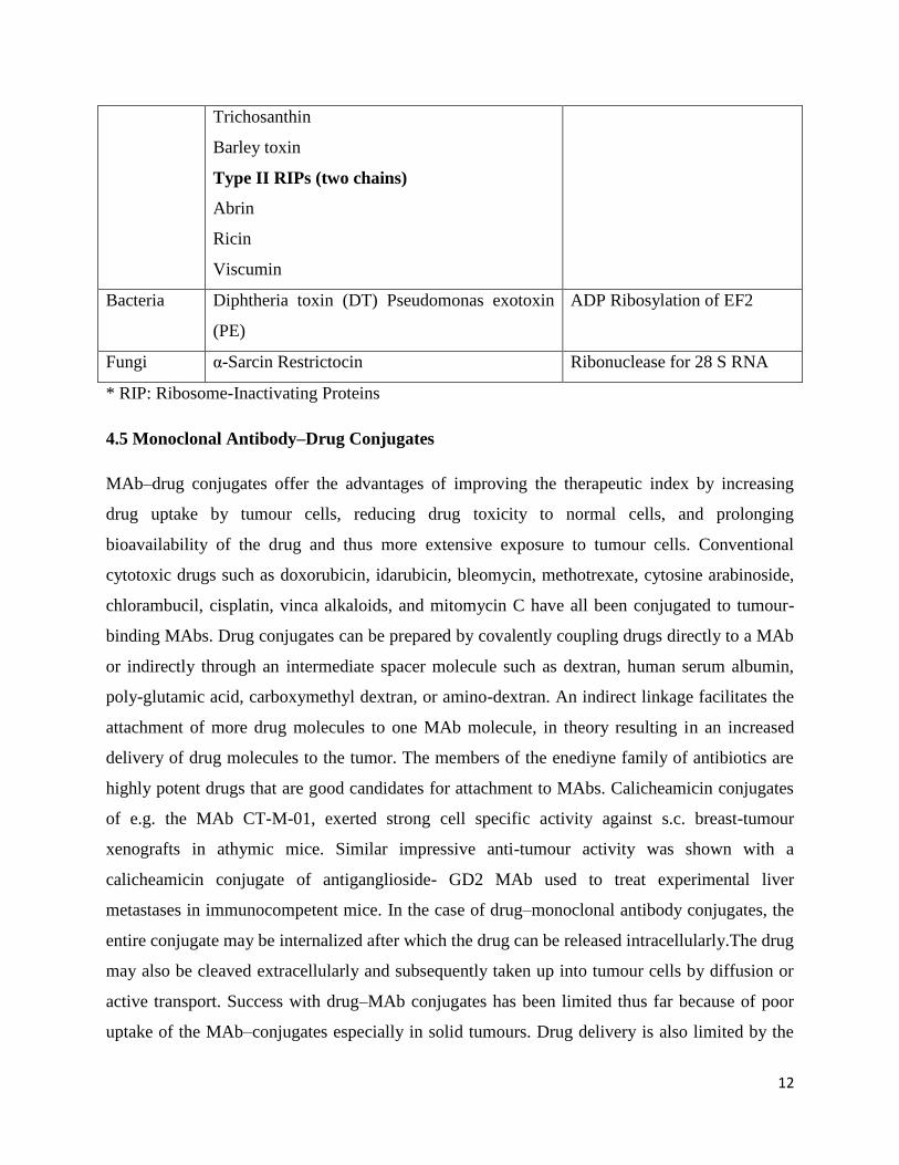
12
Trichosanthin
Barley toxin
Type II RIPs (two chains)
Abrin
Ricin
Viscumin
Bacteria Diphtheria toxin (DT) Pseudomonas exotoxin
(PE)
ADP Ribosylation of EF2
Fungi α-Sarcin Restrictocin Ribonuclease for 28 S RNA
* RIP: Ribosome-Inactivating Proteins
4.5 Monoclonal Antibody–Drug Conjugates
MAb–drug conjugates offer the advantages of improving the therapeutic index by increasing
drug uptake by tumour cells, reducing drug toxicity to normal cells, and prolonging
bioavailability of the drug and thus more extensive exposure to tumour cells. Conventional
cytotoxic drugs such as doxorubicin, idarubicin, bleomycin, methotrexate, cytosine arabinoside,
chlorambucil, cisplatin, vinca alkaloids, and mitomycin C have all been conjugated to tumour-
binding MAbs. Drug conjugates can be prepared by covalently coupling drugs directly to a MAb
or indirectly through an intermediate spacer molecule such as dextran, human serum albumin,
poly-glutamic acid, carboxymethyl dextran, or amino-dextran. An indirect linkage facilitates the
attachment of more drug molecules to one MAb molecule, in theory resulting in an increased
delivery of drug molecules to the tumor. The members of the enediyne family of antibiotics are
highly potent drugs that are good candidates for attachment to MAbs. Calicheamicin conjugates
of e.g. the MAb CT-M-01, exerted strong cell specific activity against s.c. breast-tumour
xenografts in athymic mice. Similar impressive anti-tumour activity was shown with a
calicheamicin conjugate of antiganglioside- GD2 MAb used to treat experimental liver
metastases in immunocompetent mice. In the case of drug–monoclonal antibody conjugates, the
entire conjugate may be internalized after which the drug can be released intracellularly.The drug
may also be cleaved extracellularly and subsequently taken up into tumour cells by diffusion or
active transport. Success with drug–MAb conjugates has been limited thus far because of poor
uptake of the MAb–conjugates especially in solid tumours. Drug delivery is also limited by the

13
number of drug molecules that can be efficiently carried by each antibody molecule. Furthermore
chemical conjugation is usually a complex procedure that can damage both the MAb and the
drug and MAb–conjugates require intra- or extracellularly active biochemicals and/or enzymes
to cleave the active drug from the antibody.
Class of anti-neoplastic drug Drug Disease
Antimetabolites Methotrexate Lung cancer, Colon cancer,
Teratocarcinoma, T cell
lymphoma
5-Fluorouracil B leukaemia
Cytosine arabinoside B leukaemia
Aminopterin Murine thymoma
5-Fluoro-2’-deoxyuridine Colon carcinoma
Alkylating agents Chlorambucil Murine thymoma, Murine
lymphoma, Melanoma
Melphalan Colon Cancer, Murine
thymoma
Mitomycin C Lung cancer, Various
disseminated refractory
malignancies, Gastric cancer
Cisplatinum Ovarian carcinoma
Trenimon Hepatoma
Anthracyclines Doxorubicin/adriamycin Melanoma, Ovarian
carcinoma, T-cell lymphoma,
Colon carcinoma, B-cell
lymphoma, Various
disseminated refractory
malignancies, Breast cancer,
Lung cancer, Pancreatic
cancer, Liver cancer,
Neuroblastoma

14
Daunomycin Soft-tissue sarcomas,
Mammalian carcinoma,
Hepatoma
Antimitotic agents Vinca alkaloids Lung adenocarcinoma
Miscellaneous agents Bleomycin Leukaemia
Idarubicin Murine thymoma
Maytansine Colon cancer
Calicheamicins Human breast
carcinoma xenografts
4.6 Radioimmunoconjugates
Another way of using MAbs as therapeutic agents is to couple them to radionuclides .
Radioimmunoconjugates offer many advantages in the treatment of cancer. Cell killing does not
rely on the host’s immune system and occurs by the ionizing effects of emitted radioactive
particles . These radioactive cytotoxic particles are effective over a distance of several cell
diameters, allowing eradication of antigen-negative cells by the radioimmunoconjugate bound to
the adjacent antigen-positive tumour cells. This is useful considering the heterogeneity of antigen
expression in some tumours. Finally, the amount of radioactive MAb delivered to a tumour can
be measured non-invasively by imaging.The most important factors for therapeutic efficacy of
radioimmunoconjugates are good penetration, favourable pharmacokinetics, and a prolonged
time of retention in the tumour.
Type Radioisotope
Beta-emitters lodine-131, Yttrium-90, Rhenium-188,
Rhenium-186, Copper-67
Alpha-emitters Bismuth-212, Astatine-211
Electron capture Iodine-125
4.7 Bispecific Monoclonal Antibodies
Another approach to selectively inducing tumour cell killing is by the use of bispecific
monoclonal antibodies (BsMAb).They combine the specificity of two separate antibodies within

15
one molecule and cross-link an effector killer cell or a toxic molecule with the target cell to
be destroyed. There are three major approaches for creating BsMAbs. They can be obtained by
chemical cross-linking of two MAbs, by fusing two hybridomas, or by genetic engineering. Each
method has its advantages and disadvantages. Chemical conjugates have a well-defined linkage
and can be produced in high yield. However, there is lot-to-lot variability in purity and activity.
Quadromas, produced by fusing two hybridomas, can also produce large quantities of BsMAbs.
However, in addition to the desired BsMAb, the parental MAb and every possible combination
of heavy and light chain matches and mismatches are also produced. Furthermore, quadromas
are often genetically unstable and require frequent subcloning. With recombinant fusion proteins,
it is possible to make new combinations of Fab or Fv segments or to combine human and mouse
gene segments. Yields and correct folding of the purified fusion protein can present problems as
discussed earlier. Cytotoxic drugs including toxins such as saporin, ricin A chain, vinca
alkaloids, and radioisotopes have been delivered to tumour cells with BsMAbs that bind to the
drug/toxin with one arm and to a surface molecule on the targeted cell with the other arm.This
approach has proven successful in animals as e.g. shown by Schmidt et al.. Cytotoxic effector
cells have also been cross-linked to tumour cells via BsMAb. The BsMAb activates the cytotoxic
activity of the effector cell on bridging it to the target cell. Several effector cells, including
phagocytic cells, natural killer (NK) cells and T lymphocytes, can mediate cellular cytotoxity.
Adequate pre-activation of the effector cells is an important requirement in these methods of
drug delivery. In the case of T 216 8 Strategies for Specific Drug Targeting to Tumour Cells
cells, the presence of co-stimulatory molecules such as CD28 and cytokines is a prerequisite
to achieve this, whereas granulocytes and macrophages can be activated with granulocyte
macrophage colony stimulating factor (GM-CSF). Heteroconjugates made by cross-linking two
different IgGs are twice as large as MAbs and thus are limited in their ability to penetrate
tumours, although this problem can be solved by combining two different scFvs resulting in
BsMAb formats with minimal molecular mass. As is the case for all mouse Mab-based therapies,
HAMA is generated, but with the help of humanizing and chimerizing technologies this should
become less of a problem in the future.

16
Schematic representation of bispecific antobody mediated tumor cell recognition by an
immune effector cell. Summarised are effector cell types, trigger molecules and tumor associated
antigens used as a target
4.8 Pro-drug Strategy
Antibody-directed Enzyme Pro-drug Therapy (ADEPT)
Pro-drugs in combination with enzyme–MAb conjugates can also be used to target tumour cells.
The so-called antibody-directed enzyme pro-drug therapy (ADEPT) approach involves the use of
antibody–enzyme conjugates directed against tumour-associated antigens that achieve in situ

17
activation of subsequently administered pro-drugs. Pro-drugs are inactive drug precursors that
are not readily taken up by cells and hence are less toxic to healthy cells. The pro-drug can be
converted locally in the tumour into the active drug by a specific enzyme which is covalently
linked to tumour-specific antigen-targeted MAbs.When the active form of the drug is released, it
will then distribute to the nearby tumour cells, resulting in cell death. A number of such pro-
drug/MAb–enzyme conjugates have been developed and tested in vitro and in vivo. One of the
significant advantages of this approach is that the targeted enzyme can be effective without being
endocytosed. Another beneficial aspect is that a large amount of the drug can be enzymatically
generated at the tumour site. Limitations of ADEPT include suboptimal tumour uptake due to
heterogeneity in antigen expression, development of immune responses against the enzyme
component, the risk of diffusion of the active drug away from the tumour site and the complexity
of dosing schedules.
Enzyme Pro-drug Active drug
Carboxypeptidase G2 Benzoic acid Benzoic acid
Mustard glutamates mustards
Carboxypeptidase A Methotrexate-alanine Methotrexate
Alkaline phosphatase Etoposide phosphate Etoposide
Mitomycin phosphate Mitomycin
Doxorubicin phosphate Doxorubicin
Phenolmustard phosphate Phenolmustard
Beta-glucuronidase Phenolmustard-glucuronide Phenolmustard
Epirubicin-glucuronide Epirubicin
Doxorubicin-glucuronide Doxorubicin
Penicillin amidase Palytoxin-4-hydroxyphenylacetamide
Palytoxin
Doxorubicin-phenoxyacetamide Doxorubicin
Melphalan-phenoxyacetamide Melphalan
Beta-lactamase Cephalosporin vinca alkaloid Desacetylvinblastine
hydrazide

18
Cephalosporin mustard Phenylenediamine mustard
Cephalosporin mitomycin C Mitomycin C
Cephalosporin doxorubicin Doxorubicin
Carboxylesterase Paclitaxel carbonate Paclitaxel
Carbonyloxycamptothecin Camptothecin
Cytosine deaminase 5-Fluorocytosine Fluorouracil
Plasmin Doxorubicin tripartate Doxorubicin
Daunorubicin tripartate Daunorubicin
Pro-drug Monotherapy
Another pro-drug strategy under development is the concept of ‘monotherapy’. An attractive
feature of pro-drug monotherapy, unlike ADEPT, is that antibody–enzyme conjugates are not
required. In the case of pro-drug monotherapy, local production of elevated levels of enzymes by
the tumour is exploited to release the active drug. Pro-drug monotherapy works well with
anthracycline pro-drugs that are activated by β-glucuronidase which can be found in elevated
concentrations in necrotic areas of tumour tissue. De Groot et al. also developed anthracycline
pro-drugs that can be activated by the tumour-associated protease plasmin. The plasmin system
plays a key role in tumour invasion and metastasis by its matrix degrading activity and its
involvement in tumour growth, most likely by its participation in growth factor activation and
angiogenesis. One of the limitations of pro-drug monotherapy may be the risk of diffusion of the
active drug away from the tumour site.
4.9 Synthetic co Polymers
Polymers or synthetic copolymers are believed to accumulate in solid tumours due to enhanced
vascular permeability of tumour blood vessels combined with a lack of lymphatic drainage in the
tumour tissue. Polymer-based targeting strategies can be divided into two main categories, i.e.
polymer–protein conjugates (so far the most widely studied) and polymer–drug conjugates,
particularly those containing conventional anti-tumour agents. Polymer–drug conjugation can be
used to alter the biodistribution, elimination rate and rate of metabolism of covalently bound
drugs. In the case of protein adducts, polymer conjugation can prolong the protein plasma
elimination half-life, reduce proteolytic degradation and may have the added benefit of reducing

19
immunogenicity. Polyethylene glycol (PEG) is the most widely used polymer for protein
conjugation.
Soluble polymer conjugates have also been proposed as macromolecular pro-drugs for controlled
release and targeting of various low molecular weight, (non-protein) chemicals. In this case,
polymer conjugation not only serves to alter drug biodistribution by restricting cellular capture to
the lysosomotropic route, but the polymer–drug linkage can also be designed to allow site-
specific enzymatic or hydrolytic cleavage. Thus, both the rate and the site of drug delivery can in
principle be controlled. Enhanced permeability of the microvasculature at certain sites,
particularly within solid tumours, can be exploited to facilitate site-specific accumulation of
polymer–drug conjugates. Other (co)polymers of interest besides PEG are SMANCS (styrene-
co-maleic anhydride neocarzinostatin; zinostatin stimalar) and HPMA (N-(2-hydroxypropyl)
methylacrylamide).
Targeting moieties such as sugars (galactose, mannose), proteins and antibodies have been
incorporated into the conjugates to promote receptor-mediated recognition.Thus, cell- or organ-

20
specific localization of therapy may be achieved . It should be noted however, that various cell
types in the liver and spleen are important target cells for sugar-derivatized proteins and that
hepatic clearance will always compete with extrahepatic distribution. Polymer conjugates are
most useful in the context of immuno-conjugates. Other protein constructs such as fusion
proteins can assist their future development. Soluble polymer con jugates have now also been
introduced into clinical practice.
4.10 Liposomes
Selective targeting of drugs using liposomes is expected to optimize the pharmacological
effect and toxicities of encapsulated drugs with the advantage that liposomal components are
non-toxic, non-immunogenic and biodegradable. Through encapsulation of drugs in a
macromolecular carrier such as a liposome, the volume of distribution is significantly reduced
and the concentration of drug in the tumour is increased. Under optimal conditions, the drug is
carried through the body associated with the lipid and/or aqueous moiety of the liposome. On
arrival at the tumour the system should leak at a sufficient rate for the encapsulated drug to
become bioavailable. The liposome protects the drug from metabolism and inactivation in the
plasma. Due to size limitations in the transport of large molecules across healthy endothelium,
the drug will accumulate to a reduced extent in healthy tissues. Discontinuities in the
endothelium of the tumour vasculature, on the other hand, may result in an increased
extravasation of large molecules and increased accumulation of liposomal drug in the tumour.
However, this increased penetration phenomenon may be highly dependent on the type of
tumour and the stage of tumour development. Initially, liposomes seemed to be optimal drug
carrier systems, but further research in general showed disappointing results. The clinical utility
of what are now called ‘conventional’ liposomes was limited by their rapid uptake by phagocytic
cells of the immune system, predominantly in the liver and spleen, resulting in their largely
uncontrollable properties upon administration in vivo. Interest in liposomes as drug carriers was
rejuvenated by the introduction of new ideas from membrane biophysics. Liposomes can now be
designed as non-reactive (sterically stabilized) particles, as well as cationic or fusogenic
liposomes. The non-reactive liposomes can also be designed to induce tumour-specific targeting,
while cationic or fusogenic liposomes can exhibit high specificity for nucleic acid and cell
membrane interactions. Because of their reduced recognition and uptake by the phagocytic

21
system, these liposomes are referred to as ‘stealth’ liposomes. These may prove to be useful in
cancer therapy, although it should be noted that even if distribution to macrophages is slowed
down, they will eventually find their way into these cells. In sterically-stabilized liposomes, the
lipid bilayer contains hydrophilic polymers or hydrophilic glycolipids, PEG and the ganglioside
GM being the subjects of the most detailed studies.These liposomes remain in the blood for up to
100 times longer than conventional liposomes and thus can increase the pharmacological
efficacy of encapsulated agents. Consequently, on chronic administration side-effects related to
macrophage function are certainly not excluded and can become dose limiting. The choice of the
drug for delivery via liposomes is essential. To be effective as a carrier, a liposome must be able
to efficiently balance stability in the circulation with the ability to make the drug bioavailable at
the tumour. Furthermore the drug must have adequate activity against the chosen tumour. A drug
such as doxorubicin with a relatively broad activity against a variety of tumour types is an ideal
choice in this regard. The drug also has to be efficiently loaded into the liposomal carrier.
Liposomes bearing attached antibodies or other ligands accumulate much more readily in target
cells than conventional liposomes. The encapsulation of drugs in MAb-targeted liposomes can be
used to selectively increase the concentration of drug delivered to antigen-expressing cells.
Immunoliposomes utilizing internalizing MAbs, such as anti-HER-2 or anti- CD19, can be used
to selectively deliver high concentrations of drug into the cytoplasm of antigen-expressing cells.
Encapsulation of immunomodulators, e.g. muramyl tripeptide analogues, into liposomes has
been designed to stimulate host immunity and can be used in combination with other treatment
modalities. The systemic activation of macrophages provides an additional therapeutic modality
for the eradication of cancer and cancer metastases. Liposomes have also been tested as carriers
in gene therapy. Cationic lipids in particular, can condense DNA and increase transfection yields
in vitro by several orders of magnitude. Reports on transfections in vivo stimulated intense
interest in the use of liposomes for gene therapy, but data so far have been quite disappointing.
With the generation of more sophisticated multifunctional liposomal systems containing steric
stabilization, homing devices and/or fusogenic/controlled released properties, studies on
liposomal gene delivery systems are now focused on the development of small, circulation-stable
lipid–DNA complexes. These complexes can be administered systemically and, once
accumulated at the tumour site, be specifically taken up by tumour cells via endocytosis or direct
fusion with the tumour plasma cell membrane. Perhaps the most interesting and potentially most

22
powerful therapeutic application of liposome technology for cancer therapy, may be in
combining therapeutics aimed at various (newly identified) molecular targets with conventional
cytotoxic drugs. Furthermore, optimal tumour specificity and therapeutic activity may be
achieved by synergistically combining the selectivity benefits of tumour cell molecular targets
with pharmacokinetic targeting. In fact, both therapeutic modalities can be delivered in liposomal
form.
Monoclonal antibody based products and liposome formulations registered for cancer
therapy in the USA and/or Europe.
Generic name Trade name Company Indication
Rituximaba Rituxan
(Mabthera)c
Roche Relapsed or refractory low-
grade/follicular non-
Hodgkin’s lymphoma
Trastuzumaba Herceptin Genentech-Roche Metastatic breast cancer
Edrecolomaba Panorex Glaxo-Wellcome Post-operative adjuvant therapy
Dukes C colorectal
Carcinoma
Doxorubicinb (Caelyx)
d Alza-corporation AIDS-related Kaposi’s sarcoma
Daunorubicinb
DaunoXome Nexstar
Pharmaceuticals
AIDS-related Kaposi’s sarcoma
Doxorubicinb Evacet
(Myocet)e
The Liposome
Company, Inc.
Metastatic breast cancer
a MAb-based.
b Liposome formulation.
c Rituxan is known as Mabthera in Europe.
d Doxil is known as Caelyx in Europe.
e Evacet is known as Myocet in Europe.
5. Tumour Vasculature Targeting
The architecture of a solid tumour is such that numerous layers of tumour cells are fed by one
blood vessel. This poses a significant barrier for macromolecular drug targeting preparations

23
aimed at the tumour cells that need to extravasate from the blood into the tumour tissue to reach
the target cells: the larger the chosen carrier, the less accessible the tumour tissue will be. In
contrast, endothelial cells lining the tumour vasculature are easily accessible for these
macromolecular preparations. Tumour endothelial cell specific delivery of generally toxic anti-
neoplastic or blood coagulation-inducing agents presents an attractive option for increasing
therapeutic efficacy and reducing toxic side-effects elsewhere in the body. Furthermore, the
pivotal role of endothelial cells in the maintenance of tumour cell survival and growth also
makes them an interesting target for therapeutic intervention. Tumour growth does not expand
more than several millimetres in diameter in the absence of new blood vessel formation. Many
tumours diagnosed in the clinic have a size beyond 0.5 cm in diameter and are considered pro-
angiogenic. Endothelial cells are important executioner cells in the angiogenic cascade.
Disruption of these pro-angiogenic characteristics by inhibiting pro-angiogenic signal
transduction in endothelial cells therefore provides a powerful tool with which to intervene in
tumour growth. The majority of research on inhibiting the function of tumour vasculature
focuses at the development of drugs that may have an explicit action on the endothelial cells in
the tumour. Based on their mechanism(s) of action and the fact that angiogenesis is a normal
physiological process, these drugs in theory can also interrupt the functioning of healthy
endothelial cells in the body. Recently, several angiogenesis inhibitors were withdrawn from
clinical studies, most likely as a result of lack of selectivity for tumour endothelium. This
emphasizes the need for systems that can deliver potent angiogenesis inhibitors at/in the tumour
endothelial cells only.
At present, most target epitopes capable of discriminating tumour endothelium from normal
endothelium are molecules that are expressed during angiogenesis.
5.1 Functions of Vascular Endothelial Cells in the Body
The vasculature can be considered to be one of the crucial organs in the body, extending more
than 900 m2 and playing a major role in maintaining the body’s integrity in various ways. Blood
vessels consist of endothelial cells that are directly in contact with the blood, pericytes located
beneath the endothelium, smooth muscle cells, fibroblasts, basement membrane, and
extracellular matrix (ECM). Depending on the location in the body and the organ
microenvironment, the cellular constituents, basement membrane and ECM differ in phenotype,

24
composition, and function. The endothelial cells form a monolayer in every blood vessel in the
circulation. They are actively involved in several regulatory processes in the body. Besides being
metabolically active and selectively permeable for small solutes and peptides/ proteins,
endothelial cells are actively involved in regulating haemostasis. Furthermore, they are able to
recruit cells of the immune system to specific sites of e.g. infection or inflammation by virtue of
the regulated expression of cell adhesion molecules and production of cytokines and
chemokines. In addition, endothelial cells are actively involved in vascular remodelling in for
example ovulation, wound healing, tumour growth and diabetic retinopathy.
5.2 Molecular Control of
Tumour Growth-related
Angiogenesis
Physiological stimuli during
wound healing and during the
reproductive cycle in women lead
to controlled angiogenesis.
However, pathologic conditions
such as tumour growth,
rheumatoid arthritis, and diabetic
retinopathy are also characterized
by abundant angiogenesis.
Angiogenesis is rapidly initiated
in response to hypoxic or
ischaemic conditions. In tumour
growth, this active vascular
remodelling is reflected by enhanced tumour endothelial cell proliferation to up to 20–2000
times faster than in healthy adult endothelium. In all types of angiogenesis, either under
physiological or pathologicaal conditions, endothelial cell activation is followed by matrix
degradation, cellular migration, proliferation, and ultimately neovasculature maturation
5.3 Role of Growth Factors VEGF and FGF-2
Tumor Angiogenesis

25
More than 20 cytokines from various sources have now been reported to be involved in the
processes taking place during angiogenesis.Vascular endothelial growth factor (VEGF) and basic
Fibroblast Growth Factor (bFGF or FGF-2) are the two growth factors whose roles in
angiogenesis have been most extensively studied to date. VEGF (also known as VEGF-A)
isoforms VEGF-121, 145, 165, 183, 189 and 205 are produced and the isoforms differ in their
molecular composition and weight, and biological properties. The larger forms of VEGF differ
from VEGF-121 by the presence of a heparin-binding domain. VEGF is abundantly produced by
hypoxic tumour cells, macrophages and other cells of the immune system. It induces
vasodilatation via endothelial nitric oxide production and increases endothelial cell permeability.
This allows plasma proteins to enter the tissue to form a fibrin-rich provisional network. VEGF
also induces the expression of various proteases and receptors important in cellular invasion and
tissue remodelling, it activates cellular proliferation and prevents endothelial cell apoptosis.The
two VEGF-specific tyrosine kinase receptors,VEGFR-1 (Flt-1) and VEGFR-2 (KDR/flk-1), are
expressed on vascular endothelium, and to a lesser extent on monocytes/macrophages and certain
tumour cell types. Interaction of VEGF with VEGFR-2 is a critical requirement to induce the full
spectrum of VEGF biological responses. In addition to the two VEGF receptors, VEGF-165 has
been found to bind neuropilin-1, which is also expressed on endothelial cells. A recent study
using genetic deletion methods has determined that neuropilin-1 is important for embryonic
vessel formation.
Endothelial cells exploit various proteases such as matrix metalloproteinases to penetrate into
new areas of the body by degrading the basement membrane. Furthermore,
urokinaseplasminogen activator and tissue type-plasminogen activator convert the ubiquitous
plasma protein plasminogen to plasmin. Plasmin is believed to be the most important protease
for the mobilization of FGF-2 from the ECM pool. FGF-2 induces endothelial cell motility,
proliferation and proteinase activity, and modulates integrin levels.The cellular effects of
FGFs are mediated via specific binding to high-affinity tyrosine kinase receptors. In addition,
low affinity FGF receptors consist of polysaccharide components of heparan sulfate
proteoglycans on cell surfaces and ECM. Binding to these components present in the ECM has
been proposed as a mechanism to stabilize and protect FGF from inactivation. Heparan sulfate
on cell surfaces, on the other hand, plays a more active role in displacing ECM-bound FGF-2 and
its subsequent presentation to the high affinity signal transducing receptors. Angiogenesis seems

26
exquisitely sensitive to small changes in factors such as VEGF and FGF- 2, which may have
important therapeutic implications in treatment of angiogenesis-driven disorders.
5.4 Role of Integrins
Integrins are transmembrane proteins composed of an α and β subunit in over 20 different
αβ heterodimeric combinations. They bind to ECM proteins or cell surface ligands through short
peptide sequences and are implicated in angiogenesis control. Combinations of different
integrins on (endothelial) cell surfaces allow cells to recognize and respond to a variety of
different ECM proteins. They are able to transduce signals from within the cells to the outside as
well as from the outside into the cell. Integrin-mediated cell adhesion has impact on two key
aspects of growth regulation. First, it can influence the activity of the basal cell cycle machinery
consisting of cyclin-dependent kinase complexes. Second, integrins play a vital role in
anchorage-dependent cell death or anoikis. For example, integrin αVβ3 mediates endothelial cell
adhesion to vitronectin, fibrinogen, laminin, collagen, von Willebrand Factor or osteopontin
through their exposed tripeptide Arg-Gly-Asp (RGD) moiety. Since αVβ3 is minimally expressed
on normal resting endothelium, but significantly upregulated on tumour and other activated
endothelium, it is believed to play a critical role in the process of angiogenesis. Both peptide and
antibody inhibitors of αVβ3 induced endothelial cell apoptosis, suggesting a role for this integrin
in endothelial cell survival during angiogenesis. Another αV integrin associated with
angiogenesis is αVβ5. Whereas in vivo FGF-2 or tumour necrosis factor α (TNFα) induced αVβ3-
dependent angiogenesis,VEGF or transforming growth factor β (TGF-β) initiated an
angiogenesis pathway dependent only on αVβ5.
5.5 Role of the Extracellular Matrix
Components of the ECM play an important role in the regulation of endothelial cell morphology
and function. Thrombospondin (TSP), for example, can affect endothelial cell proliferation
negatively as well as positively, depending on the endothelial microenvironment. Furthermore,
through binding to and activation of TGF-β and affecting protease activity, TSP may be able to
influence cell growth, migration and differentiation. Laminin also plays a role in cell attachment,
growth promotion, protease secretion and interactions with other ECM components. It can bind
to cell surface binding proteins including integrins which leads to integrin signalling. SPARC

27
(Secreted Protein Acidic and Rich in Cysteine), also known as BM40 or osteonectin, is a protein
whose expression is elevated under stress conditions. Transient expression of SPARC during
endothelial cell injury and cellular activation indicate a role in tissue repair, remodelling and
angiogenesis. Exogenously added SPARC or SPARC-derived peptides were able to modify
endothelial cell behaviour via the induction of proteases and inhibitors of plasmin generation.
5.6 Role of Subendothelial Support Cells
Endothelial cell interaction with ECM and mesenchymal cells is a prerequisite to form a stable
vasculature. Therefore, after endothelial cell proliferation and maturation, and the formation of
endothelial tube structures, surrounding vessel layers composed of mural cells (pericytes in small
vessels and smooth muscle cells in large vessels) need to be recruited. Endothelial cells
accomplish this via the synthesis and secretion of platelet-derived growth factor (PDGF), a
mitogen and chemoattractant for a variety of mesenchymal cells. Subsequent differentiation of
the mural precursor cells into pericytes and smooth muscle cells is believed to be a cell–cell
contact-dependent process. Upon endothelial cell–mural cell contact, a latent form of TGF-β,
produced by both endothelium and mural cells, is activated in a plasmin-mediated process.
Activated TGF-β can induce changes in myofibroblasts and pericytes which contribute to the
formation of a mature vessel, ECM production and maintenance of growth control. The
coincident investment of growing capillaries by pericytes with the deposition of basement
membrane and cessation of vessel growth during wound healing also indicates vessel growth
regulation by pericytes .The FGF-1 receptor is also implicated in endothelial cell differentiation
leading to vascular tube formation. In addition to inducing plasminogen activator, and
endothelial cell proliferation and migration, FGF-1 receptor sign
5.7 Growth Factor Receptor Targeting
Endothelial cells in normal vasculature are quiescent and divide approximately once every 6
months and about 0.01% of endothelial cells is in S-phase at any given time. In contrast, at areas
of active angiogenesis, e.g. in tumour growth, wound healing and in reproductive tissues
undergoing remodelling, endothelial cells divide rapidly. Increased proliferation in these areas is
accompanied by over-expression of growth factor receptors involved in angiogenesis. Some of
the well-characterized receptor systems involved in angiogenic response are VEGF receptors,

28
angiopoietin receptor (Tie-2), FGF receptor and endoglin.With the exception of Tie-2 receptor,
these receptor systems have all been studied for their application as targets in order to selectively
inhibit tumour endothelial cell proliferation and function.
5.8 VEGF Receptor Targeting
VEGFR-1 and VEGFR-2 are over-expressed on tumour vasculature, while being present at low
density in the surrounding normal tissues. The upregulation of VEGFR expression is mediated
by hypoxia and autocrine stimulation. Since growth factor receptors undergo endocytosis upon
ligand binding, VEGF was initially studied for its ability to deliver toxin polypeptides. In these
studies, VEGF-165 was chemically linked to a truncated form of diphtheria toxin (DT385) by a
disulfide bond. The toxin molecule used is truncated at position 385 by genetic deletion to
eliminate direct binding of diphtheria toxin to endothelial cells. The resultant molecule has the
catalytic domain (A-chain) of diphtheria toxin and the translocation domain of the B-chain.The
A-chain of diphtheria toxin possesses ADP-ribosylase activity and ribosylates elongation factor-
2 (EF-2) at a specific, post-translationally modified histidine residue called diphthamide.
Consequently, EF-2 is irreversibly inactivated leading to precipitous inhibition of protein
synthesis in the target cells.The VEGF–toxin conjugate was found to be quite effective in
inhibiting endothelial cell proliferation in vitro and experimental angiogenesis in vivo.
Cytotoxicity to endothelial cells was specific and dependent on VEGFR expression. Free toxin
molecules did not show any effect on endothelial cell viability. VEGF–toxin conjugate treatment
of tumour-bearing mice resulted in selective vascular damage in the tumour tissue and inhibited
tumour growth. Histological studies demonstrated that conjugate treatment spared the blood
vessels of normal tissues such as liver, lung and kidney from being damaged. The differential
effects of VEGFR targeting on tumour vasculature can possibly be attributed to three factors,
(1) over-expression of VEGFR in tumour vessels leading to increased homing of the VEGF–
toxin conjugate
(2) proliferation-dependent sensitivity to the effector moiety in the conjugate and
(3) polarized distribution of VEGFR.
Over-expression of VEGFR on tumour vessels has been well documented. Recent in vitro studies
suggest that only proliferating endothelial cells are sensitive to the VEGF–toxin conjugate.
Quiescent, confluent endothelial cells were found to be totally resistant to the cytotoxic activity

29
of VEGF–toxin. Endothelial cells in healthy tissues are quiescent and therefore may have
escaped the cytotoxicity of the VEGF–toxin conjugate. Definite evidence for the third possibility
is forthcoming. It is likely that the VEGFR is abluminally distributed and therefore inaccessible
to systemically circulating VEGF–toxin conjugate. At the tumour site however, increased
permeability and vascular leakage may facilitate extravasation of the VEGF–toxin conjugate. As
a result, extravascular VEGF–toxin can easily bind to the abluminally distributed receptors
Among the different splice variants, both VEGF-165 and VEGF-121 were found to be equally
efficient in delivering toxin polypeptides to endothelial cells. Conjugate treatment not only
decreased vessel density in tumour tissue but also decreased the number of branch points and
nodes. Chemical conjugates used in earlier studies were prepared by random derivatization of
VEGF with bifunctional reagents. Such methods often result in a heterogenous mixture of
different VEGF/toxin stoichiometry.To avoid batch-to-batch variations and to obtain structurally
well defined toxin conjugates, the coding region of VEGF and toxin polypeptides were fused at
the DNA level. Unlike the chemical conjugates (dimeric VEGF linked to a toxin moiety),
genetically fused proteins were expressed as monomeric VEGF fused to a toxin moiety.
Interestingly, the monomeric constructs were found to be biologically active and inhibited
tumour growth in mice. Based on these observations, it was concluded that it is possible to re-
engineer VEGF–toxin conjugates to optimize their anti-angiogenic effect. For example, VEGF
could be separated from toxin polypeptides by introducing a spacer polypeptide, which can
reduce steric hindrance. The spacer is 15 amino acids in size and its sequence is (Gly-Gly-Gly-
Gly-Ser)3.The spacer molecule provides adequate flexibility and allows unhindered interaction
between VEGF and its receptor present on the cell surface. Pharmacokinetic properties of the
construct can be improved, e.g. by fusion with human Fc fragments. Such strategies will improve
the anti-angiogenic and anti-tumour activity of vascular targeting reagents.
Inhibition of tumour neovascularization with VEGF–toxin conjugate. Schematic diagram below
showing higher levels of VEGF-receptor expression in the vascular endothelial cells of the
tumour tissue compared to normal tissue vasculature. Note that the receptors for VEGF are
located at the abluminal side of the endothelium. Both the location and density of VEGF
receptors provide a unique opportunity for targeting toxin molecules selectively to the tumour
blood vessels. VEGF–toxins administered into the systemic circulation will not affect the normal

30
vessels due to the lack of sufficient numbers of receptor molecules and their abluminal
distribution. When the conjugates reach tumour tissues, increased vascular leakage allows the
conjugates to extravasate and home onto the abluminally-located VEGF-receptors. VEGF–toxin
conjugates are internalized by receptor-mediated endocytosis and selectively inhibit endothelial
cell proliferation and tumour angiogenesis.
5.9 Endoglin Targeting
Endoglin (CD105) is a transmembrane glycoprotein, which is expressed on the surface of
vascular endothelial cells (chicken, rodent and human). Endoglin is intricately associated with
TGF-β receptor complex and is considered to be an ancillary, non-signalling receptor for TGF-β.
Genetic studies and gene deletion experiments have shown that endoglin plays a critical role in
the development of the vascular system. Specifically, endoglin modulates the communication
between vascular endothelial cells and vascular smooth muscle cells, an important step in the
maturation of blood vessels. Furthermore, endoglin binds to TGF-β1 and TGF-β3 in conjunction
with TGF-β type II receptor.TGF-β1 mediated signalling is necessary for the differentiation of

31
newly recruited mesenchymal (vascular smooth muscle) cells by endothelial cells. Genetic
deletion of TGF-β1 in mice leads to embryonic lethality with a significant defect in
developmental angiogenesis. Endoglin is over-expressed in the vasculature of tumours and other
tissues undergoing vascular remodelling. Differential upregulation of endoglin presents an
opportunity to target cytotoxic molecules to endothelial cells. Several monoclonal antibodies
specific for human endoglin have been produced. Some of the monoclonal antibodies generated
against human endoglin were also found to cross react with mouse endothelial cells. In spite of
low binding, monoclonal antibodies (SN6f and SN6j) were readily internalized by mouse
endothelial cells. Using such a cross-reactive antibody, Seon et al. chemically linked ricin A
chain or deglycosylated ricin A chain (the catalytic subunit of the plant toxin ricin). Ricin A
chain is a N-glycosidase which specifically cleaves a single adenine residue from the 28-S
ribosomal RNA. Depurination irreversibly impairs the function of ribosomes and thereby inhibits
protein synthesis. Conjugates of endoglin-specific antibody and ricin A chain showed specific
cytotoxicity against endothelial cells in vitro and inhibited experimental angiogenesis in vivo.
The anti-angiogenic properties of anti-endoglin–ricin A chain conjugate were demonstrated in a
dorsal air sac model system. Most importantly, endoglin-specific immunotoxin showed strong
anti-tumour activity in a SCID mouse tumour model. In these studies, MCF-7, a human breast
cancer cell line was transplanted into mice and then treated with endoglin specific
immunotoxin.These studies showed complete inhibition of tumour growth in all of the treated
mice without any apparent toxicity to normal tissues. Apart from intracellular targeting,
endoglin-specific antibodies were also successfully used to localize radionuclide on the cell
surface. Radioiodinated monoclonal antibodies (10 μCi) given to tumour- bearing animals
significantly inhibited tumour growth, indicating the clinical potential of targeting endoglin.
5.10 Targeting Integrins to Tumour Vasculature
Interaction between cell surface-anchored integrins and extracellular matrix components
constitutes an additional pathway necessary for angiogenesis control. In fact, studies have
identified two cytokine-mediated, integrin-dependent angiogenic pathways. One of these
pathways is associated with αVβ3 integrin, which selectively influences FGF-2 mediated
angiogenic signals. A second, non-overlapping pathway is represented by cross-talk between
αVβ5 integrin and PKC-dependent, VEGF- or TNFα-induced, signalling. Tumour angiogenesis

32
can therefore be inhibited by blocking the interaction between integrins and the RGD motif-
containing extracellular matrix proteins. Furthermore, the integrins present on tumour
endothelium can serve as target epitopes via which toxic compounds can be delivered to the
endothelial cells of the tumour. Erkki Ruoslahti and colleagues developed a novel targeting
strategy by using polypeptides capable of delivering cytotoxic drugs to integrins.An in vivo
selection of phage display libraries identified peptides that specifically home to components of
tumour blood vessels. Ruoslahti’s research group identified two major classes of peptides, one
containing the RGD motif and the other containing an NGR motif. These polypeptides were then
chemically linked to the anti-cancer drug doxorubicin. Treatment of breast carcinoma-bearing
mice with the conjugated doxorubicin caused vascular damage in the tumours and a strong anti-
tumour effect at a 10–40 x lower concentration than that of free doxorubicin, while liver and
heart toxicity was reduced compared to that observed with free doxorubicin.Whether this effect
was caused by the selective delivery of the chemotherapeutic drug to the tumour endothelial cells
and/or tumour cells, direct caspase-3 activation, or a combination, has not as yet been
established. Their results illustrate the potential of targeting therapeutic agents to integrins
expressed on the vasculature of tumours as an effective means of cancer treatment.
6. References:
1. Drug Targeting Organ-Specific Strategies edited by G. Molema, D. K. F. Meijer, Copyright ©
2001 Wiley-VCH Verlag GmbH, Methods and Principles in Medicinal Chemistry; Page no: 199-
221, 223-236, 241-245.
2. Martin’s Physical Pharmacy and Pharmaceutical Sciences-Physical Chemical and
Biopharmaceutical Principles in the Pharmaceutical Sciences, Sixth Edition, edited by Patrick J.
Sinko, PhD, RPh, Yashveer Singh, PhD; Page no: 620-621, 622-623.
3. Modren Pharmaceutics, Fifth edition, edited by Alexander T. Florence and Juergen Seipmann;
Page no: 353-377.