Embed Size (px)
Citation preview
Dr Karamallah S. Mahmood
PhD Clinical Pharmacology
1
DRUGS THAT ACT IN THE ANS
Adrenergic Agonists
Adrenergic Agonists
The adrenergic drugs affect receptors that are stimulated by norepinephrine (noradrenaline) or epinephrine (adrenaline). These receptors are known as adrenergic receptors or adrenoceptors. Adrenergic drugs that activate adrenergic receptors are termed sympathomimetics, and drugs that block the activation of adrenergic receptors are termed sympatholytics. Some sympathomimetics directly activate adrenergic receptors (direct-acting agonists), while others act indirectly by enhancing release or blocking reuptake of norepinephrine (indirect-acting agonists).
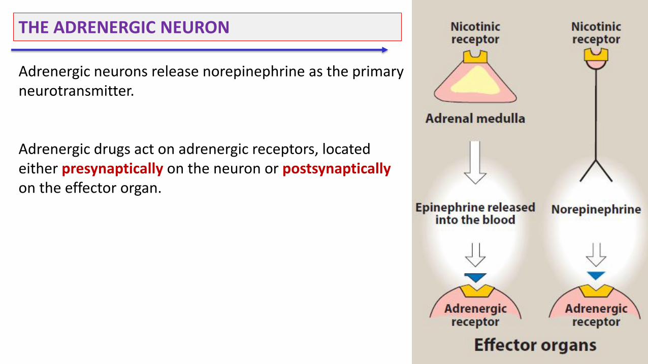
THE ADRENERGIC NEURON
Adrenergic neurons release norepinephrine as the primary neurotransmitter. Adrenergic drugs act on adrenergic receptors, located either presynaptically on the neuron or postsynaptically on the effector organ.
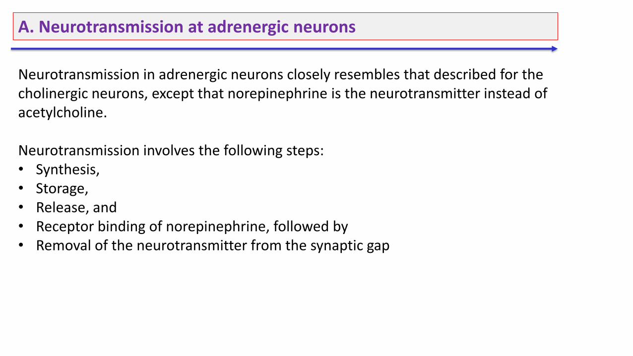
A. Neurotransmission at adrenergic neurons
Neurotransmission in adrenergic neurons closely resembles that described for the cholinergic neurons, except that norepinephrine is the neurotransmitter instead of acetylcholine. Neurotransmission involves the following steps: • Synthesis, • Storage, • Release, and • Receptor binding of norepinephrine, followed by • Removal of the neurotransmitter from the synaptic gap
5
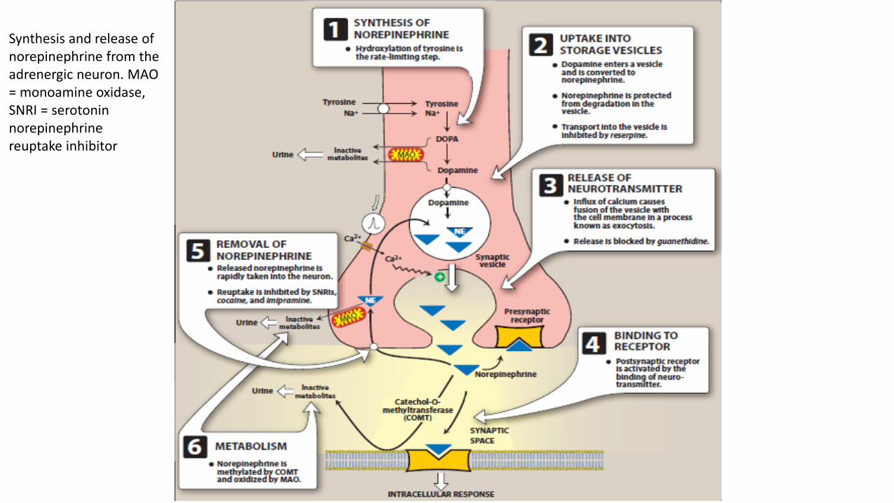
Synthesis and release of norepinephrine from the adrenergic neuron. MAO = monoamine oxidase, SNRI = serotonin norepinephrine reuptake inhibitor
A. Neurotransmission at adrenergic neurons
1. Synthesis of norepinephrine: Tyrosine is transported by a carrier into the adrenergic neuron, where it is hydroxylated to dihydroxyphenylalanine (DOPA) by tyrosine hydroxylase. This is the rate-limiting step in the formation of norepinephrine. DOPA is then decarboxylated by the enzyme aromatic I-amino acid decarboxylase to form dopamine in the presynaptic neuron. 2. Storage of norepinephrine in vesicles: Dopamine is then transported into synaptic vesicles by an amine transporter system. This carrier system is blocked by reserpine. Dopamine is next hydroxylated to form norepinephrine by the enzyme dopamine β-hydroxylase.
A. Neurotransmission at adrenergic neurons
3. Release of norepinephrine: An action potential arriving at the nerve junction triggers an influx of calcium ions from the extracellular fluid into the cytoplasm of the neuron. The increase in calcium causes synaptic vesicles to fuse with the cell membrane and to undergo exocytosis to expel their contents into the synapse. Drugs such as guanethidine block this release.
A. Neurotransmission at adrenergic neurons
4. Binding to receptors: Norepinephrine released from the synaptic vesicles diffuses into the synaptic space and binds to postsynaptic receptors on the effector organ or to presynaptic receptors on the nerve ending. Binding of norepinephrine to receptors triggers a cascade of events within the cell, resulting in the formation of intracellular second messengers that act as links (transducers) in the communication between the neurotransmitter and the action generated within the effector cell. Adrenergic receptors use both the cyclic adenosine monophosphate (cAMP) second messenger system and the phosphatidylinositol cycle to transduce the signal into an effect. Norepinephrine also binds to presynaptic receptors (mainly α2 subtype) that modulate the release of the neurotransmitter.
A. Neurotransmission at adrenergic neurons
5. Removal of norepinephrine: Norepinephrine may 1) diffuse out of the synaptic space and enter the systemic circulation; 2) be metabolized to inactive metabolites by catechol-O-methyltransferase (COMT) in the synaptic space; or 3) undergo reuptake back into the neuron. The reuptake by the neuronal membrane involves a sodium-chloride (Na+/Cl-)-dependent norepinephrine transporter (NET) that can be inhibited by tricyclic antidepressants (TCAs), such as imipramine, by serotonin–norepinephrine reuptake inhibitors such as duloxetine, or by cocaine. Reuptake of norepinephrine into the presynaptic neuron is the primary mechanism for termination of its effects.
A. Neurotransmission at adrenergic neurons
6. Potential fates of recaptured norepinephrine: Once norepinephrine reenters the adrenergic neuron, it may be taken up into synaptic vesicles via the amine transporter system and be sequestered for release by another action potential, or it may persist in a protected pool in the cytoplasm. Alternatively, norepinephrine can be oxidized by monoamine oxidase (MAO) present in neuronal mitochondria.
B. Adrenergic receptors (adrenoceptors)
12
B. Adrenergic receptors (adrenoceptors)
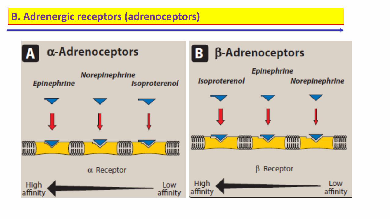
In the sympathetic nervous system, several classes of adrenoceptors can be distinguished pharmacologically. Two main families of receptors, designated α and β, are classified on the basis of their responses to the adrenergic agonists epinephrine, norepinephrine, and isoproterenol. Each of these main receptor types has a number of specific receptor subtypes that have been identified. Alterations in the primary structure of the receptors influence their affinity for various agents.
B. Adrenergic receptors (adrenoceptors)
1. α-Adrenoceptors: The α-adrenoceptors show a weak response to the synthetic agonist isoproterenol, but they are responsive to the naturally occurring catecholamines epinephrine and norepinephrine. For α receptors, the rank order of potency and affinity is epinephrine ≥ norepinephrine >> isoproterenol. The α-adrenoceptors are subdivided into two subgroups, α1 and α2, based on their affinities for α agonists and blocking drugs. For example, the α1 receptors have a higher affinity for phenylephrine than α2 receptors. Conversely, the drug clonidine selectively binds to α2 receptors and has less effect on α1 receptors.
B. Adrenergic receptors (adrenoceptors)
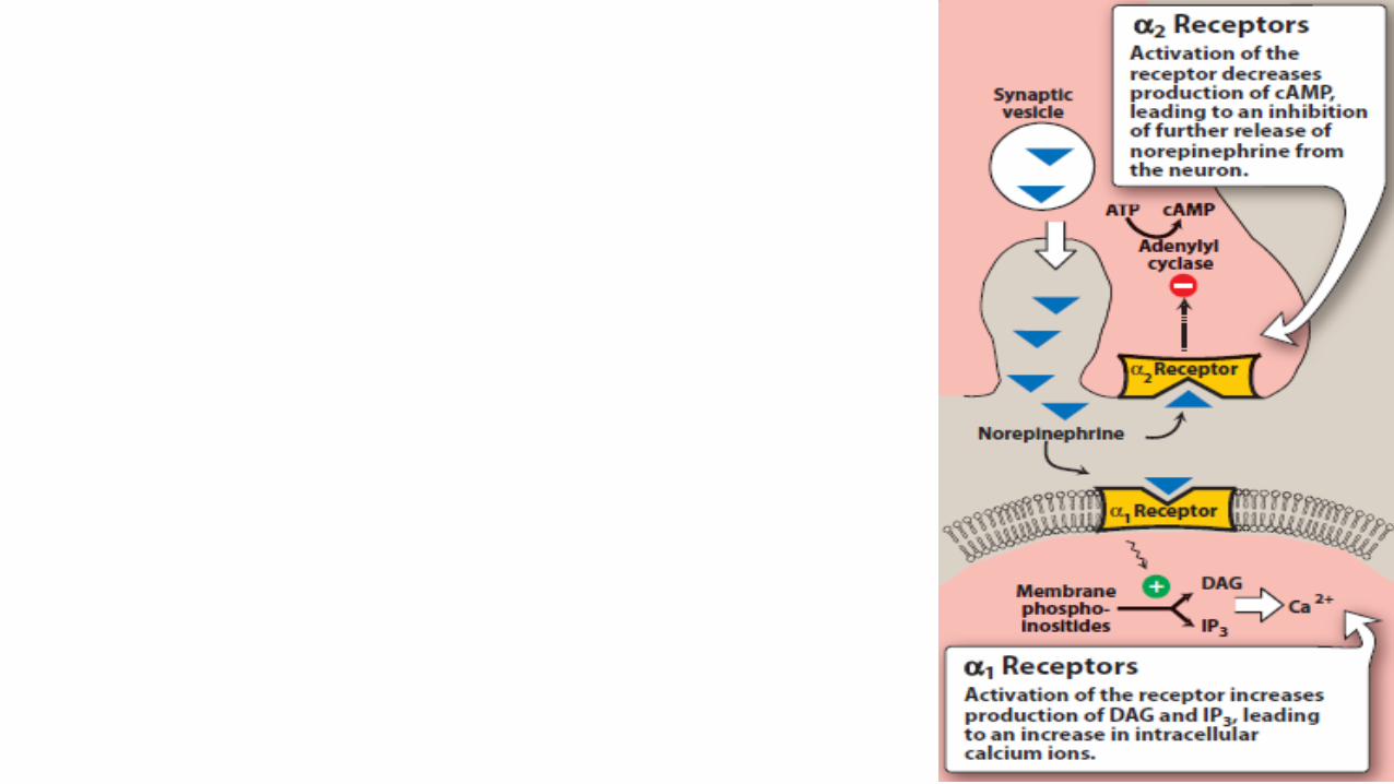
a. α1 Receptors: These receptors are present on the postsynaptic membrane of the effector organs and mediate many of the classic effects, originally designated as α-adrenergic, involving constriction of smooth muscle. Activation of α1 receptors initiates a series of reactions through the G protein activation of phospholipase C, ultimately resulting in the generation of second messengers inositol- 1,4,5-trisphosphate (IP3) and diacylglycerol (DAG). IP3 initiates the release of Ca2+ from the endoplasmic reticulum into the cytosol, and DAG turns on other proteins within the cell.
B. Adrenergic receptors (adrenoceptors)
b. α2 Receptors: These receptors are located primarily on sympathetic presynaptic nerve endings and control the release of norepinephrine. When a sympathetic adrenergic nerve is stimulated, a portion of the released norepinephrine “circles back” and reacts with α2 receptors on the presynaptic membrane. Stimulation of α2 receptors causes feedback inhibition and inhibits further release of norepinephrine from the stimulated adrenergic neuron. This inhibitory action serves as a local mechanism for modulating norepinephrine output when there is high sympathetic activity.
B. Adrenergic receptors (adrenoceptors)
b. α2 Receptors: [Note: In this instance, by inhibiting further output of norepinephrine from the adrenergic neuron, these receptors are acting as inhibitory autoreceptors.] α2 receptors are also found on presynaptic parasympathetic neurons. Norepinephrine released from a presynaptic sympathetic neuron can diffuse to and interact with these receptors, inhibiting acetylcholine release. [Note: In these instances, these receptors are behaving as inhibitory heteroreceptors.] This is another mechanism to modulate autonomic activity in a given area. In contrast to α1 receptors, the effects of binding at α2 receptors are mediated by inhibition of adenylyl cyclase and by a fall in the levels of intracellular cAMP.
B. Adrenergic receptors (adrenoceptors)
c. Further subdivisions: The α1 and α2 receptors are further divided into α1A, α1B, α1C, and α1D and into α2A, α2B, and α2C. This extended classification is necessary for understanding the selectivity of some drugs. For example, tamsulosin is a selective α1A antagonist that is used to treat benign prostatic hyperplasia. The drug has fewer cardiovascular side effects because it targets α1A subtype receptors found primarily in the urinary tract and prostate gland and does not affect the α1B subtype found in the blood vessels.
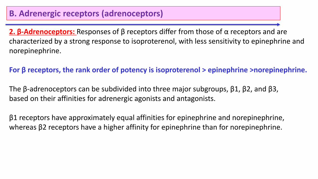
B. Adrenergic receptors (adrenoceptors)
2. β-Adrenoceptors: Responses of β receptors differ from those of α receptors and are characterized by a strong response to isoproterenol, with less sensitivity to epinephrine and norepinephrine. For β receptors, the rank order of potency is isoproterenol > epinephrine >norepinephrine. The β-adrenoceptors can be subdivided into three major subgroups, β1, β2, and β3, based on their affinities for adrenergic agonists and antagonists. β1 receptors have approximately equal affinities for epinephrine and norepinephrine, whereas β2 receptors have a higher affinity for epinephrine than for norepinephrine.
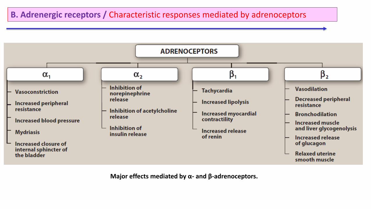
B. Adrenergic receptors / Characteristic responses mediated by adrenoceptors
Major effects mediated by α- and β-adrenoceptors.
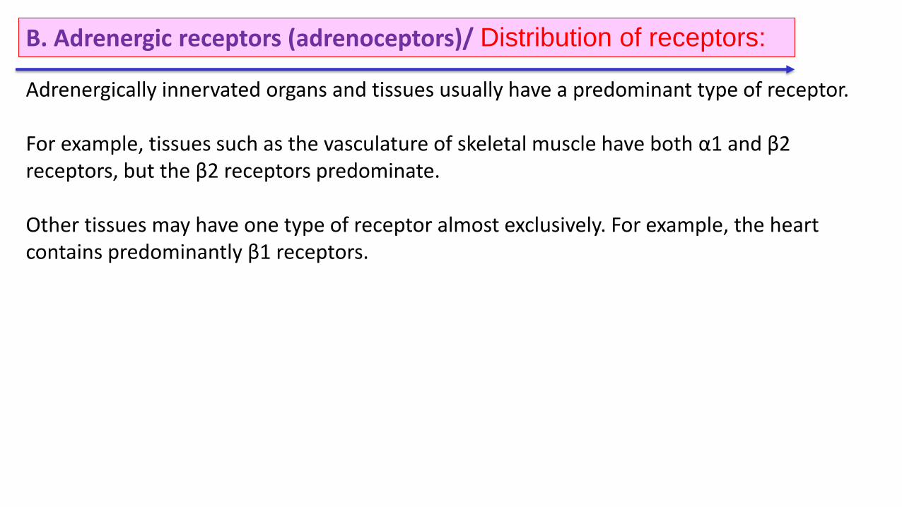
B. Adrenergic receptors (adrenoceptors)/ Distribution of receptors:
Adrenergically innervated organs and tissues usually have a predominant type of receptor. For example, tissues such as the vasculature of skeletal muscle have both α1 and β2 receptors, but the β2 receptors predominate. Other tissues may have one type of receptor almost exclusively. For example, the heart contains predominantly β1 receptors.
B. Adrenergic receptors / Characteristic responses mediated by adrenoceptors
It is useful to organize the physiologic responses to adrenergic stimulation according to receptor type, because many drugs preferentially stimulate or block one type of receptor. As a generalization, stimulation of α1 receptors characteristically produces vasoconstriction (particularly in skin and abdominal viscera) and an increase in total peripheral resistance and blood pressure. Stimulation of β1 receptors characteristically causes cardiac stimulation (increase in heart rate and contractility), whereas stimulation of β2 receptors produces vasodilation (in skeletal muscle vascular beds) and smooth muscle relaxation.
B. Adrenergic receptors / 5. Desensitization of receptors:
Prolonged exposure to the catecholamines reduces the responsiveness of these receptors, a phenomenon known as desensitization. Three mechanisms have been suggested to explain this phenomenon: 1) sequestration of the receptors so that they are unavailable for interaction with the ligand; 2) down-regulation, that is, a disappearance of the receptors either by destruction or by decreased synthesis; and 3) an inability to couple to G protein, because the receptor has been phosphorylated on the cytoplasmic side.
CHARACTERISTICS OF ADRENERGIC AGONISTS
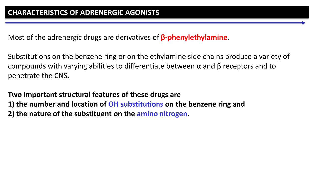
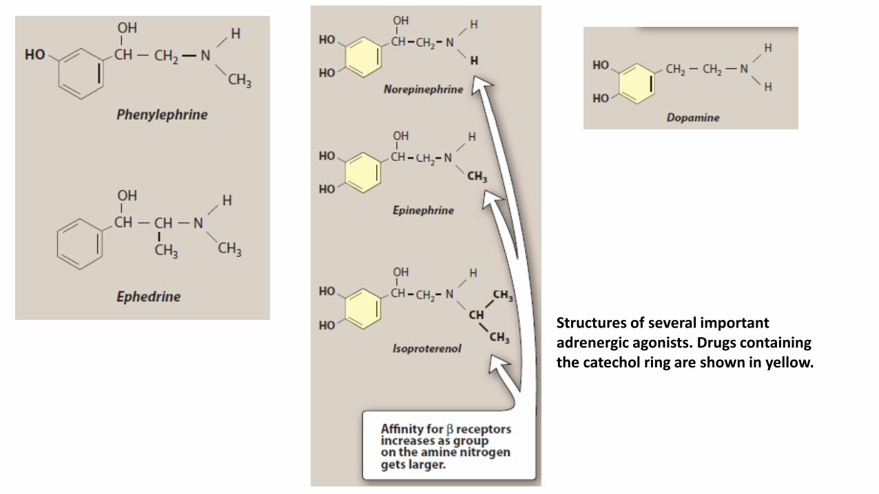
Most of the adrenergic drugs are derivatives of β-phenylethylamine. Substitutions on the benzene ring or on the ethylamine side chains produce a variety of compounds with varying abilities to differentiate between α and β receptors and to penetrate the CNS. Two important structural features of these drugs are 1) the number and location of OH substitutions on the benzene ring and 2) the nature of the substituent on the amino nitrogen.
25
Structures of several important adrenergic agonists. Drugs containing the catechol ring are shown in yellow.
CHARACTERISTICS OF ADRENERGIC AGONISTS/ A. Catecholamines
Sympathomimetic amines that contain the 3,4-dihydroxybenzene group (such as epinephrine, norepinephrine, isoproterenol, and dopamine) are called catecholamines. These compounds share the following properties: 1. High potency: Catecholamines (with –OH groups in the 3 and 4 positions on the
benzene ring) show the highest potency in directly activating α or β receptors.
2. Rapid inactivation: Catecholamines are metabolized by COMT postsynaptically and by MAO intraneuronally, as well as by COMT and MAO in the gut wall, and by MAO in the liver. Thus, catecholamines have only a brief period of action when given parenterally, and they are inactivated (ineffective) when administered orally. 3. Poor penetration into the CNS: Catecholamines are polar and, therefore, do not readily penetrate into the CNS. Nevertheless, most catecholamines have some clinical effects (anxiety, tremor, and headaches) that are attributable to action on the CNS.
CHARACTERISTICS OF ADRENERGIC AGONISTS/ B. Noncatecholamines
Compounds lacking the catechol hydroxyl groups have longer halflives, because they are not inactivated by COMT. These include phenylephrine, ephedrine, and amphetamine. These agents are poor substrates for MAO (an important route of metabolism) and, thus, show a prolonged duration of action. Increased lipid solubility of many of the noncatecholamines (due to lack of polar hydroxyl groups) permits greater access to the CNS.
CHARACTERISTICS OF ADRENERGIC AGONISTS/ Substitutions on the amine nitrogen
The nature of the substituent on the amine nitrogen is important in determining β selectivity of the adrenergic agonist. For example, epinephrine, with a –CH3 substituent on the amine nitrogen, is more potent at β receptors than norepinephrine, which has an unsubstituted amine. Similarly, isoproterenol, which has an isopropyl substituent –CH (CH3)2 on the amine nitrogen, is a strong β agonist with little α activity (Figure 6.4).
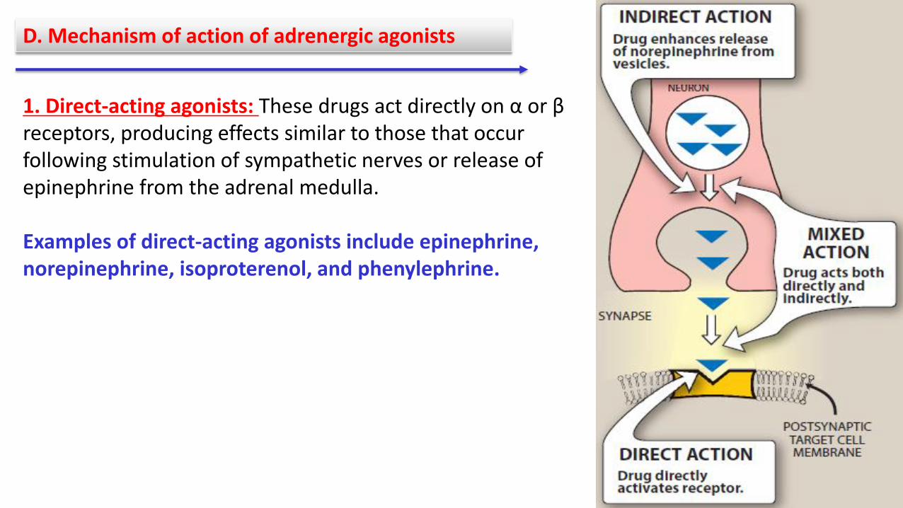
D. Mechanism of action of adrenergic agonists
1. Direct-acting agonists: These drugs act directly on α or β receptors, producing effects similar to those that occur following stimulation of sympathetic nerves or release of epinephrine from the adrenal medulla. Examples of direct-acting agonists include epinephrine, norepinephrine, isoproterenol, and phenylephrine.
D. Mechanism of action of adrenergic agonists
2. Indirect-acting agonists: These agents may block the reuptake of norepinephrine or cause the release of norepinephrine from the cytoplasmic pools or vesicles of the adrenergic neuron. The norepinephrine then traverses the synapse and binds to α or β receptors. Examples of reuptake inhibitors and agents that cause norepinephrine release include cocaine and amphetamines, respectively. 3. Mixed-action agonists: Ephedrine and its stereoisomer, pseudoephedrine, both stimulate adrenoceptors directly and release norepinephrine from the adrenergic neuron