Embed Size (px)
Citation preview

©20
11 N
atu
re A
mer
ica,
Inc.
All
rig
hts
res
erve
d.
nature CHeMICaL BIOLOGY | vol 6 | NovEMBER 2010 | www.nature.com/naturechemicalbiology 821
articlepuBLIsHed OnLIne: 3 OCtOBer 2010 | dOI: 10.1038/nCHeMBIO.452
Phosphorylation via cAMP-dependent protein kinase A (PKA) is a ubiquitous signaling mechanism that regulates many cellular processes1. PKA targets a diverse array of substrates,
including proteins localized in the cytoplasm, mitochondria, plasma membrane, sarcoplasmic reticulum membrane, nucleus, micro-tubules and actin filaments2. The architecture of this enzyme con-sists of a bean-shaped core that is conserved throughout the protein kinase family3. This core has two lobes that flank the active site: a small lobe formed by β-strands at the N terminus, which is primar-ily associated with binding and positioning ATP, and a large lobe that provides a docking surface for substrates or inhibitor proteins4. Most of the atomic resolution information on the catalytic subunit of PKA (PKA-C) is derived from X-ray crystallographic studies, which have mapped three major forms: apo (open state), nucleotide bound (closed state or binary complex) and nucleotide/inhibitor bound (closed state or ternary complex)5. The crystal structure of the closed state, which mimics the Michaelis complex has only been captured with kinase inhibitors (peptides or drugs) and in excess (inhibitory) concentrations of Mg2+ ions, precluding our understand-ing of the substrate recognition process. This problem is common to all protein kinases, whose crystal structures are trapped in inactive states with potent inhibitors. More importantly, information5,6 on protein kinase dynamics—the equilibrium fluctuations that enable the exploration of the free energy landscape—is scarce. These fluc-tuations influence enzymatic reactions because they are responsible for molecular recognition events involved in enzyme-ligand bind-ing7–13 or can regulate the catalytic cycle by limiting access to certain conformations that allow turnover12,14–16.
Using a combination of NMR spectroscopy and X-ray crystal-lography, we provide a vivid picture of PKA-C substrate recogni-tion along the reaction coordinates for the open, intermediate and closed conformational states, mapping out the dynamic landscape on fast (ps–ns) and slow (μs–ms) time scales. As a substrate, we used a peptide corresponding to the cytoplasmic domain of phos-pholamban (PLN), a single-pass membrane protein that inhibits the
sarcoplasmic reticulum membrane calcium ATPase17. We found that nucleotide binding promotes synchronous motions among residues surrounding the active site that correlate with opening and clos-ing of the enzyme’s active site cleft. In the ternary complex, some of these dynamics are redistributed, but motions persist around the active site. Although these dynamics are not correlated to the chemical step (phosphoryl transfer), they occur on the same time-scale as the rate-determining step of enzyme turnover18. Both NMR spectroscopy and X-ray crystallography indicated that the substrate, PLN, adopts an extended and dynamic conformation at the binding groove. The presence of dynamics in the substrate and at the active site of the enzyme cannot conform to an induced-fit recognition model but rather exemplify the conformational selection model of recognition7,19. Thermodynamic analysis confirmed that the bind-ing events are entropically driven, providing further support for this mechanism. The coordinated motions that open and close the cleft of the enzyme underscore the role of conformational dynamics in the slow step of catalysis.
RESULTSMalleability of the X-ray ternary complexVery few protein kinases have been crystallized with natural pep-tide substrates and even fewer with protein substrates20. Here we were able to crystallize PKA-C in complex with 5′-adenylyl-β,γ-imidodiphosphate (AMP-PNP) and a substrate peptide at 2.8-Å resolution (Fig. 1a,b and Supplementary Table 1). The peptide we used corresponded to residues 1–19 of PLN (PLN1–19)21, which com-pose the PKA-C recognition site (R13RAST17). We found that this peptide is phosphorylated by PKA-C as efficiently as the full-length protein in isotropic lipid bicelles (Supplementary Fig. 1a,b), consti-tuting a good mimic of PLN. Although the complex was crystallized under saturating ligand concentrations, two different conforma-tions of the enzyme were present in the asymmetric unit: the closed state with PKA-C bound to both AMP-PNP and PLN1–19 (ternary complex) and an open state, free of bound ligands (apo) (Fig. 1a).
1Department of Biochemistry, Molecular Biology and Biophysics, University of Minnesota, Minneapolis, Minnesota, USA. 2Department of Chemistry, University of Minnesota, Minneapolis, Minnesota, USA. 3Department of Chemistry and Biochemistry, University of San Diego, San Diego, California, USA. 4National Magnetic Resonance Facility at Madison, Department of Biochemistry, University of Wisconsin–Madison, Madison, Wisconsin, USA. *e-mail: [email protected] or [email protected]
dynamics connect substrate recognition to catalysis in protein kinase aLarry r Masterson1,2, Cecilia Cheng3, tao Yu2, Marco tonelli4, alexandr Kornev3, susan s taylor3* & Gianluigi Veglia1,2*
Atomic resolution studies of protein kinases have traditionally been carried out in the inhibitory state, limiting our current knowledge on the mechanisms of substrate recognition and catalysis. Using NMR, X-ray crystallography and thermodynamic measurements, we analyzed the substrate recognition process of cAMP-dependent protein kinase (PKA), finding that entropy and protein dynamics play a prominent role. The nucleotide acts as a dynamic and allosteric activator by coupling the two lobes of apo PKA, enhancing the enzyme dynamics synchronously and priming it for catalysis. The formation of the ternary complex is entropically driven, and NMR spin relaxation data reveal that both substrate and PKA are dynamic in the closed state. Our results show that the enzyme toggles between open and closed states, which indicates that a conformational selection rather than an induced-fit mechanism governs substrate recognition.

©20
11 N
atu
re A
mer
ica,
Inc.
All
rig
hts
res
erve
d.
822 nature CHeMICaL BIOLOGY | vol 6 | NovEMBER 2010 | www.nature.com/naturechemicalbiology
article NATURE chEMicAL biOLOgy dOI: 10.1038/nCHeMBIO.452
In the apo PKA-C molecule, the large lobe is packed against the active site face of the substrate-bound molecule, with PLN1–19 sandwiched between the two PKA-C molecules (Fig. 1a and Supplementary Fig. 2a,b). The apo conformation of PKA-C trapped in this complex is consistent with a previous apo structure3, where both lobes of the enzyme are disengaged from each other.
In the ternary complex, the PLN1–19 recognition sequence (resi-dues 12–17) is clearly positioned in the active site groove between the lobes of PKA-C (Fig. 1b). As expected for a catalytically com-petent conformation, the P-site hydroxyl group of Ser16 in PLN1–19 is aligned to accept the γ-PO4 from AMP-PNP, similar to what is observed in the PKA-C crystal structure mimicking the transi-tion state3 (Supplementary Fig. 2a). The electron densities of the PLN1–19 side chains were well defined at the active site, with a clear network of ionic interactions at the recognition site3 (Supplementary Fig. 2b). Specifically, the P – 2 and P – 3 arginine residues in PLN1–19 may form hydrogen bonds with Glu127 and Glu230 or Glu170 in PKA-C, respectively, in a manner similar to PKA–inhibitor struc-tures5. In addition, two key backbone interactions between the P – 2 and the P + 1 sites of PLN1–19 and Lys168 and Gly200 of PKA-C serve to position the substrate in the binding groove.
The crystal structure of the ternary complex indicated dynamic disorder at the small lobe when PKA-C is bound to the phosphory-latable substrate, PLN1–19. PKA-C was missing electron density for parts of the N (residues 1–16) and C termini (residues 339–342), as well as the conserved glycine-rich loop (residues 47 and 52–54) that positions the γ-phosphate. The unstructured substrate was also missing the first four residues at the N terminus and the last two res-idues at the C terminus. Strikingly, the B-factors of the small lobe in the ternary complex were significantly higher than in the apo form from the same asymmetric unit (Supplementary Fig. 3a,b). These data indicated higher malleability in the closed state relative to the open, unligated state of PKA-C.
Substrate recognition is cooperative and entropy drivenIsothermal titration calorimetry (ITC) measurements showed that PLN1–19 binds to PKA-C with a weak dissociation constant, Kd, of ~50 μM (Table 1 and Supplementary Fig. 4a,b). The binding to PLN1–19 was cooperative with the nucleotide, causing a fivefold increase in binding affinity. The cooperative effect was slightly larger than the one measured for Kemptide, a minimal recognition sequence for PKA-C22. The analysis of the titration curves shows that binding was entropically driven, with an enthalpic contribution of −1.2 ± 0.1 kcal mol−1 and an entropic penalty of −5.7 kcal mol−1 (Table 1). This entropically favored binding event is not characteristic of an
induced-fit mechanism, as enhanced ordering (driven by enthalpy from favorable intermolecular interactions) would be expected7. As indicated by the high B-factors and absence of electron density from the glycine-rich loop of the X-ray crystal structure, no significant ordering appeared to be induced in the ternary complex.
NMR analysis of amide backbone resonances for isotopically labeled PLN peptide (PLN1–20) showed negative H-X NOE values and uniformity of the ratio of the transverse and longitudinal relax-ation rates (R2/R1) (Fig. 2a,b), which indicate that when free in solu-tion the peptide is flexible, monomeric and reorients relatively fast in the NMR timescale23. Upon saturation with PKA-C, the major-ity of H-X NOE values detected across the backbone remain low (near zero), and only residues 13–17 (composing the recognition sequence) show enhanced rigidity. Although an overall enhance-ment of rigidity is detected after recognition, the average H-X NOE value is 0.54, characteristic of mobility for these amides. The calcu-lated R2/R1 ratios indicate that residues 10–18 tumble with a cor-relation time corresponding to a larger macromolecular complex23, which only slightly extends this region of interaction. Although it is difficult to relate the enhanced structural fluctuations directly with the overall increase in entropy measured by thermodynamic measurements, the NMR and ITC measurements showed a similar trend: NMR measurements suggest an increase in configurational entropy upon ligand binding, and the ITC reveals that ligand bind-ing is driven by the overall entropy.
Dynamic activation by nucleotide and substrate bindingThe effects of nucleotide and substrate binding on the backbone dynamics of the enzyme were analyzed using nuclear spin relaxation parameters (R1, R2, Rex and H-X NOE) for the three major forms of the enzyme5 (Supplementary Fig. 5a–c and Supplementary Table 2) (apo, binary complex and ternary complex), and these are mapped onto the crystal structure in Figure 3a,b. Our data showed a global decrease in R2/R1 ratios upon the transition from
αB
αC
αD
αE
αF
αHαI
αA
β3
90°
DFG
β1β2
AbsentGly loop(52–54)
Absent C terminus(339–342)
Ternary complex
Apo enzymeSm
all lobeLarge lobe
a b
Activationloop
Peptideloop
PLN1–19
PLN1–19
Figure 1 | X-ray crystal structure of the PKA-c ternary complex containing AMP-PNP and PLN1–19. (a) The asymmetric unit revealed two molecules of PKA-C, an apo (open, gray) form and a ternary (closed, tan) complex. (b) The ternary complex is missing the first 15 residues of the N terminus, part of the glycine-rich loop and C terminus, which is likely because of conformational disorder.
Table 1 | Thermodynamic parameters for the binding of PKA-c to PLN1–20 in the presence or absence of nucleotide
ΔH (kcal mol–1)
ΔS (kcal mol–1 × K)
–TΔS (kcal mol–1) Kd (mM)
Apo form −0.6 ± 0.1 0.017 ± 0.001 −5.1 ± 0.2 49 ± 8Binary complex
−1.2 ± 0.1 0.019 ± 0.001 −5.7 ± 0.1 10 ± 4
Data represent mean values ± one s.d. for triplicate measurements.

©20
11 N
atu
re A
mer
ica,
Inc.
All
rig
hts
res
erve
d.
nature CHeMICaL BIOLOGY | vol 6 | NovEMBER 2010 | www.nature.com/naturechemicalbiology 823
articleNATURE chEMicAL biOLOgy dOI: 10.1038/nCHeMBIO.452
apo to the ternary complex (Supplementary Table 2), indicating that the entire enzyme is more compact upon ligand binding, in agreement with previous findings3. However, changes in the H-X NOE and Rex values in the binary and ternary complexes indi-cated an increase in backbone dynamics of the enzyme on fast and slow timescales, with a redistribution of motion throughout the backbone.
The apo form is monomeric and well folded, with a trimmed H-X NOE average value of 0.76 (Supplementary Table 2) and an effective rotational correlation time of ~19 ns. Using H-X NOE as a proxy for the fast dynamics (ps–ns timescale), we found that the most relevant features are the marked dynamics (NOEs <0.6) in residues located between structural elements of the enzyme (Fig. 3a and Supplementary Fig. 5a). In particular, low NOE values were detected for all of the conserved loops, including the glycine-rich, catalytic, DFG, activation and P + 1 loops. Low H-X NOE values were also detected for residues within the acidic cluster (residues 328–334). In this region, Tyr330 forms electro-static interactions with an essential arginine of the substrate rec-ognition sequence (Arg13 in PLN1–20), whereas Glu333, Glu332 and Asp329 interact with the glycine-rich loop in the ligand-bound structures5.
Surprisingly, very little conformational exchange is present in the apo form on the μs–ms timescale (Rex) (Fig. 3b and Supplementary Fig. 5a). The lack of these slow dynamics is also supported by analy-sis of the NMR inverse peak heights measured at various tempera-tures (Supplementary Fig. 6a)24. Relatively small Rex values were measured throughout the entire backbone, with the majority of values distributed around zero25. Specifically, the small lobe does not show marked conformational exchange. Only a few residues interspersed throughout the large C-lobe show Rex >10 Hz.
Saturation of PKA-C with the nucleotide (AMP-PNP) changed the backbone dynamics throughout the enzyme. Although a slight overall restriction of the protein backbone motion occurred (~0.03 increase in the average H-X NOE), increases in both fast and slow dynamics were detected for regions at the active site cleft (Fig. 3a,b and Supplementary Fig. 5b). This was apparent for the glycine-rich and the DFG loops, which are directly involved in nucleotide bind-ing, as well as the activation loop, the P + 1 loop and the acidic cluster (Supplementary Fig. 5b). Other loops not directly involved in catalysis showed an increase in fast dynamics, such as the loops linking helices F and G. Other segments peripheral to the catalytic loops became more dynamically restricted.
Dynamics on a μs–ms timescale were pervasive in the presence of nucleotide, indicating conformational fluctuations in the binary complex (Fig. 3b and Supplementary Fig. 5b). Large changes in Rex at the active site cleft were observed in the glycine-rich, DFG, activation and P + 1 loops, as well as in the C-helix. Notably, the C-helix residues lying on the face oriented toward the nucleotide-binding pocket showed higher Rex values than those populating the opposite face (Supplementary Fig. 7). The conformational exchange rates detected for these residues likely reflect recruitment of the C-helix to the active site, which is a critical part of activa-tion26. Conformational exchange is also detected in regions far from the active site, such as the segments that flank the G-helix and the segment that links the conserved APE motif to the F-helix (Fig. 3b). The augmentation of dynamics was also propagated toward the C-terminal tail (residues 322–350), whose putative role is to posi-tion the small lobe for catalysis5.
Upon saturation with the substrate, the ternary complex remained dynamic, with a redistribution of fast dynamics throughout the PKA-C backbone. For the glycine-rich loop, we observed a mono-tonic increase of fast dynamics, with the average NOE value drop-ping from ~0.8 in the apo, to ~0.7 in the binary complex, to ~0.5 for the ternary complex (Fig. 3a and Supplementary Fig. 5c). However, a significant reduction in fast dynamics was detected for residues at the active site located in the P + 1, DFG and activation loops. Subtle changes occurred throughout the enzyme, indicating that the entire backbone dynamics had reorganized after substrate binding. The reorganization of dynamics in the ternary complex was also appar-ent by the redistribution of μs–ms conformational exchange for many regions (Fig. 3b). For instance, the conformational exchange detected for the C-helix in the binary complex was quenched in the ternary complex, whereas the residues forming the base of the C-spine (Ile174, Met128 and Met231) became more dynamic.
Large Rex values were observed for residues throughout the small lobe in the ternary complex. Notably, Rex values >25 Hz were observed in two regions intimately involved in the catalytic cycle, the glycine-rich and activation loops. The observed slow dynamics in these regions may reflect the conformational changes required for opening the active site for product release, the proposed rate-limiting step in the enzyme turnover18,27.
Dynamics are synchronous with the slow step of catalysisTo determine whether the conformational exchange reports on opening and closing of the enzyme, we plotted Rex versus the squared
H-X
NO
E
0
10
20
0
0.5
1.0
–3Met1
Lys3
Tyr6
Leu7
Met20
Glu19Ile18
Thr17
Arg14
Arg13
Ile12Ala11
Ser10
Arg9
Thr8Ala15
Ser16
Gln5
Val4Glu2
Met1
M
0 5 10 15 20
E K VQ Y L T R S A I R R A S T I E M
Lys3Tyr6
Leu7
Met20
Glu19Ile18Thr17
Arg14
Arg13
Ile12Ala11
Ser10Arg9
Thr8 Ala15
Ser16
Gln5
Val4Glu2
Fast dynamics Slow dynamicsH
-X N
OE
> 0.75
> 0.50
< 0.25 > Ave
> Ave + σ
> Ave + 2σ0
10
Residue number
5
R 2 /R 1
R 1 ×
R2
0
2
4
R 2 (s
–1)
R 1 (s
–1)
Figure 2 | Details of the interaction of isotopically labeled PLN1–20 with PKA-c. (a) NMR nuclear spin dynamics of PlN1–20 in the free (black) and bound to PKA-C in the presence of AMP-PNP (blue). (b) Mapping of these dynamics in the bound form shows that ordering around the recognition sequence was observed, whereas elevated R2/R1 values extend the region of interaction to residues 10–17. Data represent fitted values and associated error from nonlinear fitting.

©20
11 N
atu
re A
mer
ica,
Inc.
All
rig
hts
res
erve
d.
824 nature CHeMICaL BIOLOGY | vol 6 | NovEMBER 2010 | www.nature.com/naturechemicalbiology
article NATURE chEMicAL biOLOgy dOI: 10.1038/nCHeMBIO.452
difference of chemical shifts (Δω2) between the apo and ternary complex (open and closed states) (Fig. 4a,b; see Supplementary Fig. 8 for full labeling of peaks)28. In the fast exchange limit, if a set of nuclear spins is affected by the same conformational exchange process (for instance, open-to-closed state transitions), a linear cor-relation is expected between Rex and Δω2. Indeed, we found a linear correlation (correlation coefficient of 0.9) for many residues around the active site cleft upon addition of AMP-PNP (Fig. 4c). These residues were located in the conserved glycine-rich, DFG, activa-tion and P + 1 loops, which line the entrance to the enzyme active site. Analogous to triosephosphate isomerase28, this linear correla-tion suggested that the motions of these residues report on the same exchange process (same kex), which was the opening and closing of the enzyme active site. From the slope of the linear plot and the populations of free and bound states, it is possible to determine the exchange constant for the process, kex = 203 ± 18 s−1. This process was much slower than that of ligand (AMP-PNP) exchange between the free and bound states (kex = kon[AMP-PNP] + koff), which we obtained from NMR lineshape analysis (kex ~3.4 × 10⁴ s−1)22 (Table 2) and independently from stopped-flow kinetic measurements18. Under these conditions, koff of AMP-PNP is ~100 s−1, whereas the opening rate constant determined by the relaxation experiments is ten times slower (~10 s−1). A linear correlation between Rex and Δω2 (correlation coefficient of 0.8) was also obtained for many of the
same residues for the ternary complex (Fig. 4b). The kex in this case is 628 ± 50 s−1, with kopen = 31 ± 3 s−1. This was similar to the kcat we measured for phosphorylation of PLN1–20 using a coupled enzyme assay (~23 s−1), indicating that the slow step of catalysis is domi-nated by the opening and closing of the enzyme18,29.
Allosteric network of PKA-c shows noncontiguous pathwaysThe dynamics induced by ligand binding proceeded through con-tiguous paths throughout the active site (Figs. 3b and 4c, resi-dues colored in orange), where nearby residues are within van der Waals radii contact. However, noncontiguous effects occurred in remote regions of the enzyme (Fig. 4d, residues colored in red, Supplementary Fig. 8) that were not synchronous with the open-ing and closing motions. These dynamics are likely to report on conformational fluctuations other than the toggling between open and closed states and underscore the complexity of the allosteric network within the enzyme and its ability to funnel into dyna-mical different states. Other functional events such as binding to A-kinase anchoring proteins (AKAPs) and/or regulatory subunits may be correlated to these motions. In fact, a number of residues dynamically activated in the ternary complex are distal from the substrate-binding site and make close intermolecular contacts upon assembly with the type-I regulatory subunit30 (Fig. 4d). Therefore, the complexity of the dynamics we observed throughout
H-helix
B-helix
D-helix
β1
G-helix
E-helix
C-helixGly-rich loop
Peptide positioning
loop
DFG loopA-helix
Activationloop
β2
Acidiccluster
β4β5
F-helix
Linkerregion
a Apo Nucleotidebound
Ternarycomplex
< 0.25
> 0.50
> 0.75
H-X NOE
Smal
l lob
eLa
rge
lobe
b
< 7
> 17
> 27
Rex (Hz)
Figure 3 | Mapping of the backbone amide dynamics of PKA-c from the apo to ternary complex. Structural elements and loops are indicated. (a,b) Fast (a) and slow (b) dynamics are shown for PKA-C in the apo (left), binary complex (middle) and the ternary complex containing AMP-PNP and PlN1–20 (right).

©20
11 N
atu
re A
mer
ica,
Inc.
All
rig
hts
res
erve
d.
nature CHeMICaL BIOLOGY | vol 6 | NovEMBER 2010 | www.nature.com/naturechemicalbiology 825
articleNATURE chEMicAL biOLOgy dOI: 10.1038/nCHeMBIO.452
the backbone of PKA-C could be attributed to its ability to act as a scaffold for many other interactions within the cell.
DiScUSSiONThe enzymatic mechanism of phosphoryl transfer for PKA-C has been extensively studied18,29. Catalysis by PKA-C comprises three major steps: ligand (ATP and substrate) binding, chemical step (phosphoryl transfer) and product release. The chemical step is fast, whereas the product release constitutes the rate-determining step of the catalytic cycle18. It has been hypothesized that the latter depends on the conformational transitions of the enzyme to eject both the product and ADP18. Previous X-ray studies3,5, fluorescence spec-troscopy studies29 and studies using H-D exchange coupled to mass spectrometry31 aimed to understand the enzyme conformational dynamics along the enzymatic cycle. These studies suggest that the apo form becomes more compact as it transitions to the binary and ternary complexes. However, the binary and ternary complexes were studied in the presence of peptide inhibitors and excess con-centrations of Mg2+. As a result, it has been proposed that substrate binding by PKA-C proceeds via an induced-fit mechanism with a global rigidification of the enzyme32.
In the induced-fit mechanism, significant ordering occurs at the active site, resulting in an enthalpically driven binding process7. In contrast, our thermodynamics measurements show that the free energy of binding is dominated by a negative overall entropic penalty for the formation of the ternary complex. Moreover, the NMR data revealed substantial backbone dynamics at the active site of the enzyme that were induced upon nucleotide binding. These dynamics were not quenched upon substrate binding, indicating a significant contribution of the conformational entropy to the overall entropy for binding. We also observed a pre-existing equilibrium
of open and closed states in the binary complex before formation of the ternary complex (Fig. 4a,b). This supports the idea that the recognition of PLN1–20 is entropically driven and governed by con-formational selection rather than by an induced-fit mechanism7.
The dynamic nature of the ternary complex is complemented by the X-ray crystal structure, in which electron density is absent or poor for several dynamic regions highlighted by NMR. These regions are located in the enzyme (particularly the glycine-rich loop at the active site) and substrate (terminal regions). This phenomenon may explain why, in general, it has been so difficult to trap peptide substrates in crystal structures with PKA-C. However, it should be
60
a c d
b60
40
40
20
20
0
0
0
0
Gly-richloop
Glu248Val251
Arg134
C-subunit
R-subunit
Lys285
Gly272
Arg270Acidiccluster
180°
Larg
e lo
beSm
all l
obe
E-helixF-helix
H-helix
C-helixB-helix
G-helix
DFG loopActivation loop
Peptidepositioning
loop
β2
β1
β4β5
D-helix
0.8
0.8
0.6
0.6
0.4
0.4
0.2
0.2
∆�2 (p.p.m.2)
R ex (
s–1)
R ex (
s–1)
A188
G52
G50
T48
R56
F187
F185T51
E248
I174
G55
W196
S53
E332L103
K72
K73M118
E331
A124
A188G52
K189 G200T51
E332T88
S212
K8
E248
G214
F257
Y306
M58
I73
K72L157
L103I163
L167
F350T348
K263
Figure 4 | Opening and closing of the enzyme active site cleft. (a,b) Correlation plots of Rex for (a) AMP-PNP–bound form or (b) PlN1–20–AMP-PNP–bound form with the chemical shift differences of open and closed states. (c) The nucleotide induces opening and closing at the entrance of the enzyme that is reported by contiguous and noncontiguous pathways from the active site (residues denoted in orange). Errors in Rex were taken from error propagation of equation 1 using the r.m.s. noise of the NMR spectra. (d) In the ternary complex, residues that are not linear with opening and closing are distal and are known to interact with the regulatory subunit on the basis of the crystal structure (PDB: 2QCS).
Table 2 | Observed rate constants measured using the Rex/Dv2 correlation plot or from exchange of nucleotide involving NMR line shape analysis during ligand titration measurementsObserved kinetic constant
Ligand exchange*
Conformational exchange
k (sex1− ) 3.37 × 10⁶ ± 0.51 × 10⁶ 203 ± 18
k (s )offligand 1− 109 ± 4 –
k (s )onligand 1 *−
3.36 × 10⁶ ± 0.50 × 10⁶ –
k (s )openconf 1− – 10 ± 2
k (s )closedconf 1− – 193 ± 17
values represent fitted values and associated errors from linear least-squares analysis (conformational rates, denoted ‘conf’) or mean ± one s.d. for six residues (ligand exchange rates, denoted ‘ligand’).*The observation of a pseudo–first-order rate constant for kon is expected (for instance, k konobs
on Ligand= [ ]) .
©20
11 N
atu
re A
mer
ica,
Inc.
All
rig
hts
res
erve
d.
826 nature CHeMICaL BIOLOGY | vol 6 | NovEMBER 2010 | www.nature.com/naturechemicalbiology
article NATURE chEMicAL biOLOgy dOI: 10.1038/nCHeMBIO.452
noted that substrate recognition by this enzyme appears to be quite complex and is likely influenced by how the enzyme is funneled into a particular functional state. In fact, much stronger- binding peptides or drugs that inhibit PKA-C may invoke an induced-fit mechanism upon binding, when increased order and decreasing dynamics place the enzyme in an inhibitory state. This is likely the case with the inhibitor PKI5–24, which crystallized with PKA-C with a well-defined active site.
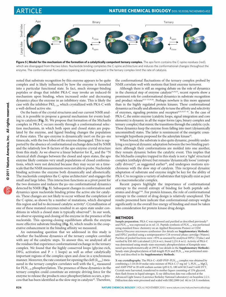
On the basis of the crystal structures and our current NMR anal-ysis, it is possible to propose a general mechanism for events lead-ing to catalysis (Fig. 5). We propose that formation of the Michaelis complex in PKA-C occurs mostly through a conformational selec-tion mechanism, in which both open and closed states are popu-lated by the enzyme, and ligand binding changes the population of these states. The apo enzyme is dynamically inert on the μs–ms timescale, with the two lobes of the enzyme disengaged. This is sup-ported by the absence of conformational exchange detected by NMR and the relatively low B-factors of the apo enzyme crystal structure from this study. As we observe a linear behavior for Rex and the 15N chemical shift changes between the closed and open states, the apo enzyme likely contains very small populations of closed conforma-tions, which were not detected because they may occur on a time-scale that the NMR measurements were not able to probe. Nucleotide binding activates the enzyme both dynamically and allosterically. The nucleotide completes the C-spine architecture4 and engages the small and large lobes. This interaction functions as a pivot point for the two domains and induces the μs–ms conformational dynamics detected by NMR (Fig. 5). Subsequent changes in conformation and dynamics upon nucleotide binding prime the active site for cataly-sis. These changes are not likely to occur without the completion of the C-spine, as shown by a number of mutations, which disrupted this region and led to decreased catalytic activity4. Crystallization of one of these mutated enzymes resulted in an open state under con-ditions in which a closed state is typically observed33. In our work, we observe opening and closing of the enzyme in the presence of the nucleotide. This opening-closing equilibrium affords the enzyme more effective substrate binding (Fig. 5), which results in the coop-erative enhancement in the binding affinity we measured.
An outstanding question that we addressed in this study is whether the backbone dynamics of the ternary complex correlate with any of the catalytic steps. To answer this, we analyzed all of the residues that experience conformational exchange in the ternary complex. We found that the highly conserved loops (glycine-rich, DFG, activation and P + 1 loops) as well as other catalytically important regions of the complex open and close in a synchronous manner. Moreover, the rate constant for opening the cleft (kopen) mea-sured in the ternary complex correlates well with the kcat measured for PLN1–20 phosphorylation. The presence of these dynamics in the ternary complex could constitute an entropic driving force for the enzyme to release the products once phosphorylation occurs, a pro-cess that has been identified as the slow step in catalysis18. Therefore,
the conformational fluctuations of the ternary complex probed by NMR correlate well with motions that limit enzyme turnover.
Although there is still an ongoing debate on the role of dynamics in the chemical step of enzyme catalysis14,34,35, recent reports show a prominent role for conformational dynamics in substrate recognition and product release9,11–13,19,36. Perhaps nowhere is this more apparent than in the highly regulated protein kinases. These conformational dynamics act locally and allosterically to tune the affinity and selectivity of enzymes, signaling proteins and receptors8,10,19,37–41. In the case of PKA-C, the entire enzyme (catalytic loops, signal integration and core elements) is dynamic in all the major forms (apo, binary complex and ternary complex) that mimic the transitions through the catalytic cycle. These dynamics keep the enzyme from falling into inert (dynamically uncommitted) states. The latter is reminiscent of the energetic coun-terweight hypothesis proposed for the adenylate kinase42.
When bound, the substrate is also quite dynamic, possibly under-lying a reciprocal dynamic adaptation between the two binding part-ners: although their conformations are molded into one another, they remain dynamic before the catalytic event. This implies that the Michaelis complex trapped in this study is not a ‘tight’ structural complex (enthalpy driven) but remains ‘dynamically loose’ (entropi-cally driven)43, as suggested earlier29, and undergoes motions syn-chronous with the slow step of catalysis. This reciprocal dynamic adaptation of substrate and enzyme might be key for the ability of PKA-C to recognize a variety of substrates that typically exist as part of a macromolecular complex.
Recent papers highlight the importance of conformational entropy to the overall entropy of binding for both peptide sub-strates and drugs9,44. For protein kinases, the role of conformational entropy in the context of drug design is relatively unexplored. The results presented here indicate that conformational entropy weighs significantly in the overall free energy of binding and must be taken into consideration for protein kinase drug design.
METhODSSample preparation. PKA-C was expressed and purified as described previously45 and PLN1–20 was expressed as in ref. 21. Peptide synthesis of PLN1–19 was performed using standard Fmoc chemistry on an Applied Biosystems Pioneer or CEM Liberty/Discover microwave synthesizer (for details see Supplementary Methods) and HPLC purified using a semipreparative C18 reversed-phase cartridge (Waters). Purities of pooled fractions were >95% as assessed by analytical HPLC (Vydac) and verified by ESI-MS (calculated 2,252.6 m/z, found 2,251.8 m/z). Activity of PKA-C was determined using steady-state enzymatic phosphorylation of Kemptide mea-sured spectrophotometrically at 299 K (see details in the Supplementary Methods). Steady-state phosphorylation of full-length PLN and PLN1–19 was performed simi-larly and described in the Supplementary Methods.
X-ray crystallography. The PKA-C–AMP-PNP–PLN1–19 complex was obtained by combining a 1:10:10:10 molar ratio mixture of PKA-C (7 mg ml−1), PLN1–19, MgCl2 and AMP-PNP in 20 mM sodium acetate (pH 6.5), 180 mM KCl and 5 mM DTT. Crystals were harvested, transferred to mother liquor consisting of 15% glycerol, then flash frozen in liquid nitrogen. X-ray diffraction data was collected at the Advanced Light Source (Lawrence Berkeley National Laboratory) on beamline 2.2.1. Diffraction data were processed and scaled with HKL2000 (ref. 46) to 2.8 Å resolution.
Apo Binary
kclose
kopen
kopen
kclose
Smal
l lob
eLa
rge
lobe
Ternary
ATP ATP ATPATPATPPLN1–20
PLN 1–20 PLN 1–20
Figure 5 | Model for the mechanism of the formation of a catalytically competent ternary complex. The apo form contains the C-spine residues (red), which are disengaged from the two lobes. Nucleotide binding completes the C-spine architecture and induces the conformational changes throughout the enzyme. The conformational fluctuations (opening and closing) present in the ternary complex limit the rate of catalysis.

©20
11 N
atu
re A
mer
ica,
Inc.
All
rig
hts
res
erve
d.
nature CHeMICaL BIOLOGY | vol 6 | NovEMBER 2010 | www.nature.com/naturechemicalbiology 827
articleNATURE chEMicAL biOLOgy dOI: 10.1038/nCHeMBIO.452
The initial data clearly fit to a primitive hexagonal lattice, and the final data were integrated and scaled in the P61 space group. Initial phases were generated by mole cular replacement using the coordinates of the PKA-C–ATP–PKI5–24 complex without the PKI peptide (PDB: 1ATP) as a search model. The ternary complex crystallized with two molecules in the asymmetric unit (Z-scores of 26.5 and 38.7) corresponding to a solvent content of 52%. The first molecule corresponds to a ternary complex containing PKA-C, AMP-PNP–Mg2+ and PLN1–19, and the second molecule corresponds to the apo form. The final R- and R-free values after data refinement (details in the Supplementary Methods) were 21.6% and 28.7%, respec-tively. The final model contained PKA-C residues 16–350 bound to AMP-PNP, two Mg2+ ions and PLN residues 5–15 in the first molecule and the apo form of PKA-C with residues 13–350 in the second molecule. Optimization of crystallization condi-tions and full details of data refinement are provided in the Supplementary Methods.
ITC measurements. ITC data were acquired on an MCS-ITC microcalorimeter (MicroCal Inc.). Stock solutions of PKA-C and PLN1–19 were dissolved in 20 mM phosphate buffer solution (pH 6.5) containing 180 mM KCl and 4 mM MgCl2 and degassed. Titrations were conducted at 27 °C using 0.1 mM PKA-C in the absence or presence of 6 mM AMP-PNP and with a stock of synthetic PLN1–19 (1.8 mM). The samples were stirred at 410 r.p.m. We used 20 injections separated by an equi-libration period of 300 s (5 μl for the first, 10 μl for each of the remaining). A one-site binding model was assumed, the data were fit using MicroCal Origin software (version 5.0) and all data were repeated in triplicate.
NMR spectroscopy and data analysis. Typical NMR samples consisted of ~500 μM PKA-C, 10 mM MgCl2, 90 mM KCl, 20 mM KH2PO4, 10 mM octanoyl-N-methylglucamide (MEGA-8), 20 mM DTT and 5% (v/v) 2H2O (uncorrected pH = 6.5). For the binary complex, PKA-C was saturated with 12 mM AMP-PNP, and the ternary complex was obtained by saturating the binary complex with 700 μM PLN1–19. Solution NMR experiments were carried out on Varian instruments operating at 600.14 or 800.29 MHz 1H Larmor frequency, using an inverse triple-axis gradient cryoprobe at 33 °C. The data were processed with NMRPIPE47 and analyzed using Sparky (www.cgl.ucsf.edu/home/sparky/). Backbone assignments were according to the procedures described in the Supplementary Methods. Nuclear spin relaxation experiments were performed according to ref. 48, with TROSY detection49. R1, R1ρ and H-X NOE experiments for PKA-C (PLN1–20) samples were conducted using a spectral width of 10,500 (6,500) Hz in the direct detected dimension and 2,200 (1,200) Hz in the indirect dimension. For R1ρ measurements, a spin-lock field strength of 1,500 Hz centered at the 15N carrier frequency was used. R1ρ values were converted into R2 as described previously50. R1 and R1ρ values were calculated by fitting intensities to a single-exponential decay, and errors were taken from repeats of single data points. H-X NOE values were taken as the ratio of intensities from experiments performed with and without saturation. Errors in the H-X NOE were calculated via error propagation using root mean square noise of the spectra.
Rex was measured using the method introduced by Palmer25, with a Hahn echo period of 2/JNH (10.8 ms). Peak intensities were used to determine Rex according to refs. 24,25:
R C Czz zzex ≈ ( ) + ( )ln lnr rb b
where Czz = (2τ)−1, Cβ = (<κ > –1) × (4τ)−1, κ = 1– 2 × lnρzz/lnρβ, ρzz = Izz/Iα and ρβ = Iβ/Iα. The intensities of the α, β and zz spin states (Iα, Ib and Izz) were obtained from two-dimensional experiments recorded in triplicate. The value <κ> was obtained from the trimmed mean of amides not showing chemical exchange. Errors in Rex were calculated by error propagation using root mean square noise of the spec-tra. The verification of residue-specific Rex was done by measuring inverse peak heights (Î) at decreasing temperatures24. The trimmed mean of resonance peak intensities (<I>) at temperatures of 22, 27 and 33 °C were used to scale individual peak heights (I) to yield Î = <I>/I. Resonances that experienced conformational exchange diminished in peak intensity more significantly with decreasing tempera-tures24 as shown in the Supplementary Results.
The linear correlation of Rex versus Δω2 was determined assuming a two-site fast exchange model (kex>Δω)28:
R p pkex
f b
ex= Δw2
where pf and pb are the fractional populations of free and bound forms at equi-librium, Δω is the chemical shift difference between the two sites, and kex is the exchange rate constant (the sum of forward and reverse kinetic rate constants). Analysis of the nucleotide-bound dynamics was done using Δω values for the transition from apo to intermediate forms, and analysis of the ternary state was done using Δω values for the transition from the apo to ternary complex. The dis-sociation constants for AMP-PNP and PLN1–20 were used to determine fractional populations to yield kex, kopen and kclosed
28. Errors in these values were propagated from the errors obtained in linear least-squares fitting to equation 2.
Accession codes. Coordinates and structure factors have been deposited in the Protein Data Bank under the accession number 3O7L.
(1)
(2)
received 8 March 2010; accepted 3 September 2010;published online 3 October 2010; corrected after print 18 april 2011
references1. Walsh, D.A. & Van Patten, S.M. Multiple pathway signal transduction by the
cAMP-dependent protein kinase. FASEB J. 8, 1227–1236 (1994).2. Shabb, J.B. Physiological substrates of cAMP-dependent protein kinase. Chem.
Rev. 101, 2381–2411 (2001).3. Taylor, S.S. et al. PKA: A portrait of protein kinase dynamics. Biochim.
Biophys. Acta 1697, 259–269 (2004).4. Kornev, A.P. & Taylor, S.S. Defining the conserved internal architecture of a
protein kinase. Biochim. Biophys. Acta 1804, 440–444 (2010).5. Johnson, D.A., Akamine, P., Radzio-Andzelm, E., Madhusudan, M. & Taylor, S.S.
Dynamics of cAMP-dependent protein kinase. Chem. Rev. 101, 2243–2270 (2001).
6. Vajpai, N. et al. Solution conformations and dynamics of ABL kinase-inhibitor complexes determined by NMR substantiate the different binding modes of imatinib/nilotinib and dasatinib. J. Biol. Chem. 283, 18292–18302 (2008).
7. Jarymowycz, V.A. & Stone, M.J. Fast time scale dynamics of protein backbones: NMR relaxation methods, applications, and functional consequences. Chem. Rev. 106, 1624–1671 (2006).
8. Popovych, N., Sun, S., Ebright, R.H. & Kalodimos, C.G. Dynamically driven protein allostery. Nat. Struct. Mol. Biol. 13, 831–838 (2006).
9. Marlow, M.S., Dogan, J., Frederick, K.K., Valentine, K.G. & Wand, A.J. The role of conformational entropy in molecular recognition by calmodulin. Nat. Chem. Biol. 6, 352–358 (2010).
10. Gsponer, J. et al. A coupled equilibrium shift mechanism in calmodulin-mediated signal transduction. Structure 16, 736–746 (2008).
11. Yao, X., Rosen, M.K. & Gardner, K.H. Estimation of the available free energy in a LOV2-J alpha photoswitch. Nat. Chem. Biol. 4, 491–497 (2008).
12. Mittag, T., Kay, L.E. & Forman-Kay, J.D. Protein dynamics and conformational disorder in molecular recognition. J. Mol. Recognit. 23, 105–116 (2009).
13. Tzeng, S.R. & Kalodimos, C.G. Dynamic activation of an allosteric regulatory protein. Nature 462, 368–372 (2009).
14. Garcia-Viloca, M., Gao, J., Karplus, M. & Truhlar, D.G. How enzymes work: Analysis by modern rate theory and computer simulations. Science 303, 186–195 (2004).
15. Beach, H., Cole, R., Gill, M.L. & Loria, J.P. Conservation of mus-ms enzyme motions in the apo- and substrate-mimicked state. J. Am. Chem. Soc. 127, 9167–9176 (2005).
16. Henzler-Wildman, K.A. et al. A hierarchy of timescales in protein dynamics is linked to enzyme catalysis. Nature 450, 913–916 (2007).
17. Traaseth, N.J. et al. Structural and dynamic basis of phospholamban and sarcolipin inhibition of ca(2+)-ATPase. Biochemistry 47, 3–13 (2008).
18. Adams, J.A. Kinetic and catalytic mechanisms of protein kinases. Chem. Rev. 101, 2271–2290 (2001).
19. Boehr, D.D., Nussinov, R. & Wright, P.E. The role of dynamic conformational ensembles in biomolecular recognition. Nat. Chem. Biol. 5, 789–796 (2009).
20. Mao, D.Y., Ceccarelli, D.F. & Sicheri, F. ‘Unraveling the tail’ of how SRPK1 phosphorylates ASF/SF2. Mol. Cell 29, 535–537 (2008).
21. Masterson, L.R. et al. Expression and purification of isotopically labeled peptide inhibitors and substrates of cAMP-dependant protein kinase A for NMR analysis. Protein Expr. Purif. 64, 231–236 (2009).
22. Masterson, L.R., Mascioni, A., Traaseth, N.J., Taylor, S.S. & Veglia, G. Allosteric cooperativity in protein kinase A. Proc. Natl. Acad. Sci. USA 105, 506–511 (2008).
23. Kay, L.E., Torchia, D.A. & Bax, A. Backbone dynamics of proteins as studied by 15N inverse detected heteronuclear NMR spectroscopy: Application to staphylococcal nuclease. Biochemistry 28, 8972–8979 (1989).
24. Fenwick, M.K. & Oswald, R.E. NMR spectroscopy of the ligand-binding core of ionotropic glutamate receptor 2 bound to 5-substituted willardiine partial agonists. J. Mol. Biol. 378, 673–685 (2008).
25. Wang, C., Rance, M. & Palmer, A.G. 3rd Mapping chemical exchange in proteins with MW > 50 kD. J. Am. Chem. Soc. 125, 8968–8969 (2003).
26. Shan, Y. et al. A conserved protonation-dependent switch controls drug binding in the abl kinase. Proc. Natl. Acad. Sci. USA 106, 139–144 (2009).
27. Lew, J., Taylor, S.S. & Adams, J.A. Identification of a partially rate-determining step in the catalytic mechanism of cAMP-dependent protein kinase: A transient kinetic study using stopped-flow fluorescence spectroscopy. Biochemistry 36, 6717–6724 (1997).
28. Massi, F., Wang, C. & Palmer, A.G. 3rd Solution NMR and computer simulation studies of active site loop motion in triosephosphate isomerase. Biochemistry 45, 10787–10794 (2006).
29. Li, F., Juliano, C., Gorfain, E., Taylor, S.S. & Johnson, D.A. Evidence for an internal entropy contributin to phosphoryl transfer: A study of domain

©20
11 N
atu
re A
mer
ica,
Inc.
All
rig
hts
res
erve
d.
828 nature CHeMICaL BIOLOGY | vol 6 | NovEMBER 2010 | www.nature.com/naturechemicalbiology
article NATURE chEMicAL biOLOgy dOI: 10.1038/nCHeMBIO.452
clossure, backbone flexibility, and the catalytic cycle of cAMP-dependent protein kinase. J. Mol. Biol. 315, 459–469 (2002).
30. Kim, C., Cheng, C.Y., Saldanha, S.A. & Taylor, S.S. PKA-I holoenzyme structure reveals a mechanism for cAMP-dependent activation. Cell 130, 1032–1043 (2007).
31. Yang, J. et al. Allosteric network of cAMP-dependent protein kinase revealed by mutation of Tyr204 in the P+1 loop. J. Mol. Biol. 346, 191–201 (2005).
32. Hyeon, C., Jennings, P.A., Adams, J.A. & Onuchic, J.N. Ligand-induced global transitions in the catalytic domain of protein kinase A. Proc. Natl. Acad. Sci. USA 106, 3023–3028 (2009).
33. Wu, J. et al. Crystal structure of the E230Q mutant of cAMP-dependent protein kinase reveals an unexpected apoenzyme conformation and an extended N-terminal A helix. Protein Sci. 14, 2871–2879 (2005).
34. Kamerlin, S.C. & Warshel, A. At the dawn of the 21st century: Is dynamics the missing link for understanding enzyme catalysis? Proteins 78, 1339–1375 (2010).
35. Schwartz, S.D. & Schramm, V.L. Enzymatic transition states and dynamic motion in barrier crossing. Nat. Chem. Biol. 5, 551–558 (2009).
36. Smock, R.G. & Gierasch, L.M. Sending signals dynamically. Science 324, 198–203 (2009).
37. Kumar, S., Ma, B., Tsai, C.J., Sinha, N. & Nussinov, R. Folding and binding cascades: Dynamic landscapes and population shifts. Protein Sci. 9, 10–19 (2000).
38. Hammes, G.G. Multiple conformational changes in enzyme catalysis. Biochemistry 41, 8221–8228 (2002).
39. Hammes-Schiffer, S. & Benkovic, S.J. Relating protein motion to catalysis. Annu. Rev. Biochem. 75, 519–541 (2006).
40. Swain, J.F. & Gierasch, L.M. The changing landscape of protein allostery. Curr. Opin. Struct. Biol. 16, 102–108 (2006).
41. Das, R. et al. Dynamically driven ligand selectivity in cyclic nucleotide binding domains. J. Biol. Chem. 284, 23682–23696 (2009).
42. Müller, C.W., Schlauderer, G.J., Reinstein, J. & Schulz, G.E. Adenylate kinase motions during catalysis: An energetic counterweight balancing substrate binding. Structure 4, 147–156 (1996).
43. Grünberg, R., Nilges, M. & Leckner, J. Flexibility and conformational entropy in protein-protein binding. Structure 14, 683–693 (2006).
44. Mauldin, R.V., Carroll, M.J. & Lee, A.L. Dynamic dysfunction in dihydrofolate reductase results from antifolate drug binding: Modulation of dynamics within a structural state. Structure 17, 386–394 (2009).
45. Masterson, L.R. et al. Backbone NMR resonance assignment of the catalytic subunit of cAMP-dependent protein kinase A in complex with AMP-PNP. Biomol. NMR Assign. 3, 115–117 (2009).
46. Minor, W., Tomchick, D. & Otwinowski, Z. Strategies for macromolecular synchrotron crystallography. Structure 8, R105–R110 (2000).
47. Delaglio, F., Grzesiek, S., Vuister, G.W., Zhu, G., Pfeifer, J. & Bax, A. NMRPipe: A multidimensional spectral processing system based on UNIX pipes. J. Biomol. NMR 6, 277–293 (1995).
48. Farrow, N.A. et al. Backbone dynamics of a free and phosphopeptide-complexed src homology 2 domain studied by 15N NMR relaxation. Biochemistry 33, 5984–6003 (1994).
49. Pervushin, K., Riek, R., Wider, G. & Wuthrich, K. Attenuated T2 relaxation by mutual cancellation of dipole-dipole coupling and chemical shift anisotropy indicates an avenue to NMR structures of very large biological macromolecules in solution. Proc. Natl. Acad. Sci. USA 94, 12366–12371 (1997).
50. Tjandra, N., Wingfield, P., Stahl, S. & Bax, A. Anisotropic rotational diffusion of perdeuterated HIV protease from 15N NMR relaxation measurements at two magnetic fields. J. Biomol. NMR 8, 273–284 (1996).
acknowledgmentsThis work was supported by the US National Institutes of Health (GM072701 and HL080081 to G.V. and GM19301 to S.S.T.). NMR data were collected at the National Magnetic Resonance Facility at Madison (NMRFAM) (US National Institutes of Health: P41RR02301, P41GM66326, RR02781 and RR08438; US National Science Foundation: DMB-8415048, OIA-9977486 and BIR-9214394) and the University of Minnesota NMR Facility (US National Science Foundation BIR-961477). We thank J.P. Loria (Yale University) for providing the TROSY Hahn echo pulse sequence, and we would also like to thank J.P. Loria, E.E. Metcalfe and G. Melacini for critical analysis of the paper.
author contributionsL.R.M., C.C., T.Y., M.T., A.K., S.S.T. and G.V. designed experiments, analyzed data and wrote the paper, and L.R.M., C.C., T.Y. and M.T. performed experiments.
Competing financial interestsThe authors declare no competing financial interests.
additional informationSupplementary information is available online at http://www.nature.com/naturechemicalbiology/. Reprints and permissions information is available online at http://npg.nature.com/reprintsandpermissions/. Correspondence and requests for materials should be addressed to S.S.T. and G.V.
nature chemical biology | VOL7 | MAY 2011 | www.nature.com/naturechemicalbiology 319
correctionsNature chemical biology
Dynamics connect substrate recognition to catalysis in protein kinase ALarry R Masterson, Cecilia Cheng, Tao Yu, Marco Tonelli, Alexandr Kornev, Susan S Taylor & Gianluigi Veglia
Nat. Chem. Biol. 6, 821–828 (2010); published online 3 October 2010; corrected after print 18 April 2011
In the version of this article initially published, the authors incorrectly acknowledged funding from the US National Institutes of Health, GM64742. The correct grant number should be GM072701. The error has been corrected in the HTML and PDF versions of the article.
corrigenDum
© 2
011
Nat
ure
Am
eric
a, In
c. A
ll ri
gh
ts r
eser
ved
.