Embed Size (px)
Citation preview
REVIEW Open Access
Dysphagia in the intensive care unit:epidemiology, mechanisms, and clinicalmanagementPatrick Zuercher1* , Céline S. Moret1, Rainer Dziewas2 and Joerg C. Schefold1
Abstract
Dysphagia may present in all critically ill patients and large-scale clinical data show that e.g. post-extubationdysphagia (PED) is commonly observed in intensive care unit (ICU) patients. Recent data demonstrate thatdysphagia is mostly persisting and that its presence is independently associated with adverse patient-centeredclinical outcomes. Although several risk factors possibly contributing to dysphagia development were proposed, theunderlying exact mechanisms in ICU patients remain incompletely understood and no current consensus exists onhow to best approach ICU patients at risk.From a clinical perspective, dysphagia is well-known to be associated with an increased risk of aspiration andaspiration-induced pneumonia, delayed resumption of oral intake/malnutrition, decreased quality of life, prolongedICU and hospital length of stay, and increased morbidity and mortality. Moreover, the economic burden on publichealth care systems is high.In light of high mortality rates associated with the presence of dysphagia and the observation that dysphagia is notsystematically screened for on most ICUs, this review describes epidemiology, terminology, and potentialmechanisms of dysphagia on the ICU. Furthermore, the impact of dysphagia on affected individuals, health caresystems, and society is discussed in addition to current and future potential therapeutic approaches.
Keywords: Deglutition disorder, ICU-acquired swallowing dysfunction, ICU-acquired weakness, Critical illness, Sepsis
BackgroundDysphagia including post-extubation dysphagia (PED) isa concern in hospitalized patients on intensive care units(ICUs). Earlier studies, which were mostly limited bystudy design, patient selection, and/or limited patientnumbers [1–6], reported conflicting and inconsistent re-sults regarding the incidence of post-extubation dyspha-gia. In fact, incidence rates ranged from 3 to 62% [7].Following systematic screening post-extubation, we re-cently published the largest prospective observationalstudy on PED and observed that the PED incidence inunselected emergency ICU admission was 18.3% [8].Further, PED persisted until ICU discharge in > 80% ofcases and > 60% of patients with impaired deglutition onICU remained dysphagic at hospital discharge [8].
Importantly, the presence of PED had an impact onmorbidity and mortality, with an excess 90-day all-causemortality rate of 9.2% [8].In general medical populations, the overall burden of
dysphagia on public health care system is consideredhigh. Dysphagia-associated complications include in-creased risk for aspiration, aspiration-induced pneumo-nia [6, 9–26], delayed resumption of oral intake/malnutrition [3, 10–13, 27, 28], decreased quality of life[21, 27], prolonged ICU and/or hospital length of stay[3, 8, 11, 14, 29], and increased morbidity and mortality[3, 6, 8, 9, 13, 27, 30–33].Considering that post-extubation dysphagia is not rou-
tinely screened for in most ICUs [34], maybe due to lim-ited awareness, PED appears a rather poorly recognizedhealth care problem. Years following the latest system-atic reviews on incidence and mechanisms of swallowingdisorders in critically ill ICU patients [4, 7, 35], weembarked to update respective available data in the
© The Author(s). 2019 Open Access This article is distributed under the terms of the Creative Commons Attribution 4.0International License (http://creativecommons.org/licenses/by/4.0/), which permits unrestricted use, distribution, andreproduction in any medium, provided you give appropriate credit to the original author(s) and the source, provide a link tothe Creative Commons license, and indicate if changes were made. The Creative Commons Public Domain Dedication waiver(http://creativecommons.org/publicdomain/zero/1.0/) applies to the data made available in this article, unless otherwise stated.
* Correspondence: [email protected] of Intensive Care Medicine, Inselspital, Bern University Hospital,University of Bern, 3010 Bern, CH, SwitzerlandFull list of author information is available at the end of the article
Zuercher et al. Critical Care (2019) 23:103 https://doi.org/10.1186/s13054-019-2400-2
context of dysphagia epidemiology, potential mecha-nisms leading to dysphagia, screening approaches, andcurrent and future treatment modalities.
MethodsA systematic online literature search in PubMed was per-formed using Boolean logic combining and including theterms “dysphagia,” “swallowing dysfunction,” or “degluti-tion disorder” and terms reflecting “critical illness” in thetitles and excluding malignancies (dysphagia[Title] ORswallowing dysfunction[Title] OR swallowing impair*[Ti-tle] OR swallowing disord*[Title] OR deglutition dysfunc-tion[Title] OR deglutition disord* OR deglutition impair*)AND (ICU[Title] OR critical illness[Title] OR intensivecare[Title] OR critical care[Title] OR critically ill[Title]OR intubation[Title] OR post-extubation[Title])NOT car-cin*[Title] NOT malign*[Title] NOT cancer*[Title] NOTTumor*[Title] NOT neopla*[Title] NOT palliat*[Title]).In total, 123 articles were included following an initialsearch strategy using the above stated search string. In de-tail, n = 103 articles were identified by the given searchstrategy which was followed by a search within the identi-fied articles (identification of an additional n = 58in-article citations). Thirty-eight articles were excluded
from the final analysis (n = 12 focus on airway and/or anes-thetics, n = 11 focus not on dysphagia, n = 8 pediatric and/orneonatal investigations, n = 3 not accessible, n = 2 other lan-guages, n = 1 neuropsychiatric, n = 1 veterinarian investiga-tion). Publications were screened for until December 2018.
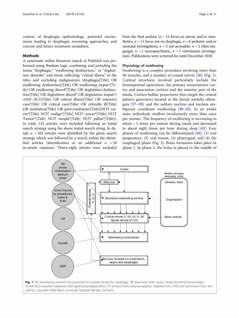
Physiology of swallowingSwallowing is a complex procedure involving more than50 muscles, and a number of cranial nerves [36] (Fig. 1).Cortical structures involved particularly include thefrontoparietal operculum, the primary sensorimotor cor-tex and association cortices and the anterior part of theinsula. Cortico-bulbar projections then target the centralpattern generators located in the dorsal medulla oblon-gata [37–39], and the solitary nucleus and nucleus am-biguous coordinate swallowing [40–42]. In an awakestate, individuals swallow involuntarily more than onceper minute. The frequency of swallowing is increasing toabout > 5 times per minute during meals and decreasedto about eight times per hour during sleep [43]. Fourphases of swallowing can be differentiated [44]: (1) oralpreparatory, (2) oral transit, (3) pharyngeal, and (4) theesophageal phase (Fig. 2). Bolus formation takes place inphase 1. In phase 2, the bolus is placed in the middle of
Fig. 1 The swallowing network and presumed ICU-related factors for dysphagia. TBI (traumatic brain injury), stroke (ischemic/hemorrhagic),ICUAW (ICU-acquired weakness), GER (gastroesophageal reflux, CIP (critical illness polyneuropathy). Adapted from [139] with permission from theauthors, copyright Heike Blum, University Hospital Münster, Germany
Zuercher et al. Critical Care (2019) 23:103 Page 2 of 11
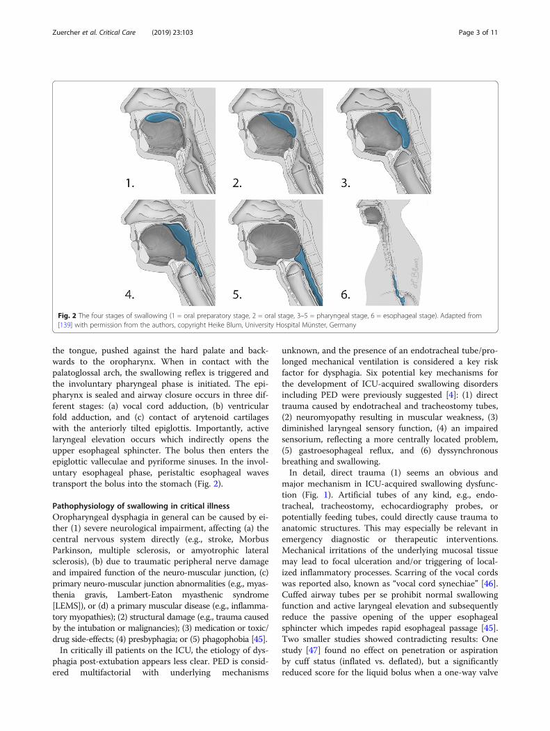
the tongue, pushed against the hard palate and back-wards to the oropharynx. When in contact with thepalatoglossal arch, the swallowing reflex is triggered andthe involuntary pharyngeal phase is initiated. The epi-pharynx is sealed and airway closure occurs in three dif-ferent stages: (a) vocal cord adduction, (b) ventricularfold adduction, and (c) contact of arytenoid cartilageswith the anteriorly tilted epiglottis. Importantly, activelaryngeal elevation occurs which indirectly opens theupper esophageal sphincter. The bolus then enters theepiglottic valleculae and pyriforme sinuses. In the invol-untary esophageal phase, peristaltic esophageal wavestransport the bolus into the stomach (Fig. 2).
Pathophysiology of swallowing in critical illnessOropharyngeal dysphagia in general can be caused by ei-ther (1) severe neurological impairment, affecting (a) thecentral nervous system directly (e.g., stroke, MorbusParkinson, multiple sclerosis, or amyotrophic lateralsclerosis), (b) due to traumatic peripheral nerve damageand impaired function of the neuro-muscular junction, (c)primary neuro-muscular junction abnormalities (e.g., myas-thenia gravis, Lambert-Eaton myasthenic syndrome[LEMS]), or (d) a primary muscular disease (e.g., inflamma-tory myopathies); (2) structural damage (e.g., trauma causedby the intubation or malignancies); (3) medication or toxic/drug side-effects; (4) presbyphagia; or (5) phagophobia [45].In critically ill patients on the ICU, the etiology of dys-
phagia post-extubation appears less clear. PED is consid-ered multifactorial with underlying mechanisms
unknown, and the presence of an endotracheal tube/pro-longed mechanical ventilation is considered a key riskfactor for dysphagia. Six potential key mechanisms forthe development of ICU-acquired swallowing disordersincluding PED were previously suggested [4]: (1) directtrauma caused by endotracheal and tracheostomy tubes,(2) neuromyopathy resulting in muscular weakness, (3)diminished laryngeal sensory function, (4) an impairedsensorium, reflecting a more centrally located problem,(5) gastroesophageal reflux, and (6) dyssynchronousbreathing and swallowing.In detail, direct trauma (1) seems an obvious and
major mechanism in ICU-acquired swallowing dysfunc-tion (Fig. 1). Artificial tubes of any kind, e.g., endo-tracheal, tracheostomy, echocardiography probes, orpotentially feeding tubes, could directly cause trauma toanatomic structures. This may especially be relevant inemergency diagnostic or therapeutic interventions.Mechanical irritations of the underlying mucosal tissuemay lead to focal ulceration and/or triggering of local-ized inflammatory processes. Scarring of the vocal cordswas reported also, known as “vocal cord synechiae” [46].Cuffed airway tubes per se prohibit normal swallowingfunction and active laryngeal elevation and subsequentlyreduce the passive opening of the upper esophagealsphincter which impedes rapid esophageal passage [45].Two smaller studies showed contradicting results: Onestudy [47] found no effect on penetration or aspirationby cuff status (inflated vs. deflated), but a significantlyreduced score for the liquid bolus when a one-way valve
Fig. 2 The four stages of swallowing (1 = oral preparatory stage, 2 = oral stage, 3–5 = pharyngeal stage, 6 = esophageal stage). Adapted from[139] with permission from the authors, copyright Heike Blum, University Hospital Münster, Germany
Zuercher et al. Critical Care (2019) 23:103 Page 3 of 11

was placed in comparison to in- or deflated cuff condi-tions (n = 14). Another study with a limited sample size(n = 7) [48] observed no significant alteration in hyoidbone movement and laryngeal excursion in cases when atracheotomy tube was placed. Further, long-term intub-ation may lead to dislocation or even subluxation ofarytenoid cartilages, resulting in an impaired glottis clos-ure during swallowing [49]. Further, traumatic laryngos-copy was shown to lead to hypoglossal nerve palsy andto cause dysphagia [50–53]. Peripheral damage of the re-current laryngeal nerve, e.g., caused by tube cuff com-pression (or as a complication during surgery) can resultin vocal cord paresis/paralysis and may prohibit compe-tent airway protection.Another relevant aspect is the presence of ICU ac-
quired weakness (ICUAW) [54]. In critically ill patientswith ICUAW, general muscular weakness and muscularatrophy was reported, which may affect the swallowingapparatus [54–56]. ICUAW may be a consequence of“disuse” in patients receiving long-term intubation,long-term (analgo)-sedation, and/or neuromuscularblocking agents [28, 54, 57, 58]. Further, specificswallow-related muscular weakness was recently sug-gested in previously orally intubated acute respiratorydistress syndrome patients (n = 11, median duration ofintubation 14 days) after examination by videofluoro-scopic swallowing study (VFSS) [59]. In addition, inICU-acquired ventilator-induced diaphragmatic dysfunc-tion (VIDD) [55], cough strength could be diminishedleading to limited glottic clearance. Importantly, reducedlocal sensation appears as an additional key problem inICU-acquired dysphagia. Either caused by direct mech-anical damage, local inflammation/edema, or by criticalillness polyneuropathy (CIP) [54, 56], afferent sensorypathways may be impaired leading to swallowing dys-function [60–63]. Clinically, this may become apparentwhen a bolus reaches the reflex trigger zone in the pala-toglossal arch but the afferent input is impaired resultingin a delayed swallow response and pre-deglutitive aspir-ation. However, the exact role of sensory impairment incritically ill patients appears unclear. In a recent study,no nerve conduction abnormalities were demonstrated,questioning the role of CIP in dysphagia [27].Central (cerebral) problems in ICU-acquired swallow-
ing disorders are mostly caused by direct damage to thecentral nervous system, e.g., in traumatic brain injury,stroke/hemorrhage, and/or inflammatory disorders.Contributing to this, reduced qualitative (e.g., in delir-ium) or quantitative level of consciousness further in-creases the risk for aspiration [64] and may delaytherapeutic measures for dysphagia. Moreover,drug-induced effects (e.g., (analgo-) sedatives or variousneurotropic medications) may affect swallowing eithercentrally (mostly via reduced consciousness) or
peripherally (mostly at the neuro-muscular junction). Inthis context, another potential mechanism was suggested[4], i.e., exact coordination of laryngeal closure, apnea,and opening of the upper esophageal sphincter may beimpaired. In critically ill patients, this is referred to as“dyssynchrony” between respiration and swallowing [4].Furthermore, in critically ill patients with respiratory dis-tress, the apnoeic period during swallowing is shortenedwith potential premature opening of the larynx beforethe bolus has passed into the esophagus [65].
Terminology of dysphagia on the ICUDifferent terms are used to assess dysphagia. In the 10threvision of the International Statistical Classification ofDisease and Related Health Problems (ICD-10,WHO-Version 2016), dysphagia (R13) is listed in “ChapterXVIII: symptoms, signs and abnormal clinical and labora-tory findings,” not elsewhere classified and more specifiedunder R10–19 in “Symptoms and signs involving the di-gestive system and abdomen.” Dysphagia, swallowing dis-order, or deglutition disorder/dysfunction are often usedsynonymously. In 2013, the term ICU-acquired swallow-ing disorder was introduced [4] suggesting multiple poten-tial pathomechanisms in critical illness leading to acquireddysphagia in a previously dysphagia-naïve patient. Inter-national consensus on dysphagia definitions is lackingwhich may negatively impact on data comparability. Wetherefore recently proposed a delphi procedure with theaim to harmonize respective terminology [66].
Epidemiology of dysphagia on the ICUA systematic review on the incidence of PED publishedin 2010 included a total of 14 studies with a total of3520 individuals (mean of approximately 251 patientsper study, median of 67) and concluded that the inci-dence rate ranges from 3 to 62% [7]. Study design,patient selection (e.g., assessment of patientspost-aspiration), and/or limited patient numbers in re-spective included studies introduced a high risk of biasand showed reduced quality of evidence [1–3, 5, 6, 67].In a subsequent retrospective observational cohortstudy, a dysphagia prevalence of up to 84% was reported[67]. A recent larger study (DYnAMICS) performed byus included 1304 medical and surgical ICU patients withpotential PED risk reported an incidence rate of 12.4%(18.3% in unselected emergency admissions) after sys-tematic screening [8]. In DYnAMICS, the incidence waslikely underestimated due to exclusion of patients leav-ing the ICU alive with tracheostomy (no extubation/decannulation) [8].
Risk factors for dysphagia on the ICURisk factors for dysphagia might theoretically be inferredfrom the abovementioned pathomechanisms. However,
Zuercher et al. Critical Care (2019) 23:103 Page 4 of 11
studies focusing on risk factors for dysphagia followingendotracheal intubation are scarce and provide conflict-ing results. Studies are mostly of limited sample size andeither supporting or rejecting respective factors, includ-ing factors such as age [12, 19, 20, 68–75], decreasedcardiac output [19, 70], intubation duration [19, 20, 22,32, 70, 71, 73, 74], postoperative pulmonary complica-tions [70, 74], tube feeding [19, 22, 32, 74], sepsis [6, 32,72], transesophageal echocardiogram (TEE) [70, 71],perioperative stroke [3, 32, 69–71], or gastroesophagealreflux [1, 19, 76]. More consistently rejected as potentialrisk factors are APACHE II and SOFA scores [1, 3, 6, 9,22, 67]; BMI [1, 6, 9]; gender [1, 6, 9, 32, 70, 75, 77, 78];comorbidities such as arterial hypertension, kidney dis-ease, diabetes, COPD, myocardial infarction, or heartfailure [3, 19, 32, 67, 69, 70, 72], as well as smoking [32,72]; and endotracheal tube size [3, 67, 75]. These contra-dicting results reflect bias due to patient selection, differ-ing study/screening protocols, and limited patientnumbers. However, despite controversial discussions, itappears that most presumed dysphagia risk factor wouldbe duration of intubation/mechanical ventilation [1, 3, 4,12, 32, 67, 68, 70, 78–80]. In addition, rather accepted riskfactors [4] may include the presence of pre-existing dys-phagia, local malignancy/post-surgical medical conditionsaffecting anatomic structures of the swallowing tract, and/or considerable quantitative/qualitative reduction of con-sciousness. Overall, large-scale clinical data is missing andstrongly warranted in order to potentially reduce thenumber of patients affected by preventional measures.
Assessment of dysphagia in the critically illIn stroke patients, early dysphagia detection minimizesthe risk of aspiration [81] and systematic dysphagiascreening reduces stroke-associated pneumonia rates[82]. This suggests that a systematic routine screeningapproach should be performed in all patients at riskwithout limitation to selected patient cohorts (e.g.,stroke patients). Non-instrumental [66] and instrumentalmeasures are available for timely assessment of dyspha-gia in the critically ill [83]. Non-instrumental assess-ments for dysphagia are typically performed by trainedspecialists (e.g., speech-language therapists, physiothera-pists, or occupational therapists). The following clinicalexaminations were previously proposed for general popu-lations of hospitalized (mostly non-ICU) patients: the bed-side swallowing evaluation (BSE) [84], the VolumeViscosity Swallowing Test (V-VST) [85], the MannAssessment of Swallowing Ability (MASA, K-MASA,MASA-C, MMASA) [86–89], the McGill Ingestive Swal-lowing Assessment (MISA, MISA-DK) [90, 91], theGugging Swallowing Screen (GUSS) [92], the Northwest-ern Dysphagia Patient Check Sheet (NDPCS) [93], theDysphagia Disorder Survey (DDS) [94], the Practical
Aspiration Screening Scheme (PASS) [95], the Kuchi-KaraTaberu Index (KT Index) [96], and the Practical Assess-ment of Dysphagia [97] test (reviewed in [66]).Instrumental tests, such as the flexible endoscopic
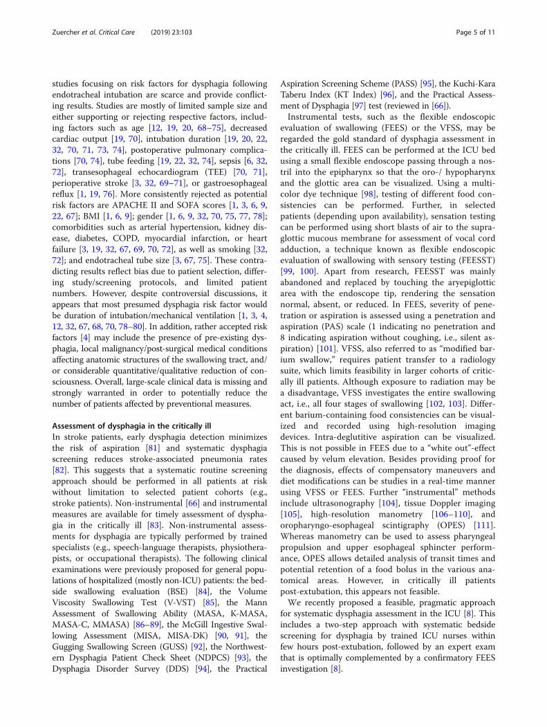
evaluation of swallowing (FEES) or the VFSS, may beregarded the gold standard of dysphagia assessment inthe critically ill. FEES can be performed at the ICU bedusing a small flexible endoscope passing through a nos-tril into the epipharynx so that the oro-/ hypopharynxand the glottic area can be visualized. Using a multi-color dye technique [98], testing of different food con-sistencies can be performed. Further, in selectedpatients (depending upon availability), sensation testingcan be performed using short blasts of air to the supra-glottic mucous membrane for assessment of vocal cordadduction, a technique known as flexible endoscopicevaluation of swallowing with sensory testing (FEESST)[99, 100]. Apart from research, FEESST was mainlyabandoned and replaced by touching the aryepiglotticarea with the endoscope tip, rendering the sensationnormal, absent, or reduced. In FEES, severity of pene-tration or aspiration is assessed using a penetration andaspiration (PAS) scale (1 indicating no penetration and8 indicating aspiration without coughing, i.e., silent as-piration) [101]. VFSS, also referred to as “modified bar-ium swallow,” requires patient transfer to a radiologysuite, which limits feasibility in larger cohorts of critic-ally ill patients. Although exposure to radiation may bea disadvantage, VFSS investigates the entire swallowingact, i.e., all four stages of swallowing [102, 103]. Differ-ent barium-containing food consistencies can be visual-ized and recorded using high-resolution imagingdevices. Intra-deglutitive aspiration can be visualized.This is not possible in FEES due to a “white out”-effectcaused by velum elevation. Besides providing proof forthe diagnosis, effects of compensatory maneuvers anddiet modifications can be studies in a real-time mannerusing VFSS or FEES. Further “instrumental” methodsinclude ultrasonography [104], tissue Doppler imaging[105], high-resolution manometry [106–110], andoropharyngo-esophageal scintigraphy (OPES) [111].Whereas manometry can be used to assess pharyngealpropulsion and upper esophageal sphincter perform-ance, OPES allows detailed analysis of transit times andpotential retention of a food bolus in the various ana-tomical areas. However, in critically ill patientspost-extubation, this appears not feasible.We recently proposed a feasible, pragmatic approach
for systematic dysphagia assessment in the ICU [8]. Thisincludes a two-step approach with systematic bedsidescreening for dysphagia by trained ICU nurses withinfew hours post-extubation, followed by an expert examthat is optimally complemented by a confirmatory FEESinvestigation [8].
Zuercher et al. Critical Care (2019) 23:103 Page 5 of 11
Clinical consequences of dysphagia in the critically illIn the critically ill, only few studies analyzed the impactof PED on patient-centered clinical outcomes. A recentretrospective study found an independent association ofdysphagia with a composite endpoint of pneumonia,reintubation, or death [67]. In addition, dysphagia wasassociated with longer hospitalization, more dischargesto a nursing home, and increased need for placement ofa feeding tube [67, 77]. In a recent large prospective ob-servational study (DYnAMICS) in a mixed population of1304 critically ill patients with systematic bedsidescreening for dysphagia post-extubation, we observed anindependent association of PED with 28-day and 90-daymortality after adjustment for typical confounders. Anexcess of 9.2% of a 90-day mortality rate was observedin patients with dysphagia [8].In summary, the clinical consequences of dysphagia in
the critically ill are important, with prolonged length ofhospitalization, increased resource use, increased treat-ment costs, and increased mortality [8, 31, 67, 112].Early identification of patients at risk seems warrantedin an effort to minimize respective burdens.
Therapeutic considerationsOverviewThe body of evidence for dysphagia treatment, especiallyin dysphagia-positive ICU patients, is limited. Generally,three major therapeutic pillars for dysphagia treatmentare considered [113]: dietary texture modifications, pos-tural changes/compensatory maneuvers, and interven-tions aiming to improve swallowing function (e.g.,devices using neuromuscular stimulation).
Dietary texture modification and compensatory maneuversAdaptation, a term mostly used in the German-speakingliterature [45], is referring to as texture modification ac-cording to the deglutition pathology and use of technicalaids, e.g., prosthetics to account for velopharyngeal de-fects, in an effort to optimize swallowing. Compensationis referring to either compensatory maneuvers and/orpostural changes to address swallowing deficiencies. Spe-cial swallowing techniques, e.g., supraglottic swallowing,may support patients with delayed swallowing reflex orincomplete laryngeal closure (e.g., after cordectomy).The patient would then be trained to hold the breath be-fore and during swallowing and forced to cough imme-diately afterwards to optimize glottic/throat clearance[114, 115]. For patients with impaired laryngeal eleva-tion, reduced tongue force, or dysfunctional opening ofthe upper esophageal sphincter (UES), the Mendelsohnmaneuver can be applied. In this maneuver, during oralpreparatory stage, the patient presses the bolus as force-fully as possible against the hard palate for up to 3 s.This elevates the larynx and improves UES opening and
clearance of food residuals [116]. Postural change “chindown” reduces the distance between to the tongue basisand pharyngeal dorsal wall, hence narrowing the airwayand therefore reducing the risk for leaking (bolus entersprematurely the pharynx) or aspiration within patientsknown for a delayed swallowing reflex. Moreover, theepiglottic vallecula gets distended, facilitating esophagealbolus passage [117–119].Head movements (backwards, lateralization towards/
away to the side of the paresis/palsy) may also be usefulin transporting the bolus to the swallow reflex triggerarea [117] or facilitating bolus passage via the healthypiriform recess [117, 120]. Functional dysphagia therapywas found successful in improving swallowing functionin patients suffering from neurogenic dysphagia [121].Apparently, an intensive therapy approach with fivetrainings a week seems to be more effective in the acutestage of swallowing dysfunction [122]. In addition,sphincter myotomy is an irreversible option in patientswith a functionally obstructing of the upper esophagealsphincter to facilitate pharyngo-esophageal bolus propul-sion [123, 124]. Medialization thyreoplasty can furtherbe applied in patients with unilateral vocal cord paresisand suffering from aspiration to improve cough andthroat clearance [125]. A laryngectomy poses a last re-sort for patients with persisting aspiration and suffer-ing from repeated severe consequences. In doing this,breathing and alimentary pathways become com-pletely separated.
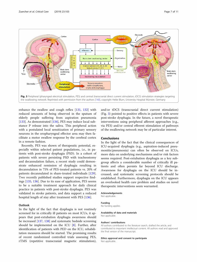
Interventional/technological approaches: pharyngealelectrical stimulationRecently, pharyngeal electrical stimulation (PES) (Fig. 3)was proposed as a novel treatment modality using a gas-tric feeding tube-like stimulation catheter to enhanceneuromuscular pharyngeal stimulation, targeted to theindividual patient. Stimulation levels are personalized atthe start of the treatment to ensure that optimal levelsof stimulation are delivered. PES is considered to targetthe afferent sensory feedback within the swallowing net-work that seems crucial for swallowing safety and effi-cacy of motor execution [126, 127]. PES may involvetwo postulated key modes of action: (a) facilitation ofcortico-bulbar pathways [128] and (b) increase of swal-lowing processing efficiency in respective central ner-vous system areas [129], e.g., the right primary andsecondary sensorimotor cortex and the right supplemen-tary motor area. Data also demonstrate an increase ofpharyngeal cortical representation and motor excitabilityfor more than half an hour after 10 min of PES treat-ment. A dose-response study ([130]) showed optimalcost-effectiveness when applying a PES protocol withone cycle of 10-min stimulation per day for a total of 3consecutive days. Further, substance P is known to
Zuercher et al. Critical Care (2019) 23:103 Page 6 of 11
enhance the swallow and cough reflex [131, 132] withreduced amounts of being observed in the sputum ofelderly people suffering from aspiration pneumonia[133]. As demonstrated [134], PES may induce local sub-stance P release into the saliva. This peripheral actionwith a postulated local sensitization of primary sensoryneurons in the oropharyngeal effector area may then fa-cilitate a motor swallow response by the cerebral cortexin a remote fashion.Recently, PES was shown of therapeutic potential, es-
pecially within selected patient populations, i.e., in pa-tients with post-stroke dysphagia (PSD). In a cohort ofpatients with severe persisting PSD with tracheostomyand decannulation failure, a recent study could demon-strate enhanced remission of dysphagia resulting indecannulation in 75% of PES-treated patients vs. 20% ofpatients decannulated in sham-treated individuals [129].Two recently published studies support respective find-ings [135, 136]. Due to its ease of application, PES seemsto be a suitable treatment approach for daily clinicalpractice in patients with post-stroke dysphagia. PES wasvalidated in stroke patients, and data support a reducedhospital length of stay after treatment with PES [136].
OutlookIn the light of the fact that dysphagia is not routinelyscreened for in critically ill patients on most ICUs, it ap-pears that post-extubation dysphagia awareness shouldbe increased [137, 138] and systematic bedside screeningshould be implemented on the ICU [8]. Further, afteridentification of patients with PED on the ICU, rehabili-tation measures should be started. The promising resultsof recent randomized controlled trials assessing PES,rTMS (repetitive transcranial magnetic stimulation),
and/or tDCS (transcranial direct current stimulation)(Fig. 3) pointed to positive effects in patients with severepost-stroke dysphagia. In the future, a novel therapeuticinterventions using peripheral afferent approaches (e.g.,via PES) and/or central efferent stimulation of pathwaysof the swallowing network may be of particular interest.
ConclusionsIn the light of the fact that the clinical consequences ofICU-acquired dysphagia (e.g., aspiration-induced pneu-monitis/pneumonia) can often be observed on ICUs,more data on underlying mechanisms and/or risk factorsseems required. Post-extubation dysphagia as a key sub-group affects a considerable number of critically ill pa-tients and often persists far beyond ICU discharge.Awareness for dysphagia on the ICU should be in-creased, and systematic screening protocols should beestablished. Furthermore, dysphagia on the ICU appearsan overlooked health care problem and studies on noveltherapeutic interventions seem warranted.
AcknowledgementsNot applicable.
FundingNo funding applies
Availability of data and materialsNot applicable.
Authors’ contributionsAll authors contributed to the literature search, drafted the article, andcontributed to important intellectual content. All authors read and approvedthe final version of the manuscript.
Ethics approval and consent to participateNot applicable.
Fig. 3 Peripheral (pharyngeal electrical stimulation, PES) and central (transcranial direct current stimulation, tDCS) stimulation strategies targetingthe swallowing network. Reprinted with permission from the authors [140], copyright Heike Blum, University Hospital Münster, Germany
Zuercher et al. Critical Care (2019) 23:103 Page 7 of 11
Consent for publicationNot applicable.
Competing interestsDrs. Zuercher, Moret, and Schefold declare that the Department of IntensiveCare Medicine, Inselspital, Bern, has received research or other grants from(full disclosure) Orion Pharma, Abbott Nutrition International, B. BraunMedical AG, CSEM AG, Edwards Lifesciences Services GmbH, Kenta BiotechLtd., Maquet Critical Care AB, Omnicare Clinical Research AG, Nestle, PierreFabre Pharma AG, Pfizer, Bard Medica S.A., Abbott AG, Anandic MedicalSystems, Pan Gas AG Healthcare, Bracco, Hamilton Medical AG, FreseniusKabi, Getinge Group Maquet AG, Dräger AG, Teleflex Medical GmbH, GlaxoSmith Kline, Merck Sharp and Dohme AG, Eli Lilly and Company, Baxter,Astellas, Astra Zeneca, CSL Behring, Novartis, Covidien, and Nycomed outsidethe submitted work. The money was paid into departmental funds. Nopersonal financial gain applies. Dr. Dziewas declares no competing interests.
Publisher’s NoteSpringer Nature remains neutral with regard to jurisdictional claims inpublished maps and institutional affiliations.
Author details1Department of Intensive Care Medicine, Inselspital, Bern University Hospital,University of Bern, 3010 Bern, CH, Switzerland. 2Department of Neurology,University Hospital Münster, Münster, Germany.
Received: 1 February 2019 Accepted: 18 March 2019
References1. Brodsky MB, Gellar JE, Dinglas VD, Colantuoni E, Mendez-Tellez PA,
Shanholtz C, Palmer JB, Needham DM. Duration of oral endotrachealintubation is associated with dysphagia symptoms in acute lung injurypatients. J Crit Care. 2014;29(4):574–9.
2. Brown CV, Hejl K, Mandaville AD, Chaney PE, Stevenson G, Smith C.Swallowing dysfunction after mechanical ventilation in trauma patients. JCritical Care. 2011;26(1):108 e109–113.
3. Macht M, King CJ, Wimbish T, Clark BJ, Benson AB, Burnham EL, Williams A,Moss M. Post-extubation dysphagia is associated with longer hospitalizationin survivors of critical illness with neurologic impairment. Crit Care. 2013;17(3):R119.
4. Macht M, Wimbish T, Bodine C, Moss M. ICU-acquired swallowing disorders.Crit Care Med. 2013;41(10):2396–405.
5. Moraes DP, Sassi FC, Mangilli LD, Zilberstein B, de Andrade CR. Clinicalprognostic indicators of dysphagia following prolonged orotrachealintubation in ICU patients. Crit Care. 2013;17(5):R243.
6. Zielske J, Bohne S, Brunkhorst FM, Axer H, Guntinas-Lichius O. Acute andlong-term dysphagia in critically ill patients with severe sepsis: results of aprospective controlled observational study. Eur Arch Otorhinolaryngol. 2014;271(11):3085–93.
7. Skoretz SA, Flowers HL, Martino R. The incidence of dysphagia followingendotracheal intubation: a systematic review. Chest. 2010;137(3):665–73.
8. Schefold JC, Berger D, Zurcher P, Lensch M, Perren A, Jakob SM, ParviainenI, Takala J. Dysphagia in mechanically ventilated ICU patients (DYnAMICS): aprospective observational trial. Crit Care Med. 2017;45(12):2061–9.
9. Brodsky MB, Huang M, Shanholtz C, Mendez-Tellez PA, Palmer JB,Colantuoni E, Needham DM. Recovery from dysphagia symptoms after oralendotracheal intubation in acute respiratory distress syndrome survivors. A5-year longitudinal study. Ann Am Thorac Soc. 2017;14(3):376–83.
10. Christensen M, Trapl M. Development of a modified swallowing screeningtool to manage post-extubation dysphagia. Nurs Crit Care. 2018;23(2):102-107. https://doi.org/10.1111/nicc.12333. Epub 2017 Dec 28..
11. Medeiros GC, Sassi FC, Zambom LS, Andrade CR. Correlation between theseverity of critically ill patients and clinical predictors of bronchial aspiration.J Bras Pneumol. 2016;42(2):114–20.
12. Oliveira ACM, Friche AAL, Salomao MS, Bougo GC, Vicente LCC. Predictivefactors for oropharyngeal dysphagia after prolonged orotracheal intubation.Braz J Otorhinolaryngol. 2018;84(6):722–28. https://doi.org/10.1016/j.bjorl.2017.08.010. Epub 2017 Sep 13.
13. Scheel R, Pisegna JM, McNally E, Noordzij JP, Langmore SE. Endoscopicassessment of swallowing after prolonged intubation in the ICU setting.Ann Otol Rhinol Laryngol. 2016;125(1):43–52.
14. See KC, Peng SY, Phua J, Sum CL, Concepcion J. Nurse-performed screeningfor postextubation dysphagia: a retrospective cohort study in critically illmedical patients. Crit Care. 2016;20(1):326.
15. Loeb M, McGeer A, McArthur M, Walter S, Simor AE. Risk factors forpneumonia and other lower respiratory tract infections in elderly residentsof long-term care facilities. Arch Intern Med. 1999;159(17):2058–64.
16. Marik PE, Kaplan D. Aspiration pneumonia and dysphagia in the elderly.Chest. 2003;124(1):328–36.
17. Martin BJ, Corlew MM, Wood H, Olson D, Golopol LA, Wingo M, Kirmani N.The association of swallowing dysfunction and aspiration pneumonia.Dysphagia. 1994;9(1):1–6.
18. Vergis EN, Brennen C, Wagener M, Muder RR. Pneumonia in long-term care:a prospective case-control study of risk factors and impact on survival. ArchIntern Med. 2001;161(19):2378–81.
19. Ajemian MS, Nirmul GB, Anderson MT, Zirlen DM, Kwasnik EM. Routinefiberoptic endoscopic evaluation of swallowing following prolongedintubation: implications for management. Archives Surgery (Chicago, Ill :1960). 2001;136(4):434–7.
20. Barquist E, Brown M, Cohn S, Lundy D, Jackowski J. Postextubationfiberoptic endoscopic evaluation of swallowing after prolongedendotracheal intubation: a randomized, prospective trial. Crit Care Med.2001;29(9):1710–3.
21. Ekberg O, Hamdy S, Woisard V, Wuttge-Hannig A, Ortega P. Social andpsychological burden of dysphagia: its impact on diagnosis and treatment.Dysphagia. 2002;17(2):139–46.
22. El Solh A, Okada M, Bhat A, Pietrantoni C. Swallowing disorders postorotracheal intubation in the elderly. Intensive Care Med. 2003;29(9):1451–5.
23. Holas MA, DePippo KL, Reding MJ. Aspiration and relative risk of medicalcomplications following stroke. Arch Neurol. 1994;51(10):1051–3.
24. Marik PE. Aspiration pneumonitis and aspiration pneumonia. N Engl J Med.2001;344(9):665–71.
25. Schmidt J, Holas M, Halvorson K, Reding M. Videofluoroscopic evidence ofaspiration predicts pneumonia and death but not dehydration followingstroke. Dysphagia. 1994;9(1):7–11.
26. Tolep K, Getch CL, Criner GJ. Swallowing dysfunction in patients receivingprolonged mechanical ventilation. Chest. 1996;109(1):167–72.
27. Ponfick M, Linden R, Nowak DA. Dysphagia--a common, transient symptomin critical illness polyneuropathy: a fiberoptic endoscopic evaluation ofswallowing study*. Crit Care Med. 2015;43(2):365–72.
28. DeVita MA, Spierer-Rundback L. Swallowing disorders in patients withprolonged orotracheal intubation or tracheostomy tubes. Crit Care Med.1990;18(12):1328–30.
29. Daly E, Miles A, Scott S, Gillham M. Finding the red flags: swallowingdifficulties after cardiac surgery in patients with prolonged intubation. J CritCare. 2016;31(1):119–24.
30. Medeiros GC, Sassi FC, Mangilli LD, Zilberstein B, Andrade CR. Clinicaldysphagia risk predictors after prolonged orotracheal intubation. Clinics.2014;69(1):8–14.
31. Altman KW, Yu GP, Schaefer SD. Consequence of dysphagia in thehospitalized patient: impact on prognosis and hospital resources. ArchOtolaryngol Head Neck Surg. 2010;136(8):784–9.
32. Barker J, Martino R, Reichardt B, Hickey EJ, Ralph-Edwards A. Incidence andimpact of dysphagia in patients receiving prolonged endotrachealintubation after cardiac surgery. Can J Surg. 2009;52(2):119–24.
33. Smithard DG, O'Neill PA, Parks C, Morris J. Complications and outcome afteracute stroke. Does dysphagia matter? Stroke. 1996;27(7):1200–4.
34. Brodsky MB, Gonzalez-Fernandez M, Mendez-Tellez PA, Shanholtz C,Palmer JB, Needham DM. Factors associated with swallowingassessment after oral endotracheal intubation and mechanicalventilation for acute lung injury. Ann Am Thorac Soc. 2014;11(10):1545–52.
35. Macht M, White SD, Moss M. Swallowing dysfunction after critical illness.Chest. 2014;146(6):1681–9.
36. Reiter R, Brosch S. Update oropharyngeal dysphagia part 1: physiology,pathology and diagnosis. Laryngorhinootologie. 2012;91(4):224–7.
37. Hamdy S, Mikulis DJ, Crawley A, Xue S, Lau H, Henry S, Diamant NE. Corticalactivation during human volitional swallowing: an event-related fMRI study.Am J Phys. 1999;277(1 Pt 1):G219–25.
Zuercher et al. Critical Care (2019) 23:103 Page 8 of 11
38. Hamdy S, Rothwell JC, Brooks DJ, Bailey D, Aziz Q, Thompson DG.Identification of the cerebral loci processing human swallowing withH2(15)O PET activation. J Neurophysiol. 1999;81(4):1917–26.
39. Riecker A, Gastl R, Kuhnlein P, Kassubek J, Prosiegel M. Dysphagia due tounilateral infarction in the vascular territory of the anterior insula. Dysphagia.2009;24(1):114–8.
40. Kumar S. Swallowing and dysphagia in neurological disorders. Rev NeurolDis. 2010;7(1):19–27.
41. Lang IM. Brain stem control of the phases of swallowing. Dysphagia. 2009;24(3):333–48.
42. Prosiegel M, Holing R, Heintze M, Wagner-Sonntag E, Wiseman K. Thelocalization of central pattern generators for swallowing in humans--aclinical-anatomical study on patients with unilateral paresis of the vagalnerve, Avellis’ syndrome, Wallenberg’s syndrome, posterior fossa tumoursand cerebellar hemorrhage. Acta Neurochir Suppl. 2005;93:85–8.
43. Lear CS, Flanagan JB Jr, Moorrees CF. The frequency of deglutition in man.Arch Oral Biol. 1965;10:83–100.
44. Dodds WJ, Stewart ET, Logemann JA. Physiology and radiology of thenormal oral and pharyngeal phases of swallowing. AJR Am J Roentgenol.1990;154(5):953–63.
45. Reiter R, Brosch S. Update oropharyngeal dysphagia part 2: etiology andtherapy. Laryngorhinootologie. 2012;91(5):291–9.
46. Stauffer JL, Olson DE, Petty TL. Complications and consequences ofendotracheal intubation and tracheotomy. A prospective study of 150critically ill adult patients. Am J Med. 1981;70(1):65–76.
47. Suiter DM, McCullough GH, Powell PW. Effects of cuff deflation and one-way tracheostomy speaking valve placement on swallow physiology.Dysphagia. 2003;18(4):284–92.
48. Terk AR, Leder SB, Burrell MI. Hyoid bone and laryngeal movementdependent upon presence of a tracheotomy tube. Dysphagia. 2007;22(2):89–93.
49. Sue RD, Susanto I. Long-term complications of artificial airways. Clin ChestMed. 2003;24(3):457–71.
50. Hong SJ, Lee JY. Isolated unilateral paralysis of the hypoglossal nerve aftertransoral intubation for general anesthesia. Dysphagia. 2009;24(3):354–6.
51. Bramer S, Koscielny S, Witte OW, Terborg C. Bilateral hypoglossal nerve palsyfollowing intubation. Nervenarzt. 2006;77(2):204–7.
52. Batjom E, Coron T, Mercier F, Benhamou D. Hypoglossal nerve palsy, a rarecomplication of orotracheal intubation. Ann Fr Anesth Reanim. 2006;25(5):541–2.
53. Dziewas R, Ludemann P. Hypoglossal nerve palsy as complication of oralintubation, bronchoscopy and use of the laryngeal mask airway. Eur Neurol.2002;47(4):239–43.
54. Schefold JC, Bierbrauer J, Weber-Carstens S. Intensive care unit-acquiredweakness (ICUAW) and muscle wasting in critically ill patients with severesepsis and septic shock. J Cachexia Sarcopenia Muscle. 2010;1(2):147–57.
55. Berger D, Bloechlinger S, von Haehling S, Doehner W, Takala J, Z'GraggenWJ, Schefold JC. Dysfunction of respiratory muscles in critically ill patientson the intensive care unit. J Cachexia Sarcopenia Muscle. 2016;7(4):403–12.
56. Jolley SE, Bunnell AE, Hough CL. ICU-acquired weakness. Chest. 2016;150(5):1129–40.
57. Goldsmith T. Evaluation and treatment of swallowing disorders followingendotracheal intubation and tracheostomy. Int Anesthesiol Clin. 2000;38(3):219–42.
58. Feng X, Todd T, Lintzenich CR, Ding J, Carr JJ, Ge Y, Browne JD, KritchevskySB, Butler SG. Aging-related geniohyoid muscle atrophy is related toaspiration status in healthy older adults. J Gerontol A Biol Sci Med Sci. 2013;68(7):853–60.
59. Brodsky MB, De I, Chilukuri K, Huang M, Palmer JB, Needham DM.Coordination of pharyngeal and laryngeal swallowing events during singleliquid swallows after Oral endotracheal intubation for patients with acuterespiratory distress syndrome. Dysphagia. 2018;33(6):768–77.
60. Aviv JE. Clinical assessment of pharyngolaryngeal sensitivity. Am J Med.2000;108(Suppl 4a):68S–72S.
61. Aviv JE, Spitzer J, Cohen M, Ma G, Belafsky P, Close LG. Laryngeal adductorreflex and pharyngeal squeeze as predictors of laryngeal penetration andaspiration. Laryngoscope. 2002;112(2):338–41.
62. Bradley RM. Sensory receptors of the larynx. Am J Med. 2000;108(Suppl 4a):47S–50S.
63. Shaker R, Hogan WJ. Reflex-mediated enhancement of airway protectivemechanisms. Am J Med. 2000;108(Suppl 4a):8S–14S.
64. Leder SB, Suiter DM, Lisitano Warner H. Answering orientation questionsand following single-step verbal commands: effect on aspiration status.Dysphagia. 2009;24(3):290–5.
65. Boden K, Cedborg AI, Eriksson LI, Hedstrom HW, Kuylenstierna R, SundmanE, Ekberg O. Swallowing and respiratory pattern in young healthyindividuals recorded with high temporal resolution. NeurogastroenterolMotil. 2009;21(11):1163–e1101.
66. Perren A, Zurcher P, Schefold JC. Clinical approaches to assess post-extubation dysphagia (PED) in the critically ill. Dysphagia. 2019. https://doi.org/10.1007/s00455-019-09977-w.
67. Macht M, Wimbish T, Clark BJ, Benson AB, Burnham EL, Williams A, Moss M.Postextubation dysphagia is persistent and associated with poor outcomesin survivors of critical illness. Crit Care. 2011;15(5):R231.
68. Bordon A, Bokhari R, Sperry J, Testa D, Feinstein A, Ghaemmaghami V.Swallowing dysfunction after prolonged intubation: analysis of risk factors intrauma patients. Am J Surg. 2011;202(6):679–82 discussion 682-673.
69. Ferraris VA, Ferraris SP, Moritz DM, Welch S. Oropharyngeal dysphagia aftercardiac operations. Ann Thorac Surg. 2001;71(6):1792–5 discussion 1796.
70. Hogue CW Jr, Lappas GD, Creswell LL, Ferguson TB Jr, Sample M, Pugh D, BalfeD, Cox JL, Lappas DG. Swallowing dysfunction after cardiac operations.Associated adverse outcomes and risk factors including intraoperativetransesophageal echocardiography. J Thorac Cardiovasc Surg. 1995;110(2):517–22.
71. Rousou JA, Tighe DA, Garb JL, Krasner H, Engelman RM, Flack JE 3rd, DeatonDW. Risk of dysphagia after transesophageal echocardiography during cardiacoperations. Ann Thorac Surg. 2000;69(2):486–9 discussion 489-490.
72. Skoretz SA, Yau TM, Ivanov J, Granton JT, Martino R. Dysphagia andassociated risk factors following extubation in cardiovascular surgicalpatients. Dysphagia. 2014;29(6):647–54.
73. de Larminat V, Montravers P, Dureuil B, Desmonts JM. Alteration inswallowing reflex after extubation in intensive care unit patients. Crit CareMed. 1995;23(3):486–90.
74. Leder SB, Cohn SM, Moller BA. Fiberoptic endoscopic documentation of thehigh incidence of aspiration following extubation in critically ill traumapatients. Dysphagia. 1998;13(4):208–12.
75. Elpern EH, Scott MG, Petro L, Ries MH. Pulmonary aspiration in mechanicallyventilated patients with tracheostomies. Chest. 1994;105(2):563–6.
76. Metheny NA, Clouse RE, Chang YH, Stewart BJ, Oliver DA, Kollef MH.Tracheobronchial aspiration of gastric contents in critically ill tube-fed patients:frequency, outcomes, and risk factors. Crit Care Med. 2006;34(4):1007–15.
77. Omura K, Komine A, Yanagigawa M, Chiba N, Osada M. Frequency andoutcome of post-extubation dysphagia using nurse-performed swallowingscreening protocol. Nurs Crit Care. 2019;24(2):70–75. https://doi.org/10.1111/nicc.12359. Epub 2018 Jul 3..
78. Sassi FC, Medeiros GC, Zambon LS, Zilberstein B, Andrade CRF. Evaluationand classification of post-extubation dysphagia in critically ill patients. RevCol Bras Cir. 2018;45(3):e1687.
79. Kim MJ, Park YH, Park YS, Song YH. Associations between prolongedintubation and developing post-extubation dysphagia and aspirationpneumonia in non-neurologic critically ill patients. Ann Rehabil Med. 2015;39(5):763–71.
80. Kwok AM, Davis JW, Cagle KM, Sue LP, Kaups KL. Post-extubation dysphagiain trauma patients: it's hard to swallow. Am J Surg. 2013;206(6):924–7discussion 927-928.
81. Doggett DL, Tappe KA, Mitchell MD, Chapell R, Coates V, Turkelson CM.Prevention of pneumonia in elderly stroke patients by systematic diagnosisand treatment of dysphagia: an evidence-based comprehensive analysis ofthe literature. Dysphagia. 2001;16(4):279–95.
82. Teuschl Y, Trapl M, Ratajczak P, Matz K, Dachenhausen A, Brainin M.Systematic dysphagia screening and dietary modifications to reduce stroke-associated pneumonia rates in a stroke-unit. PLoS One. 2018;13(2):e0192142.
83. Marvin S, Thibeault S, Ehlenbach WJ: Post-extubation dysphagia: doestiming of evaluation matter? Dysphagia. 2019;34(2):210–19. https://doi.org/10.1007/s00455-018-9926-3. Epub 2018 Jul 24.
84. Lynch YT, Clark BJ, Macht M, White SD, Taylor H, Wimbish T, Moss M. Theaccuracy of the bedside swallowing evaluation for detecting aspiration insurvivors of acute respiratory failure. J Crit Care. 2017;39:143–8.
85. Clave P, Arreola V, Romea M, Medina L, Palomera E, Serra-Prat M. Accuracyof the volume-viscosity swallow test for clinical screening of oropharyngealdysphagia and aspiration. Clin Nutr. 2008;27(6):806–15.
86. Antonios N, Carnaby-Mann G, Crary M, Miller L, Hubbard H, Hood K,Sambandam R, Xavier A, Silliman S. Analysis of a physician tool for evaluating
Zuercher et al. Critical Care (2019) 23:103 Page 9 of 11
dysphagia on an inpatient stroke unit: the modified Mann Assessment ofSwallowing Ability. J Stroke Cerebrovasc Dis. 2010;19(1):49–57.
87. Carnaby GD, Crary MA. Development and validation of a cancer-specificswallowing assessment tool: MASA-C. Support Care Cancer. 2014;22(3):595–602.
88. Gonzalez-Fernandez M, Sein MT, Palmer JB. Clinical experience using theMann assessment of swallowing ability for identification of patients at riskfor aspiration in a mixed-disease population. Am J Speech-Lang Pathol.2011;20(4):331–6.
89. Oh JC, Park JH, Jung MY, Yoo EY, Chang KY, Lee TY. Relationship betweenquantified instrumental swallowing examination and comprehensive clinicalswallowing examination. Occup Ther Int. 2016;23(1):3–10.
90. Hansen T, Lambert HC, Faber J. Validation of the Danish version of theMcGill Ingestive Skills Assessment using classical test theory and the Raschmodel. Disabil Rehabil. 2012;34(10):859–68.
91. Lambert HC, Gisel EG, Groher ME, Abrahamowicz M, Wood-Dauphinee S.Psychometric testing of the McGill Ingestive Skills Assessment. Am J OccupTher. 2006;60(4):409–19.
92. Trapl M, Enderle P, Nowotny M, Teuschl Y, Matz K, Dachenhausen A, BraininM. Dysphagia bedside screening for acute-stroke patients: the GuggingSwallowing Screen. Stroke. 2007;38(11):2948–52.
93. Logemann JA, Veis S, Colangelo L. A screening procedure for oropharyngealdysphagia. Dysphagia. 1999;14(1):44–51.
94. Sheppard JJ, Hochman R, Baer C. The dysphagia disorder survey: validationof an assessment for swallowing and feeding function in developmentaldisability. Res Dev Disabil. 2014;35(5):929–42.
95. Zhou Z, Salle J, Daviet J, Stuit A, Nguyen C. Combined approach in bedsideassessment of aspiration risk post stroke: PASS. Eur J Phys Rehabil Med.2011;47(3):441–6.
96. Maeda K, Shamoto H, Wakabayashi H, Enomoto J, Takeichi M, Koyama T.Reliability and validity of a simplified comprehensive assessment tool forfeeding support: Kuchi-Kara Taberu Index. J Am Geriatr Soc. 2016;64(12):e248–52.
97. Lee KM, Kim HJ. Practical assessment of dysphagia in stroke patients. AnnRehabil Med. 2015;39(6):1018–27.
98. Hacki T, Kramer H, Kleinjung C, Perez-Alvarez C, Schmid J. Endoscopic multi-color deglutition study. Laryngorhinootologie. 2001;80(6):335–40.
99. Aviv JE, Kaplan ST, Thomson JE, Spitzer J, Diamond B, Close LG. The safetyof flexible endoscopic evaluation of swallowing with sensory testing(FEESST): an analysis of 500 consecutive evaluations. Dysphagia. 2000;15(1):39–44.
100. Schindler A, Ginocchio D, Peri A, Felisati G, Ottaviani F. FEESST in therehabilitation of dysphagia after partial laryngectomy. Ann Otol RhinolLaryngol. 2010;119(2):71–6.
101. Rosenbek JC, Robbins JA, Roecker EB, Coyle JL, Wood JL. A penetration-aspiration scale. Dysphagia. 1996;11(2):93–8.
102. Hannig C, Wuttge-Hannig A, Hess U. Analysis and radiologic staging of thetype and severity of aspiration. Radiologe. 1995;35(10):741–6.
103. Palmer JB, Kuhlemeier KV, Tippett DC, Lynch C. A protocol for thevideofluorographic swallowing study. Dysphagia. 1993;8(3):209–14.
104. Fanucci A, Cerro P, Ietto F, Brancaleone C, Berardi F. Physiology of oralswallowing studied by ultrasonography. Dentomaxillofac Radiol. 1994;23(4):221–5.
105. Manabe N, Haruma K, Nakato R, Kusunoki H, Kamada T, Hata J. Newultrasonographic screening method for oropharyngeal dysphagia: tissueDoppler imaging. Am J Physiol Gastrointest Liver Physiol. 2018;314(1):G32–8.
106. Castell JA, Castell DO. Modern solid state computerized manometry of thepharyngoesophageal segment. Dysphagia. 1993;8(3):270–5.
107. Cook IJ, Dodds WJ, Dantas RO, Kern MK, Massey BT, Shaker R, HoganWJ. Timing of videofluoroscopic, manometric events, and bolus transitduring the oral and pharyngeal phases of swallowing. Dysphagia. 1989;4(1):8–15.
108. Dodds WJ, Hogan WJ, Lydon SB, Stewart ET, Stef JJ, Arndorfer RC.Quantitation of pharyngeal motor function in normal human subjects. JAppl Physiol. 1975;39(4):692–6.
109. Dodds WJ, Logemann JA, Stewart ET. Radiologic assessment of abnormaloral and pharyngeal phases of swallowing. AJR Am J Roentgenol. 1990;154(5):965–74.
110. McConnel FM. Analysis of pressure generation and bolus transit duringpharyngeal swallowing. Laryngoscope. 1988;98(1):71–8.
111. Fattori B, Grosso M, Bongioanni P, Nacci A, Cristofani R, AlSharif A, Licitra R,Matteucci F, Rossi B, Rubello D, et al. Assessment of swallowing by
oropharyngoesophageal scintigraphy in patients with amyotrophic lateralsclerosis. Dysphagia. 2006;21(4):280–6.
112. Kozlow JH, Berenholtz SM, Garrett E, Dorman T, Pronovost PJ. Epidemiologyand impact of aspiration pneumonia in patients undergoing surgery inMaryland, 1999–2000. Crit Care Med. 2003;31(7):1930–7.
113. Rassameehiran S, Klomjit S, Mankongpaisarnrung C, Rakvit A. Postextubationdysphagia. Proc (Bayl Univ Med Cent). 2015;28(1):18–20.
114. Hirst LJ, Sama A, Carding PN, Wilson JA. Is a ‘safe swallow’ really safe? Int JLang Commun Disord. 1998;33(Suppl):279–80.
115. Ohmae Y, Logemann JA, Kaiser P, Hanson DG, Kahrilas PJ. Effects of twobreath-holding maneuvers on oropharyngeal swallow. Ann Otol RhinolLaryngol. 1996;105(2):123–31.
116. Kahrilas PJ, Logemann JA, Krugler C, Flanagan E. Volitional augmentation ofupper esophageal sphincter opening during swallowing. Am J Phys. 1991;260(3 Pt 1):G450–6.
117. Logemann JA. Treatment of oral and pharyngeal dysphagia. Phys MedRehabil Clin N Am. 2008;19(4):803–16 ix.
118. Shanahan TK, Logemann JA, Rademaker AW, Pauloski BR, Kahrilas PJ. Chin-down posture effect on aspiration in dysphagic patients. Arch Phys MedRehabil. 1993;74(7):736–9.
119. Welch MV, Logemann JA, Rademaker AW, Kahrilas PJ. Changes inpharyngeal dimensions effected by chin tuck. Arch Phys Med Rehabil. 1993;74(2):178–81.
120. Logemann JA, Kahrilas PJ, Kobara M, Vakil NB. The benefit of head rotation onpharyngoesophageal dysphagia. Arch Phys Med Rehabil. 1989;70(10):767–71.
121. Prosiegel M, Heintze M, Wagner-Sonntag E, Hannig C, Wuttge-Hannig A,Yassouridis A. Deglutition disorders in neurological patients. A prospectivestudy of diagnosis, pattern of impairment, therapy and outcome.Nervenarzt. 2002;73(4):364–70.
122. Carnaby G, Hankey GJ, Pizzi J. Behavioural intervention for dysphagia inacute stroke: a randomised controlled trial. Lancet Neurol. 2006;5(1):31–7.
123. Kos MP, David EF, Klinkenberg-Knol EC, Mahieu HF. Long-term results ofexternal upper esophageal sphincter myotomy for oropharyngealdysphagia. Dysphagia. 2010;25(3):169–76.
124. Shama L, Connor NP, Ciucci MR, McCulloch TM. Surgical treatment ofdysphagia. Phys Med Rehabil Clin N Am. 2008;19(4):817–35 ix.
125. Flint PW, Purcell LL, Cummings CW. Pathophysiology and indications formedialization thyroplasty in patients with dysphagia and aspiration.Otolaryngol Head Neck Surg. 1997;116(3):349–54.
126. Muhle P, Claus I, Marian T, Schroder JB, Wollbrink A, Pantev C, Warnecke T,Dziewas R, Suntrup-Krueger S. Introducing a virtual lesion model ofdysphagia resulting from pharyngeal sensory impairment. Neurosignals.2018;26(1):1–10.
127. Teismann IK, Steinstraeter O, Stoeckigt K, Suntrup S, Wollbrink A, Pantev C,Dziewas R. Functional oropharyngeal sensory disruption interferes with thecortical control of swallowing. BMC Neurosci. 2007;8:62.
128. Hamdy S, Rothwell JC, Aziz Q, Singh KD, Thompson DG. Long-termreorganization of human motor cortex driven by short-term sensorystimulation. Nat Neurosci. 1998;1(1):64–8.
129. Suntrup S, Marian T, Schroder JB, Suttrup I, Muhle P, Oelenberg S, HamacherC, Minnerup J, Warnecke T, Dziewas R. Electrical pharyngeal stimulation fordysphagia treatment in tracheotomized stroke patients: a randomizedcontrolled trial. Intensive Care Med. 2015;41(9):1629–37.
130. Jayasekeran V, Singh S, Tyrrell P, Michou E, Jefferson S, Mistry S, Gamble E,Rothwell J, Thompson D, Hamdy S. Adjunctive functional pharyngealelectrical stimulation reverses swallowing disability after brain lesions.Gastroenterology. 2010;138(5):1737–46.
131. Imoto Y, Kojima A, Osawa Y, Sunaga H, Fujieda S. Cough reflex induced bycapsaicin inhalation in patients with dysphagia. Acta Otolaryngol. 2011;131(1):96–100.
132. Jin Y, Sekizawa K, Fukushima T, Morikawa M, Nakazawa H, Sasaki H.Capsaicin desensitization inhibits swallowing reflex in guinea pigs. Am JRespir Crit Care Med. 1994;149(1):261–3.
133. Nakagawa T, Ohrui T, Sekizawa K, Sasaki H. Sputum substance P inaspiration pneumonia. Lancet. 1995;345(8962):1447.
134. Suntrup-Krueger S, Bittner S, Recker S, Meuth SG, Warnecke T, Suttrup I,Marian T, Dziewas R. Electrical pharyngeal stimulation increases substance Plevel in saliva. Neurogastroenterol Motil. 2016;28(6):855–60.
135. Dziewas R, Mistry S, Hamdy S, Minnerup J, Van Der Tweel I, Schabitz W, BathPM, Investigators P-T. Design and implementation of Pharyngeal electricalStimulation for early de-cannulation in TRACheotomized (PHAST-TRAC)
Zuercher et al. Critical Care (2019) 23:103 Page 10 of 11
stroke patients with neurogenic dysphagia: a prospective randomizedsingle-blinded interventional study. Int J Stroke. 2017;12(4):430–7.
136. Dziewas R, Stellato R, van der Tweel I, Walther E, Werner CJ, Braun T, CiterioG, Jandl M, Friedrichs M, Notzel K, et al. Pharyngeal electrical stimulation forearly decannulation in tracheotomised patients with neurogenic dysphagiaafter stroke (PHAST-TRAC): a prospective, single-blinded, randomised trial.Lancet Neurol. 2018;17(10):849–59.
137. Marian T, Dunser M, Citerio G, Kokofer A, Dziewas R. Are intensive carephysicians aware of dysphagia? The MAD(ICU) survey results. Intensive CareMed. 2018;44(6):973–5.
138. van Snippenburg W, Kroner A, Flim M, Hofhuis J, Buise M, Hemler R, SpronkP. Awareness and management of dysphagia in Dutch intensive care units:a nationwide survey. Dysphagia. 2019;34(2):220–28. https://doi.org/10.1007/s00455-018-9930-7. Epub 2018 Aug 1.
139. Dziewas R, Glahn J. Dysphagiemanagement, in: Stefan Schwab, PeterSchellinger, Andreas Unterberger, Christian Werner, Werner Hacker (Hrsg),NeuroIntensiv, 3. Auflage, Springer Verlag, Berlin, Heidelberg. 2015. p. 108–14.
140. PanEuropean Networks; Science technology (24) 65: firing up theswallowing network, 188–89. Copyright Heike Blum, Department ofNeurology, University Hospital Münster, Germany
Zuercher et al. Critical Care (2019) 23:103 Page 11 of 11