Embed Size (px)
Citation preview
E2f3b plays an essential role in myogenicdifferentiation through isoform-specificgene regulation
Patrik Asp, Diego Acosta-Alvear, Mary Tsikitis, Chris van Oevelen, and Brian David Dynlacht1
New York University School of Medicine, New York University Cancer Institute, New York, New York 10016, USA
Current models posit that E2F transcription factors can be divided into members that either activate or represstranscription, in part through collaboration with the retinoblastoma (pRb) tumor suppressor family. The E2f3locus encodes E2f3a and E2f3b proteins, and available data suggest that they regulate cell cycle-dependent geneexpression through opposing transcriptional activating and repressing activities in growing and quiescent cells,respectively. However, the role, if any, of E2F proteins, and in particular E2f3, in myogenic differentiation is notwell understood. Here, we dissect the contributions of E2f3 isoforms and other activating and repressing E2Fs tocell cycle exit and differentiation by performing genome-wide identification of isoform-specific targets. We showthat E2f3a and E2f3b target genes are involved in cell growth, lipid metabolism, and differentiation in an isoform-specific manner. Remarkably, using gene silencing, we show that E2f3b, but not E2f3a or other E2F familymembers, is required for myogenic differentiation, and that this requirement for E2f3b does not depend on pRb.Our functional studies indicate that E2f3b specifically attenuates expression of genes required to promotedifferentiation. These data suggest how diverse E2F isoforms encoded by a single locus can play opposing roles incell cycle exit and differentiation.
[Keywords: E2F; E2f3b; cell cycle; ChIP-on-chip; myogenic differentiation]
Supplemental material is available at http://www.genesdev.org.
Received August 14, 2008; revised version accepted November 17, 2008.
The decision to differentiate or proliferate representsa critical point in the development of multicellularorganisms. Mammalian cells must cease to proliferateand enter the G0 phase of the cell cycle either right beforeor during differentiation, and this terminal cell cycle exitis regulated by a complex network of extra- and intracel-lular signaling pathways and checkpoints. The retino-blastoma tumor suppressor family (pRb, p107, and p130),also known as pocket proteins, act as transcriptionalregulators in concert with E2F to control cell cycle entryand progression (Ren et al. 2002; Cam and Dynlacht 2003;Stevens and La Thangue 2003). It is well established thatthe pRb and E2F proteins, as well as their Drosophilahomologs, also regulate genes involved in DNA repair,chromatin structure, G2M checkpoints, and differentia-tion (Ren et al. 2002; Dimova et al. 2003; Zhu 2005).
Pocket proteins regulate E2F activity by binding to thetrans-activation domain of E2F (Sherr 2004; Giacinti andGiordano 2006). Formation of pocket protein/E2F com-plexes on the promoter leads to both passive and activerepression of E2F target genes, resulting from interactionsthat both physically block the trans-activation domain of
E2F and promote recruitment of chromatin modifyingenzymes, such as HDACs. Thus, pocket proteins func-tion by modifying chromatin, leading to subsequentchanges in expression (De Falco et al. 2006; Macalusoet al. 2006).
The E2F protein family consists of eight members,E2F1–8, and their heterodimeric DNA-binding partners,DP1 and DP2. They have been classified into activatorsand repressors, depending on the transcriptional responsewhen bound to promoters. E2F1–3 are generally regardedas activators, while E2F4–6 are repressors (Blais andDynlacht 2007). E2F7–8 were identified recently and areless well characterized, although their activities appear tobe primarily inhibitory (Moon and Dyson 2008). E2Fs andpocket proteins exhibit tissue and developmental stagespecificity. For example, E2F2 has been shown to playa role in the hematopoietic compartment (Cloud et al.2002; Dirlam et al. 2007). Recently, E2f3 has been in-directly implicated in the maintenance of trophoblaststem cells in the placenta and has been shown to playa role in neuronal migration (McClellan et al. 2007;Wenzel et al. 2007). In addition, E2f3�/� mice die ofcongestive heart failure, suggesting a defect in heartmuscle development (Cloud et al. 2002). In contrast toheart development, the role of E2Fs, if any, in skeletalmuscle differentiation, has not been thoroughly explored.
1Corresponding author.E-MAIL [email protected]; FAX (212) 263-6157.Article is online at http://www.genesdev.org/cgi/doi/10.1101/gad.1727309.
GENES & DEVELOPMENT 23:37–53 � 2009 by Cold Spring Harbor Laboratory Press ISSN 0890-9369/09; www.genesdev.org 37
Cold Spring Harbor Laboratory Press on June 13, 2018 - Published by genesdev.cshlp.orgDownloaded from
The mouse E2f3 locus gives rise to two transcriptsencoding longer (E2f3a) and shorter (E2f3b) isoforms thatare distinguished exclusively by 122 and six unique N-terminal residues, respectively. Both isoforms possessidentical DNA-binding, dimerization, and trans-activa-tion domains that allow binding of heterodimeric part-ners and pocket proteins, facilitating recruitment tocanonical E2F target genes. The two isoforms are highlyrelated, thus explaining their biological redundancy (Heet al. 2000; Leone et al. 2000; Danielian et al. 2008; Tsaiet al. 2008). However, the two proteins do not showcomplete functional overlap. For example, E2f3a is pre-dominantly expressed in growing cells, with enrichmentin S phase, while E2f3b is expressed throughout the cellcycle and in quiescent cells (Leone et al. 2000). Further,E2f3a has been shown to be essential for proper neuraldevelopment in the mouse retina, while E2f3b has beenshown to play a role in oncogene surveillance via tran-scriptional repression of the Arf locus (Aslanian et al.2004; Chen et al. 2007). It has been demonstrated recentlythat E2f3a alone is sufficient for embryonic and postnataldevelopment in the absence of E2f1, E2f2, and E2f3b (Tsaiet al. 2008). Interestingly, the same is also true whenexpressing E2f1 or E2f3b from the E2f3a locus. Clearly,the ability to drive embryonic and postnatal developmentis not a unique property of the E2f3a protein itself but isa result of spatial and temporal expression mediated bythe E2f3a promoter, which could explain the apparenttissue-specific roles for different E2Fs.
Despite these findings, it is clear that E2F familymembers exhibit considerable redundancy, and knockoutstudies are complicated by the occurrence of functionalcompensation that can occur during development, mak-ing it difficult to elucidate the contribution of individualisoforms. Thus, distinct roles for the E2f3 isoforms havenot been thoroughly investigated. Here, we took advan-tage of isoform-specific target identification and acutegene ablation to uncover novel, isoform-specific func-tions for E2f3.
We show that E2f3b plays a unique and essential role inmyogenic differentiation, distinguishing it from E2f1,E2f2, and E2f3a, which are dispensable. Furthermore, lossof E2f4, another repressor E2F, has a considerably less-pronounced effect. Genome-wide identification of iso-form-specific E2f3 target genes suggests that E2f3a playsa role in activating genes involved in cell cycle pro-gression. In sharp contrast, E2f3b is recruited to genesinvolved in differentiation and development, suggestinga role in these processes. We also find that E2F3, espe-cially E2f3b, binds to an unexpectedly large number oflipid metabolism genes. Surprisingly, loss of E2f3 has noovert effect on the expression of cell cycle genes, but ithas a pronounced impact on E2f3b-specific, differentia-tion-associated genes. Unexpectedly, our data reveal thatrecruitment of E2f3a and E2f3b isoforms to promoters canbe restored through ectopic expression, promoting tran-scriptional regulation of target genes in a remarkablyisoform-specific manner. In some instances, E2f3a isinappropriately recruited to E2f3b targets upon enforcedexpression, although it is incapable of regulating these
genes, suggesting an additional layer of control subse-quent to promoter recruitment. Interestingly, the role ofE2f3b in myogenic differentiation and gene-repression ispRb-independent. Thus, we report the novel finding thatE2f3 isoforms can discriminate between specific targetgenes, and we implicate E2f3b as a unique E2F familymember that plays an essential role in mammaliandifferentiation and development.
Results
E2f3 is critical for myogenic differentiation
We sought to determine the role, if any, of E2f1, E2f2,E2f3, and E2f4 in myogenic differentiation. Expression ofE2f1 and E2f3a was strongly down-regulated during thisprocess, reaching barely detectable levels in maturemyotubes, while E2f2, E2f3b, and E2f4 were maintainedat constant levels or were slightly up-regulated (Fig. 1A).We next investigated the contribution of individual E2Fproteins to myogenic differentiation by suppressing theirexpression using RNAi. We infected growing C2C12myoblasts with retroviruses expressing microRNAs(miRNA) (Dickins et al. 2005; Silva et al. 2005) engineeredto specifically target E2f1, E2f2, both isoforms of E2f3,and E2f4, and showed that each miRNA specifically andpotently ablated expression of each individual protein. Asa control, we expressed a miRNA targeting luciferase (Fig.1B). We confirmed that miRNA-mediated protein abla-tion remained constant throughout differentiation byinducing differentiation and analyzing samples after 96h, at which point myogenic differentiation was essen-tially complete (Fig. 1B). In contrast to studies in knock-out mouse embryonic fibroblasts (MEFs) (Kong et al.2007), acute knockdown did not induce strong compen-satory changes in E2F protein levels, enabling us to studythe contribution of each individual E2F to the process ofmuscle differentiation (Fig. 1B).
Next, miRNA-transduced cells were induced to differ-entiate and myogenic differentiation was examined bydetecting the expression of myosin heavy chain (MHC),a differentiation marker (Fig. 1C). Expression of thecontrol miRNA did not affect differentiation as comparedwith untreated cells (Fig. 1C). Depletion of E2f1 or E2f2had no discernible effect on differentiation, as infectedmyoblasts expressing either miRNA fused to form highlyorganized and elongated myotubes comparable with non-transduced and control cells (Fig. 1C). In sharp contrast,depletion of E2f3 resulted in substantial inhibition ofmyogenic differentiation (Fig. 1C). The MHC-positivecells that arose during differentiation resembled short,stunted myotubes and were either mononucleated orconsisted of few nuclei (as compared with normal myo-tubes) (Fig. 1C). A second, independent E2F3-directedmiRNA gave indistinguishable results (data not shown).
Depletion of E2f4 also adversely affected the normaldifferentiation program and gave rise to a less-dense layerof myotubes compared with control cells. However, thephenotype was clearly more subtle than the one provokedby E2f3 deficiency, since E2f4-deficient myotubes were
Asp et al.
38 GENES & DEVELOPMENT
Cold Spring Harbor Laboratory Press on June 13, 2018 - Published by genesdev.cshlp.orgDownloaded from
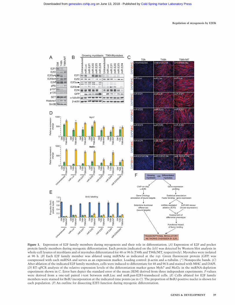
Figure 1. Expression of E2F family members during myogenesis and their role in differentiation. (A) Expression of E2F and pocketprotein family members during myogenic differentiation. Each protein (indicated on the left) was detected by Western blot analysis inwhole-cell lysates of myoblasts and of myotubes differentiated for 48 or 96 h (T48h and T96h/MT, respectively). Myotubes were isolatedat 96 h. (B) Each E2F family member was ablated using miRNAs as indicated at the top. Green fluorescent protein (GFP) wascoexpressed with each miRNA and serves as an expression marker. Loading control: b-actin and a-tubulin. (*) Nonspecific bands. (C)After ablation of the indicated E2F family members, cells were induced to differentiate for 48 and 96 h and stained with MHC and DAPI.(D) RT–qPCR analysis of the relative expression levels of the differentiation marker genes Myh7 and Mef2c in the miRNA-depletionexperiment shown in C. Error bars depict the standard error of the mean (SEM) derived from three independent experiments. P-valueswere derived from a one-tail paired t-test between miR.Luc and miR.pan-E2F3-transduced cells. (E) Cells ablated for E2F familymembers were stained for BrdU incorporation at the indicated time points (as in C). The proportion of BrdU-positive nuclei is shown foreach population. (F) An outline for dissecting E2F3 function during myogenic differentiation.
Regulation of myogenesis by E2f3b
GENES & DEVELOPMENT 39
Cold Spring Harbor Laboratory Press on June 13, 2018 - Published by genesdev.cshlp.orgDownloaded from
well organized and multinucleated, consisting of a nearlynormal complement of nuclei (Fig. 1C). We also sawa significant increase in the number of larger, hypertro-phic myotubes among the E2f4-depleted population (Fig.1C). Simultaneous depletion of both E2f3 and E2f4 pro-voked a differentiation defect qualitatively similar to theone observed for E2f3 alone, and further exacerbation ofthe E2f3 ablation phenotype was not seen (Fig. 1C). Thissuggested that E2f4 did not additively contribute to thefunction of E2f3 in myogenic differentiation within thescope of this analysis. In addition, we surveyed the ex-pression of two other myogenic differentiation markergenes, Myh7 and Mef2c, which were strongly up-regulatedduring myogenesis. We found that depletion of E2f3, E2f4,or both, but not E2f1 or E2f2, led to significantly lowerexpression levels of both genes, in agreement with otherdifferentiation markers and mirroring the observed dif-ferentiation defect (Fig. 1D).
We next initiated efforts to determine the mechanismof the differentiation defect. The phenotypes observedafter depleting E2f3 (or E2f4) were not a result of varia-tions in cell density at the time of induction of differen-tiation, as the densities were the same for eachpopulation as determined by DAPI staining (data notshown). Furthermore, BrdU incorporation assays did notreveal aberrant DNA replication upon induction of dif-ferentiation, indicating that cells were indeed able to exitthe cell cycle, despite loss of E2f3 or E2f4 (Fig. 1E). Wedid not detect changes in cell cycle profiles of growingmyoblasts using FACS analysis, nor did we observechanges in myoblast growth rates (Supplemental Fig.S1A,B). We conclude that E2f3, but not E2f1 or E2f2, isessential for proper myogenic differentiation in a mannerindependent of E2f4. Further, the phenotype observed uponE2f3 ablation appears to be a result of impaired differenti-ation rather than from a failure to exit the cell cycle.
Identification of E2f3 isoform-specific target genes
The foregoing experiments did not address whetherE2f3a, E2f3b, or both, play a role in myogenic differenti-ation. Thus, we sought to dissect the role of each E2f3protein in myogenic differentiation by employing a two-pronged genomic strategy (Fig. 1F). Chromatin immuno-precipitation (ChIP)-on-chip analysis was used to identifyE2f3 target genes and, if possible, identify isoform-specifictarget genes. A combination of miRNA knockdown, ex-pression profiling, and isoform-specific rescue experi-ments was used to investigate specific contributions togene transcription and differentiation. We then integratedthe results of these two approaches and dissected the roleof E2f3a and E2f3b in muscle differentiation.
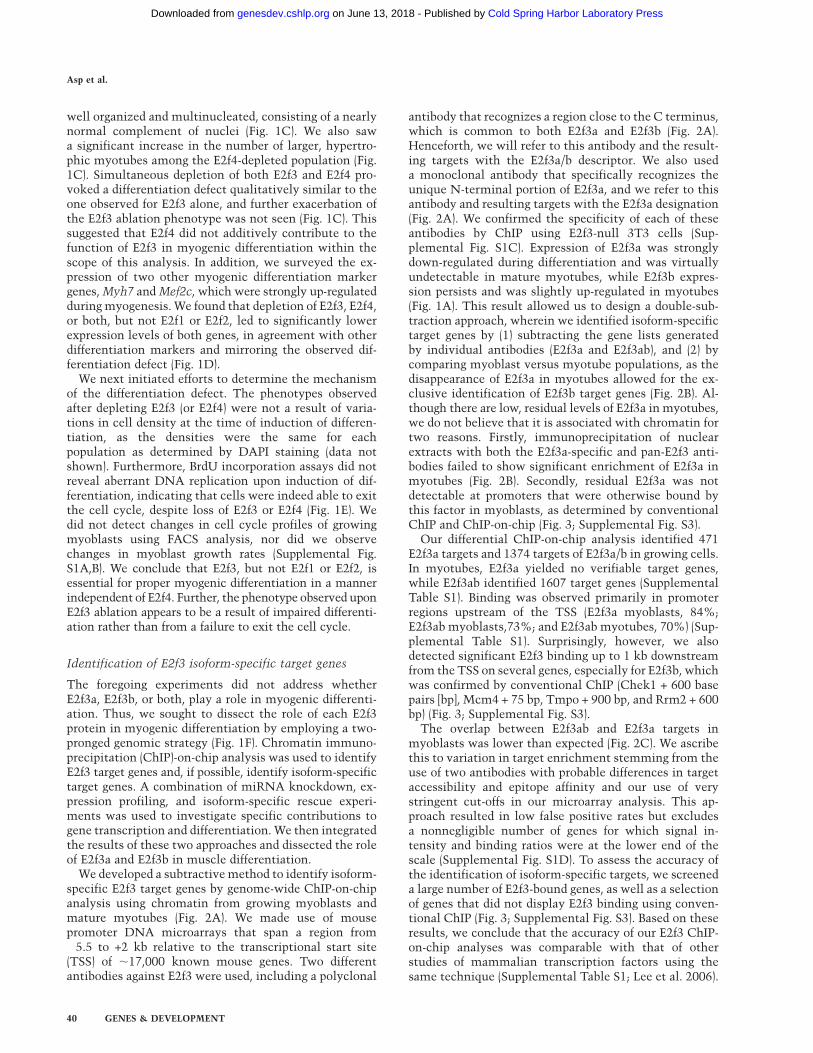
We developed a subtractive method to identify isoform-specific E2f3 target genes by genome-wide ChIP-on-chipanalysis using chromatin from growing myoblasts andmature myotubes (Fig. 2A). We made use of mousepromoter DNA microarrays that span a region from�5.5 to +2 kb relative to the transcriptional start site(TSS) of ;17,000 known mouse genes. Two differentantibodies against E2f3 were used, including a polyclonal
antibody that recognizes a region close to the C terminus,which is common to both E2f3a and E2f3b (Fig. 2A).Henceforth, we will refer to this antibody and the result-ing targets with the E2f3a/b descriptor. We also useda monoclonal antibody that specifically recognizes theunique N-terminal portion of E2f3a, and we refer to thisantibody and resulting targets with the E2f3a designation(Fig. 2A). We confirmed the specificity of each of theseantibodies by ChIP using E2f3-null 3T3 cells (Sup-plemental Fig. S1C). Expression of E2f3a was stronglydown-regulated during differentiation and was virtuallyundetectable in mature myotubes, while E2f3b expres-sion persists and was slightly up-regulated in myotubes(Fig. 1A). This result allowed us to design a double-sub-traction approach, wherein we identified isoform-specifictarget genes by (1) subtracting the gene lists generatedby individual antibodies (E2f3a and E2f3ab), and (2) bycomparing myoblast versus myotube populations, as thedisappearance of E2f3a in myotubes allowed for the ex-clusive identification of E2f3b target genes (Fig. 2B). Al-though there are low, residual levels of E2f3a in myotubes,we do not believe that it is associated with chromatin fortwo reasons. Firstly, immunoprecipitation of nuclearextracts with both the E2f3a-specific and pan-E2f3 anti-bodies failed to show significant enrichment of E2f3a inmyotubes (Fig. 2B). Secondly, residual E2f3a was notdetectable at promoters that were otherwise bound bythis factor in myoblasts, as determined by conventionalChIP and ChIP-on-chip (Fig. 3; Supplemental Fig. S3).
Our differential ChIP-on-chip analysis identified 471E2f3a targets and 1374 targets of E2f3a/b in growing cells.In myotubes, E2f3a yielded no verifiable target genes,while E2f3ab identified 1607 target genes (SupplementalTable S1). Binding was observed primarily in promoterregions upstream of the TSS (E2f3a myoblasts, 84%;E2f3ab myoblasts,73%; and E2f3ab myotubes, 70%) (Sup-plemental Table S1). Surprisingly, however, we alsodetected significant E2f3 binding up to 1 kb downstreamfrom the TSS on several genes, especially for E2f3b, whichwas confirmed by conventional ChIP (Chek1 + 600 basepairs [bp], Mcm4 + 75 bp, Tmpo + 900 bp, and Rrm2 + 600bp) (Fig. 3; Supplemental Fig. S3).
The overlap between E2f3ab and E2f3a targets inmyoblasts was lower than expected (Fig. 2C). We ascribethis to variation in target enrichment stemming from theuse of two antibodies with probable differences in targetaccessibility and epitope affinity and our use of verystringent cut-offs in our microarray analysis. This ap-proach resulted in low false positive rates but excludesa nonnegligible number of genes for which signal in-tensity and binding ratios were at the lower end of thescale (Supplemental Fig. S1D). To assess the accuracy ofthe identification of isoform-specific targets, we screeneda large number of E2f3-bound genes, as well as a selectionof genes that did not display E2f3 binding using conven-tional ChIP (Fig. 3; Supplemental Fig. S3). Based on theseresults, we conclude that the accuracy of our E2f3 ChIP-on-chip analyses was comparable with that of otherstudies of mammalian transcription factors using thesame technique (Supplemental Table S1; Lee et al. 2006).
Asp et al.
40 GENES & DEVELOPMENT
Cold Spring Harbor Laboratory Press on June 13, 2018 - Published by genesdev.cshlp.orgDownloaded from
The subtractive analysis of differences in ChIP-on-chip,as outlined above, together with confirmatory ChIPexperiments, revealed the existence of five distinct cate-gories of E2f3 target genes (Fig. 2C). Current models forE2f3 function posit that E2f3a is a transcriptional activa-
tor, while E2f3b acts as a transcriptional repressor inquiescent and differentiated cells, wherein E2f3a is notexpressed and E2f3b interacts with pRb (Kong et al. 2007).We verified that this is also true in muscle and showedthat E2f3b, but not E2f3a, interacts with pRb in fully
Figure 2. Identification of E2f3a and E2f3b targets. (A) Strategy for genome-wide discovery E2F3 targets and the epitopes of the twoE2F3 antibodies used. (B) Enrichment test of E2F3 isoforms. (Top) Nuclear extracts from myoblasts and myotubes were immunopre-cipitated with the indicated antibodies and detected by Western blotting with antibodies against E2f3a/b. (C) Venn diagrams indicatingthe intersection between target genes in growing myoblasts and myotubes (see the text for details). Isoform-specific E2F3 target groupsare labeled A–E. (D) Analysis of the percent overlap between E2F3 and E2F4 targets in myotubes expressing the percentage of boundtargets as a function of E2F4 target genes bound by E2F3 in each group. Corresponding Venn diagrams are shown in SupplementalFig S3. (E) Gene Ontology (GO) analysis of E2F3 target genes expressed as the percentage of the total number of independent GOannotations within each group. Groups are indicated on the right, and GO categories on the bottom.
Regulation of myogenesis by E2f3b
GENES & DEVELOPMENT 41
Cold Spring Harbor Laboratory Press on June 13, 2018 - Published by genesdev.cshlp.orgDownloaded from
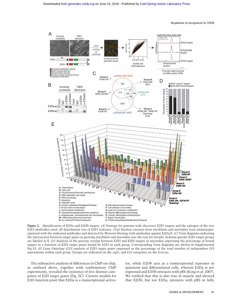
differentiated myotubes (Supplemental Fig. S5A). As pRbhas a well-established role in terminal cell cycle exit andsilencing of cell cycle genes, some E2f3-regulated genesmight therefore be expected to undergo a transition inwhich E2f3a is recruited to promoters in growing myo-blasts, whereas E2f3b would be recruited to the samegenes in myotubes, concomitant with gene repression.Indeed, when we compared the list of E2f3a targets inmyoblasts with the list of E2f3b targets in myotubes, wefound 214 genes that belonged to this ‘‘switching’’ cate-gory, designated as group A (Fig. 2C; Supplemental TableS2). We confirmed this E2f3a to E2f3b switching on severalgenes by conventional ChIP analysis (Fig. 3; Supplemental
S3). We also identified a set of genes exclusively bound byE2f3a in growing myoblasts, referred to as group B genes,which did not recruit E2f3b in myotubes (Fig. 2C; Supple-mental Table S2). Confirmatory ChIP analysis verifiedthat these genes were indeed E2f3a-specific (Smyd4 andTop2a in Fig. 3; Supplemental Fig. S3).
We applied the subtractive approach described aboveand were able to identify E2f3b-specific targets in bothmyoblasts and myotubes. E2f3a target genes (in myo-blasts) were subtracted from those bound by E2f3a/b ineither condition, allowing enrichment of E2f3b-specifictarget genes. We then compared the lists of presumptiveE2f3b targets in myoblasts and myotubes and identified
Figure 3. Verification of isoform-specific tar-gets by conventional ChIP. qPCR analysis ofa select number of genes representing the fiveE2F3 isoform-specific gene categories. Enrich-ment >0.05 is significant. Error bars depict thestandard error of the mean (SEM) derived fromthree independent experiments.
Asp et al.
42 GENES & DEVELOPMENT
Cold Spring Harbor Laboratory Press on June 13, 2018 - Published by genesdev.cshlp.orgDownloaded from

792 genes uniquely bound by E2f3b in myoblasts, desig-nated group C. We observed 314 genes that were consti-tutively bound by E2f3b in both conditions, which wedesignated as group D. Lastly, we identified 1079 genesthat were myotube-specific targets of E2f3b, designated asgroup E (Fig. 2C; Supplemental Table S2). To validate themicroarray data, we analyzed a select number of genesfrom each group by conventional ChIP, which confirmedthem to be (1) E2f3b-specific and (2) bound by E2f3b ina condition-specific manner (myoblasts and/or myotubes)(Fig. 3;Supplemental S3).
Given the pervasive role of E2f4 recruitment to cellcycle genes in differentiated myotubes (Blais et al. 2007),we also identified E2f4 targets in myotubes by performingChIP-on-chip using identical promoter arrays (van Oeve-len et al. 2008). We then compared the list of targetsbound by E2f4 in myotubes with each of the five classesof E2f3 targets described above. Interestingly, we founda striking overlap between E2f4 and E2f3a target genes ingroups A and B. In sharp contrast, the absolute numberand percentage of E2f3b-specific targets in groups C–Ethat were commonly bound by E2f4 was significantlylower (Fig. 2D; Supplemental Fig S2).
Functional classification of E2f3 target genes
The identification of E2F3 isoform-specific target genessuggested that E2f3a and E2f3b have distinct biologicalfunctions. We analyzed the genes in each of the fivegroups by clustering them according to Gene Ontology(GO) using NIH-DAVID and extensive manual curation(Fig. 2E; Supplemental Table S2). We found that genes ingroups A (switching group) and group B (E2f3a bound inmyoblasts only) exhibited very similar GO profiles andwere enriched in genes involved in growth and cell cyclecontrol, transcription, RNA processing, and chromosomestructure (Fig. 2E), suggesting that E2f3a is primarilyinvolved in cell growth control.
Interestingly, the E2f3b-specific categories (groups C–E)contained significantly fewer genes involved in cell cyclecontrol and proliferation (Fig. 2E), and group E (E2f3bbound in myotubes) was particularly depleted of suchgenes. Instead, these groups were very similar to eachother and were enriched for several unique functionalcategories (such as ER redox and homeostasis, synapse/synaptogenesis, and axon guidance) that were not repre-sented within groups A and B (Fig. 2E). Of special interest,we identified a number of genes bound by E2f3b involvedin cellular differentiation/development, and muscle dif-ferentiation/physiology (Fig. 2E), specifically linkingE2f3b, but not E2f3a, to the differentiation defect ob-served when depleting E2f3 (Fig. 1C). We also noted thatgroups C–E contained genes involved in lipid/cholesterolmetabolism, although with a small number of such genesalso present in group B (Fig. 2E).
With our subtractive approach, we were able to clearlyidentify and verify E2f3a- and E2f3b-specific genes, and byclustering these genes into functional groups, a pictureemerges in which E2f3a is associated with cell cycle andproliferation, while E2f3b-specific genes are primarily
associated with differentiation and development. Theprocesses of proliferation and differentiation are inti-mately linked, and our data places the two E2f3 isoformsat the center of fundamental crossroads in myogenicdevelopment.
Correlation between E2f3 binding and gene expression
Transcription factor binding does not necessarily predictgene regulation, and therefore, we sought to correlateE2f3 isoform binding with expression by merging ourfactor-binding data with gene expression profiles ob-tained from growing and differentiated C2C12 myo-blasts (Blais et al. 2005). Data were presented as theratio between expression levels in myotubes and grow-ing myoblasts (MT/GM) in order to correlate E2f3 bind-ing with dynamic changes in gene expression duringmyogenesis.
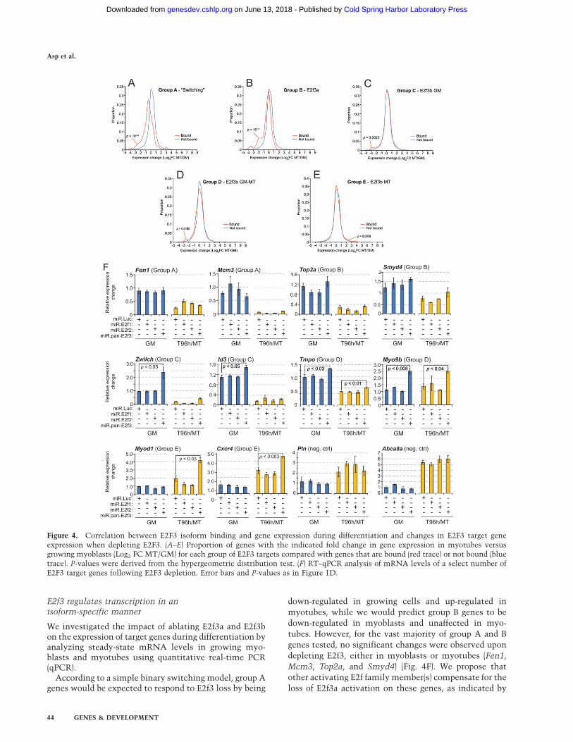
We found that groups A and B genes were stronglyrepressed during differentiation (Fig. 4A,B) in agreementwith the fact that these groups primarily consisted of cellcycle- and proliferation-associated genes such as Aurkb,Cdc6, and Uhr, which were strongly down-regulatedduring differentiation (Supplemental Fig. S4A). Thehypergeometric distribution test confirmed these corre-lations to be statistically significant (P < 10�10) (Fig. 4,B).This correlation agreed with a model where E2f3a pro-motes expression of group A genes in growing myoblastsand E2f3b functions as a transcriptional repressor inmyotubes. However, the differentiation-associated re-pression of group B genes, such as Top2a (SupplementalFig. S4A), cannot be attributed to E2f3b and must bemediated by some other repressor, possibly E2f4, as theoverlap between group B genes and E2f4 targets isextensive.
The E2f3b-specific target genes in groups C–E did notexhibit such a strong correlation between expression anddifferentiation, but had an almost even distribution ofboth up- and down-regulated transcripts (Fig. 4C–E). Incontrast to groups A and B, these groups were representedby considerably more functionally diverse categories,such as myogenic differentiation, signal transduction,metabolic pathways, and transcription (Fig. 2E), poten-tially explaining the highly variable target gene expres-sion patterns that we observed. For example, Smyd1,Tnnc2, and Chrng were induced during differentiation,while Id3 and Ccnb1were down-regulated (SupplementalFig. S4A).
With the exception of the behavior of group A genes,which constituted a small fraction of the total number ofE2f3 targets we identified, the correlation between E2f3aand E2F3b binding and gene expression during differenti-ation was not fully consistent with a model in whichE2f3b functions as a strong transcriptional repressor.Together with the apparent E2f3-independent suppres-sion of group B genes in myotubes, and the diverseexpression profiles of groups C–E, our data indicates thatregulation of E2f3 target genes is more complex thananticipated, and that E2f3b may not function as a simplebinary switch.
Regulation of myogenesis by E2f3b
GENES & DEVELOPMENT 43
Cold Spring Harbor Laboratory Press on June 13, 2018 - Published by genesdev.cshlp.orgDownloaded from
E2f3 regulates transcription in anisoform-specific manner
We investigated the impact of ablating E2f3a and E2f3bon the expression of target genes during differentiation byanalyzing steady-state mRNA levels in growing myo-blasts and myotubes using quantitative real-time PCR(qPCR).
According to a simple binary switching model, group Agenes would be expected to respond to E2f3 loss by being
down-regulated in growing cells and up-regulated in
myotubes, while we would predict group B genes to be
down-regulated in myoblasts and unaffected in myo-
tubes. However, for the vast majority of group A and B
genes tested, no significant changes were observed upon
depleting E2f3, either in myoblasts or myotubes (Fen1,
Mcm3, Top2a, and Smyd4) (Fig. 4F). We propose that
other activating E2f family member(s) compensate for the
loss of E2f3a activation on these genes, as indicated by
Figure 4. Correlation between E2F3 isoform binding and gene expression during differentiation and changes in E2F3 target geneexpression when depleting E2F3. (A–E) Proportion of genes with the indicated fold change in gene expression in myotubes versusgrowing myoblasts (Log2 FC MT/GM) for each group of E2F3 targets compared with genes that are bound (red trace) or not bound (bluetrace). P-values were derived from the hypergeometric distribution test. (F) RT–qPCR analysis of mRNA levels of a select number ofE2F3 target genes following E2F3 depletion. Error bars and P-values as in Figure 1D.
Asp et al.
44 GENES & DEVELOPMENT
Cold Spring Harbor Laboratory Press on June 13, 2018 - Published by genesdev.cshlp.orgDownloaded from

recent studies (Danielian et al. 2008; Tsai et al. 2008). Ina similar manner, the differentiation-associated down-regulation of these genes in the absence of E2f3b could bemediated by E2f4, as the majority of group A and B genesare also targets of this E2F (Fig. 2D).
Interestingly, group C genes displayed a stronger re-sponse to E2f3 depletion than group A and B genes,exhibiting significant up-regulation exclusively in myo-blasts after knockdown (Fig. 4F). This coincided with thefact that group C genes were targets of E2f3b in myoblastsbut not in myotubes. Our data further suggest that E2f3blimits the rate of transcription of these genes, functioningas an attenuator of transcription rather than acting asa strong repressor, as these genes were significantlyexpressed even in the presence of E2f3b. The correlationbetween E2f3b and transcriptional attenuation was evenmore apparent for the constitutive E2f3b target genes ofgroup D, which, as a consequence, were significantly up-regulated in both myoblasts and myotubes after E2f3ablation (Tmpo and Myo9b) (Fig. 4F). Interestingly, themyotube-specific E2f3b target genes in group E had thestrongest response to E2f3 loss and were significantly up-regulated in myotubes. Expression of these genes inmyoblasts was unaffected by E2f3 depletion, furtherstrengthening the correlation between E2f3b and attenu-ation of transcription (Myod1 and Cxcr4) (Fig 4F).
The observed expression changes appear to be specificfor E2f3, since depletion of E2f1 or E2f2 did not showa corresponding effect on expression of any of the genestested in myoblasts or myotubes, in agreement withour observation that these family members are notessential for muscle differentiation (Figs. 1C, 4F). Theseresults also suggest that E2f3b is a more critical regulatorof transcription than E2f3a during myogenic differentia-tion, since we observe stronger responses to E2f3b loss inmyotubes as compared with E2f3a loss in myoblasts.We propose that other activating E2Fs, such as E2f1 andE2f2, may compensate for the loss of E2f3a on groups Aand B genes in myoblasts, whereas E2f3b plays a role thatis not entirely fulfilled by other E2Fs in myotubes.Furthermore, these data reinforce the notion that E2f3bhas unique biological properties distinct from those ofE2f3a.
E2f3b is essential for myogenic differentiation
Our functional analysis of isoform-specific target genesshowed that E2f3a was associated with cell cycle andproliferation genes in myoblasts, whereas E2f3b wasassociated with developmental and differentiation genesin myotubes, particularly those involved in myogenesis.This suggested that E2f3b has a more important role inmyogenic differentiation than E2f3a. To test this, weassessed the ability of both isoforms to individuallyrescue the abortive differentiation associated with E2f3loss. Attempts to differentially ablate the two isoformswere unsuccessful due to the limited amount of sequencethat could be targeted discriminatively (344 bp for E2f3aand 46 bp for E2f3b). Therefore, we took an alternativeapproach in which we attempted to rescue the differen-
tiation phenotype by individually expressing RNAi-resistant, HA-tagged E2F3 isoforms in E2f3 miRNA-expressing cells (Materials and Methods). Expression ofE2f3a or E2f3b in untreated cells lead to distinctlyelevated protein levels, while the levels of E2f3a andE2f3b observed after infecting E2F3 knockdown cellswere comparable with those found in untreated myo-blasts and myotubes (Supplemental Fig S4B).
First, we gaged the impact of overexpressing eitherisoform in untreated myoblasts. Unexpectedly, expres-sion of E2f3a had no detectable adverse effects on normaldifferentiation as compared with untreated and controltransduced cells (Supplemental Fig. S5B). This resultsuggested that there are cellular mechanisms capable ofoverriding inappropriate E2f3a activity. The same is alsotrue for E2f3b, since its forced expression in untreatedcells did not interfere with differentiation (SupplementalFig. S5B).
Next, we attempted to rescue the differentiation defectsobserved after E2f3 depletion by expressing E2f3a or E2f3b.Expression of E2f3a in E2f3-depleted cells was not able torescue the abortive differentiation, but rather, causeda phenotype even more severe than loss of E2f3 alone (cf.Figs. 5A and 1C). In striking contrast, expression of E2f3b inE2f3-deficient cells lead to near complete rescue of differ-entiation and resulted in organized and dense bundles ofmyotubes (cf. Figs. 5A and 1C). These data strongly sug-gested that E2f3b, but not E3f3a, was essential for myogenicdifferentiation, and this conclusion agreed with the func-tional categories assigned to our E2f3b target genes.
E2f3b promotes myogenic differentiation ina pRb-independent manner
The role of pocket proteins in myogenic differentiationhas been intensively studied both in mouse and in cellculture models, and it is clear that pRb is essential formyogenic differentiation (Novitch et al. 1996, 1999;Kobayashi et al. 1998; de Bruin et al. 2003; Wu et al.2003). It has been shown that a population of E2f3bspecifically interacts with pRb in quiescent and post-mitotic cells, and we showed that this is also observedin myotubes (Supplemental Fig. S5A; Kong et al. 2007).It has also been shown that pRb is associated withlong-term repression of transcription in myotubes (Blaiset al. 2007). However, the fact that E2f3b target geneswere not exclusively down-regulated during differentia-tion suggested that E2f3b function in the context ofmuscle differentiation could be at least partially pRbindependent.
To test this hypothesis, we introduced a point mutationinto our miRNA-resistant E2f3a and E2f3b expressionconstructs (Y436H in E2f3a and Y309H in E2f3b) (Materi-als and Methods). The targeted residue is conservedamong E2F1–3, and this substitution has been shown todisrupt the interaction between E2f1 and pRb whilepreserving trans-activation function (Shan et al. 1996).Expression of these miRNA-resistant HA-tagged E2f3mutants resulted in levels that approximated both theirwild-type counterparts and the endogenous proteins
Regulation of myogenesis by E2f3b
GENES & DEVELOPMENT 45
Cold Spring Harbor Laboratory Press on June 13, 2018 - Published by genesdev.cshlp.orgDownloaded from
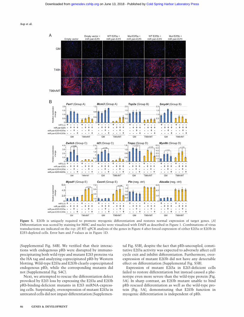
(Supplemental Fig. S4B). We verified that their interac-tions with endogenous pRb were disrupted by immuno-precipitating both wild-type and mutant E2f3 proteins viathe HA tag and analyzing coprecipitated pRb by Westernblotting. Wild-type E2f3a and E2f3b clearly coprecipitatedendogenous pRb, while the corresponding mutants didnot (Supplemental Fig. S4C).
Next, we attempted to rescue the differentiation defectprovoked by E2f3 loss by expressing the E2f3a and E2f3bpRb-binding-deficient mutants in E2f3 miRNA-express-ing cells. Surprisingly, overexpression of mutant E2f3a inuntreated cells did not impair differentiation (Supplemen-
tal Fig. S5B), despite the fact that pRb-uncoupled, consti-tutive E2f3a activity was expected to adversely affect cellcycle exit and inhibit differentiation. Furthermore, over-expression of mutant E2f3b did not have any detectableeffect on differentiation (Supplemental Fig. S5B).
Expression of mutant E2f3a in E2f3-deficient cellsfailed to restore differentiation but instead caused a phe-notype even more severe than the wild-type protein (Fig.5A). In sharp contrast, an E2f3b mutant unable to bindpRb rescued differentiation as well as the wild-type pro-tein (Fig. 5A), demonstrating that E2f3b function inmyogenic differentiation is independent of pRb.
Figure 5. E2f3b is uniquely required to promote myogenic differentiation and restores normal expression of target genes. (A)Differentiation was scored by staining for MHC and nuclei were visualized with DAPI as described in Figure 1. Combinations of virustransductions are indicated on the top. (B) RT–qPCR analysis of the genes in Figure 4 after forced expression of either E2f3a or E2f3b inE2F3-depleted cells. Error bars and P-values as in Figure 1D.
Asp et al.
46 GENES & DEVELOPMENT
Cold Spring Harbor Laboratory Press on June 13, 2018 - Published by genesdev.cshlp.orgDownloaded from

E2f3b restores expression of target genes
We investigated the expression of E2f3 target genes in theisoform-specific rescue experiments described above byanalyzing steady-state mRNA levels using qPCR beforeand after rescue. Forced expression of E2f3a in E2f3-depleted cells did not lead to dramatic changes in expres-sion levels of any E2f3 target genes investigated, neitherin myoblasts nor myotubes, and the weak changesdetected were not statistically significant (Fig. 5B). How-ever, the ability of E2f3b to rescue differentiation coin-cided with significant transcriptional down-regulationand restoration of normal transcript levels of E2f3btargets (Fig. 5B). This effect was most prominent forE2f3b target genes in myotubes (Myod1 and Cxcr4) (Fig.5B) suggesting a context-dependent role for E2f3b inagreement with its ability to promote differentiation. Insharp contrast, expression of group A and B genes did notchange significantly upon restoration of E2f3b (Fen 1,Mcm3, Top2a, and Smyd4) (Fig. 5B), which was also thecase when total E2f3 was depleted (Fig. 4F).
Although E2f3a is expressed at slightly higher levels thanE2f3b in the rescue experiment (Supplemental Fig. S4), ourdata showed remarkably specific regulation by E2f3 iso-forms, since even these higher levels of E2f3a were unableto significantly affect transcription of the E2f3 target genesanalyzed. In contrast, the lower levels of ectopically pro-duced E2f3b were clearly able to attenuate expression of itstargets, restoring their expression to normal levels.
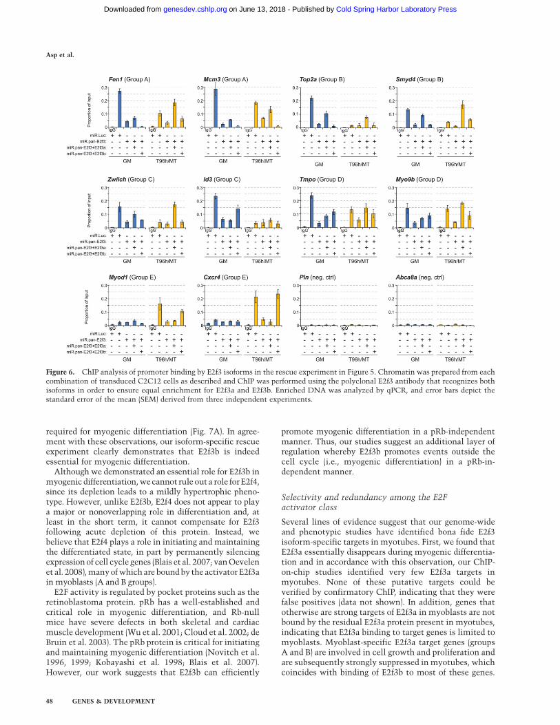
Gene-specific variations in E2f3 isoform targeting
We performed conventional E2f3 ChIP analysis on therescue experiments described above to correlate theobserved restoration of normal expression with E2f3isoform-specific promoter binding.
Exogenous E2f3a was efficiently recruited to groups Aand B genes in myoblasts, even though it did not inducestatistically significant changes in expression (Fen1,Mcm3, Top2a, and Smyd4 in Fig. 6). This agrees withthe proposed redundancy between E2f-1, E2f-2, and E2f-3aon these genes, ensuring full expression even in theabsence of one of these activators. We also detectedexogenous E2f3a binding upon ectopic expression of thisisoform in myotubes, wherein there is normally no E2f3aprotein present. However, this nonphysiological bindingwas not functional, since it did not result in altered geneexpression. Moreover, even though we could only detectsignificant recruitment of E2f3b to two group A and Bgenes out of the four analyzed (Fen and Smyd4) (Fig. 6)binding was only observed in myotubes but not inmyoblasts, although the protein was expressed at levelsequivalent to those of E2f3a. However, E2f3b binding wasnot crucial for the proper expression of these genes asthey were all down-regulated during differentiation inboth E2f3-depleted and E2f3b-rescued cells. This supportsour conclusion that there is functional overlap betweenE2f3b and E2f4 on these genes and that E2f4 alone isenough to ensure differentiation-associated transcrip-tional repression and cell cycle exit.
For genes in groups C and D we detected significantrecruitment of both E2f3a and E2f3b in the rescued cells.As for group A and B genes, binding of E2f3a occurredin both myoblasts and myotubes, but no significantchanges in expression were associated with this binding(Fig. 6). For group C genes, E2f3b binding was onlydetected in myoblasts and coincided with the timewhen endogenous E2f3b would normally be recruited tothese genes (Zwilch, Id3 in Fig. 6) and similarly, E2f3brecruitment to group D genes mirrored the pattern ofthe endogenous protein and was detected in both myo-blasts and myotubes (Tmpo, Myo9b in Fig. 6). Forboth group C and group D genes, recruitment of E2f3brestored expression to normal levels compared with E2f3depleted cells.
Strikingly, group E genes showed a very high degree ofspecificity for E2f3b, and in no case did we detect bindingof E2f3a to these genes. Recruitment of E2f3b mirroredprecisely the normal pattern of E2f3b recruitment, sincewe exclusively observed strong binding in myotubes(Myod1 and Cxcr4 in Fig. 6). As shown in Figure 5B,E2f3b binding to these genes was accompanied by com-plete restoration of normal expression levels.
Thus, E2f3b exhibited remarkably specific and physio-logical patterns of promoter recruitment and regulationof its targets. Likewise, E2f3a recruitment to promoterswas enabled by re-expression of this isoform, except ongroup E genes. In all cases, however, inappropriate generegulation was not observed, attesting to additionalmechanisms that limit or prevent inappropriate activityof E2f3a on these genes. The mechanisms that restrictE2f3a and E2f3b binding and activity are currently un-known, but the fact that we only detect transcriptionalchanges associated with E2f3b binding on E2f3b-specificgenes supports our conclusion that E2f3b is both func-tionally unique and essential for myogenesis.
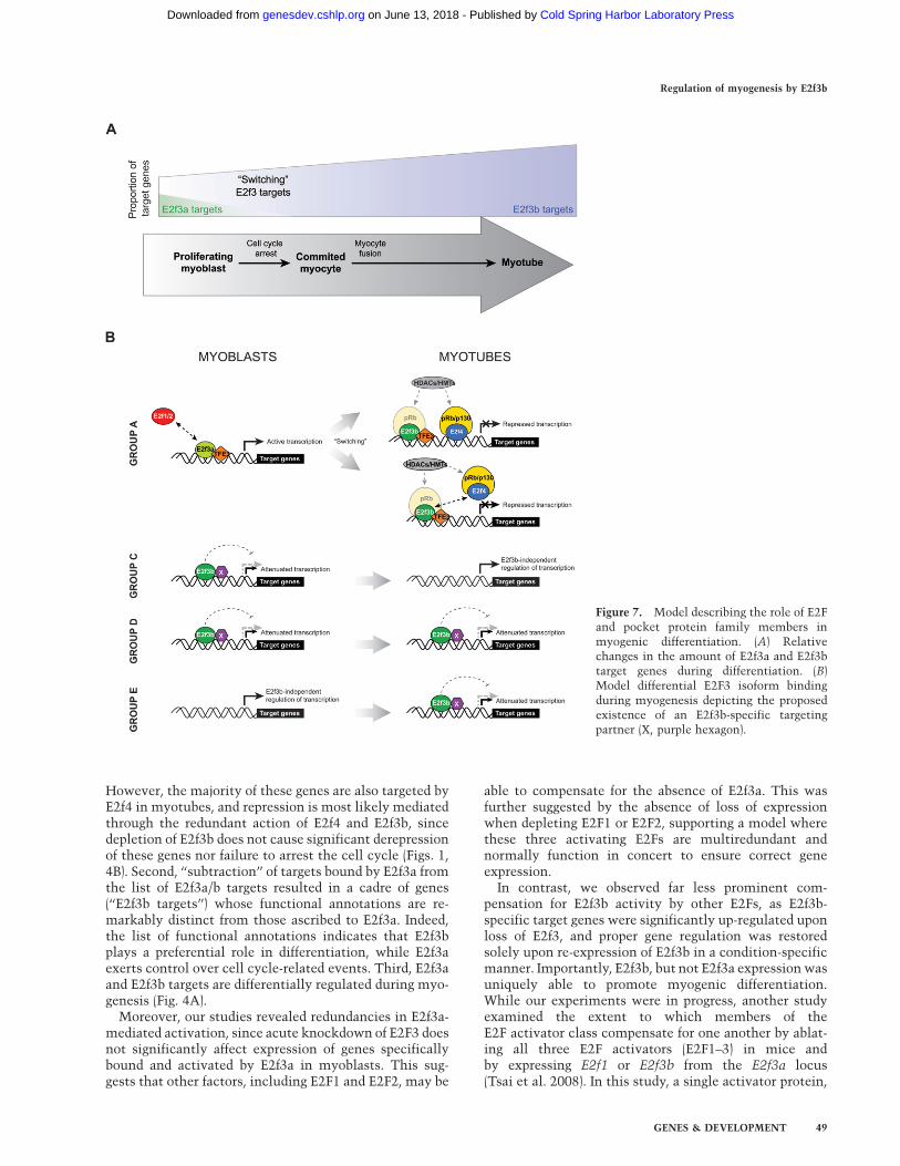
Discussion
Using methods that enhance the ability to identify familymember-specific functions (isoform-specific ChIP-on-chip and acute ablation), our work highlights both spec-ificity and redundancy within the E2F family. Here, wedefined a previously unknown and critical role for E2f3bin myogenic differentiation. Identification of E2f3 iso-form-specific target genes uncovered a hitherto unknowncomplexity in E2F3 function that vastly extends thenetwork of gene categories controlled by the E2F tran-scription factor family.
E2F factors in myogenic differentiation
Our data show that E2f3b plays an indispensable role inmyogenic differentiation, while, in striking contrast,E2f1, E2f2, and E2f3a do not play an obvious role in ourin vitro differentiation model. Genes targeted by E2f3aare heavily enriched in proliferation-associated genes,while E2f3b targets are heavily enriched in genes involvedin differentiation, signaling, lipid biosynthesis, and celladhesion and motility, suggesting that E2f3b is uniquely
Regulation of myogenesis by E2f3b
GENES & DEVELOPMENT 47
Cold Spring Harbor Laboratory Press on June 13, 2018 - Published by genesdev.cshlp.orgDownloaded from
required for myogenic differentiation (Fig. 7A). In agree-ment with these observations, our isoform-specific rescueexperiment clearly demonstrates that E2f3b is indeedessential for myogenic differentiation.
Although we demonstrated an essential role for E2f3b inmyogenic differentiation, we cannot rule out a role for E2f4,since its depletion leads to a mildly hypertrophic pheno-type. However, unlike E2f3b, E2f4 does not appear to playa major or nonoverlapping role in differentiation and, atleast in the short term, it cannot compensate for E2f3following acute depletion of this protein. Instead, webelieve that E2f4 plays a role in initiating and maintainingthe differentiated state, in part by permanently silencingexpression of cell cycle genes (Blais et al. 2007; van Oevelenet al. 2008), many of which are bound by the activator E2f3ain myoblasts (A and B groups).
E2F activity is regulated by pocket proteins such as theretinoblastoma protein. pRb has a well-established andcritical role in myogenic differentiation, and Rb-nullmice have severe defects in both skeletal and cardiacmuscle development (Wu et al. 2001; Cloud et al. 2002; deBruin et al. 2003). The pRb protein is critical for initiatingand maintaining myogenic differentiation (Novitch et al.1996, 1999; Kobayashi et al. 1998; Blais et al. 2007).However, our work suggests that E2f3b can efficiently
promote myogenic differentiation in a pRb-independentmanner. Thus, our studies suggest an additional layer ofregulation whereby E2f3b promotes events outside thecell cycle (i.e., myogenic differentiation) in a pRb-in-dependent manner.
Selectivity and redundancy among the E2Factivator class
Several lines of evidence suggest that our genome-wideand phenotypic studies have identified bona fide E2f3isoform-specific targets in myotubes. First, we found thatE2f3a essentially disappears during myogenic differentia-tion and in accordance with this observation, our ChIP-on-chip studies identified very few E2f3a targets inmyotubes. None of these putative targets could beverified by confirmatory ChIP, indicating that they werefalse positives (data not shown). In addition, genes thatotherwise are strong targets of E2f3a in myoblasts are notbound by the residual E2f3a protein present in myotubes,indicating that E2f3a binding to target genes is limited tomyoblasts. Myoblast-specific E2f3a target genes (groupsA and B) are involved in cell growth and proliferation andare subsequently strongly suppressed in myotubes, whichcoincides with binding of E2f3b to most of these genes.
Figure 6. ChIP analysis of promoter binding by E2f3 isoforms in the rescue experiment in Figure 5. Chromatin was prepared from eachcombination of transduced C2C12 cells as described and ChIP was performed using the polyclonal E2f3 antibody that recognizes bothisoforms in order to ensure equal enrichment for E2f3a and E2f3b. Enriched DNA was analyzed by qPCR, and error bars depict thestandard error of the mean (SEM) derived from three independent experiments.
Asp et al.
48 GENES & DEVELOPMENT
Cold Spring Harbor Laboratory Press on June 13, 2018 - Published by genesdev.cshlp.orgDownloaded from
However, the majority of these genes are also targeted byE2f4 in myotubes, and repression is most likely mediatedthrough the redundant action of E2f4 and E2f3b, sincedepletion of E2f3b does not cause significant derepressionof these genes nor failure to arrest the cell cycle (Figs. 1,4B). Second, ‘‘subtraction’’ of targets bound by E2f3a fromthe list of E2f3a/b targets resulted in a cadre of genes(‘‘E2f3b targets’’) whose functional annotations are re-markably distinct from those ascribed to E2f3a. Indeed,the list of functional annotations indicates that E2f3bplays a preferential role in differentiation, while E2f3aexerts control over cell cycle-related events. Third, E2f3aand E2f3b targets are differentially regulated during myo-genesis (Fig. 4A).
Moreover, our studies revealed redundancies in E2f3a-mediated activation, since acute knockdown of E2F3 doesnot significantly affect expression of genes specificallybound and activated by E2f3a in myoblasts. This sug-gests that other factors, including E2F1 and E2F2, may be
able to compensate for the absence of E2f3a. This wasfurther suggested by the absence of loss of expressionwhen depleting E2F1 or E2F2, supporting a model wherethese three activating E2Fs are multiredundant andnormally function in concert to ensure correct geneexpression.
In contrast, we observed far less prominent com-pensation for E2f3b activity by other E2Fs, as E2f3b-specific target genes were significantly up-regulated uponloss of E2f3, and proper gene regulation was restoredsolely upon re-expression of E2f3b in a condition-specificmanner. Importantly, E2f3b, but not E2f3a expression wasuniquely able to promote myogenic differentiation.While our experiments were in progress, another studyexamined the extent to which members of theE2F activator class compensate for one another by ablat-ing all three E2F activators (E2F1–3) in mice andby expressing E2f1 or E2f3b from the E2f3a locus(Tsai et al. 2008). In this study, a single activator protein,
Figure 7. Model describing the role of E2Fand pocket protein family members inmyogenic differentiation. (A) Relativechanges in the amount of E2f3a and E2f3btarget genes during differentiation. (B)Model differential E2F3 isoform bindingduring myogenesis depicting the proposedexistence of an E2f3b-specific targetingpartner (X, purple hexagon).
Regulation of myogenesis by E2f3b
GENES & DEVELOPMENT 49
Cold Spring Harbor Laboratory Press on June 13, 2018 - Published by genesdev.cshlp.orgDownloaded from
E2f3a, was sufficient to ensure embryonic and post-natal development. Further, these knock-in experimentssuggested that E2F family members are interchangeable,since expression of either E2f1 or E2f3b rescued pheno-types associated with E2f3a inactivation, but timing ofexpression dictated by E2f3a regulatory sequenceswas paramount. We showed that in the context ofmuscle differentiation, E2f3b generally functions asan attenuator of transcription and not as an activator,which is difficult to reconcile with the presumptiveability of E2f3b to rescue the E2f3a phenotype describedby Tsai et al. (2008). However, we did not examine allE2f3b target genes, and therefore we cannot exclude thepossibility that there might be instances whenE2f3b functions as an activator of critical developmentalgenes.
No muscle defects were described in the knock-out/knock-in study by Tsai et al. (2008) nor was a musclephenotype detected in a recent publication describingE2F3 isoform knockout mice in an otherwise wild-typeE2F background (Danielian et al. 2008). We believe thata likely explanation pertains to the manner in which E2f3was ablated. In our case, ablation was acute, whereas theearlier study took advantage of germline knockouts,a situation in which significant compensation by otherE2F factors is known to occur (Kong et al. 2007). Inpreliminary studies, we tested this possibility by continu-ously cultivating myoblasts depleted for E2F3 for 2–3 wkbefore inducing differentiation. Interestingly, silencing ofE2F3 expression persists for the entire duration of thisexperiment, but after such extended culture, these myo-blasts partially regain their ability to differentiate, albeitnot as well as wild-type cells (data not shown). Thisprovides evidence for the differences between acute andlong-term loss of E2F factors and suggests that care mustbe taken when analyzing such data.
In the mouse knockout/knock-in models, it is conceiv-able that most cells are able to compensate for lack ofE2f3a, and thereby enable the animal to complete em-bryogenesis. However, it is clear that this is not the casefor all tissues, since E2f3a�/� mice are deficient in whiteadipose tissue. Intriguingly, we find that both E2F3 iso-forms are recruited to a substantial number of genesinvolved in lipid metabolism (Fig. 2E), prompting futureexperiments designed to investigate whether deregula-tion of these target genes could, in part, account for themutant phenotype.
Factors underlying E2f3a and E2f3b specificity
It has been shown that E2f3a synergistically interactswith the E-box transcription factor TFE3 and that thisinteraction promotes specific recruitment of E2F3 to thep68 promoter and activates transcription (Giangrandeet al. 2003). This activation can be negated by coexpres-sion of E2f3b, which agrees with our experiments thatshow that E2f3b is able to attenuate gene expression, mostprominently in myotubes (Figs. 5B, 7). Given this result, itis interesting to speculate that a specificity factor akin toTFE3 could also operate in recruiting E2f3b to target genes
(Fig. 7B). Isoform specificity could then be imparted by thedisappearance of E2f3a (and presence of E2f3b) as cellsdifferentiate. Such a mechanism has been observed for theArf promoter, which is normally specifically bound byE2f3b, but becomes occupied by E2f3a in the absence ofE2f3b (Danielian et al. 2008). However, this model doesnot explain the existence of E2f3b-specific target genes inmyoblasts, wherein both isoforms are present, whichsuggests that E2f3b can be uniquely targeted to promotersas suggested in our model (Fig. 7B).
The combined analysis of isoform recruitment andexpression in our rescue experiment clearly demonstratesboth specific and nonspecific recruitment and function ofisoforms to E2f3 target genes. E2f3a is far more pro-miscuous than E2f3b and is able to bind to most genesin both myoblasts and myotubes, with the exception ofgroup E genes that appear to be resistant to E2f3a bind-ing. On the other hand, E2f3b is highly specific in itsbinding to chromatin, and the ectopically expressed pro-tein precisely mirrored the binding normally seenwith endogenous E2f3b. Interestingly, despite being ableto bind to most genes investigated, E2f3a had no detect-able effect on the transcription of these genes. For groupsA and B genes, this can be explained by the proposedfunctional redundancy with E2f1 and E2f2, whichwould ensure that these genes are always transcribed atoptimal levels irrespective of E2f3a. However, sucha model does not explain why E2f3a binding to groupsA and B genes in myotubes has no affect on transcriptionor why groups C and D genes are similarly resistant totranscriptional activation by E2f3a. Currently, we donot know the mechanism behind this selectivity. ForE2f3b, the situation is strikingly different, and binding bythe exogenous protein restores normal expressionlevels in all cases investigated, except for group A genesin myotubes, where E2f3b binding appears to have noeffect on transcription. We believe that the proposedredundancy with E2f4 allows for the differentiation-associated repression of these genes in the presence orabsence of E2f3b. The discovery that group E genes arespecific for E2f3b, with respect to both binding andgene regulation, strongly supports our conclusion thatE2f3b is essential for myogenic differentiation, since thisgroup contains myotube-specific E2f3b targets involvedin differentiation.
The work reported here documents that regulation byE2f3a and E2f3b is remarkably robust and highly specific.We propose that function and recruitment of a specificisoform depends on the individual properties of E2F3target promoters. Indeed, preliminary analyses have re-vealed striking differences between groups A and B pro-moters on the one hand and groups C–E promoters on theother. For instance, while consensus E2f-binding sites areabundant in the former group, recognizable E2f-bindingsites are essentially absent in the latter (data not shown).We are currently performing detailed computational andfunctional analyses on each group of target promoters in anattempt to identify isoform-specific binding partners andto determine the mechanism by which E2f3a and E2f3bspecifically regulate transcription (Fig. 7B).
Asp et al.
50 GENES & DEVELOPMENT
Cold Spring Harbor Laboratory Press on June 13, 2018 - Published by genesdev.cshlp.orgDownloaded from
Materials and methods
Tissue culture
C2C12 myoblasts were obtained from Sigma and cell culture,differentiation, and selective harvesting of myotubes were asdescribed previously (Blais et al. 2007).
Plasmids
Standard molecular biology methods were used for all DNAmanipulations and all constructs were verified by sequencingand extensive restriction digestions. For RNAi we used a modi-fied version of the miRNA expression system available throughOpen Biosystems (Dickins et al. 2005; Silva et al. 2005). Wemodified the pMSCV-TMP vector by replacing the Tet-responsivepromoter with a constitutive mouse E1a promoter. miRNAsequences targeting different E2Fs were obtained through theRNAi Codex database (http://codex.cshl.edu/scripts/newmain.pl) and target specificity was verified by BLAST searches of themouse genome. The 97-mer miRNA sequences given by thedatabase were obtained from Operon and cloned into the miRNAvector according to the instructions from Open Biosystems.The ORFs of E2f3a and E2f3b were PCR-amplified from C2C12myoblast cDNA and cloned into pBABE-puro. We created miRNA-resistant expression vectors by modifying the miRNA targetingsequence by introducing three silent base changes by PCR. Theseconstructs were further modified by PCR to generate pointmutations that disrupted the interaction with pRb as describedpreviously (Shan et al. 1996). Primer and miRNA-oligo sequencesare available upon request.
Viral infections
Retroviral particles were produced in the packaging cellline Phoenix-Eco as described previously (Acosta-Alvear et al.2007). C2C12 cells were transduced with 3 3 1 mL of viralsupernatant every 4 h supplemented with 8 mg/mL polybrene.The last infection was left overnight and replaced with freshmedium the next morning, and cells were allowed to recoverfor 10 h, after which puromycin was added at a concentration of2 mg/mL. Cells were selected for 3 d and split as needed tokeep them as growing myoblasts, after which they were dividedinto aliquots and stored in liquid nitrogen. For double infections,the total volume of virus supernatant was kept constant at 1 mL,and cells were transduced simultaneously with two differentviruses.
FACS analysis
FACS profiling was performed as described previously (Tsanget al. 2007).
ChIP and ChIP-on-chip
ChIP and ChIP-on-chip were performed as described previously(Blais et al. 2005; Acosta-Alvear et al. 2007). Conventional ChIPanalyses were performed three to four times and ChIP-on-chipexperiments were performed in duplicates, in all cases usingchromatin samples from independent experiments. Microarrayswere scanned using an Agilent DNA Microarray Scanner (modelG2565Ba). The data was processed using the Agilent featureextraction software (version 9.5.2) and Agilent ChIP analyticsversion 1.31. Data were analyzed using a heuristic peak-findingalgorithm developed in our laboratory for promoter and tiling
arrays, and highly stringent cut-offs were used to determinesignificant binding (Supplemental Fig. S1D).
Immunoprecipitation
Nuclear extracts were prepared as described previously (Cavellanet al. 2006) with the following modifications: Hypotonic homo-genization buffer was adjusted to 7 mM KCl and nuclearextraction buffer was adjusted to 0.42 M KCl. A total of 1 mgof nuclear extract was immunoprecipitated with 1 mg of anti-bodies cross-linked to a mix of protein A/G sepharose andwashed with nuclear extraction buffer containing 0.15 M KCl.Precipitated material was separated on an 8% SDS-PAGE to-gether with 50 mg of nuclear extract as input and blotted to PVDF.
RT–PCR
RNA isolation and reverse transcription were performed asdescribed previously (Acosta-Alvear et al. 2007).
Real-time PCR
Conventional ChIP and RT–PCR analysis were performed byqPCR using SYBR green and the Bio-Rad iCycler (version 4.0006).All primer pairs were tested using dilution series of template(chromatin or cDNA) to verify specificity and determine theiramplification efficiency (E-value). E-values were used to calcu-late amplification fold changes in accordance with the Pfafflmethod (Pfaffl 2001). Conventional ChIP enrichment was calcu-lated as the relative amplification fold change to respective input(myoblasts or myotubes) and expressed as a proportion of theinput amplification. Inputs represent 0.25% of the total chroma-tin in the ChIP (25 mg). An enrichment of 0.05% or higher wasempirically determined to represent significant binding. Expres-sion data were calculated relative to untreated myoblasts,normalized against Rps26, and expressed as proportion foldchange relative to untreated myoblasts. Primer sequences forChIP and RT–PCR are available upon request.
Immunofluorescence microscopy and BrdU labeling
Immunofluorescence staining of MHC and BrdU labeling anddetection were performed as described (Blais et al. 2007), exceptthat BrdU labeling time was shortened to 1 h.
Antibodies
Antibodies for ChIP and ChIP-on-chip: aE2f3a (MS-1063, Neo-markers) and E2f3a/b (sc-878, Santa Cruz Biotechnologies). Anti-bodies for Western blot: aE2F1 (sc-193, Santa CruzBiotechnologies), aE2F2 (sc-633, Santa Cruz Biotechnologies),aE2F4 (sc-512, Santa Cruz Biotechnologies), aGFP (G1546,Sigma-Aldrich) a-bActin (A1978, Sigma-Aldrich) and a-a-tubulin(T5168, Sigma-Aldrich). Antibodies for protein immunoprecipi-tation: aHA (12CA5). Antibodies for immunofluorescence:aMHC (MF20, Developmental Studies Hybridoma Bank [DSHB])and aBrdU (G3G4, DSHB).
Acknowledgments
We thank current and previous members of the Dynlachtlaboratory for support, insightful questions, and comments. Thiswork was supported by grants from the NIH to B.D.D, anAmerican Heart Association Heritage post-doctoral fellowshipawarded to P.A., and a Susan G. Komen post-doctoral fellowshipawarded to C.v.O.
Regulation of myogenesis by E2f3b
GENES & DEVELOPMENT 51
Cold Spring Harbor Laboratory Press on June 13, 2018 - Published by genesdev.cshlp.orgDownloaded from
References
Acosta-Alvear, D., Zhou, Y., Blais, A., Tsikitis, M., Lents, N.H.,Arias, C., Lennon, C.J., Kluger, Y., and Dynlacht, B.D. 2007.XBP1 controls diverse cell type- and condition-specific tran-scriptional regulatory networks. Mol. Cell 27: 53–66.
Aslanian, A., Iaquinta, P.J., Verona, R., and Lees, J.A. 2004.Repression of the Arf tumor suppressor by E2F3 is requiredfor normal cell cycle kinetics. Genes & Dev. 18: 1413–1422.
Blais, A. and Dynlacht, B.D. 2007. E2F-associated chromatinmodifiers and cell cycle control. Curr. Opin. Cell Biol. 19:658–662.
Blais, A., Tsikitis, M., Acosta-Alvear, D., Sharan, R., Kluger, Y.,and Dynlacht, B.D. 2005. An initial blueprint for myogenicdifferentiation. Genes & Dev. 19: 553–569.
Blais, A., van Oevelen, C.J., Margueron, R., Acosta-Alvear, D.,and Dynlacht, B.D. 2007. Retinoblastoma tumor suppressorprotein-dependent methylation of histone H3 lysine 27 isassociated with irreversible cell cycle exit. J. Cell Biol. 179:1399–1412.
Cam, H., and Dynlacht, B.D. 2003. Emerging roles for E2F:Beyond the G1/S transition and DNA replication. Cancer
Cell 3: 311–316.Cavellan, E., Asp, P., Percipalle, P., and Farrants, A.K. 2006. The
WSTF–SNF2h chromatin remodeling complex interacts withseveral nuclear proteins in transcription. J. Biol. Chem. 281:16264–16271.
Chen, D., Opavsky, R., Pacal, M., Tanimoto, N., Wenzel, P.,Seeliger, M.W., Leone, G., and Bremner, R. 2007. Rb-mediatedneuronal differentiation through cell-cycle-independent reg-ulation of E2f3a. PLoS Biol. 5: e179, doi:10.1371/journal.-pbio.0050179.
Cloud, J.E., Rogers, C., Reza, T.L., Ziebold, U., Stone, J.R.,Picard, M.H., Caron, A.M., Bronson, R.T., and Lees, J.A.2002. Mutant mouse models reveal the relative roles ofE2F1 and E2F3 in vivo. Mol. Cell. Biol. 22: 2663–2672.
Danielian, P.S., Friesenhahn, L.B., Faust, A.M., West, J.C., Caron,A.M., Bronson, R.T., and Lees, J.A. 2008. E2f3a and E2f3bmake overlapping but different contributions to total E2f3activity. Oncogene 27: 6561–6570.
de Bruin, A., Wu, L., Saavedra, H.I., Wilson, P., Yang, Y., Rosol,T.J., Weinstein, M., Robinson, M.L., and Leone, G. 2003. Rbfunction in extraembryonic lineages suppresses apoptosis inthe CNS of Rb-deficient mice. Proc. Natl. Acad. Sci. 100:6546–6551.
De Falco, G., Comes, F., and Simone, C. 2006. pRb: Master ofdifferentiation. Coupling irreversible cell cycle withdrawalwith induction of muscle-specific transcription. Oncogene
25: 5244–5249.Dickins, R.A., Hemann, M.T., Zilfou, J.T., Simpson, D.R., Ibarra,
I., Hannon, G.J., and Lowe, S.W. 2005. Probing tumorphenotypes using stable and regulated synthetic microRNAprecursors. Nat. Genet. 37: 1289–1295.
Dimova, D.K., Stevaux, O., Frolov, M.V., and Dyson, N.J. 2003.Cell cycle-dependent and cell cycle-independent control oftranscription by the Drosophila E2F/RB pathway. Genes &Dev. 17: 2308–2320.
Dirlam, A., Spike, B.T., and Macleod, K.F. 2007. Deregulated E2f-2 underlies cell cycle and maturation defects in retinoblas-toma null erythroblasts. Mol. Cell. Biol. 27: 8713–8728.
Giacinti, C. and Giordano, A. 2006. RB and cell cycle pro-gression. Oncogene 25: 5220–5227.
Giangrande, P.H., Hallstrom, T.C., Tunyaplin, C., Calame, K.,and Nevins, J.R. 2003. Identification of E-box factor TFE3 asa functional partner for the E2F3 transcription factor. Mol.
Cell. Biol. 23: 3707–3720.
He, Y., Armanious, M.K., Thomas, M.J., and Cress, W.D. 2000.Identification of E2F-3B, an alternative form of E2F-3 lackinga conserved N-terminal region. Oncogene 19: 3422–3433.
Kobayashi, M., Yamauchi, Y., and Tanaka, A. 1998. Stableexpression of antisense Rb-1 RNA inhibits terminal dif-ferentiation of mouse myoblast C2 cells. Exp. Cell Res.239: 40–49.
Kong, L.J., Chang, J.T., Bild, A.H., and Nevins, J.R. 2007.Compensation and specificity of function within the E2Ffamily. Oncogene 26: 321–327.
Lee, T.I., Jenner, R.G., Boyer, L.A., Guenther, M.G., Levine, S.S.,Kumar, R.M., Chevalier, B., Johnstone, S.E., Cole, M.F.,Isono, K., et al. 2006. Control of developmental regulatorsby Polycomb in human embryonic stem cells. Cell 125: 301–313.
Leone, G., Nuckolls, F., Ishida, S., Adams, M., Sears, R., Jakoi, L.,Miron, A., and Nevins, J.R. 2000. Identification of a novelE2F3 product suggests a mechanism for determining speci-ficity of repression by Rb proteins. Mol. Cell. Biol. 20: 3626–3632.
Macaluso, M., Montanari, M., and Giordano, A. 2006. Rb familyproteins as modulators of gene expression and new aspectsregarding the interaction with chromatin remodelingenzymes. Oncogene 25: 5263–5267.
McClellan, K.A., Ruzhynsky, V.A., Douda, D.N., Vanderluit, J.L.,Ferguson, K.L., Chen, D., Bremner, R., Park, D.S., Leone, G.,and Slack, R.S. 2007. Unique requirement for Rb/E2F3 inneuronal migration: Evidence for cell cycle-independentfunctions. Mol. Cell. Biol. 27: 4825–4843.
Moon, N.S., and Dyson, N. 2008. E2F7 and E2F8 keep the E2Ffamily in balance. Dev. Cell 14: 1–3.
Novitch, B.G., Mulligan, G.J., Jacks, T., and Lassar, A.B. 1996.Skeletal muscle cells lacking the retinoblastoma proteindisplay defects in muscle gene expression and accumulatein S and G2 phases of the cell cycle. J. Cell Biol. 135: 441–456.
Novitch, B.G., Spicer, D.B., Kim, P.S., Cheung, W.L., and Lassar,A.B. 1999. pRb is required for MEF2-dependent gene expres-sion as well as cell-cycle arrest during skeletal muscledifferentiation. Curr. Biol. 9: 449–459.
Pfaffl, M.W. 2001. A new mathematical model for relativequantification in real-time RT–PCR. Nucleic Acids Res. 29:e45.
Ren, B., Cam, H., Takahashi, Y., Volkert, T., Terragni, J., Young,R.A., and Dynlacht, B.D. 2002. E2F integrates cell cycleprogression with DNA repair, replication, and G(2)/M check-points. Genes & Dev. 16: 245–256.
Shan, B., Durfee, T., and Lee, W.H. 1996. Disruption of RB/E2F-1interaction by single point mutations in E2F-1 enhances S-phase entry and apoptosis. Proc. Natl. Acad. Sci. 93: 679–684.
Sherr, C.J. 2004. Principles of tumor suppression. Cell 116: 235–246.
Silva, J.M., Li, M.Z., Chang, K., Ge, W., Golding, M.C., Rickles,R.J., Siolas, D., Hu, G., Paddison, P.J., Schlabach, M.R., et al.2005. Second-generation shRNA libraries covering themouse and human genomes. Nat. Genet. 37: 1281–1288.
Stevens, C., and La Thangue, N.B. 2003. E2F and cell cyclecontrol: A double-edged sword. Arch. Biochem. Biophys. 412:157–169.
Tsai, S.Y., Opavsky, R., Sharma, N., Wu, L., Naidu, S., Nolan, E.,Feria-Arias, E., Timmers, C., Opavska, J., de Bruin, A., et al.2008. Mouse development with a single E2F activator.Nature 454: 1137–1141.
Tsang, W.Y., Wang, L., Chen, Z., Sanchez, I., and Dynlacht, B.D.2007. SCAPER, a novel cyclin A-interacting protein thatregulates cell cycle progression. J. Cell Biol. 178: 621–633.
Asp et al.
52 GENES & DEVELOPMENT
Cold Spring Harbor Laboratory Press on June 13, 2018 - Published by genesdev.cshlp.orgDownloaded from
van Oevelen, C., Wang, J., Asp, P., Yan, Q., Kaelin Jr., W.G.,Kluger, Y., and Dynlacht, B.D. 2008. A role for mam-malian Sin3 in permanent gene silencing. Mol. Cell 32:359–370.
Wenzel, P.L., Wu, L., de Bruin, A., Chong, J.L., Chen, W.Y.,Dureska, G., Sites, E., Pan, T., Sharma, A., Huang, K., et al.2007. Rb is critical in a mammalian tissue stem cellpopulation. Genes & Dev. 21: 85–97.
Wu, L., Timmers, C., Maiti, B., Saavedra, H.I., Sang, L., Chong,G.T., Nuckolls, F., Giangrande, P., Wright, F.A., Field, S.J.,et al. 2001. The E2F1–3 transcription factors are essential forcellular proliferation. Nature 414: 457–462.
Wu, L., de Bruin, A., Saavedra, H.I., Starovic, M., Trimboli, A.,Yang, Y., Opavska, J., Wilson, P., Thompson, J.C., Ostrowski,M.C., et al. 2003. Extra-embryonic function of Rb is essentialfor embryonic development and viability. Nature 421: 942–947.
Zhu, L. 2005. Tumour suppressor retinoblastoma protein Rb: Atranscriptional regulator. Eur. J. Cancer 41: 2415–2427.
Regulation of myogenesis by E2f3b
GENES & DEVELOPMENT 53
Cold Spring Harbor Laboratory Press on June 13, 2018 - Published by genesdev.cshlp.orgDownloaded from
10.1101/gad.1727309Access the most recent version at doi: 23:2009, Genes Dev.
Patrik Asp, Diego Acosta-Alvear, Mary Tsikitis, et al. isoform-specific gene regulationE2f3b plays an essential role in myogenic differentiation through
Material
Supplemental
http://genesdev.cshlp.org/content/suppl/2009/01/07/23.1.37.DC1
References
http://genesdev.cshlp.org/content/23/1/37.full.html#ref-list-1
This article cites 40 articles, 16 of which can be accessed free at:
License
ServiceEmail Alerting
click here.right corner of the article or
Receive free email alerts when new articles cite this article - sign up in the box at the top
Copyright © 2009 by Cold Spring Harbor Laboratory Press
Cold Spring Harbor Laboratory Press on June 13, 2018 - Published by genesdev.cshlp.orgDownloaded from