Embed Size (px)
Citation preview
RESEARCH ARTICLE Open Access
Early detection of sensorineural hearingloss in Muckle-Wells-syndromeJasmin B. Kuemmerle-Deschner1*†, Assen Koitschev2†, Pascal N. Tyrrell3, Stefan K. Plontke4, Norbert Deschner5,Sandra Hansmann1, Katharina Ummenhofer1, Peter Lohse6, Christiane Koitschev2 and Susanne M. Benseler1,7
Abstract
Background: Muckle-Wells-syndrome (MWS) is an autoinflammatory disease characterized by systemic and organ-specific inflammation due to excessive interleukin (IL)-1 release. Inner ear inflammation results in irreversiblesensorineural hearing loss, if untreated. Early recognition and therapy may prevent deafness. The aims of the studywere to characterize the spectrum of hearing loss, optimize the otologic assessment for early disease anddetermine responsiveness to anti-IL-1-therapy regarding hearing.
Methods: A single center prospective cohort study of children and adults with MWS was performed. Standardizedclinical, laboratory and otologic assessments including standard pure tone audiometry, additional high tonethresholds, vestibular organ testing, tinnitus evaluation and functional disability classes were determined serially.Pure-tone-average models were developed and evaluated. Risk factors for hearing loss and the impact of anti-IL-1treatment were determined.
Results: A total of 23 patients with genetically confirmed MWS were included, of whom 63 % were females; 52 %were children. At baseline all patients had active MWS; 91 % reported clinically impaired hearing with 74 % havingan abnormal standard assessment (0.5–4 kHz). In contrast, high frequency pure tone averages (HF-PTA) wereabnormal in all symptomatic patients including those with early hearing loss (sensitivity 100 %). Females were athighest risk for hearing loss even after adjustment for age (p = 0.008). Treatment with IL-1 blockade resulted inimproved or stable hearing in 91 % of patients.
Conclusions: Early inner ear inflammation in MWS primarily affects the high frequencies, beyond the range ofstandard otologic assessment tools. The HF-PTA is a sensitive tool to detect imminent hearing loss and monitortreatment response.
Keywords: NLRP3 mutation, Muckle-Wells-Syndrome, Cryopyrin-associated periodic syndrome, Autoinflammatorysyndromes, Hearing loss, Inner ear, Pure tone average
BackgroundMuckle-Wells syndrome (MWS) is an autosomal domin-ant autoinflammatory disease in the clinical spectrum ofcryopyrin-associated periodic syndrome (CAPS). CAPScomprise the mildest form, familial cold autoinflamma-tory syndrome (FCAS), the intermediate MWS and themost severe phenotype chronic infantile neurological cu-taneous and articular syndrome (CINCA) or neonatal-
onset multisystem inflammatory disease (NOMID) [1].First described in 1962, MWS was characterized by thetriad of urticaria, deafness and reactive amyloid A (AA)amyloidosis [2]. In 2001, Hoffman et al., reported gain-of-function mutations in the NLRP3-gene (CIAS1) onchromosome 1q44 encoding the protein NLRP3 (cryo-pyrin) in MWS [3–5]. Subsequently NLRP3/cryopyrinwas identified to be a key protein of the multiproteincytoplasmic complex named inflammasome [6]. InCAPS patients, impaired NLRP3/cryopyrin results in ex-cessive release of the active form of interleukin (IL)-1β[7], causing severe inflammatory symptoms includingfever, rash, conjunctivitis, headache, arthralgia/arthritis
* Correspondence: [email protected] B. Kuemmerle-Deschner and Assen Koitschev are co-first authors.†Equal contributors1Department of Pediatrics, Division of Pediatric Rheumatology, UniversityHospital Tuebingen, Hoppe-Seyler-Str. 1, D-72076 Tuebingen, GermanyFull list of author information is available at the end of the article
© 2015 Kuemmerle-Deschner et al. Open Access This article is distributed under the terms of the Creative CommonsAttribution 4.0 International License (http://creativecommons.org/licenses/by/4.0/), which permits unrestricted use, distribution,and reproduction in any medium, provided you give appropriate credit to the original author(s) and the source, provide a linkto the Creative Commons license, and indicate if changes were made. The Creative Commons Public Domain Dedicationwaiver (http://creativecommons.org/publicdomain/zero/1.0/) applies to the data made available in this article, unless otherwisestated.
Kuemmerle-Deschner et al. Pediatric Rheumatology (2015) 13:43 DOI 10.1186/s12969-015-0041-9
and fatigue [8]. Devastating organ disease of MWS in-cludes amyloidosis and deafness [9].Sensorineural hearing loss in MWS often rapidly pro-
gresses from mild high-tone deficits to complete deaf-ness [10, 11]. Early hearing loss primarily affects highfrequencies of ≥ 6 kHz reflecting the characteristic highsensitivity pattern of hair cells to injury as described inother systemic conditions such as rheumatoid arthritisand diabetes [12, 13]. Goldbach-Mansky and co-workerswere able to visualize the inflammatory injury in CAPSon MRI studies [14, 15]. Early inner ear inflammationand hearing loss may initially not impact communicationin quiet. Reports suggest the reversibility of early innerear inflammation and improved hearing with IL-1 block-ade in MWS patients [11, 16–20]. MWS treatment op-tions include anakinra [17], a short acting IL-1 receptorantagonist and canakinumab, a fully human monoclonalantibody providing selective and prolonged IL-1β block-ade [21] and rilonacept, an IL-1 trap fusion protein [16].Early detection of imminent hearing loss is crucial, yet
challenging. Traditionally, pediatric and adult screeningaudiograms determine individual hearing thresholds atthe frequencies 0.5, 1.0, 2, and 3 or 4 kHz reflectingthose frequencies most relevant for speech discrimin-ation. Hearing thresholds at each frequency are deter-mined, and averaged in a single value, the so-called 4pure tone average (4PTA: 0.5, 1, 2, and 4 kHz). Thiscommonly used approach has significant limitations forthe early detection of hearing loss in MWS, since thefrequencies affected earliest are above the test range andtherefore not included in the evaluation. However, earlydetection of imminent hearing loss and immediate initi-ation of targeted therapy may prevent progression todeafness in children and adults with MWS. Thus, a tai-lored assessment tool for detection of early hearing lossin MWS is urgently needed.Therefore, the aims of the study were 1) to characterize
the distinct pattern of hearing loss at diagnosis of MWS,2) to modify the established standard 4PTA assessmenttool to the hearing loss characteristics of MWS patientsand assess its sensitivity in detecting hearing loss and 3) todetermine risk factors associated with hearing loss inMWS and the effects of IL-1-inhibition.
MethodsA single-center cohort study of consecutive patients di-agnosed with MWS between 4/2004 and 12/2007 wasperformed. Pediatric and adult patients had to have aclinical diagnosis of MWS and genetic confirmation of aNRLP3 mutation. The clinical diagnosis was based onthe presence of MWS typical features of fever, non-purulent conjunctivitis, urticaria-like rash, sensorineuralhearing loss, arthralgia/arthritis, fatigue coupled withraised inflammatory markers. Mutations were determined
as previously described [22]. Exclusion criteria were 1) sig-nificant medical conditions impacting on hearing otherthan MWS and 2) age <3 years at diagnosis. Informedconsent was obtained from all patients for the DNA se-quence analysis of their NLRP3 gene. The study was ap-proved by the local ethics committee (Clinical EthicsCommittee at the University Hospital Tuebingen, REB No326/2007B01).
Clinical features and disease activityAll MWS patients were diagnosed and followed accord-ing to a standardized protocol by an interdisciplinarytertiary care team. Demographic information includedage at diagnosis, gender, ethnicity and type of NLRP3gene mutation. Clinical characteristics were documentedas previously reported [23]. Review of systems capturedconstitutional symptoms of fever (pattern and duration)and fatigue. Organ specific clinical characteristics includ-ing headache, ocular symptoms of conjunctivitis, uveitis,papilledema and other eye symptoms, sensorineuralhearing loss, oral ulcers, abdominal pain, arthralgia, arth-ritis and myalgia and skin symptoms including erythema-tous and cold-induced rash were collected. Proteinuria,hematuria and renal failure requiring organ replacementtherapy were recorded. Inflammatory markers including C-reactive protein (CRP) and serum amyloid A (SAA) weredetermined in all patients. Disease activity was captured inthe previously reported MWS-Disease Activity Score(MWS-DAS) [24]. MWS-DAS captures active MWS in 10domains, nine of which reflect organ involvement of MWSincluding fever, headache, eye involvement, hearing impair-ment, oral ulcers, abdominal pain, renal disease, musculo-skeletal disease, and rash in addition to the Patient GlobalAssessment Score. The MWS-DAS attributes 0, 1, or 2points to each level of disease activity. Two points are givenfor severe symptoms, one point for mild symptoms, andzero points for the absence of symptoms in a domain. Aspreviously determined, a MWS-DAS score of <10 pointsreflects overall mild MWS disease, whereas a score >10points is an indicator of severe MWS disease activity.
Hearing assessmentAll patients had serial comprehensive otolaryngologicassessments. Pure-tone audiometry was serially per-formed for each ear separately using conventional, playor behavioral audiograms. All adult patients were add-itionally tested using speech audiometry, caloric vestibu-lar stimulation and tinnitus handicap questioning. Theaudiograms of members of the same family were com-pared in order to capture the family-specific risk of pro-gressive hearing loss. The audiologic examinationincluded air and bone conduction threshold for puretone frequencies of 0.125, 0.25, 0.5, 1, 2, 4, 6 and 8 kHzand middle ear functional testing by impedance
Kuemmerle-Deschner et al. Pediatric Rheumatology (2015) 13:43 Page 2 of 9
audiometry. Hearing threshold is defined as the dB valueat which sound is perceived at a given frequency. Inorder to account for normal age-related hearing loss,measured age-adjusted hearing thresholds were calcu-lated (Additional file 1: Table S1).
Hearing assessment toolsPure Tone Average – 4PTAPure tone thresholds at 0.5, 1, 2 and 4 kHz were mea-sured at each time point for each ear. The thresholdswere summed and divided by 4 giving each frequencyequal weight in the score. An average was calculated foreach ear and examination time point (4PTA). Valueswere considered abnormal if they differed by ≥ 10 dBfrom normative hearing threshold levels (Additional file 1:Table S1) established from large groups of individualsmatched in respect to age and frequency [25]. Comparisonto normative data has been used in rare disease popula-tions before [26]. Because of very minor gender differ-ences especially in the younger age groups, male andfemale data were combined.
High frequency pure tone average – HF-PTAHigh frequency pure tone average was determined bymeasuring pure tone thresholds at 6 and 8 kHz andaveraging these thresholds giving each frequency equalweight in the score (HF-PTA). Values were considered
abnormal if they differed by ≥ 10 dB from normativehearing threshold levels (Additional file 2: Table S2)established from large groups of individuals matchedwith respect to age and frequency [25]. Because of veryminor genders differences especially in the younger agegroups, male and female control data were combined.
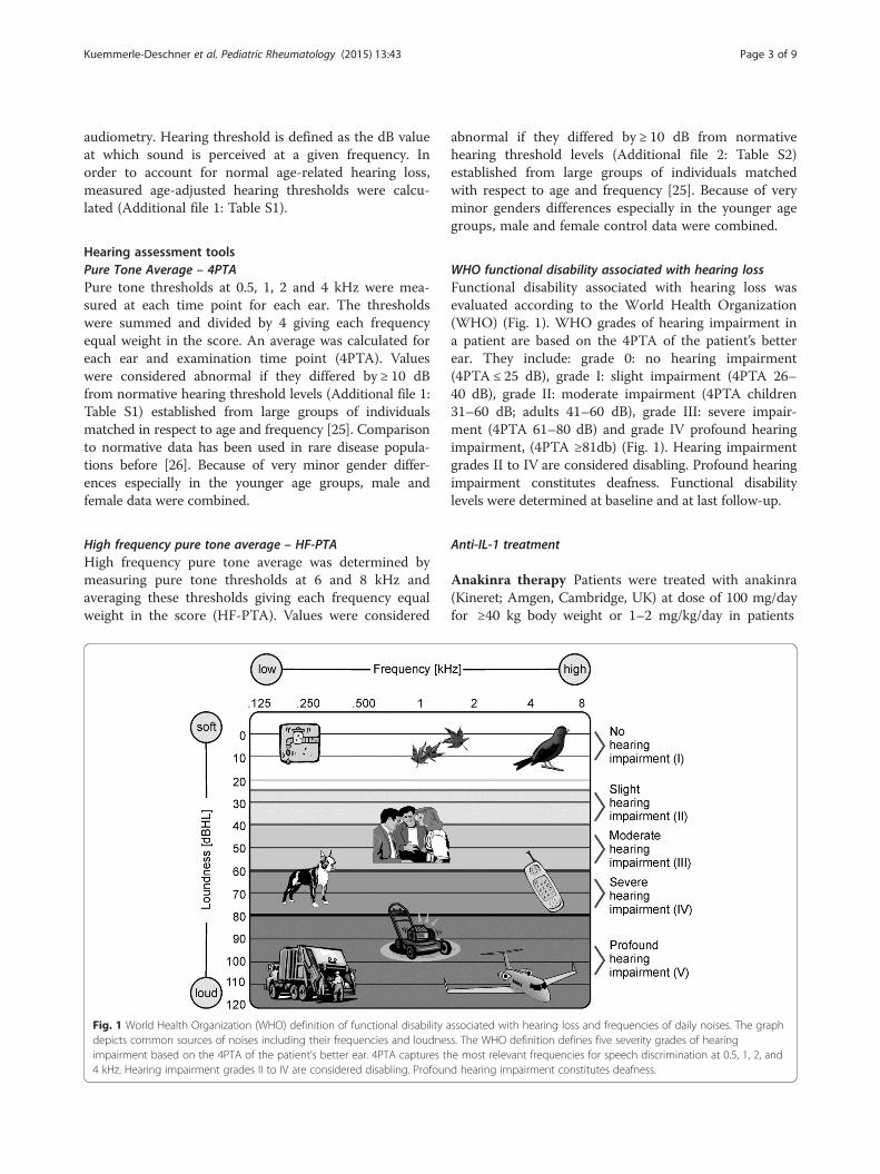
WHO functional disability associated with hearing lossFunctional disability associated with hearing loss wasevaluated according to the World Health Organization(WHO) (Fig. 1). WHO grades of hearing impairment ina patient are based on the 4PTA of the patient’s betterear. They include: grade 0: no hearing impairment(4PTA ≤ 25 dB), grade I: slight impairment (4PTA 26–40 dB), grade II: moderate impairment (4PTA children31–60 dB; adults 41–60 dB), grade III: severe impair-ment (4PTA 61–80 dB) and grade IV profound hearingimpairment, (4PTA ≥81db) (Fig. 1). Hearing impairmentgrades II to IV are considered disabling. Profound hearingimpairment constitutes deafness. Functional disabilitylevels were determined at baseline and at last follow-up.
Anti-IL-1 treatment
Anakinra therapy Patients were treated with anakinra(Kineret; Amgen, Cambridge, UK) at dose of 100 mg/dayfor ≥40 kg body weight or 1–2 mg/kg/day in patients
Fig. 1 World Health Organization (WHO) definition of functional disability associated with hearing loss and frequencies of daily noises. The graphdepicts common sources of noises including their frequencies and loudness. The WHO definition defines five severity grades of hearingimpairment based on the 4PTA of the patient’s better ear. 4PTA captures the most relevant frequencies for speech discrimination at 0.5, 1, 2, and4 kHz. Hearing impairment grades II to IV are considered disabling. Profound hearing impairment constitutes deafness.
Kuemmerle-Deschner et al. Pediatric Rheumatology (2015) 13:43 Page 3 of 9
<40 kg body weight. In children with persistent disease ac-tivity, the anakinra dose was stepwise increased to a max-imum of 8 mg/kg/day. Anakinra was self-administered bysubcutaneous injection once daily.
Canakinumab therapy Patients received canakinumabsubcutaneously 150 mg or 2 mg/kg for body weight<40 kg every 8 weeks. In case of residual symptoms,patients were maintained on a more intense dosing regi-men (increase dose up to 600 mg s.c. or 8 mg/kg s.cand/or increase of dosing frequency).
Outcome
Primary outcome Hearing loss was defined as increasedthreshold of ≥10 dB for any of the age-adjusted pure toneaverage for the frequencies 0.5, 1, 2 and 4 kHz (4-PTA)and the two high frequencies 6 and 8 kHz (HF-PTA).
Secondary outcomes 1) Improved or stable hearing wasdefined as a) decrease in hearing threshold by ≥ 20 dB inone frequency or by ≥10 dB in two, or more consecutivefrequencies for improved hearing or b) absence of wors-ening of hearing at last follow-up. 2) Worsening of hear-ing was defined as increase in hearing thresholdby ≥20 dB in one frequency, or by ≥10 dB in two ormore consecutive frequencies, in accordance with defini-tions used in previous studies [15].
Statistical analysisDescriptive statistics were performed; results were re-ported as frequencies with percentages, means withstandard deviations, or medians with ranges. Univariateparametric analyses utilized Fisher’s exact and chi-squareanalyses for categorical data and students T-test for con-tinuous variables. Non-parametric tests were performedwhere appropriate. Paired univariable analyses wereconducted to determine treatment responses. Repeatedmeasures linear regression analysis was performed forcomparative and predictive modeling of hearing thresh-olds to account for paired ears within patients. All testsconsidered p <0.05 as statistically significant. Statisticalanalyses were performed using SAS statistical softwarefor Windows Version 9.3 (SAS Institute, Cary, NorthCarolina, USA).
ResultsA total of 23 patients with clinically and genetically con-firmed MWS were included in the study; 65 % were fe-males. The median age at diagnosis was 18 years with arange from 3–72 years. Fifty-two percent were childrenage <18 years. Three NLRP3 mutations were found inthis cohort: 61 % had the c.937G>A (p.Glu313Lys)formerly known as E311K mutation, 22 % the c.1049C>T
(p.Thr350Met), formerly known as T348M and 17 % thec.598G>A (p.Val200Met), formerly known as V198Mmutation (Table 1).
Clinical features, disease activity and treatmentAll patients had active disease at diagnosis reflected in amedian MWS-DAS of 8. Most commonly seen clinicalfeatures were arthralgia (96 %), eye involvement (91 %)and headache (83 %). Clinical hearing loss was reportedby 91 % of patients. Other features included oral ulcers(78 %), rash (65 %) and myalgia (52 %). At baseline, themean SAA level was 68.7 mg/dl, mean CRP level2.2 mg/dl (Table 1). Anakinra cohort: 10 patients (43 %)received anakinra treatment; these were three males andseven females. The median age at diagnosis was 17 yearswith a range from 3 to 44 years. The most common mu-tation was the c.937G>A (p.Glu313Lys) mutationpresent in 5 (50 %) patients; 3 (30 %) had the c.1049C>T(p.Thr350Met) and 2 (20 %) the c.598G>A (p.Val200-Met) mutation. Canakinumab cohort: 13 patients (57 %),5 males and 8 females, were treated with canakinumab.The median age at diagnosis was 34 years with a rangebetween 3 and 72 years. Three different NLRP3 muta-tions were found: c.937G>A (p.Glu313Lys) in nine andc.1049C>T (p.Thr350Met) and c.598G>A (p.Val200Met)in two respectively.
Table 1 Demographic, clinical and laboratory findings andcharacteristic patterns of hearing loss in patients with Muckle-Wells syndrome (MWS)
Patients
N = 23
Demographics
Median age at diagnosis of MWS [years] (range) 18 (3–72)
Children ≤18 years at diagnosis (%) 12 (52)
Females (%) 15 (65)
NLRP3 mutation
• c.937G>A (p.Glu313Lys) (%) 14 (61)
• c.1049C>T (p.Thr350Met) (%) 5 (22)
• c.598G>A (p.Val200Met) (%) 4 (17)
Overall disease activity
Median MWS-DAS at baseline (range) 8 (6–16)
Laboratory markers
Mean SAA at baseline in mg/l (std), (nl* <1) 6.87 (1.43)
Mean CRP at baseline in mg/l (std), (nl* <0.05) 0.22 (0.17)
Treatment
IL-1 Inhibition (%) 23/23 (100)
Anakinra (%) 10 (43)
Canakinumab (%) 13 (57)
*nl: normal value
Kuemmerle-Deschner et al. Pediatric Rheumatology (2015) 13:43 Page 4 of 9
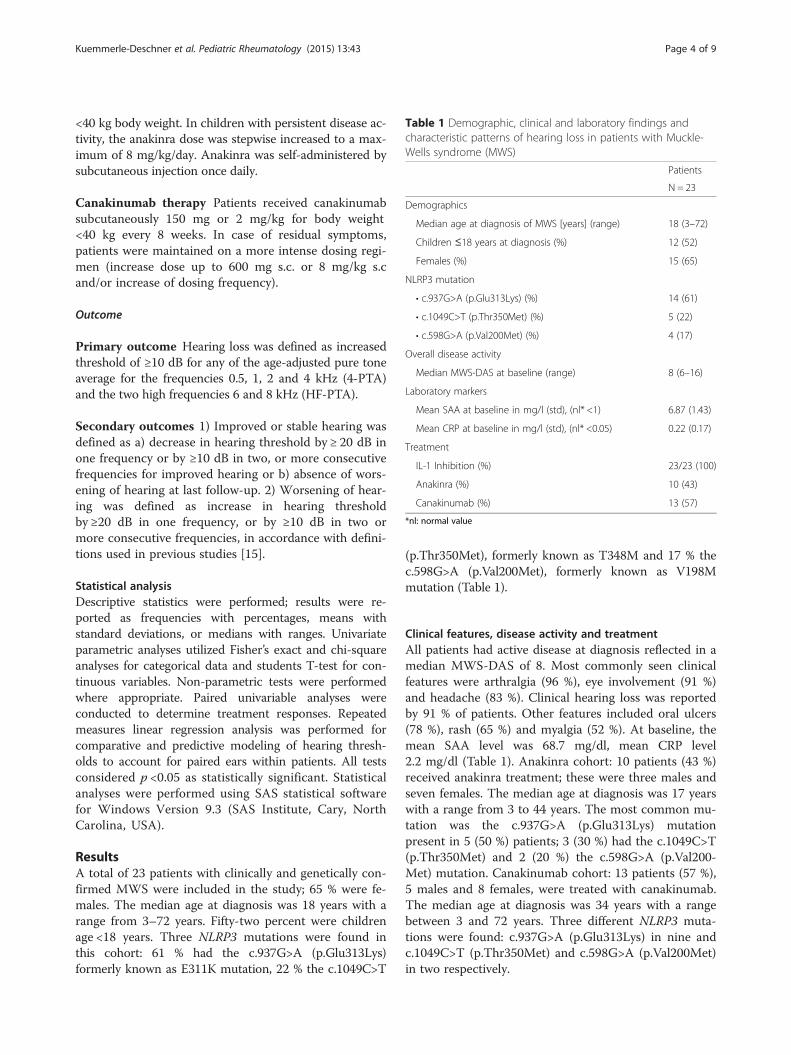
Hearing assessmentAt baseline, 21/23 MWS patients (91 %) had sensori-neural hearing loss by audiogram, correspondingly 42/46hearing assessments (single ears) were abnormal. All 11adults had abnormal hearing assessments (100 %, 22/22ears), while only 10/12 children (83 %, 20/24 ears) hadevidence of sensorineural hearing loss. Hearing thresh-olds at baseline varied significantly across ages and
frequencies (Fig. 2). Comparison of hearing thresholdsby frequency demonstrated significant differences be-tween lower and higher frequencies, even after adjust-ing for age and gender (p <0.001). All patients hadnormal tympanic membrane mobility, normal middleear pressure by impedance audiometry and normalcaloric vestibular stimulation. Seven adult patients re-ported tinnitus.
a
b
Fig. 2 Variation of hearing thresholds across ages and frequencies in patients with Muckle-Wells syndrome (MWS). a Comparison of hearingthreshold within a family with MWS. Audiogram of a 5 year-old child with MWS (a-1): Standard hearing assessment in the 4PTA range including0.5, 1, 2, and 4 kHz was normal (light gray area). In contrast the proposed HF-PTA captured a dramatic early high frequency hearing loss with increasedhearing thresholds at 6 and 8 kHz (dark grey area). The child’s 37-year old father reported hearing impairment. The corresponding audiogram (a-11)revealed abnormally increased hearing thresholds across all frequencies (light and dark grey areas). In the father advanced MWS associatedhearing loss is captured not only by HF-PTA but also by standard 4PTA. b Comparison of hearing thresholds of standard 4 pure-tone-average (4PTA) and proposed high frequency pure-tone-average (HF-PTA) in children and adults with MWS and age-matched healthycontrols across the age spectrum. Normal hearing thresholds captured in 4PTA and HF-PTA increase with age (4PTA grey line, HF-PTAblack line). MWS patients of all ages have significantly higher 4PTAs (black circles) and HF-PTAs (grey triangles) even after adjusting forage-specific normal hearing thresholds
Kuemmerle-Deschner et al. Pediatric Rheumatology (2015) 13:43 Page 5 of 9
4PTA assessmentsAt baseline, 34/46 (74 %) MWS hearing assessmentsdemonstrated an abnormal 4PTA, including 22/22 adultpatient hearing assessments (100 %) and 12/24 (50 %)children ears. The median baseline 4PTA was 28 (range0–72); at last follow-up, the median 4PTA was 26 (range0–56) (Table 2).
HF-PTAAt baseline, 42 MWS patient assessments demonstratedan abnormal HF-PTA (91%). An abnormal HF-PTA wasfound in 22/22 adult assessments (100 %) and in 20/24(83 %) in children. The median baseline HF-PTA was 45(range 0–114); at last follow-up, the median HF-PTAwas 43 (range 0–114) (Table 2).
Sensitivity of PTA assessmentsThe sensitivity of the 4PTA to detect clinical hearingloss was 81 %. The 4PTA assessment exclusively missed
children with early severe hearing loss affecting primarilyhigh frequencies. The HF-PTA testing identified 100 %of patients.
WHO functional disability associated with hearing lossAt baseline, 8 patients – all children – had no hearingimpairment (WHO grade 0), two patients, both adults,showed slight impairment (WHO grade I), eight – fourchildren and four adults – had moderate (grade II), fourhad severe hearing impairment (WHO grade III), allwere adults, and one adult had profound hearing impair-ment (WHO grade IV).At last follow-up, 9 patients – all children – presented
with no hearing impairment (WHO grade 0), one adultshowed slight (WHO grade I), nine patients, six adultsand three children, had moderate (grade II), three pa-tients, all adults (WHO grade III), and one adult stillhad profound hearing impairment (WHO grade IV). Soone child reversed hearing loss during treatment.
a
b
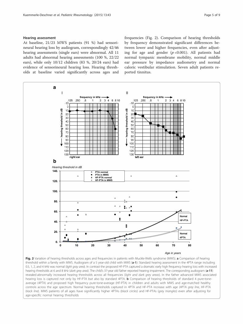
Fig. 3 Effect of anti-IL-1 therapy on hearing assessments in patients with MWS. a Comparison of two different anti-IL-1 therapy regimens. A totalof 11 assessments (ears) of 9 patients improved with anti-IL1 therapy; 4/9 were children, five were adults. None of the patients had worsening ofthe contralateral ear. Improvement occurred with both treatment regimens. All but two patients with hearing improvement were females. Overall,worsening was exclusively observed in adult patients, a male and a female. Both patients had received anakinra therapy. b Responsiveness tochange: Comparison between 4PTA and HF-PTA follow-up assessments after exposure to anti-IL-1 therapy. 4PTA and HF-PTA was calculated in allMWS patients following anti-IL-1 therapy. Hearing improved in five children and six adults, most commonly captured by HF-PTA (10/11), either byHF-PTA only (5/10, light grey bars) or also by the standard 4PTA (5/10, medium grey bars). Hence, a total of 91 % of improved assessments weredetected by HF-PTA, while just in one patient improvement was detected by standard 4PTA only. Worsening was not documented in children,but deteriorated in two adults. In both, this was detected by standard 4PTA, in one also in the HF-PTA
Kuemmerle-Deschner et al. Pediatric Rheumatology (2015) 13:43 Page 6 of 9

Risk factors for hearing loss in MWSFemale gender (p = 0.008), earlier age at diagnosis (p<0.001), and affection of high compared to low frequen-cies (p = <0.001) were found to be significantly associ-ated with hearing loss in a repeated measures linearregression model to account for paired ears within pa-tients. Young age was primarily associated with high fre-quency hearing loss, while older patients had evidenceof abnormal hearing thresholds across all frequencies.Female gender was an independent risk factor for hear-ing loss in MWS.
Effect of anti-IL-1 therapy on hearing lossA total of 44/46 (96 %) patient ears had improved orstable hearing assessments as defined above when
treated with anti-IL1 therapy. Improvement was docu-mented in 11 ears, six of which were treated with cana-kinumab and five with anakinra. Stable hearing wasdocumented for 33 ears, 20 of which were treated withcanakinumab and 13 with anakinra. Only two ears wors-ened significantly; these include one of an 18-year-oldmale and one of a 37-year-old female, both treated withanakinra. The contralateral ears for the two patientsremained stable (Fig. 3).
DiscussionThis is the first study to systematically analyze thespectrum of hearing loss in a large prospective cohort ofMWS patients across all ages. A key finding is that earlyinner ear inflammation in MWS primarily affects highfrequencies above 4 kHz and remains undetected whenusing standard assessment approaches (4PTA). There-fore, optimized testing for early hearing loss in MWShas to include high frequency hearing thresholds of 6and 8 kHz (HF-PTA) in order to significantly increasethe sensitivity of the hearing assessment and to allow forthe recognition of early MWS associated hearing lossdue to inflammatory injury. This is particularly import-ant in children, since early on in life MWS solely affectshigh frequencies. In fact, the study determined that earl-ier age at diagnosis was significantly associated with thedevelopment of hearing loss. Rapid onset of therapy maycontrol the inflammation thus greatly reducing the riskof inner ear damage and deafness in MWS [24]. Thestudy confirmed that anti-IL-1 therapy is effective in sta-bilizing or even improving sensorineural hearing loss,particularly in younger patients. Improved hearing isbest confirmed when utilizing the optimized assessmenttool HF-PTA in addition to standard testing. Reversibil-ity of hearing loss follows the MWS pattern and is mostcommonly seen in high frequencies. The study alsohighlighted that female MWS patients of all ages are atsignificantly higher risk for hearing loss and deafnesscompared to males. Importantly, females of all ages hada good response to anti-IL-1 therapy.Onset and progression of hearing loss in MWS has a
distinct pattern (Fig. 2a). The inflammatory attack pri-marily affects high frequencies. A similar hearing losspattern has been described in other inflammatory dis-eases including rheumatoid arthritis [12], metabolic dis-eases including diabetes [13, 27] and thyroid disease [28]and exposure to ototoxic drugs [29]. Hearing thresholdsin the high frequencies of the pure tone audiogram areparticularly relevant for speech discrimination and com-munication, especially in a noisy environment [30]. Af-fected MWS patients report impaired hearing; howeverstandard evaluation may remain normal. Detection ofearly hearing loss in MWS mandates testing of high fre-quency hearing thresholds. Early inner ear injury may
Table 2 Audiologic findings in a cohort of pediatric and adultMWS patients at baseline and following treatment with anti-IL-1-therapy utilizing the standard 4PTA and the proposed highfrequency (HF-PTA) instrument
Patients N = 23
Assessments(ears) N = 46
Hearing loss at baseline
Patients with clinical hearing loss (%) 21/23 (91)
Ears (assessments) with hearing loss (total) (%) 42/46 (91)
Adults with hearing loss (%) 11/11 (100)
• Abnormal assessments (adults) (%) 22/22 (100)
Children with hearing loss (%) 10/12 (83)
•Abnormal assessments (children) (%) 20/24 (83)
Hearing thresholds at baseline
4-frequency pure tone average (4PTA),median (range)*
28 (0–72)
Number of abnormal 4PTA assessments (%)* 34/46 (74)
•Adult assessments with abnormal 4PTA (%) 22/22 (100)
•Pediatric assessments with abnormal 4PTA (%) 12/24 (50)
High Frequency HF-PTA median (range)* 45 (0–114)
Abnormal HF-PTA hearing assessments (%) 42/46 (91)
•Adult assessments with abnormal HF-PTA (%) 22/22 (100)
•Pediatric assessments with abnormal HF-PTA (%) 20/24 (83)
Hearing thresholds at last follow-up
4 Pure Tone Average (4PTA); median (range)* 26 (0–56)
Abnormal 4PTA assessments (%) 34/46 (74)
High Frequency HF-PTA; median (range)* 43 (0–114)
Abnormal HF-PTA assessments (%) 42/46 (83)
Sensitivity of hearing assessments
Detection of clinical hearing loss by 4PTA 81 %
Detection of clinical hearing loss by HF-PTA 100 %
* Each patient is represented twice, since each ear had a separate hearingassessment; all values are adjusted for age (Additional files 1 and 2: Tables S1and S2)
Kuemmerle-Deschner et al. Pediatric Rheumatology (2015) 13:43 Page 7 of 9
remain unnoticed when using the current testing routine(4PTA). The proposed HF-PTA has a higher sensitivityfor early disease and should therefore be considered inall ages, but particularly in younger MWS patients. Anextended hearing threshold testing (0.5-8 kHz) is in-creasingly used for noise induced hearing loss [31] andwhen monitoring for potential ototoxicity with antibi-otics and chemotherapeutic agents [29]. Overall, MWSpatients require comprehensive otolaryngologic assess-ments. Serial pure tone thresholds including air- andbone conduction thresholds for frequencies of 0.125,0.25, 0.5, 1, 2, 4, 6 and 8 kHz should be determined bypure-tone audiometry using conventional, play or behav-ioral audiograms. Speech audiometry, caloric vestibularstimulation, tinnitus assessments and middle ear func-tional testing by impedance audiometry should beincluded.Prevention of deafness as a result of inner ear inflam-
mation in children and adults is a key task when treatingpatients with MWS. Anti-IL-1 therapy was effective instabilizing progression of hearing loss in this cohort,similarly to what was reported by others caring for pa-tients with CAPS [15, 32–34]. In our study, reversibilityof inner ear disease was found to follow a characteristicpattern with early improvement in the high frequencies.Change was best detected by HF-PTA and may bemissed when testing standard frequencies only. Overall,it appears that there is a window of opportunity at thetime of primary involvement of high frequencies. Cor-respondingly, younger patients were most likely to re-spond to treatment. The window may vary betweendifferent NLRP3 mutations [11]. Overall, younger agewas clearly identified to be a risk factor associated withsensorineural hearing loss. Females with MWS appear tobe at higher risk for hearing loss at all ages, correspond-ingly they were previously found to have overall moresevere disease as captured by the MWS disease activityscore (MWS-DAS) [24]. Reasons for this observation areunknown. The unbalanced gender ratio in this studyonly reflects the gender of the consecutive patients in-cluded into this study at time of inclusion. As the genderdistribution in other larger cohorts of CAPS patients isroughly balanced (own complete CAPS cohort: 55 %males, β-confident registry: 54 % females) in CAPS nogender preference can be observed. Also, type of muta-tion is associated with different degrees of hearing loss,as described previously [11]. Interestingly, in this studyall but two patients with evidence of hearing improve-ment after anti-IL-1 therapy were females.There are several imitations to the study. The number
of patients tested is small, only 23 children and adultswere included. However, MWS is a very rare condition. Acomprehensive, standardized assessment and therapyprotocol was applied prospectively to all patients allowing
for the capture of detailed clinical and audiological data atbaseline and following therapy. The age spectrum waswide with relatively few patients per age group threateningthe generalizability of the results. However, the clinicalspectrum of CAPS is also wide and varies further betweenNLRP3 gene mutation types regarding impact on hearing[24]. The study included three different mutations and al-most balanced numbers of children and adults. The studydid not include additional objective measures of hearingsuch as otoacoustic emissions, brainstem evoked responseaudiometry and gadolinium-enhanced magnetic reson-ance imaging (MRI) studies. However, these tests havelimited utility as generalizable screening and monitoringtools for centers worldwide. In addition, MRI studies ofthe inner ear in children commonly require anesthesia.
ConclusionsDiagnostic evaluation and monitoring of hearing abilitiesin patients with MWS mandates a comprehensive oto-laryngologic approach. The addition of HF-PTA to thestandard 4PTA pure tone threshold testing allows forearly recognition of imminent hearing loss and shouldprompt the rapid start of anti-IL-1 therapy for preven-tion of damage in particular in the high risk MWSpopulations.
Additional files
Additional file 1: Table S1. Median level of normal hearing across agegroups and frequencies (modified from Spoor [25]). (DOCX 24 kb)
Additional file 2: Table S2. Age-group specific median limits of normalhearing (calculated from Spoor 1967 [25], for the 4-frequency pure toneaverage (4PTA0.5-4kHz) and the proposed high-frequency pure tone average(HF-PTA6,8kHz). Because of very minor gender differences, especially in theyounger age groups, male and female data were combined. (DOC 51 kb)
Competing interestsDrs. Kuemmerle-Deschner and Koitschev have received consulting fees,speaking fees, and/or honoraria from Novartis. The University of Tuebingenhas received funds from Novartis Pharma for salary support for Dr. Hansmann’swork on trials of canakinumab in the treatment of cryopyrin associated periodicsyndrome. Dr. Plontke has received consulting fees from Otonomy Inc. andspeaking fees from ENT Academy. All authors are employed and funded bytheir respective institutions.
Authors’ contributionsJKD and AK conceived of the study, and participated in its design andcoordination and helped to draft the manuscript. PNT participated in thedesign of the study and performed the statistical analysis. SKP, ND, and SMBparticipated in the design oft he study and helped draft the manuscript. SHand KU conducted the data acquisition and patient examination. CKperformed the otolaryngologic examinations. PL carried out the moleculargenetic studies. All authors read and approved the final manuscript.
AcknowledgementsThe authors want to acknowledge Christine Michler and Iris Haug for help inthe management of the study and Katja Ambjoernsen for help in dataacquisition and otolaryngologic examinations.
Kuemmerle-Deschner et al. Pediatric Rheumatology (2015) 13:43 Page 8 of 9
Author details1Department of Pediatrics, Division of Pediatric Rheumatology, UniversityHospital Tuebingen, Hoppe-Seyler-Str. 1, D-72076 Tuebingen, Germany.2Department of Otorhinolaryngology, Head and Neck Surgery, KlinikumStuttgart, Stuttgart, Germany. 3Department of Medical Imaging, University ofToronto, Toronto, Canada. 4Department of Otorhinolaryngology, Head andNeck Surgery, University Hospital Halle (Saale), Halle (Saale), Germany.5Department of Anesthesiology and Intensive Care Medicine, UniversityHospital Tuebingen, Tuebingen, Germany. 6CeGaT, Center for Genomics andTranscriptomics, Tuebingen, Germany. 7Rheumatology, Department ofPediatrics, Alberta Children’s Hospital, University of Calgary, Calgary, Canada.
Received: 13 July 2015 Accepted: 27 October 2015
References1. Aksentijevich I, D Putnam C, Remmers EF, Mueller JL, Le J, Kolodner RD,
et al. The clinical continuum of cryopyrinopathies: novel CIAS1 mutations inNorth American patients and a new cryopyrin model. Arthritis Rheum.2007;56(4):1273–85.
2. Muckle TJ, Wells M. Urticaria, deafness, and amyloidosis: a new heredo-familial syndrome. Q J Med. 1962;31:235–48.
3. Hoffman HM, Mueller JL, Broide DH, Wanderer AA, Kolodner RD. Mutationof a new gene encoding a putative pyrin-like protein causes familial coldautoinflammatory syndrome and Muckle-Wells syndrome. Nat Genet.2001;29(3):301–5.
4. Dode C, Le Du N, Cuisset L, Letourneur F, Berthelot JM, Vaudour G, et al.New mutations of CIAS1 that are responsible for Muckle-Wells syndromeand familial cold urticaria: a novel mutation underlies both syndromes. AmJ Hum Genet. 2002;70(6):1498–506.
5. Feldmann J, Prieur AM, Quartier P, Berquin P, Certain S, Cortis E, et al.Chronic infantile neurological cutaneous and articular syndrome is causedby mutations in CIAS1, a gene highly expressed in polymorphonuclear cellsand chondrocytes. Am J Hum Genet. 2002;71(1):198–203.
6. Tschopp J, Martinon F, Burns K. NALPs: a novel protein family involved ininflammation. Nat Rev Mol Cell Biol. 2003;4(2):95–104.
7. Agostini L, Martinon F, Burns K, McDermott MF, Hawkins PN, Tschopp J.NALP3 forms an IL-1beta-processing inflammasome with increased activityin Muckle-Wells autoinflammatory disorder. Immunity. 2004;20(3):319–25.
8. Glaser RL, Goldbach-Mansky R. The spectrum of monogenicautoinflammatory syndromes: understanding disease mechanisms and useof targeted therapies. Curr Allergy Asthma Rep. 2008;8(4):288–98.
9. Hawkins PN, Lavender JP, Pepys MB. Evaluation of systemic amyloidosis byscintigraphy with 123I-labeled serum amyloid P component. N Engl J Med.1990;323(8):508–13.
10. Koitschev A, Gramlich K, Hansmann S, Benseler S, Plontke SK, Koitschev C,et al. Progressive familial hearing loss in Muckle-Wells syndrome. ActaOtolaryngol. 2012;132(7):756–62.
11. Kuemmerle-Deschner JB, Koitschev A, Ummenhofer K, Hansmann S, PlontkeSK, Koitschev C, et al. Hearing loss in Muckle-Wells syndrome. ArthritisRheum. 2013;65(3):824–31.
12. Pascual-Ramos V, Contreras-Yanez I, Enriquez L, Valdes S, Ramirez-AnguianoJ. Hearing impairment in a tertiary-care-level population of Mexicanrheumatoid arthritis patients. J Clin Rheumatol. 2012;18(8):393–8.
13. Lerman-Garber I, Cuevas-Ramos D, Valdes S, Enriquez L, Lobato M, OsornioM, et al. Sensorineural hearing loss–a common finding in early-onset type 2diabetes mellitus. Endocr Pract. 2012;18(4):549–57.
14. Ahmadi N, Brewer CC, Zalewski C, King KA, Butman JA, Plass N, et al.Cryopyrin-associated periodic syndromes: otolaryngologic and audiologicmanifestations. Otolaryngol Head Neck Surg. 2011;145(2):295–302.
15. Sibley CH, Plass N, Snow J, Wiggs EA, Brewer CC, King KA, et al. Sustainedresponse and prevention of damage progression in patients with neonatal-onset multisystem inflammatory disease treated with anakinra: a cohortstudy to determine three- and five-year outcomes. Arthritis Rheum.2012;64(7):2375–86.
16. Hoffman HM, Throne ML, Amar NJ, Sebai M, Kivitz AJ, Kavanaugh A, et al.Efficacy and safety of rilonacept (interleukin-1 Trap) in patients withcryopyrin-associated periodic syndromes: results from two sequentialplacebo-controlled studies. Arthritis Rheum. 2008;58(8):2443–52.
17. Hawkins PN, Lachmann HJ, McDermott MF. Interleukin-1-receptor antagonistin the Muckle-Wells syndrome. N Engl J Med. 2003;348(25):2583–4.
18. Goldbach-Mansky R. Blocking interleukin-1 in rheumatic diseases. Ann N YAcad Sci. 2009;1182:111–23.
19. Rynne M, Maclean C, Bybee A, McDermott MF, Emery P. Hearingimprovement in a patient with variant Muckle-Wells syndrome in responseto interleukin 1 receptor antagonism. Ann Rheum Dis. 2006;65(4):533–4.
20. Klein AK, Horneff G. Improvement of sensoneurinal hearing loss in a patientwith Muckle-Wells syndrome treated with anakinra. Klin Padiatr.2010;222(4):266–8.
21. Lachmann HJ, Kone-Paut I, Kuemmerle-Deschner JB, Leslie KS, Hachulla E,Quartier P, et al. Use of canakinumab in the cryopyrin-associated periodicsyndrome. N Engl J Med. 2009;360(23):2416–25.
22. Hoffman HM, Wanderer AA, Broide DH. Familial cold autoinflammatorysyndrome: phenotype and genotype of an autosomal dominant periodicfever. J Allergy Clin Immunol. 2001;108(4):615–20.
23. Kuemmerle-Deschner JB, Wittkowski H, Tyrrell PN, Koetter I, Lohse P,Ummenhofer K, et al. Treatment of Muckle-Wells syndrome: analysis of twoIL-1-blocking regimens. Arthritis Res Ther. 2013;15(3):R64.
24. Kummerle-Deschner JB, Tyrrell PN, Reess F, Kotter I, Lohse P, Girschick H,et al. Risk factors for severe Muckle-Wells syndrome. Arthritis Rheum.2010;62(12):3783–91.
25. Spoor A. Presbyacusis values in relation to noise induced hearing loss. IntAudiol. 1967;6(1):48–7.
26. Hanisch F, Rahne T, Plontke SK. Prevalence of hearing loss in patients withlate-onset Pompe disease: audiological and otological consequences. Int JAudiol. 2013;52(12):816–23.
27. Sunkum AJ, Pingile S. A clinical study of audiological profile in diabetesmellitus patients. Eur Arch Otorhinolaryngol. 2013;270(3):875–9.
28. Berker D, Karabulut H, Isik S, Tutuncu Y, Ozuguz U, Erden G, et al. Evaluation ofhearing loss in patients with Graves' disease. Endocrine. 2012;41(1):116–21.
29. Abujamra AL, Escosteguy JR, Dall'Igna C, Manica D, Cigana LF, Coradini P, et al.The use of high-frequency audiometry increases the diagnosis ofasymptomatic hearing loss in pediatric patients treated with cisplatin-basedchemotherapy. Pediatr Blood Cancer. 2013;60(3):474–8.
30. Wilson RH. Clinical experience with the words-in-noise test on 3430veterans: comparisons with pure-tone thresholds and word recognition inquiet. J Am Acad Audiol. 2011;22(7):405–23.
31. Mehrparvar AH, Mirmohammadi SJ, Ghoreyshi A, Mollasadeghi A,Loukzadeh Z. High-frequency audiometry: a means for early diagnosis ofnoise-induced hearing loss. Noise Health. 2011;13(55):402–6.
32. Imagawa T, Nishikomori R, Takada H, Takeshita S, Patel N, Kim D, et al.Safety and efficacy of canakinumab in Japanese patients with phenotypesof cryopyrin-associated periodic syndrome as established in the first open-label, phase-3 pivotal study (24-week results). Clin Exp Rheumatol.2013;31(2):302–9.
33. Aksentijevich I, Nowak M, Mallah M, Chae JJ, Watford WT, Hofmann SR, et al.De novo CIAS1 mutations, cytokine activation, and evidence for geneticheterogeneity in patients with neonatal-onset multisystem inflammatorydisease (NOMID): a new member of the expanding family of pyrin-associated autoinflammatory diseases. Arthritis Rheum. 2002;46(12):3340–8.
34. Weegerink NJ, Schraders M, Leijendeckers J, Slieker K, Huygen PL, HoefslootL, et al. Audiometric characteristics of a Dutch family with Muckle-Wellssyndrome. Hear Res. 2011;282(1–2):243–51.
Submit your next manuscript to BioMed Centraland take full advantage of:
• Convenient online submission
• Thorough peer review
• No space constraints or color figure charges
• Immediate publication on acceptance
• Inclusion in PubMed, CAS, Scopus and Google Scholar
• Research which is freely available for redistribution
Submit your manuscript at www.biomedcentral.com/submit
Kuemmerle-Deschner et al. Pediatric Rheumatology (2015) 13:43 Page 9 of 9