Embed Size (px)
Citation preview
can cycle from one state to another when main-tained under conditions that support stem cells. This highlights the fact that stochastic influences may be at work within stem-cell populations8. Roesch and colleagues’ results will be crucial if the same principles apply to patients’ primary tumours that have not been maintained in culture.
The field of cancer-stem-cell research is young and emerging, so it is not surprising that many questions remain. Each study must be carefully assessed, particularly when consider-ing the experimental methodology and models used. One point that should always be borne
in mind is the relevance of a study to tumour growth in patients. ■
Peter Dirks is at the Hospital for Sick Children, University of Toronto, Toronto, Ontario M5G 1X8, Canada.e-mail: [email protected]
1. Boiko, A. D. et al. Nature 466, 133–137 (2010).2. Roesch, A. et al. Cell 141, 583–594 (2010).3. Quintana, E. et al. Nature 456, 593–598 (2008).4. Takahashi, K. & Yamanaka, S. Cell 126, 663–676 (2006).5. Hamburger, A. W. & Salmon, S. E. Science 197, 461–463 (1977).6. Lee, J. et al. Cancer Cell 9, 391–403 (2006).7. Hayashi, K., Lopes, S. M., Tang, F. & Surani, M. A. Cell Stem
Cell 3, 391–401 (2008).8. Chang, H. H. et al. Nature 453, 544–547 (2008).
EARLY LIFE
Origins of multicellularityPhilip C. J. Donoghue and Jonathan B. Antcliffe
Interpreting truly ancient fossils is an especially tricky business. The conclusion that 2.1-billion-year-old structures from Gabon are the remains of large colonial organisms will get palaeobiologists talking.
It is a peculiar but widely held view that Charles Darwin used the palaeontological record as one of the principal lines of evi-dence for biological evolution. He did not. To modern eyes, On the Origin of Species presents a shocking account of the fossil record as an archive of evolutionary history.
For instance, Darwin highlights the idea that the then earliest-known fossil-bearing rocks, from the Cambrian period, beginning about 542 million years ago, contain records of modern groups — implying an extensive prehistory teeming with life. One-and-a-half centuries of subsequent research have revealed a vast microscopic fossil record of unicellular protists and bacteria extending, some would argue, as far back as there are sedimentary rocks from which they could be recovered. But although fossils of millimetre- to metre-scale multicellular organisms characterize the 90 mil-lion years of the Ediacaran period that precedes the Cambrian, pre-Ediacaran macroscopic fossils are exceedingly rare.
Hence the excitement that will surround the report on page 100 of this issue1, in which El Albani and colleagues describe macroscopic fossils from 2.1-billion-year-old rocks from southeastern Gabon. These centimetre-scale fossils, which the authors interpret as repre-sentatives of multicellular organisms, push the record of macroscopic life back more than 200 million years into the Palaeoproterozoic, an interval of Earth’s history so alien to us that we would not be able to breathe the air. Even though these fossils postdate the Great Oxida-tion Event — the notable rise in atmospheric oxygen 2.4 billion years ago — the atmosphere was still a toxic mix of greenhouse gases, with oxygen making up only a few per cent of
Time (billions of years ago)
Great OxidationEvent
Oxy
gen
leve
l (lo
g sc
ale)Atmosphere
Oceans
0
–2
–4
–6
4 3 2 1 0
Gabon fossils
Earliest stromatolitesOldest certain bacterial fossils
Hitherto oldest record of macroscopic multicellularityProbable eukaryotic fossils
Ediacara biotaOldest unequivocal animal fossils
Anoxic Sulphidic Oxic
The rocks have been dated with great precision (to 2,100 ± 30 million years old), and the pos-sibility that the structures are a younger min-eral assemblage that grew within the rock has been excluded by sulphur-isotope analyses. These indicate that the fossil-replacing min-eral pyrite (an iron sulphide) was precipitated by sulphate-reducing bacteria as the sediments were deposited. Further, contrasts between the carbon-isotope signature of the structures and their surrounding sediment indicate that the pyrite grew in an organic framework.
However, it is the fossil morphology, revealed through X-ray microtomography, that provides the most compelling evidence of biogenicity. The fossils take the form of three-dimensional sheets with scalloped margins defined by radial slits and an internal radial fabric (as seen in Fig. 4 of the paper on page 102). The authors interpret this structure as evidence of coordi-nated growth. The relative complexity of the fossils, and their co-occurrence with solu-ble organic compounds containing sterane (derived from sterol precursors), lead El Albani and colleagues to conclude that they are unlike any living bacterium. They don’t rule out the possibility that these remains might even rep-resent the earliest multicellular eukaryotes — that is, colonies of organisms with a mem-brane-bound nucleus that represent a distinct, more complex form of life from bacteria.
The null hypothesis, however, has to be that these remains represent bacterial colo-nies. Future work must determine whether the sterane signature, a hallmark of eukary-otes, is derived from soluble organics gener-ated within the sediments, or whether they migrated into these sediments from younger rock sequences.
In the interim, however, is there anything in the geometry of these presumed multicellular sheets that sets them apart from bacterial colonies? Multicellularity represents one
modern levels. This bacterial world was under-going the greatest episode of climate change in the history of the planet: pumping out oxygen, drawing down carbon dioxide, slowly trans-forming the Earth into the world we know.
The fossils are not much to look at — yet, given their antiquity, the fact that they can be seen by the naked eye is astonishing. Out of their geological context, these structures are unremarkable and would probably have been ignored. But the geological context is as remark-able as are the potential evolutionary implica-tions. El Albani and colleagues1 have, therefore, been painstaking in attempting to demonstrate the age and biological nature of the structures.
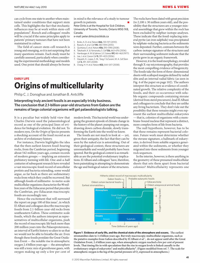
Figure 1 | Evidence of early life, and the chemical state of the atmosphere and oceans. The earliest stromatolites date to 3.4 billion years ago. But truly macroscopic multicellular organisms, such as the putative examples from Gabon described by El Albani et al.1, do not appear until after the Great Oxidation Event, 2.4 billion years ago, when atmospheric oxygen reached a few per cent of present levels. That timing fits in with speculation that the rise in oxygen levels is linked causally to the evolutionary origin of eukaryotes5 and multicellularity6. (Figure modified from ref. 7. The scale for atmospheric oxygen is the log of the partial pressure of O2 expressed in atmospheres.)
41
NATURE|Vol 466|1 July 2010 NEWS & VIEWS
© 20 Macmillan Publishers Limited. All rights reserved10

of the principal thresholds in evolutionary history2. This threshold has been exceeded tens of times3, perhaps because much of the requisite molecular machinery to facilitate cell–cell coordination is a shared primitive feature of living organisms4, but also because some definitions of multicellularity encompass everything from simple bacterial colonies to badgers. Stricter definitions of multicellularity are met in far fewer instances. Although the fossils are macroscopic, they do not seem to represent anything other than the basic type of multicellularity, which occurs earlier in time in the form of stromatolites.
Nevertheless, the size, form and thickness of these sheets may be significant. It has been argued that the paucity of ancient records of macroscopic multicellularity could reflect the physiological limitations enforced by atmos-pheric and oceanic chemistry during the first 2 billion years of Earth’s history5,6. The proxim-ity in the age of these fossils to the timing of the Great Oxidation Event (Fig. 1) fits elegantly with speculative hypotheses on the co-evolu-tion of life and the chemistry of the oceans.
This latest discovery raises more questions
than it answers. But within the confines of a very patchy global record of Proterozoic and Archaean rocks, which extend from about 3.8 billion years ago to the beginning of the Cambrian, these remains contribute to a fossil record that belies the dated caricature painted in the Origin. It was Darwin’s view that absence of organisms in these early intervals of Earth’s history would prove his theory of biological evolution wrong. The discovery and continu-ing elucidation of the Precambrian fossil record has met Darwin’s predictions on the extent and structure of evolutionary history. ■
Philip C. J. Donoghue and Jonathan B. Antcliffe are in the Department of Earth Sciences, University of Bristol, Wills Memorial Building, Queen’s Road, Bristol BS8 1RJ, UK.e-mail: [email protected]
1. El Albani, A. et al. Nature 466, 100–104 (2010).2. Maynard Smith, J. & Szathmáry, E. The Major Transitions in
Evolution (Oxford Univ. Press, 1997).3. Butterfield, N. J. Precambr. Res. 173, 201–211 (2009).4. Shapiro, J. A. Annu. Rev. Microbiol. 52, 81–104 (1998).5. Anbar, A. D. & Knoll, A. H. Science 297, 1137–1142 (2002).6. Bengtson, S., Rasmussen, B. & Krapez, B. Paleobiology 33,
351–381 (2007).7. Anbar, A. D. Science 322, 1481–1483 (2008).
DRUG DISCOvERY
Pulled from a protein’s embraceWilliam L. Jorgensen
It is hard to predict how strongly a small molecule will bind to a protein, but this is a crucial goal of computer-aided drug discovery. A new approach models the forcible removal of molecules from a protein’s active site.
Most drugs are small ligand molecules that bind to a protein. Some disrupt protein–protein interactions, whereas many others — enzyme inhibitors — inhibit reactions carried out by proteins. Computational methods for accu-rately predicting the binding affinity of a ligand for a protein are therefore central to rational drug design. Reporting in the Journal of the American Chemical Society, Colizzi et al.1 describe a stimulating advance in this field: a computational method for predicting protein–ligand binding affinities that works by model-ling the force that is required to pull inhibitors out of their complexes with proteins.
Traditionally, drugs were discovered by trial and error. Powders made from willow bark, for example, have been known to be useful for treating headaches, pains and fevers since the time of Hippocrates. The active ingredi-ent was eventually isolated in the 1800s, and a close analogue became aspirin in 1899. Drug discovery evolved to be increasingly deliberate and, with the advent of structural biology in the 1960s, the rational design of enzyme inhibitors was made possible. Given the three-dimensional structure of a target enzyme, ‘structure-based design’ can be carried
out, whereby an inhibitor is constructed to be complementary to the enzyme’s active site. The minimum requirements are the target’s struc-ture and computer-graphics tools that enable one to build and examine how molecules fit into the active site. Additional insight provided by evaluating the molecular energetics of the binding process is, however, crucial to most current activities in structure-based design.
So what is the goal of structure-based design? Simply, it is to assist in the discovery of safe and effective drugs. To achieve this, it is essential to design inhibitors that bind tightly to active sites (with a large, negative ‘free energy of binding’, ΔGb, to use the thermodynamics jargon). This helps the inhibitors to exert their biological effect at low doses. It also promotes selectiv-ity for the target enzyme, which helps prevent un desirable side effects that might arise if other enzymes are inhibited. The development of methods for accurately computing ΔGb is therefore an active area of research.
The most sophisticated approaches for calculating ΔGb use molecular dynamics or ‘Monte Carlo’ statistical mechanics to model the enzyme and the inhibitor in water, with all atoms represented. A direct calculation of the
change in free energy as an enzyme and inhibi-tor are brought together is difficult, owing to the flexibility of the two components and the changes in their hydration (the arrangement of water molecules around them) that occur on binding2. So alternative approaches are being explored. In a popular one, known as double decoupling, the free-energy changes associated with making the inhibitor ‘disap-pear’ from the complex with the enzyme and for making the inhibitor disappear in bulk water are computed. The difference in these results yields ΔGb. However, such calculations are still computationally taxing and are often imprecise for compounds as large as typical drugs. It is easier to compute the differences in ΔGb for structurally similar inhibitors, and this approach has been used with much suc-cess in guiding the discovery of, for example, potent anti-HIV agents3. Nevertheless, there is a pressing need for new methods that address the binding problem.
It is against this backdrop that Colizzi et al.1 report their new approach for gaining insight into protein–ligand binding affinities. Mimick-ing single-molecule pulling experiments and simulations4, they used ‘steered’ molecular dynamics (SMD) to compute the force that is required to extract inhibitors from complexes with enzymes. To model each extraction, the authors carried out molecular-dynamics simulations to predict what would happen if a harmonic spring was attached to the inhibi-tor and then pulled at constant vel ocity into the surrounding water (Fig. 1a). The force that was applied to the inhibitor was determined from the extension of the spring and recorded as a function of time, producing a force profile (Fig. 1b). Strongly bound inhibitors yield pro-files with higher peak forces, whereas the force profiles of weaker inhibitors are flatter.
To illustrate their approach, Colizzi et al. investigated the binding of five structurally related compounds to the protein FabZ from the malaria-causing parasite Plasmodium fal-ciparum, an enzyme that is a potential target for antimalarial drugs5. The tested compounds included two (luteolin and myricetin) that inhibit FabZ and three that do not. The authors began by computationally generating putative structures for the FabZ–luteolin complex by using protein–ligand docking methods. Two alternative binding orientations for luteolin emerged, but the authors’ SMD results demon-strated that one of these modes was preferred. They then used the preferred mode in SMD calculations to generate force profiles for the five compounds.
The resultant profiles for luteolin and myri-cetin differed qualitatively from those for the three inactive analogues, with peak forces that were twice as large as those of the inac-tive compounds — as would be expected if the inhibitors bind to FabZ more strongly than the non-inhibitors. Emboldened by this suc-cess, the authors went on to use their method to predict the activity of another structurally
42
NATURE|Vol 466|1 July 2010NEWS & VIEWS
© 20 Macmillan Publishers Limited. All rights reserved10