Embed Size (px)
Citation preview

1
© American College of Medical Genetics and Genomics Original research article
introductionAutosomal recessive cerebellar ataxias (ARCAs) are a group of neurodegenerative disorders characterized by a tremendous clinical and genetic heterogeneity.1,2 Early-onset ARCAs rep-resent a subgroup for which several disease genes have been identified as playing a role in processes such as DNA damage response, DNA repair, DNA transcription, DNA replication, RNA processing, protein folding and modification, and neu-rotransmitter metabolism, of which glutamate is an example.1
Glutamate receptors were recently implicated in two sub-forms of early-onset ARCA. First, mutations in GRM1, encod-ing the metabotropic glutamate receptor 1, were found in patients of Roma origin with ARCA, characterized by develop-mental delay, intellectual disability, and a small brain.3
Second, a homozygous partial deletion of GRID2, encod-ing the ionotropic glutamate receptor delta 2, was found in a consanguineous Turkish family with early-onset ARCA, nys-tagmus, hypotonia with developmental delay in gross motor skills, encephalopathy with cerebellar ataxia, oculomotor apraxia, and pyramidal tract involvement.4 Another recent
study reported additional partial biallelic GRID2 deletions in patients with early-onset ARCA, eye movement abnormali-ties, delayed speech, and intellectual disability.5 In addition, a heterozygous deletion of exon 1 and the upstream region of GRID2 was found in an adult with spastic paraplegia, ataxia, frontotemporal dementia, and lower motor neuron involve-ment.6 Prior evidence implicating GRID2 in early-onset cerebellar ataxia was provided by two studies dealing with autoantibodies against the receptor after acute viral infection in two children.7,8
Apart from these few human cases, the link between GRID2 and ARCA was already apparent in natural mutants in mouse. Indeed hotfoot (ho) mice are characterized by cerebellar ataxia and jerky movements of the hind limbs.9 The hotfoot locus has been mapped to chromosome 6,10,11 leading to the identifica-tion of the Grid2 gene as the underlying disease gene.12 The Grid2 gene is a hotspot for deletions; at least 20 different reces-sive hotfoot alleles have been described,13 most of which are deletions disrupting parts of the coding region of Grid2.14 This hypermutability of the Grid2 locus can probably be ascribed to
Submitted 26 March 2014; accepted 19 June 2014; advance online publication 14 August 2014. doi:10.1038/gim.2014.95
Genet Med
00
00
2014
Genetics in Medicine
10.1038/gim.2014.95
Original Research Article
00
00
26March2014
19June2014
© American College of Medical Genetics and Genomics
14August2014
Purpose: The aim of this study was to identify the genetic cause of early-onset autosomal recessive cerebellar ataxia associated with reti-nal dystrophy in a consanguineous family.
Methods: An affected 6-month-old child underwent neurologi-cal and ophthalmological examinations. Genetic analyses included homozygosity mapping, copy number analysis, conventional poly-merase chain reaction, Sanger sequencing, quantitative polymerase chain reaction, and whole-exome sequencing. Expression analysis of GRID2 was performed by quantitative polymerase chain reaction and immunohistochemistry.
results: A homozygous deletion of exon 2 of GRID2 (p.Gly30_Glu-81del) was identified in the proband. GRID2 encodes an ionotropic glutamate receptor known to be selectively expressed in cerebellar Purkinje cells. Here, we demonstrated GRID2 expression in human adult retina and retinal pigment epithelium. In addition, Grid2
expression was demonstrated in different stages of murine retinal development. GRID2 immunostaining was shown in murine and human retina. Whole-exome sequencing in the proband did not pro-vide arguments for other disease-causing mutations, supporting the idea that the phenotype observed represents a single clinical entity.
conclusion: We identified GRID2 as an underlying disease gene of early-onset autosomal recessive cerebellar ataxia with retinal dystro-phy, expanding the clinical spectrum of GRID2 deletion mutants. We demonstrated for the first time GRID2 expression and localization in human and murine retina, providing evidence for a novel functional role of GRID2 in the retina.
Genet Med advance online publication 14 August 2014
Key Words: autosomal recessive cerebellar ataxia; glutamate receptor; GRID2; hotfoot; retinal dystrophy
1Center for Medical Genetics, Ghent University and Ghent University Hospital, Ghent, Belgium; 2Department of Pediatric Ophthalmology, Queen Fabiola Children’s University Hospital, Brussels, Belgium; 3Department of Ophthalmology, University of Cologne, Cologne, Germany; 4Unite Troubles de l’Equilibré et Vertiges, CHU Brugmann, Brussels, Belgium; 5Department of Pediatric Neurology, Queen Fabiola Children’s University Hospital, Brussels, Belgium. Correspondence: Elfride De Baere ([email protected])
Early-onset autosomal recessive cerebellar ataxia associated with retinal dystrophy: new human hotfoot phenotype
caused by homozygous GRID2 deletion
Kristof Van Schil, MSc1, Françoise Meire, MD, PhD2, Marcus Karlstetter, PhD3, Miriam Bauwens, MSc1, Hannah Verdin, MSc1, Frauke Coppieters, PhD1, Eva Scheiffert, BSc3, Christian Van Nechel, MD, PhD4,
Thomas Langmann, PhD3, Nicolas Deconinck, MD, PhD5 and Elfride De Baere, MD, PhD1
Genetics in medicine
2
VAN SCHIL et al | Human GRID2 deletion in ARCA and retinal dystrophyOriginal research article
the large size of the gene (1.4 Mb) and, more specifically, of the second intron.
All hotfoot deletions remove parts of the N-terminal extra-cellular leucine/isoleucine/valine binding protein–like domain of the protein. This domain is crucial for correct trafficking of the protein out of the endoplasmic reticulum to the surface of Purkinje cells,13,15 where it is selectively expressed16 and thought to play an important role in the organization of parallel fiber–Purkinje cell synapses and associated long-term depression. However, the wild-type protein does not seem to form func-tional ion channels, nor does it bind to glutamate.15
In the reported patients with GRID2-associated ARCA, the first mutation is the counterpart of hotfoot mutant ho8J/ho13J, caused by a homozygous deletion of GRID2 exons 3 and 4,4 and the second mutation is compatible with mouse mutant ho15J, caused by a compound heterozygous exon 2 deletion. Two other human deletions, one of exon 4 and the other of exon 1, do not have mouse counterparts.
Here we report a fifth human deletion mutant, characterized by a homozygous GRID2 deletion and mim-icking the mouse ho15J mutant. This was found in a pro-band of consanguineous origin with early-onset ARCA and retinal dystrophy. Whole-exome sequencing in the pro-band demonstrated no defects other than the GRID2 dele-tion. Moreover, we demonstrated GRID2 messenger RNA (mRNA) and protein expression in both murine and human retina, suggesting a novel functional role of this receptor in the retina. Our study expands the expression domain of GRID2 and the clinical spectrum of hotfoot deletion mutants in human, which thus far involved only cerebellar and no retinal phenotypes. Finally, we provided further evidence for evolutionary conservation of a hotfoot fragile site between mice and humans.
MAtEriALS And MEtHodSPatientsThe proband is a child of Moroccan origin with cerebellar ataxia and eye motility disorder and originates from a consanguine-ous family. Both parents and two healthy siblings participated in this study. Ophthalmological examination was carried out with fundoscopy, assessment of eye motility, and electroreti-nography (ERG). The child underwent neurological assessment including magnetic resonance imaging. Informed consent was obtained, and research protocols adhered to the tenets of the Declaration of Helsinki.
Molecular characterization of GRID2 deletionIdentity-by-descent and homozygosity mapping. Identity-by-descent and homozygosity mapping was performed using genome-wide single-nucleotide polymorphism chip analysis using the HumanCytoSNP-12 BeadChip platform (Illumina, San Diego, CA). Identity-by-descent regions (>1 Mb) were identified using PLINK software17 integrated in arrayCGHbase.18 Resulting homozygous regions were ranked according to their length and number of consecutive
homozygous single-nucleotide polymorphisms, as described by Coppieters et al.23
Copy number variation analysis. Copy number variation analysis in the index patient was performed using the aforementioned single-nucleotide polymorphism chips (log R ratio + B allele frequency). Subsequently, quantitative polymerase chain reaction was used to perform segregation analysis of the identified GRID2 deletion in the healthy family members. Primers and conditions can be found in Supplementary Table S2 online; design was done as previously described.19 qBasePlus software (Biogazelle, Zwijnaarde, Belgium) was used for data analysis,20 and two reference genes were used for normalization of the relative quantities. Two positive controls with known copy numbers were used as a reference to calculate the copy numbers.19
Delineation of deletion. Further delineation of the deletion was done by iterative rounds of conventional polymerase chain reaction (for primers and conditions, see Supplementary Table S2 online) in both the 5′ and 3′ breakpoint regions. A junction product was obtained by combining the GRID2del_4Aa_F and GRID2del_14Ea_R primers and was sequenced (BigDye Terminator v3.1 Cycle Sequencing Kit on an ABI 3730XL genetic analyzer; Applied Biosystems, Foster City, CA). Sequences were analyzed with Seqscape software (Applied Biosystems).
Bioinformatics deletion. An extensive bioinformatic analysis has been performed on both breakpoint regions of four different molecularly characterized human GRID2 deletions, as previously described21 (see Supplementary Text S1 online).
Whole-exome sequencing. Whole-exome sequencing was performed in the index patient and one healthy sibling. For exome enrichment, the Nextera Rapid Capture Exome kit (Illumina) was used, followed by paired-end sequencing on a HiSeq 2000 (Illumina) by Aros AB (Aarhus, Denmark). The CLC Genomics Workbench version 6.4 (Aarhus, Denmark) was used to read mapping against the human genome reference sequence (National Center for Biotechnology Information, GRCh37/hg19), removal of duplicate reads after mapping, coverage analysis, and quality-based variant calling, followed by further variant annotation using Alamut-HT (Interactive Biosoftware, Rouen, France).
Grid2 expression and localizationmRNA expression analysis. The following human complementary DNAs were assayed: adult and fetal cerebellum, adult cerebral cortex, adult retina, and adult retinal pigment epithelium (Biochain, Newark, CA). The following mouse tissues were assayed: developmental stages P0 to P60 of the retina. Preparation details of murine complementary DNA can be found in Supplementary Text S1 online. An exon-spanning assay was designed for both human GRID2 and mouse Grid2; primers and conditions
Genetics in medicine

3
Human GRID2 deletion in ARCA and retinal dystrophy | VAN SCHIL et al Original research article
can be found in Supplementary Table S2 online. qBasePlus software was used for data analysis.20 Two reference genes validated by the geNorm experiment have stable expression in all tested human tissues, so intertissue comparisons can be made. For murine tissues, β-actin served as the reference gene.
Retinal samples of human donors. Retinal samples of human donors were obtained from the Eye Bank of the Center of Ophthalmology, University of Cologne, Germany. After dissection of the anterior segment, the remaining tissue included the posterior pole. Remaining vitreous humor was removed to obtain retinal tissue before further processing.
The research followed the tenets of the Declaration of Helsinki.
Immunohistochemistry. Human and mouse retinal sections were used for immunofluorescence analysis, which was performed as previously described22 (see Supplementary Text S1 online).
rESuLtSPhenotype of the probandThe 2-year-old patient of Moroccan origin was born to consan-guineous married parents. He displayed the following anthro-pometric measurements at birth: weight P97, height P75, and
Figure 1 clinical presentation of the proband. (a,b) Magnetic resonance imaging. (a) (Left) Sagittal T1-weighted axial images of the patient at the age of 1 year showing cerebellar atrophy particularly prominent in the vermis. Supratentorial brain architecture is normal with a complete corpus callosum although appearing slightly thin. (Right) T2-weighted axial image of the patient at the same age, confirming cerebellar atrophy with prominent cerebellar folia also mainly localized in the vermis. (b) Corresponding pictures from control child, 18 months of age. (c) Photopic, scotopic, maximal, and flicker responses on electroretinogram from the affected child compared with responses obtained in the same conditions from a 2-year-old control child. An amplitude deficit can be observed for photopic as well as scotopic responses. At the same scale, the amplitude deficit is obvious between the recordings made at 6 months (upper traces) and 24 months (middle traces) compared with the control subject (lower traces).
Photopic
6 m
onth
sP
atie
nt
Con
trol
40 ms/div 25 ms/div 20 ms/div 20 ms/div
40 ms/div 25 ms/div 20 ms/div 20 ms/div
40 ms/div 25 ms/div 20 ms/div 20 ms/div
50 µ
V/d
iv
50 µ
V/d
iv
50 µ
V/d
iv
25 µ
V/d
iv
50 µ
V/d
iv
50 µ
V/d
iv
50 µ
V/d
iv
25 µ
V/d
iv
50 µ
V/d
iv
50 µ
V/d
iv
50 µ
V/d
iv
25 µ
V/d
iv
Scotopic Max Flicker
24 m
onth
sC
ontr
ol
a
c
b
Genetics in medicine
4
VAN SCHIL et al | Human GRID2 deletion in ARCA and retinal dystrophyOriginal research article
head circumference P90. General physical examination showed pectus excavatum, the presence of some joint laxity, and no sig-nificant dysmorphism. On neurological examination, the pro-band had nystagmus, first recognized at the age of 2 months. Cerebellar system involvement was observed, with truncal hypotonia and ataxic movements of the upper arms. He had abnormal backward head movements especially accompanied by a staring gaze, and his speech was dysarthric. Brain magnetic
resonance imaging at 1 year demonstrated cerebellar atrophy particularly prominent in the vermis region (Figure 1). An extensive metabolic workup was normal, as were serum con-centrations of albumin, α-fetoprotein, creatine kinase, lactate, pyruvate, vitamin E, and cholesterol. Visual evoked potentials, brainstem evoked potentials, electroencephalography, and brain auditory evoked potentials were normal. The neurological course of the disease was rather static, with marked oculomotor
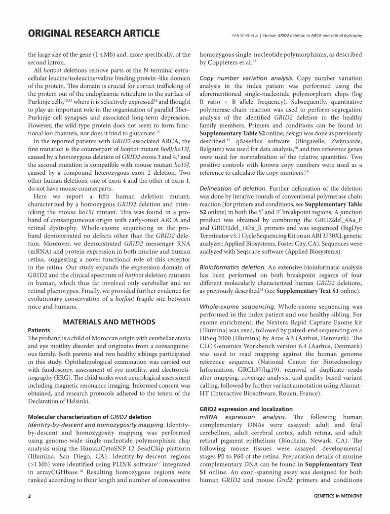
Figure 2 identification and characterization of GRID2 deletion. (a) Pedigree of the family. Genotypes are indicated for all family members for whom segregation analysis was performed. (b) Segregation analysis of the deletion in all family members. One healthy sibling (IV:1) does not carry the deletion, both parents (III:1 and III:2) and the other healthy sibling (IV:2) carry one copy of the deletion, and the proband (IV:3) has two copies. (c) ArrayCGH profile indicating the presence of a deletion on chromosome 4 (chr.4). (d) Further delineation of the deletion by conventional polymerase chain reaction (PCR). Gray and red boxes and connecting lines indicate respectively nondeleted and deleted exons and introns. Short horizontal lines correspond to designed PCR amplicons and are used to delineate the deletion. The black dotted line indicates the junction product. (e) Sanger sequencing of the junction product led to the delineation of the deletion at nucleotide level, chr4: g.93422866_93754032del. del, allele with deleted exon 2; wt, wild-type allele.
I:1
II:1
III:1wt/del
III:2wt/del
III:1 III:2 IV:1 IV:2
GRID2 - intron 1
GRID2 - exon 2
GRID2 - intron 2
IV:3IV:1wt/wt
chr. 4
5
Log
R r
atio
4
3
2
1
0
93 Mb 93.1 Mb 93.2 Mb
700 bp
Amp 4
Exon Intron
Deletedexon
Deletedintron
DelineationPCR amplicon
Amp 14Eb
AGGTC 331 kb AACTC
Amp 14EaAmp 4A
Amp 4Aa Amp 14E
PCR
2 31
331.167 bp 800 bp
93.3 Mb 93.4 Mb 93.5 Mb 93.6 Mb 93.7 Mb 93.8 Mb 93.9 Mb 94.0 Mb
−1
−2
−3
−4
−5
IV:2wt/del
IV:3del/del
2.0
1.5
Rel
ativ
e fr
eque
ncie
s
1.0
0.5
II:2 II:3 II:4
I:2
a
c
d e
b
Genetics in medicine

5
Human GRID2 deletion in ARCA and retinal dystrophy | VAN SCHIL et al Original research article
apraxia and difficulties in fixing gaze. Deep reflexes were always normal, with no sign of pyramidal tract involvement. Slight ankle flexion and inversion contractures were observed.
Ophthalmological examination at the age of 2 months showed visual fixation delay together with a multidirectional nystag-mus. An ERG at the age of 6 months showed a severe reduc-tion of both rod and cone responses (Figure 1). At 24 months, all ERG amplitudes, already significantly reduced at 6 months, were further impaired, with an additional average ampli-tude loss of ~30% for both scotopic and photopic responses (Figure 1). No fundus abnormalities were observed. Further ophthalmological examination at the age of 24 months revealed a manifest eye movement disorder, mainly tonic upgaze. Both voluntary pursuit and saccadic eye movements were impossi-ble. The child could only observe objects in his peripheral visual fields by turning his head. A nystagmus with small-amplitude beats was best observed when the child attempted fixation (Supplementary Movies S1–S4 online). His visual acuity is estimated to be at least 0.05.
A summary of the clinical findings of the index case is pre-sented in Supplementary Table S1 online. The oculomotor impairment can be observed in the Supplementary Movies S1–S4 online.
identification and characterization of homozygous GRID2 deletionTo identify the underlying genetic cause of the ARCA and Retinal dystrophy (RD) phenotype in the proband, homozy-gosity mapping combined with copy number variation analysis revealed a homozygous deletion of exon 2 of the GRID2 gene (Figure 2), located on chromosome 4. Breakpoints of the dele-tion were situated between single-nucleotide polymorphisms rs7684294 and rs7659159 at the 5′ end of the deletion and between rs6852643 and rs1456362 at the 3′ end. Further delin-eation of the deletion by iterative rounds of conventional poly-merase chain reaction led to refinement of the 5′ breakpoint region to 130 bp and 260 bp for the 3′ region. Subsequently, a deletion junction of 650 bp was obtained in the patient but not in the control (Figure 2d). Sanger sequencing of this junc-tion product led to the delineation of the deletion at nucleo-tide level: chr4:g.93422866_93754032del (Figure 2e). This deletion removes the entire second coding exon of the GRID2 gene, resulting in an in-frame deletion (p.Gly30_Glu81del). Segregation analysis by quantitative polymerase chain reaction confirmed the homozygous deletion in the index patient and demonstrated that both parents and one of the healthy siblings are heterozygous carriers of the deletion, whereas the other sib-ling is homozygous for the wild-type allele (Figure 2b).
To assess the underlying mechanism of the four character-ized GRID2 deletions found in our proband and in the ARCA patients reported by Hills et al.,5 a bioinformatics analysis21 was performed on both breakpoint regions of these dele-tions. A summary of the bioinformatics findings is given in Table 1; visualization of microhomology can be found in Supplementary Figure S1 online. Based on the results of this ta
ble
1 S
um
mar
y o
f b
ioin
form
atic
an
alys
is o
f h
um
an G
RID
2 d
elet
ion
bre
akp
oin
ts
del
etio
nSt
art
(hg
19)
End
(h
g19
)Si
ze
(kb
)M
icro
hom
olog
y (b
p)
inse
rtio
n,
del
etio
n, o
r su
bst
itu
tio
n
5′ B
reak
po
int
reg
ion
3′ B
reak
po
int
reg
ion
Pote
nti
al
mo
lecu
lar
mec
han
ism
rep
etit
ive
elem
ent
nu
mb
er
of
seq
uen
ce
mo
tifs
nu
mb
er o
f n
on
-B d
nA
co
nfo
rmat
ion
p
red
icti
on
m
oti
fsr
epet
itiv
e el
emen
t
nu
mb
er
of
seq
uen
ce
mo
tifs
nu
mb
er o
f n
on
-B d
nA
co
nfo
rmat
ion
p
red
icti
on
m
oti
fs
E2 d
elet
ion
(thi
s st
udy)
9342
2866
9375
4032
331
1–
–4
–A
luSx
5–
Repl
icat
ive/
NH
EJ
E2
mat
erna
lly
inhe
rited
de
letio
n (H
ills
et a
l.5 )
9348
1110
9353
1257
502
?A
T_ric
h3
2M
LT1J
52
Repl
icat
ive/
NH
EJ
E2 d
e no
vo
dele
tion
(Hill
s et
al.5 )
9341
2943
9374
8082
335
–?
–5
1C
harli
e15
1N
HEJ
E4 d
elet
ion
(Hill
s et
al.5 )
9401
9842
9405
6765
373
?–
81
–4
-Re
plic
ativ
e/N
HEJ
bp, b
ase
pairs
; E, e
xon;
NH
EJ, n
onho
mol
ogou
s en
d-jo
inin
g.
*Non
-B D
NA
con
form
atio
ns s
houl
d be
loca
ted
at b
oth
side
s of
the
junc
tion
or o
verla
ppin
g th
e ju
nctio
n. R
eplic
ativ
e st
ands
for r
eplic
ativ
e-ba
sed
mec
hani
sms
and
incl
udes
fork
sta
lling
and
tem
plat
e sw
itchi
ng (F
oSTe
S),
mic
roho
mol
ogy-
med
iate
d br
eak-
indu
ced
repl
icat
ion
(MM
BIR)
, ser
ial r
eplic
atio
n sl
ippa
ge (S
RS),
and
brea
k-in
duce
d SR
S (B
ISRS
).
Genetics in medicine
6
VAN SCHIL et al | Human GRID2 deletion in ARCA and retinal dystrophyOriginal research article
extensive analysis, the studied GRID2 deletions with micro-homology may be caused by nonhomologous end-joining (<5-bp microhomology) or a replicative-based repair mechanism, favoring the latter because an information scar, typical of non-homologous end-joining, was not present at the junction for our deletion and was not reported for the deletions described by Hills et al.5 The deletion without microhomology is more likely to be caused by nonhomologous end-joining. The pres-ence of sequence motifs can result in genomic instability and the formation of a deletion.
To date, GRID2 deletions in humans and Grid2 deletions in mice have only been associated with early-onset ARCA and not with retinal involvement. To gain further evidence that the phenotype observed in the proband represents a single clinical entity, whole-exome sequencing was performed in the index patient and one healthy sibling. An overview of the vari-ant filtering, performed as previously described,23 is given in Supplementary Table S3 online. Variant filtering in 209 RetNet genes (https://sph.uth.edu/retnet/) resulted in 47 remain-ing variants with a potential pathogenic effect. Subsequently, assuming the occurrence of a homozygous mutation because
of the consanguinity in this family, all variants located in a homozygous region (>1 Mb) were filtered, resulting in 48 vari-ants with a potential pathogenic effect. However, based on identity-by-descent and exome data of the nonaffected siblings, variant allele frequency, minor allele frequency, literature or database searches, and exclusion by Sanger sequencing, none of the retained variants in known RD genes or located in iden-tity-by-descent regions could be linked to an RD phenotype (Supplementary Table S3 online). An overview of these vari-ants can be found in Supplementary Tables S4 and S5 online.
Expression analysis of Grid2 transcript and protein in mice and humansIn previous mouse and rat studies, Grid2 expression was shown selectively in cerebellar Purkinje cells, more specifically at par-allel fiber synapses. The recent study by Hills et al.5 demon-strated conservation of this selective expression in the human cerebellum. As a first step toward explaining the RD phenotype observed here, we performed expression analysis in human complementary DNA of adult and fetal cerebellum, adult cere-bral cortex, adult retina, and adult retinal pigment epithelium,
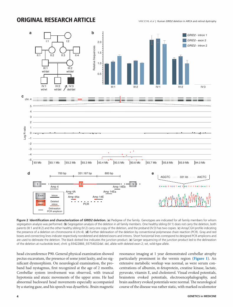
Figure 3 Grid2 expression. (a) GRID2 messenger RNA (mRNA) expression in human complementary DNA (cDNA) of adult and fetal cerebellum, adult cerebral cortex, adult retina, and adult retinal pigment epithelium (RPE), with the strongest expression in cerebellum, similar expression in retina and cerebral cortex, and lower expression in RPE. (b) Grid2 mRNA expression at different developmental stages of retina from neonate (P0) and adult mice (P60). Increasing Grid2 gene expression with progressing development, reaching constant expression levels in adult mice, consistent with gene expression profiles of other retinal genes. (c,d) Representative fluorescent images of horizontal cross-sections of (c) human and (d) mouse retina stained with anti-GRID2 antibody (red, 1:250). Retinal counterstaining was performed with 4′,6-diamidino-2-phenylindole (DAPI) (blue). (c) GRID2 immunoreactivity is detected in the photoreceptor inner segments (IS), the outer plexiform layer (OPL), and the ganglion cell layer (GCL). (d) Grid2 immunoreactivity in the murine retina is more globally distributed along photoreceptor IS, the inner nuclear layer (INL)/inner plexiform layer (IPL) margin, and the GCL. ONL, outer nuclear layer; OS, outer segments.
Cerebellum(adult)
Cerebellum(fetal)
Cerebralcortex
Retina
Human Mouse
OS
ONL
OPL
INL
IPL
GCL
IS OS
ONL
OPL
INL
IPL
GCL
IS
RPE Lympho-cytes
P0
0.5
1.0
1.5
2.0
P3 P7 P10 P18 P21
MouseHuman
0.1
1.0
10
Rel
ativ
e G
RID
2 m
RN
A le
vels
Rel
ativ
e G
rid2
mR
NA
leve
ls
P60
a b
c d
Genetics in medicine

7
Human GRID2 deletion in ARCA and retinal dystrophy | VAN SCHIL et al Original research article
demonstrating GRID2 mRNA expression in all of these tissues (Figure 3a). Expression was strongest in cerebellum, followed by similar expression levels in retina and cerebral cortex and lower expression in retinal pigment epithelium. In addition, mRNA expression was assessed at different developmental stages of murine retina, including very young developing reti-nas from neonatal mice (P0) and adult retinas (P60). Grid2 gene expression increased with progressing development and reached constant expression levels in adult mice, which is con-sistent with the gene expression profile of other retinal genes (Thomas Langmann and Marcus Karlstetter, personal commu-nication) (Figure 3b).
Next, we performed immunohistochemistry to investigate GRID2 protein localization in the retina. Our results dem-onstrate that GRID2 protein is expressed in both the human and murine retina. More specifically, GRID2 localized to pho-toreceptor inner segments, the outer plexiform layer, and the ganglion cell layer in human and mouse. Faint Grid2 immu-noreactivity also was observed at the inner nuclear layer/inner plexiform layer margin in the mouse retina. Grid2 expression in the murine retina was globally distributed, whereas human GRID2 expression was restricted to single cells (Figure 3c,d). A negative control staining without primary antibody can be seen in Supplementary Figure S2 online.
diScuSSionIn this study we aimed to identify the underlying genetic cause of early-onset ARCA, eye movement disorder, and RD in a child of consanguineous origin. Our genetic studies revealed a 331-kb homozygous deletion removing the second exon of GRID2, mimicking the deletion in ho15J mice; a recent study showed that dysfunction of cerebellar neuronal circuits underlie the charac-teristic abnormality of hindlimb movements during locomotion in mice with this deletion.24 Involvement of GRID2 mutations in human disease has been reported only recently, when our study was in a final stage. Utine et al.4 reported a homozygous dele-tion of exons 3 and 4 in a consanguineous Turkish family with ARCA and cerebellar atrophy, whereas Hills et al.5 described
two additional biallelic GRID2 deletions of exon 2 (compound heterozygous) and exon 4 (homozygous) associated with ARCA and eye movements, mainly tonic upgaze. Maier et al.6 reported a heterozygous deletion of exon 1 and the upstream region of GRID2 in a 24-year-old man with spastic paraplegia, ataxia, frontotemporal dementia, and lower motor neuron involve-ment. Four of these six different human deletions have a con-cordant hotfoot allele in mice (Figure 4), and their associated neurological phenotypes show significant overlap, which is summarized as follows: onset before the age of 1 year, a static neurological course, oculomotor impairment characterized by tonic upgaze, nystagmus, oculomotor dyspraxia, gross motor delay, a less affected mental developmental delay, and progres-sive cerebellar atrophy on brain magnetic resonance imaging (Supplementary Table S1 and Figure S3 online). Importantly, in our patient with ARCA an early-onset RD was shown, docu-mented by decreased scotopic and photopic responses on ERG. Because this was not documented in the previously reported patients, we cannot completely rule out retinal involvement in these patients. The association of ARCA, cerebellar atrophy, and early-onset RD might point to a GRID2 deletion as an under-lying cause, which might prompt high-resolution copy number variation analysis. Likewise, ERG might be a valuable tool in the differential diagnosis of early onset ARCAs with cerebellar atro-phy (Supplementary Figure S3 online).
To support the hypothesis that the neurological and retinal phenotypes in the proband are a single entity, we performed whole-exome sequencing, but no obvious disease-caus-ing mutations in known RD or other genes could be found (Supplementary Tables S4 and S5 online). Moreover, expres-sion of GRID2 mRNA could be demonstrated, for the first time, in human adult retina and retinal pigment epithelium and in different developmental stages of murine retina. Similar to other retina-associated genes, low Grid2 mRNA expression was found at early postnatal stages, followed by an increase during development, and reaching a steady state around adulthood. In addition, GRID2 immunostaining was shown both in human and murine retina, localizing to photoreceptor inner segments,
Figure 4 overview of murine and human hotfoot mutants. Structure of the GRID2 gene; boxes represent exons, connecting lines introns. Colored boxes represent the exons encoding the leucine/isoleucine/valine binding protein–like domain of the protein. Horizontal bars represent GRID2 deletions in mice and humans, depicted in the upper and lower parts, respectively. Corresponding deletions between humans and mice are colored. The red bars represent a homozygous deletion of exon 2, found in ho15J mice, in the patients described by Hills et al.5 as a compound heterozygous deletion, and our proband. The blue bars represent a homozygous deletion removing both exons 3 and 4, as described in ho8/13J and tpr mice and the patients reported by Utine et al.4 The human deletion of exon 1 is a heterozygous deletion, reported by Maier et al.6
ho15J
Motohashi, 2007ho8/13J, tpr
ho7J
ho11/12J
ho9J
Hills, 2013Hills, 2013
GRID2
Mouse
Human
1 2 3 4 6 7 8 9 10 11 12 13 14 15 16
ExonLIVBP-like
domainIntron
Glutamate receptor, ionotropic, delta 2
Grid2 - chr. 6: 63.256.857 − 64.666.279 (mm10)GRID2 - chr. 4: 93.225.453 − 94.695.707 (hg19)
Deletion, this studychr.4: g.93422866_93754032del
Utine, 2013This study
Maier, 2014
ho4J
Genetics in medicine

8
VAN SCHIL et al | Human GRID2 deletion in ARCA and retinal dystrophyOriginal research article
the outer plexiform layer, and the ganglion cell layer. This pattern is in agreement with previous histological and physi-ological analyses, indicating that photoreceptor, bipolar, and ganglion cells use glutamate as their neurotransmitter. Many glutamate receptor types are known to be expressed in the ret-ina. To the best of our knowledge, only one of them has been linked with retinal disease when mutated. Indeed, mutations in GRM6, encoding the metabotrope glutamate receptor mGluR6, were found in autosomal recessive congenital night blindness.25
Ionotropic glutamate receptors (iGluRs) represent another subtype, directly gating ion channels and mediating rapid syn-aptic transmission through either kainate/AMPA (α-amino- 3-hydroxy-5-methyl-4-isoxazolepropionic acid) or N-methyl-D-aspartate receptors. Glutamate binding on iGluRs opens cation channels, depolarizing the postsynaptic cell membrane. Neurons within the OFF pathway (horizontal cells, OFF bipolar cells, amacrine cells, and ganglion cells) express functional iGluRs.26 The more restricted expression observed in the human retinal section might be ascribed to the more peripheral, rod-rich location of the section. In addi-tion, this might also suggest a differential expression in rods and cones.
Interestingly, Grid1, the first delta subunit of the iGluRs, was also shown to display sensorial expression, more specifically in the inner ear.27 In line with this, mice lacking Grid1 showed impaired hearing.28 In addition, Grid1 mRNA was detected in ganglion and bipolar retinal cells,29 reminiscent of the Grid2 immunostaining profile.
One of the drivers of retinal expression of GRID2 might be a cis-regulatory element bound by CRX, a retina-specific transcription factor of which the regulome was characterized recently in mouse retina.30 Indeed, two cis-regulatory elements bound by CRX are located in the GRID2 region, and one of them is removed by the homozygous deletion in our proband (Supplementary Figure S4 online). Although we cannot exclude RD and concordant ERG changes in the GRID2 dele-tion mutants based on the clinical data reported by Utine et al.,4 Hills et al.,5 and Maier et al.,6 the absence of a retinal phenotype in these patients might be explained by intact cis-regulatory ele-ments bound by CRX on at least one of the two alleles.
Of note, the murine ho15J mutant,13 which is the counterpart of our human deletion, displays retinal degeneration, which is attributed to its genetic background characterized by homo-zygosity for the retinal degeneration 1 mutation of the Pde6b gene (C3HJ).5 Hills et al.5 examined eye movements in both ho15J and Grid2 knockout mice, both showing larger sponta-neous eye movements than their littermates. Therefore retinal phenotyping and immunostaining of the Grid2 knockout mice, having a C57BL/6 background, might shed more light on the consequences of Grid2 disruption in mouse retina.
With the identification of the new hotfoot mutant in humans, we provide further evidence for evolutionary conservation of a fragile site between mice and humans, which is likely attrib-uted to the large size of the second intron in mice and humans. The hypermutability of this region also is illustrated by the
high number of scattered structural variants, especially in the second intron, found in genomic databases such as Ensembl. Despite the large number of GRID2 deletion alleles, their allele frequency is estimated to be very low, taking into account the small number of hotfoot mutants in human reported so far. A bioinformatics study of the breakpoint regions of four different, molecularly characterized deletion alleles in human, including our proband, led to the conclusion that nonhomologous end-joining or a replicative-based repair mechanism are the most plausible mechanisms underlying the deletions, which is in agreement with the scattered location of the breakpoints.
In conclusion, we identified a new human hotfoot mutant associated with retinal involvement. We identified GRID2 as the underlying genetic cause of this entity, thereby providing further evidence for evolutionary conservation of a hotfoot fragile site between mouse and human. Moreover, we demon-strated GRID2 expression in both murine and human retina, providing evidence for a novel functional role of this iGluR in the retina. Our study expands the expression domain of GRID2 and the clinical spectrum of GRID2 hotfoot deletion mutants in humans, which thus far involved only cerebellar and no retinal phenotypes.
SUPPLEMENTARY MATERIALSupplementary material is linked to the online version of the paper at http://www.nature.com/gim
ACKNOWLEDGMENTSThis work was supported by grants from the Research Foundation Flanders (FWO) (FWO11/KAN/013-31524611; FWO 3G079711 to E.D.B.; FWO/KAN/1520913N to F.C.), Research Fund of Ghent University (BOF10/STA/055 to E.D.B.), the IAP project P7/43 (Belspo), Belgian Medical Genomics Initiative (BeMGI) to E.D.B., and Funds for Research in Ophthalmology (FRO) to K.V.S. K.V.S. is a doctoral fellow from the Institute for Innovation by Science and Technology. F.C. is a postdoctoral fellow, H.V. was a doctoral fellow, M.B. is a doctoral fellow, and E.D.B. is a senior clinical investigator of the FWO. The sponsor or funding organization had no role in the design or conduct of this research.
DISCLOSUREThe authors declare no conflict of interest.
REFERENCES 1. Fogel BL, Perlman S. Clinical features and molecular genetics of autosomal
recessive cerebellar ataxias. Lancet Neurol 2007;6:245–257. 2. Anheim M, Tranchant C, Koenig M. The autosomal recessive cerebellar ataxias.
N Engl J Med 2012;366:636–646. 3. Guergueltcheva V, Azmanov DN, Angelicheva D, et al. Autosomal-recessive
congenital cerebellar ataxia is caused by mutations in metabotropic glutamate receptor 1. Am J Hum Genet 2012;91:553–564.
4. Utine GE, Haliloglu G, Salanci B, et al. A homozygous deletion in GRID2 causes a human phenotype with cerebellar ataxia and atrophy. J Child Neurol 2013;28:926–932.
5. Hills LB, Masri A, Konno K, et al. Deletions in GRID2 lead to a recessive syndrome of cerebellar ataxia and tonic upgaze in humans. Neurology 2013;81: 1378–1386.
Genetics in medicine
9
Human GRID2 deletion in ARCA and retinal dystrophy | VAN SCHIL et al Original research article 6. Maier A, Klopocki E, Horn D, et al. De novo partial deletion in GRID2 presenting
with complicated spastic paraplegia. Muscle Nerve 2014;49:289–292. 7. Shiihara T, Kato M, Konno A, Takahashi Y, Hayasaka K. Acute cerebellar
ataxia and consecutive cerebellitis produced by glutamate receptor delta2 autoantibody. Brain Dev 2007;29:254–256.
8. Shimokaze T, Kato M, Yoshimura Y, Takahashi Y, Hayasaka K. A case of acute cerebellitis accompanied by autoantibodies against glutamate receptor delta2. Brain Dev 2007;29:224–226.
9. Guastavino JM, Sotelo C, Damez-Kinselle I. Hot-foot murine mutation: behavioral effects and neuroanatomical alterations. Brain Res 1990;523: 199–210.
10. Southard JL. Linkage data. Mouse News Lett 1981;64:60. 11. Lalouette A, Christians E, Guénet JL, Vriz S. Construction of a high-resolution
genetic map encompassing the hotfoot locus. Mamm Genome 1997;8:903–906.
12. Lalouette A, Guénet JL, Vriz S. Hotfoot mouse mutations affect the delta 2 glutamate receptor gene and are allelic to lurcher. Genomics 1998;50: 9–13.
13. Motohashi J, Kakegawa W, Yuzaki M. Ho15J: a new hotfoot allele in a hot spot in the gene encoding the delta2 glutamate receptor. Brain Res 2007;1140: 153–160.
14. Wang Y, Matsuda S, Drews V, Torashima T, Meisler MH, Yuzaki M. A hot spot for hotfoot mutations in the gene encoding the delta2 glutamate receptor. Eur J Neurosci 2003;17:1581–1590.
15. Yuzaki M. The delta2 glutamate receptor: a key molecule controlling synaptic plasticity and structure in Purkinje cells. Cerebellum 2004;3:89–93.
16. Araki K, Meguro H, Kushiya E, Takayama C, Inoue Y, Mishina M. Selective expression of the glutamate receptor channel delta 2 subunit in cerebellar Purkinje cells. Biochem Biophys Res Commun 1993;197:1267–1276.
17. Purcell S, Neale B, Todd-Brown K, et al. PLINK: a tool set for whole-genome association and population-based linkage analyses. Am J Hum Genet 2007;81:559–575.
18. Menten B, Pattyn F, De Preter K, et al. arrayCGHbase: an analysis platform for comparative genomic hybridization microarrays. BMC Bioinformatics 2005;6:124.
19. D’haene B, Vandesompele J, Hellemans J. Accurate and objective copy number profiling using real-time quantitative PCR. Methods 2010;50:262–270.
20. Hellemans J, Mortier G, De Paepe A, Speleman F, Vandesompele J. qBase relative quantification framework and software for management and automated analysis of real-time quantitative PCR data. Genome Biol 2007;8:R19.
21. Verdin H, D’haene B, Beysen D, et al. Microhomology-mediated mechanisms underlie non-recurrent disease-causing microdeletions of the FOXL2 gene or its regulatory domain. PLoS Genet 2013;9:e1003358.
22. Hlawatsch J, Karlstetter M, Aslanidis A, et al. Sterile alpha motif containing 7 (samd7) is a novel crx-regulated transcriptional repressor in the retina. PLoS ONE 2013;8:e60633.
23. Coppieters F, Van Schil K, Bauwens M, et al. Identity-by-descent-guided mutation analysis and exome sequencing in consanguineous families reveals unusual clinical and molecular findings in retinal dystrophy. Genet Med 2014; e-pub ahead of print 13 March 2014 (doi:10.1038/gim.2014.24)..
24. Takeuchi E, Sato Y, Miura E, Yamaura H, Yuzaki M, Yanagihara D. Characteristics of gait ataxia in d2 glutamate receptor mutant mice, ho15J. PLoS ONE 2012;7:e47553.
25. Dryja TP, McGee TL, Berson EL, et al. Night blindness and abnormal cone electroretinogram ON responses in patients with mutations in the GRM6 gene encoding mGluR6. Proc Natl Acad Sci USA 2005;102: 4884–4889.
26. Connaughton V. Glutamate and glutamate receptors in the vertebrate retina. In: Webvision: The Organization of the Retina and Visual System. 2005. http://webvision.med.utah.edu/book/part-v-phototransduction-in-rods-and-cones/glutamate-and-glutamate-receptors-in-the-vertebrate-retina/. Accessed 6 November 2013.
27. Safieddine S, Wenthold RJ. The glutamate receptor subunit delta1 is highly expressed in hair cells of the auditory and vestibular systems. J Neurosci 1997;17:7523–7531.
28. Gao J, Maison SF, Wu X, et al. Orphan glutamate receptor delta1 subunit required for high-frequency hearing. Mol Cell Biol 2007;27: 4500–4512.
29. Jakobs TC, Ben Y, Masland RH. Expression of mRNA for glutamate receptor subunits distinguishes the major classes of retinal neurons, but is less specific for individual cell types. Mol Vis 2007;13:933–948.
30. Corbo JC, Lawrence KA, Karlstetter M, et al. CRX ChIP-seq reveals the cis-regulatory architecture of mouse photoreceptors. Genome Res 2010;20: 1512–1525.
Genetics in medicine