Embed Size (px)
Citation preview
EASL Clinical Practice Guidelines on the management of benign liver tumors
Presented by Dr.neda rad, fellowship of Taleghani hospital
Benign liver tumors are a heterogeneous group of lesions with different cellular origins, These lesions are frequently found incidentally as a consequence of the widespread use of imaging tests and often have a benign course.
Liver nodules are often identified initially on an abdominal ultrasound scan (US). An US may be performed to investigate a symptom, such as abdominal pain or weight loss, a sign such as hepatomegaly, a finding such as abnormal liver function tests, or possibly an unrelated condition (e.g., a urinary tract infection).
Current patient history should cover the presenting complaint and past medical history and should determine whether the individual has any condition associated with the development of liver lesions. These may include a previous cancer or constitutional symptoms (anorexia, weight loss, asthenia) or fever which may point to malignancy or an infection.
A history of foreign travel or dysentery may be important if an amoebic abscess is suspected. A systemic enquiry should explore if there are symptoms or signs to support a primary malignancy elsewhere, such as altered bowel habit, a breast lump or a skin lesion. A medication history is always important, but in the context of a ‘liver lump’ should specifically establish use of oral contraceptive pills (OCPs).
In addition, direct questioning should identify any risk factors for chronic liver disease or cancer. These include a known history of viral hepatitis or cirrhosis, history of transfusion, tattoos, intravenous drug abuse, family history of liver disease or liver tumour, alcohol excess, smoking, features of the metabolic syndrome (obesity, type 2 diabetes mellitus, hypertension, cardiovascular disease) and a drug history, which may identify those such as methotrexate, tamoxifen or androgens.
Following examination and baseline investigations, which should aim to exclude underlying chronic liver disease, contrast enhanced (CE) imaging for tumour characterization is indicated, with options including CE ultrasound (CEUS), computer tomography (CT) and magnetic resonance imaging (MRI). If cancer is suspected, a CT scan would provide a rapid assessment and is widely available.
. MRI may take longer and induces more anxiety in individuals with claustrophobia, but unlike CT, does not use ionizing radiation. Based on the water content and magnetic properties, MRI provides a more detailed assessment of tissues. MRI is therefore preferable as a first line assessment when a benign lesion is suspected, especially in a young individual.
. In association with an unremarkable baseline history, examination and blood tests, imaging is frequently sufficient to establish a diagnosis of a benign liver tumour and inform subsequent management decisions. It is important, however, not to misdiagnose a malignancy. If there is significant doubt, biopsy or resection may be appropriate. However, these are invasive procedures associated with risk and should only be pursued after consideration by an experienced multidisciplinary team (MDT).
The team should be one with expertise in the management of benign liver lesions and should include a hepatologist, a hepatobiliary surgeon, diagnostic and interventional radiologists and a pathologist.
Hepatic haemangiomas are the most common primary liver tumours. Haemangiomas are present in 0.4–20% of the general population, and are typically discovered incidentally during evaluation of non-specific abdominal complaints .The prevalence of haemangiomas is generally estimated to be around 5% in imaging series [6], but has been reported as high as 20% in autopsy series.

. Haemangioma can be diagnosed in all age groups but are more frequently diagnosed in women between 30–50 years. Reported female to male gender ratios are variable, ranging from as low as 1.2:1 and as .high as 6:1 .

Hepatic haemangiomas are frequently small (<4cm ) and solitary, although they can reach 20 cm in diameter. Even when they are large, most patients are asymptomatic.
Hepatic haemangiomas belong to the group of non-epithelial lesions. They are very commonly observed in surgical specimen resected for other reasons. Haemangiomas measuring 10 cm or more, referred to as ‘‘giant haemangiomas”, may be symptomatic, including pain and features of an inflammatory reaction syndrome and coagulopathy named Kasabach-Merritt syndrome (KMS).
. The pathogenesis of haemangioma is an ill understood, possibly congenital disorder with possible hormonal dependence .KMS refers to any vascular lesion associated with thrombocytopenia and a consumptive coagulopathy and purpura. Although KMS might complicate any haemangioma, as has been classically described, epidemiological data suggest that it is more likely to be associated with large haemangiomas (>5 cm).
. KMS is related to platelet trapping, activation and consumption within the abnormal vascular structure . In these vascular lesions, breaches in endothelial integrity occur, leading to exposure to subendothelial collagen and tissue factors, and culminating in platelet aggregation and activation of the coagulation cascade.
Macroscopic examination of haemangiomas demonstrates well-delineated, flat lesions of red-blue colour that may partially collapse on sectioning. Sizes range from <3cm(‘‘capillary haemangiomas”) to up to 10 cm (‘‘cavernous or giant haemangiomas”). Irregular borders and presence of multiple haemangioma-like vessels in the liver parenchyma adjacent to the vascular mass have been reported in cavernous haemangioma. Some degrees of fibrosis, calcification and thrombosis may be observed, most commonly in larger lesions
Small haemangiomas may become entirely fibrous, appearing as a solitary fibrous nodule and reported as an hepatic sclerosed haemangioma. These can occasionally be misdiagnosed as a malignant fibrous tumour.
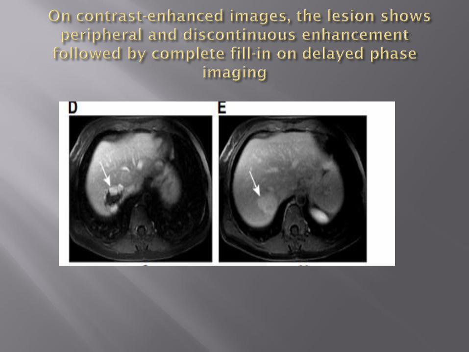
Upon US, the classic appearance of an haemangioma is that of a homogenous hyperechoic mass, measuring less than 3 cm in diameter with acoustic enhancement and sharp margins. CE examinations (CEUS, CT or MRI) are required when US is atypical. They show peripheral and globular enhancement of the lesion followed by a central enhancement on delayed phases

MRI is the key imaging modality in liver haemangiomas and also shows typical findings on pre-contrast imaging (hypointense on T1-weighted sequences and strongly hyperintense on heavily T2-weighted sequences) .
. Haemangiomas, especially those with high-flow, might show atypical features using gadoxetic acid (hepatobiliary MR contrast agents) – with relatively low signal intensity relative to the surrounding normal liver parenchyma during the equilibrium (3 min delay) phase.
This pseudo washout can mimic hypervascular hepatic tumours. However, they can be diagnosed by observing very strong signal intensity on T2-weighted imaging and enhancement on arterial phase-dominant imaging .

The two most common imaging atypias correspond to rapidly filling haemangiomas and giant haemangiomas. Both types of heamangioma are easily diagnosed on MRI The diagnosis of rapidly filling haemangioma is based on strong hyper intensity on T2-weighted images, the enhancement concomitant with that of arterial structures, and the persistent enhancement on delayed phase imaging.
Occasionally haemangiomas are cystic, pedunculated, have a fluid-fluid level or are associated with capsular retraction. In these very rare situations, imaging, including MRI, is less reliable. MRI has the highest sensitivity and specificity for diagnosing liver haemangiomas with values over 90% .
When the diagnosis cannot be achieved with imaging, percutaneous biopsy may be required. Provided that a cuff of normal hepatic parenchyma is interposed between the capsule and the margin of haemangioma, needle biopsy is not contraindicated and allows a diagnosis with an overall accuracy of 96% .

Haemangiomas are most often asymptomatic incidental discoveries that may change in size during long term follow-up .There is no relationship between the size of haemangiomas and complications, with little relationship between symptoms and characteristics of haemangiomas. Whether patients with large lesions, or lesions with mild symptoms, benefit from surgery is debatable.
. No randomized trials are available showing a superior effect of resection as compared to conservative treatment .For the majority of patients, a conservative approach is appropriate. Pregnancy and the use of OCPs are not contraindicated in the presence of stable asymptomatic haemangioma. Incidental reports described the development of KMS during pregnancy in females with liver haemangiomas larger than 5 cm.

Symptomatic or giant haemangiomas are not common and affected individuals should be referred to a benign liver tumour MDT. Again, surgical resection is rarely indicated ,except in the presence of KMS .Transcatheter hepatic embolizationcan be considered to manage the KMS ,as can medical therapy with corticosteroids or vincristine .Rarely, for complicated, large or extensive unresectable tumours, liver transplantation may be indicated.
In patients with a normal or healthy liver, a hyperechoic lesion is very likely to be a liver haemangioma. With typical radiology (homogeneous hyperechoic, sharp margin, posterior enhancement, and absence of halo sign) in a lesion less than 3 cm, ultrasound is sufficient to establish the diagnosis (evidence level II-2, grade of recommendation 1)
In oncology patients or those with underlying liver disease, contrast enhanced imaging (CEUS, CT or MRI) is required (evidence level II-2, grade of recommendation 1).
The diagnosis by contrast enhanced imaging is based on a typical vascular profile characterized by peripheral and globular enhancement on arterial phase followed by a central enhancement on delayed phases. (evidence level II-2, grade of recommendation 1)
Due to its benign course, imaging follow-up is not required for typical haemangioma (evidence level II-2, grade of recommendation 1).
Pregnancy and oral contraceptives are not contraindicated (evidence level III; grade of recommendation 2).
Conservative management is appropriate for typical cases (evidence level II-2, grade of recommendation 1).
In the presence of Kasabach-Merrit syndrome, growing lesions or lesions symptomatic by compression - refer to benign liver tumour MDT (evidence level III, grade of recommendation 1).
FNH accounts for the second most frequent benign tumour of the liver. In unselected autopsy series there is an estimated prevalence of 0.4–3%, although this is reduced to 0.03% considering clinically relevant prevalence. . There is a marked female preponderance (up to 90%), with the average age at presentation between 35 and 50 years. In most cases FNH is solitary and smaller than 5 cm, although tumours may be larger.

FNH are multiple in 20–30% of cases and associated with liver haemangioma in 20% of cases .Association of FNH with hepatocellular adenomas (HCA) is less common (although conversely, FNH are relatively common in patients with established adenomas).
FNH is thought to represent a proliferative cell response to an aberrant dystrophic artery and may be associated with other conditions characterized by arterial damage, such as hereditary haemorrhagic telangiectasia or previously treated solid tumours in children . Pregnancy and OCPs have not been demonstrated to play a role in development or progression of FNH.
FNH is a polyclonal hepatocellular proliferation, considered as a hyperplastic reaction resulting from arterial malformation. This theory is strongly supported by the absence in FNH of somatic mutations.
. Compared to other neoplastic disorders, the size of FNH is stable over time in the vast majority of cases. Case series of FNH showing that in the vast majority of cases the lesions remain stable, also indicate that the majority are asymptomatic, and that complications are extremely rare.
FNH is typically a solitary wellcircumscribed, unencapsulated mass, showing a central fibrous scar, which contains dystrophic arterial vessels. Histologically, FNH is composed of benign-appearing hepatocytes arranged in nodules that are usually partially delineated by fibrous septa originating from the central scar. Several degrees of ductular proliferation and inflammatory cells may be observed in the fibrous septa. Besides the typical form, several atypical forms of FNH are recognized.
FNH without a central scar is the most common of these; mostly absent in lesions <3cm . FNH with significant steatosis are also recognized . Molecular analysis identified upregulation of extracellular matrix genes associated with activation of the transforming growth factor beta (TGF-b) signaling pathway and overexpression of Wnt/b-catenin target genes, including GLUL, coding for glutamine synthase.
Such b-catenin activation without b-catenin activating mutations results in a typical map-like pattern of glutamine synthase (GS) overexpression in the periphery of the nodules close to the vessels .This map-like pattern of GS expression is specific to FNH and GS immunohistochemical staining is commonly used to help for pathological diagnosis in difficult cases .


Multiple FNH may be observed in specific clinical context, especially in patients with underlying vascular liver diseases, such as Budd-Chiari syndrome, obliterative portal venopathy and congenital disorders, including hereditary haemorrhagic telangiectasia, portal vein agenesis.

Imaging features of FNH resemble closely to pathologic findings. On US, FNH is usually slightly hypo- or isoechoic and very rarely hyperechoic. Sometimes the lesion is only detected by visualization of a pseudocapsule, which is due to compression of the surrounding liver tissue or vessels. Typically, on colour doppler, central arteries have a spoke-wheel pattern. Regardless of the imaging modality.
FNH usually associates several findings: i) lesion homogeneity except the central scar, ii) slightly different from the adjacent liver on pre-contrast US, CT or MRI. strong and homogeneous enhancement on arterial phase CEUS, CT or MR with a central vascular supply, which becomes similar to adjacent liver on portal and delayed phases . central scar best seen on MRI (hypointense on pre-contrast T1-weighted images, strongly hyperintense on T2-weighted images, and becoming hyperintense on delayed phase using extracellular MR contrast agents because of the accumulation of contrast material in the fibrous tissue .
The diagnosis of FNH is based on a combination of these imaging features but none of them is completely specific to FNH. On diffusion weighted MRI, FNH may appear hyperintense on high b-values corresponding to mild diffusion restriction. Nevertheless, ADC values are usually close to that of the liver.
MRI has the highest sensitivity compared to ultrasound and CT and a specificity of almost 100% for the diagnosis of FNH. Yet, its sensitivity is lower (70–80%) especially in small FNHs where central scar is often missing. When all features are not met, combination of CEUS and MRI yields the highest diagnostic accuracy . CEUS is more accurate than MRI in FNH smaller than 3 cm whereas the opposite is true in larger FNH .
Hepatobiliary MR contrast agents can be used to highlight the hepatocellular origin of the lesions. Most FNHs are iso-or hyperintense on hepatobiliary phase, some having rim-accentuated enhancement .With hepatobiliary MR contrast agents, the sensitivity for diagnosing FNH has increased up to 90%.
Among the atypias seen in FNH, one of the most common ones is the steatotic FNH, which can mimic HCA. Steatotic FNH seems to be more often observed in patients with liver steatosis. The diagnosis of steatotic FNH can be reached on imaging with very high specificity as long as all typical imaging findings are seen in the lesion .Other atypical findings include strong hyperintensity on T2-weighted imaging, pseudocapsule that can mimic true capsule, and washout. In atypical cases on imaging, liver biopsy is indicated.
There is insufficient evidence to support or refute elective surgery for FNH .but in the absence of symptoms and given the rarity of complications, a conservative approach is recommended. There is a poor correlation between FNH and symptoms and therefore even in the case of symptoms, treatment is rarely indicated. Treatment is only pursued in exceptional cases (e.g., pedunculated, expanding, exophytic) and resection is the treatment of choice. Non-surgical treatments should be reserved for those unfit for resection.
Where the diagnosis is firm and the individual asymptomatic, follow-up imaging is not required and the patients can be discharged, as summarized in Fig. 3. There is no indication for discontinuing OCPs and follow-up during pregnancy is not necessary. If the diagnosis of FNH is not firmly established on imaging, or the individual is symptomatic (relating to pain or compression), the patient should be referred to a benign liver MDT.
CEUS, CT, or MRI can diagnose FNH with nearly 100% specificity when typical imaging features are seen in combination (evidence level II-2, grade of recommendation 1)
MRI has the highest diagnostic performance overall. The highest diagnostic accuracy by CEUS is achieved in FNH less than 3 cm (evidence level II-2, grade of recommendation 1)
For a lesion typical of FNH follow-up is not necessary, unless there is underlying vascular liver disease (evidence level III, grade of recommendation 2)
Treatment is not recommended (evidence level II-3, grade of recommendation 2)
If imaging is atypical, or the patients is symptomatic, refer to a benign liver tumour MDT (evidence level III, grade of recommendation 1)
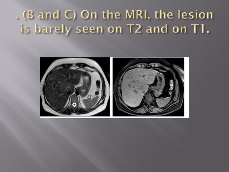


Incidence and prevalence data for HCA are not well established, although the reported prevalence is between 0.001 and 0.004% .HCA is approximately 10 times less common than FNH and is frequently diagnosed in women age 35–40 years, with a reported female:male ratio of 10:1. Several studies have supported the potential role of sex hormones in the development of HCA. A 30–40 fold increase in the incidence of HCA has been assumed in long term users of OCPs.
Notably, the incidence of HCA has increased in males associated with the increase in the use of anabolic substances related to sport or after the use of anabolic androgenic steroids by body builders .HCA are associated with the use of androgenic steroid therapy for aplastic anemia or paroxysmal nocturnal hemoglobinuria .]. There are sporadic case reports of HCA in patients with elevated levels of endogenously produced androgens or sex hormone imbalance (e.g., polycystic ovary, Klinefelter syndrome)
The recent increase in the HCA prevalence is noticeably associated with the rising prevalence of obesity and the metabolic syndrome .
HCAs encompass various types of clonal benign hepatocellular proliferations including several molecular subgroups. These are associated with specific morphological features and significant risks of complications, mostly haemorrhage and malignant transformation [104,105]. HCA are usually solitary, sometimes pedunculated and of various size. The size ranges from several millimeters to 30 cm
Large subcapsular vessels are commonly found on macroscopic examination. On cut sections, the tumour is well-delineated, sometimes encapsulated, of fleshy appearance ranging in colour from white to brown. HCA may display heterogeneous areas of necrosis and/or haemorrhage. Histologically, HCA consist of a proliferation of benign hepatocytes arranged in a trabecular pattern. Small thin vessels are usually found throughout the tumour
Unlike other benign liver lesions, HCA have the potential for haemorrhage and malignant transformation . In nearly all cases of spontaneous rupture or haemorrhage the lesion is >5 cm.

transformation is relatively rare, but is more common in HCA with activating mutations in b-catenin .while HNF-1a mutated HCA rarely undergo malignant transformation . In practical terms, the course of HCA diagnosed in women is more often benign, while HCA diagnosed in men have a significantly higher incidence of malignant transformation .which at least partly reflects the differences in molecular subtypes in men and women
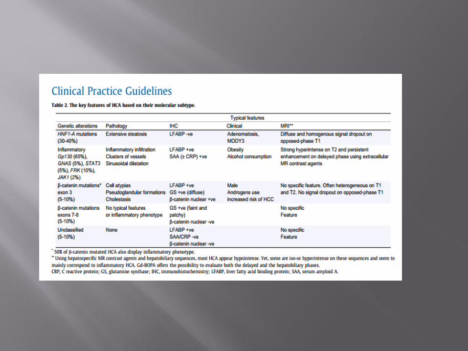
1. HCA inactivated for HNF-1a (H-HCA), accounting for 30 to 40% of HCA. H-HCA are defined by inactivation of HNF-1a, a transcription factor involved in hepatocyte differentiation and metabolism control .In H-HCA, HNF-1a mutations are somatic in most of cases, while germline mutations are observed in patients with adenomatosis and MODY3 diabetes, potentially in familial context .
Morphologically, H-HCAs are characterized by prominent steatosis [104], usually of marked intensity. However, steatosis may be mild in some H-HCA and significant in other subgroups of H-HCA, especially in inflammatory ones (I-HCA). The hallmark of H-HCA is the absence of expression in tumour hepatocytes of genes controlled by HNF-1a, among them, liver fatty acid binding protein (LFABP), which is in contrast highly expressed in non-tumour hepatocytes
2. Inflammatory Adenomas (I-HCA), accounting for 40 to 55% of HCA. I-HCAs represent a heterogeneous subgroup of HCA regarding the variety of gene mutations, although all described mutations result in the activation of the JAK/STAT pathway .Indeed, mutations of gp130 (IL6ST), FRK, STAT3, GNAS and JAK1 have been identified in around 65%, 10%, 5%, 5% and 2% of I-HCA, respectively
Almost all of these mutations are mutually exclusive. I-HCA are more often observed in patients with obesity and/or metabolic syndrome, as well as in the context of a high alcohol consumption. Systemic inflammatory syndrome, demonstrated by increased serum C-reactive protein (CRP) and fibrinogen levels, can regress following HCA removal. Morphologically, I-HCA, initially described as ‘‘telangiectatic form of FNH”, further reclassified as ‘‘telangiectatic HCA”, are characterized by the presence of clusters of small arteries surrounded by extracellular matrix and inflammatory infiltrates associated with foci of sinusoidal dilatation .
By immunohistochemistry, tumour hepatocytes exhibit cytoplasmic expression of serum amyloid A (SAA) and CRP, two proteins of the acute phase of inflammation induced by STAT3 activation. CRP immunostaining appears to be more sensitive but less specific, since non-tumour hepatocytes may be positive in the adjacent normal liver counterpart. As previously mentioned, I-HCA may show some degree of steatosis and also features of b-HCA related to additional b-catenin mutations.
3. b-catenin activated HCA (b-HCA), accounting for 10 to 20% of HCA. b-HCA are defined by b-catenin activation within the tumours. Mutations of the b-catenin gene (CTNNB1) were initially localized at hot spots in exon 3, and more recently in exons 7 and 8 [
While b-catenin mutations are exclusive of HNF-1a mutations, they can be combined with a JAK/STAT activating mutation defining the subgroup of I-HCA; and up to 50% of b-HCA are also inflammatory .b-HCA are over represented in males and display a higher risk of malignant transformation towards hepatocellular carcinoma (HCC).
Morphologically, b-HCAs are characterized by the presence of cellular atypias, pseudoglandular formations and cholestasis. Tumoural hepatocytes show a specific immunophenotypical profile including diffuse, usually strong, GS positivity (a b-catenin target) as well as a nuclear expression of b-catenin. Although both markers have a very good specificity for b-catenin mutations, their sensitivity is insuffi- cient, especially for b-catenin expression as a biomarker, since very few nuclei may be b-catenin positive
4. Unclassified HCA, accounting for 5% to 10% of HCA. A small subset of HCA do not display any specific morphological features nor do they have any of the gene mutations previously described.
HCA molecular classification has markedly contributed to the understanding of the oncogenic pathways involved in liver tumorigenesis. While the size of HCA, with the accepted clinically relevant size cut off of 5 cm correlating with the risk of complications – both haemorrhage and HCC development – the molecular subtyping is highly associated with the risk of malignant transformation into HCC. Among the different subgroups, b-HCAs exhibit the highest risk for malignancy, including those with dual b-catenin and inflammatory phenotype.
. As b-HCAs are enriched in male patients, this could at least in part explain the high risk of malignant transformation reported in men. Methods for the molecular analysis of HCA are not presently sensitive enough for widespread application.
On imaging, HCA is no longer a unique entity and imaging features reflect the tumour subtypes. As the most striking pathologic features are the presence of fat or telangiectatic component, imaging should be fat sensitive and should use contrast agents to look for dilated vascular spaces. CEUS, CT or MRI are able to detect the dilated vascular spaces. On CEUS, HCA usually shows homogeneous contrast enhancement in the arterial phase, typically with rapid complete centripetal filling.

In the early portal venous phase, it usually becomes isoechoic or, more rarely, remains slightly hyperechoic. CEUS can differentiate HCA from FNH because of the absence of the central spoke-wheel pattern in HCA, but is not sufficiently accurate to subtype HCA .
HNF-1a inactivated HCAs are characterized by the presence of marked steatosis on pathology. They appear homogeneous on MRI and have a variable signal on T2-sequences: usually slightly hyperintense on non-fat suppressed sequence and iso-or hypointense on fat suppressed T2-weighted sequence.

They are usually moderately hypervascular and often show washout on portal and/or delayed phase using extracellular MR contrast agents. On high b-values diffusion-weighted MRI, they are iso-or moderately hyperintense.
The two referenced series have included only hepatocellular adenomas with 50 and 44 cases, respectively. Inflammatory HCAs are characterized on MRI by their telangiectatic features. They show a strong hyperintense signal on T2-weighted images (as strong as the signal of the spleen), which may be either diffuse or as a rim-like band in the periphery of the lesion and defines the atoll sign
As HCAs have the potential for haemorrhage and malignant transformation .
On baseline diagnostic imaging the size of an HCA is important to note, as is an exophytic characteristic if it is present, given the associations of haemorrhage with size >5 cm and exophytic protrusion
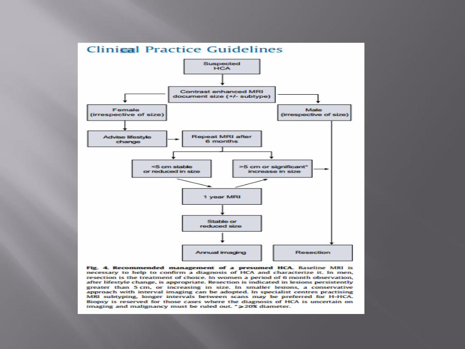
Irrespective of size, however, resection or curative treatment is recommended for all HCA diagnosed in men because of a significantly higher incidence of malignant transformation .HCA in women that are less than 5 cm on the baseline scan rarely rupture and malignant transformation less common. In women, lifestyle change is recommended and should include the discontinuation of OCPs and control of body weight.
For all presumed HCA, a reassessment with CE-MRI is advised after 6 months. HCA persistently greater than 5 cm, or increasing in size >20% diameter – should be considered for resection or curative treatment – irrespective of their molecular or histological subtype – because of the risk of haemorrhage.
Biopsy may be considered within a benign liver tumour MDT to exclude malignancy. In the case of tissue availability obtained for diagnostic purpose, curative intervention is advised for the activated b-catenin mutated HCA, irrespective of size.
. Follow-up imaging 6 monthly to establish growth patterns and monitor for malignant transformation is advisable. There is no robust data on the timeline to define stable disease. For lesions stable after 12 months, annual followup is acceptable. US is cost effective and may be preferred in well seen lesions. For lesions stable or reducing in size after 5 years, biannual imaging can be proposed
The recommended first line therapy is resection of larger (>5 cm) or growing lesions, aiming to remove the whole tumour and all risk of malignant transformation. Non-surgical modalities, such as embolization for larger lesions or ablation for smaller lesions can be pursued as an alternative to resection, but this would only be the treatment of choice in poor surgical candidates. For smaller indeterminate lesions, ablation without confirmation of diagnosis is not recommended. In these cases, biopsy should be considered.

. Small foci of haemorrhage within HCA are often observed and are not an indication for clinical intervention (case series, evidence level 4). If clinically evident haemorrhage occurs, admission for close observation and CE-CT scan is appropriate. In cases of major haemorrhage, resuscitation with blood products and transfer to a centre where embolization can be performed to control active bleeding is appropriate . Further investigation once stable should be pursued to exclude malignancy and secure appropriate follow-up.
HCA in a pregnant woman requires close follow-up using frequent US (every 6–12 weeks) to monitor size.

embolization can be considered. Prior to 24 weeks, surgery maybe preferred, especially for smaller resections located at the periphery of the liver anatomy, as the ionizing radiation exposure and the use of intravenous contrast agents associated with radiologically guided transarterial embolization may be harmful to the fetus
MRI is superior to all other imaging modalities and due to its intrinsic properties to detect fat and vascular spaces it offers an opportunity to subtype HCA up to 80% (evidence level II-2, grade of recommendation 1) •
The positive identification of HNF-1α HCA or inflammatory HCA is achievable with MRI with >90% specificity. By contrast, identification of β-catenin activated HCA and its distinction with unclassified HCA and hepatocellular carcinoma is not possible by any imaging technique (evidence level II-2, grade of recommendation 1)
Treatment decisions are based on gender, size and pattern of progression (evidence level III, grade of recommendation 2)
• Upon HCA diagnosis, lifestyle changes such as discontinuation of OCP as well as weight loss should be advised (evidence level II-2, grade of recommendation 1)
HCA resection is recommended irrespective of size in men and in any instance of proven β-catenin mutation (evidence level II-3, grade of recommendation 2)
• In women, a period of 6 months observation after lifestyle change is advised and resection is indicated for nodules equal or greater than 5 cm and those continuing to grow (evidence level II-3, grade of recommendation 2)
In women, lesions less than 5 cm should be reassessed at 1 year, and annual imaging adopted thereafter (evidence level III, grade of recommendation 2)
• A bleeding HCA with haemodynamic instability should be embolized and residual viable lesion on follow-up imaging is an indication for resection (evidence level III, grade of recommendation 2)
In retrospectively collected surgical series of patients, HCA presented as multinodular disease in up to a half all patients, was noted to be more frequent in OCP users and those with features of the metabolic syndrome, while being exceptionally rare in men .
. The term liver adenomatosis, that in the past meant the presence of more than 10 HCAs (case series, evidence level 4), has now been replaced with the term multiple HCAs – recognizing the fact that precise counting of HCA by imaging can be challenging. In patients with widespread HCAs involving both lobes, microscopic adenomatous foci escaping radiological detection have been found in up to 20% of the resected livers
The clinical presentation and risk of bleeding and malignant transformation in patients with multiple HCAs do not differ from those in patients with a single HCA, being driven by the size of the largest nodule, rather than the number of nodules [89,110]. Regression of tumour burden has been reported to occur in up to a third of patients complying with lifestyle changes – such as withdrawal from OCPs or weight reduction, while progression of HCA is associated with obesity
With these two things in mind, we recommend the management of patients with multiple HCA should be based on the size of the largest tumour.
Those individuals with unilobular disease can be treated with hepatic resection. For those with more widespread HCA, resection of the largest adenomas may be an option [137]. Because it often is impossible to resect all tumours in patients with multiple HCAs, liver transplantation has been proposed, but should only be considered in patients with more than 10 lesions and underlying liver disease
The management of patients with multiple HCA should be based on the size of the largest tumour (evidence level III, grade of recommendation 2) • Hepatic resection might be considered in unilobular disease, and in those cases with more widespread HCA, resection of the largest adenomas may be an option (evidence level III, grade of recommendation 2) • Liver transplantation is not recommended in multiple HCA, but might be considered in individuals with underlying liver disease (evidence level III, grade of recommendation 2)


![EASL Clinical Practical Guidelines: Management of ... · PDF fileEASL Clinical Practical Guidelines: Management of Alcoholic ... and 15.2 for primary biliary cirrhosis [1]. ... trend](https://img.pdfslide.net/doc/110x75/5a9d93557f8b9a21688c86a1/easl-clinical-practical-guidelines-management-of-clinical-practical-guidelines.jpg)