Embed Size (px)
Citation preview

neonatalINTENSIVE CARE
Vol. 29 No. 1Winter 2016

888-825-9640
NeoPAP® infant respiratory support system
Simple designEasy to useEffective delivery NeoPAP’s innovative design is literally a breeze... · CPAP, high flow and resuscitation modes are built into one easy to use device, improving workflow efficiency and reducing costs.· Baby-Trak® leak compensation and sophisticated alarms give you peace of mind and the ability to focus on the specialized needs of your RDS patients.· The unique bonnet and patient-friendly interface support developmental care and may help reduce costly skin damage.
See what NeoPAP can do for your NICU... Learn more at www.circadiance.com or contact us at [email protected]
www.circadiance.com101463 REV 1
Who knows better what ventilatory support a patient needs
than the patient? The new SERVO-n® neonatal ventilator, with
NAVA® (Neurally Adjusted Ventilatory Assist) and non-invasive NAVA
standard*, lets the baby’s own physiological signal control the exact
timing and amount of assist for every breath. This same signal also
provides the clinician insight into the baby’s breathing drive for
diagnostics and weaning.
Give your neonatal patient the support they need to breathe easier,
sleep better and grow stronger with SERVO-n and NAVA®.
Baby knows best
Maq
uet
is a
reg
iste
red
tra
dem
ark
of M
aque
t G
mb
H •
Cop
yrig
ht M
aque
t M
edic
al S
yste
ms
US
A o
r its
aff
iliat
es. •
C
AU
TIO
N: F
eder
al
(US
) law
res
tric
ts t
his
dev
ice
to s
ale
by
or o
n th
e or
der
of a
phy
sici
an. R
efer
to
Inst
ruct
ions
for
Use
for
curr
ent
ind
icat
ions
, war
ning
s,
cont
rain
dic
atio
ns, a
nd p
reca
utio
ns. M
CV
0003
9355
RE
VA
www.maquetusa.comAt NEO 2016, visit Maquet Booth #400
*Excludes Edi module.
NIC Feb 2016 SERVO-n Ad MCV00039355 REVA.indd 1 1/25/16 4:37 PM

Arie L. Alkalay, MDClinical Professor of PediatricsDavid Geffen School of MedicinePediatrician, Cedars-SinaiLos Angeles, CA
M. A. Arif, MDProfessor of Pediatrics & Head, NeonatologyNational Institutes of Child HealthKarachi, Pakistan
Muhammad Aslam, MDAssociate Professor of PediatricsUniversity of California, IrvineNeonatologist, UC Irvine Medical CenterOrange, California
Edward Austin, MDAustin-Hernandez Family Medical CenterCompton, CA
Richard L. Auten, MDAssistant Professor of PediatricsDuke University Medical CenterDurham, NC
Bruce G. Bateman, MDDepartment of Obstetrics & GynecologyUniversity of VirginiaCharlottesville, VA
Sandy Beauman, MSN, RNC-NICCNC ConsultingAlbuquerque, NM
David D. Berry, MDWake Forest University School of MedicineWinston-Salem, NC
Melissa K. Brown, BS, RRT-NPS, RCPFaculty, Respiratory Therapy ProgramGrossmont CollegeEl Cajon, CA
D. Spencer Brudno, MDAssociate Professor of PediatricsMedical Director, Pediatric TherapyMedical College of GeorgiaAugusta, GA
Curtis D. Caldwell, NNPUNM School of Medicine, Dept of PediatricsAlbuquerque, NM
Ed Coombs, MA RRT-NPS, ACCS, FAARCMarketing Director – Intensive CareKey Application Field Manager – Respiratory Care, Draeger MedicalTelford, PA
Jonathan Cronin, MDAssistant Professor of PediatricsHarvard Medical School ChiefNeonatology and Newborn Medicine UnitDepartment of Pediatrics Massachusetts General Hospital for ChildrenBoston, MA
Michael P. Czervinske, RRTNeonatal and Pediatric Critical CareUniversity of Kansas Medical CenterKansas City, KS
Professor Adekunle H. DawoduDirector, International Patient Care and Education, Cincinnati Children’s HospitalCincinnati, OH
Jayant Deodhar, MDAssociate Professor of Clinical PediatricsChildren’s Hospital CenterCincinnati, OH
Leonard Eisenfeld, MDAssociate Professor of PediatricsUniversity of Connecticut School of MedicineDivision of NeonatologyConnecticut Children’s Medical CenterHartford, CT
Sami Elhassani, MDNeonatologistSpartanburg, SC
Ivan Frantz, III, MDChariman of Department of PediatricsChief, Division of Newborn MedicineTufts University School of MedicineBoston, MA
Philippe S. Friedlich, MDAssociate Professor of Clinical PediatricsChildren’s Hospital of Los AngelesLos Angeles, CA
G. Paolo Gancia, MDNeonatologist, Terapia IntensivaNeonatale-NeonatologiaCuneo, Italy
George A. Gregory, MDProfessor of Pediatrics and AnesthesiaUniversity of CaliforniaSan Francisco, CA
Charles J. Gutierrez, PhD, RRT, FAARCNeurorespiratory Clinical Specialist, J.A. Haley VA Hospital and Assistant Professor, Pulmonary, Critical Care & Sleep Medicine,Morsani College of Medicine, University of South Florida, Tampa, FL
William R. Halliburton, RRT, RCPNeonatal Respiratory Care CoordinatorDepartment of Respiratory CareHillcrest Baptist Medical CenterWaco, TX
Mary Catherine Harris, MDAssociate Professor of PediatricsDivision of NeonatologyUniversity of Pennsylvania School of MedicineThe Children’s Hospital of PhiladelphiaPhiladelphia, PA
David J. Hoffman, MDClinical Associate Professor of PediatricsPenn State College of MedicineStaff NeonatologistThe Reading Hospital and Medical CenterWest Reading, PA
Michael R. Jackson, RRTNewborn Intensive Care Unit Beth Israel Hospital Boston, MA
Chang-Ryul Kim, MDAssociate Professor of PediatricsCollege of MedicineHanyang University Kuri HospitalSeoul, South Korea
David M. Kissin, BS, RRTPerinatal/Pediatric SpecialistMaine Medical Center, Portiand, ME
Sheldon Korones, MDDirector of Newborn CenterCollege of Medicine, Memphis, TN
Scott E. Leonard, MBA, BA, RRTDirector of Respiratory Therapy, EEG, NeurophysiologyGeorge Washington University HospitalWashington, DC
Raymond Malloy, MHA, RRTDirector of Pulmonary CareThomas Jefferson University HospitalPhiladelphia, PA
Paul J. Mathews, PhD, RRT, FCCM, FCCP, FAARCAssociate Professor of Respiratory CareUniversity of Kansas Medical CenterKansas City, KS
William Meadow, MDProfessor of PediatricsCo-Section Chief, NeonatologyComer Children’s HospitalThe University of ChicagoChicago, IL
David G. Oelberg, MDCenter for Pediatric ResearchEastern Virginia Medical SchoolChildren’s Hospital of The King’s DaughtersNorfolk, VA
Rahmi Ors, MDDirector, Department of Neonatology and PediatricsProfessor of Pediatrics and NeonatologistMeram Medical FacultyNecmettin Erbakan UniversityKonya, Turkey
T. Michael O’Shea, MD, MPHChief, Neonatology DivisionWake Forest University School of MedicineWinston-Salem, NC
Lisa Pappas, RRT-NPSRespiratory Clinical Coordinator NICUUniversity of Utah HospitalSalt Lake City, UT
G. Battisita Parigi, MDAssociate Professor of Pediatric SurgeryUniversity of Pavia, Italy
Richard Paul, MDChief, Maternal & Fetal MedicineDepartment of Obstetrics & GynecologyUniversity of Southern CaliforniaLos Angeles, CA
Max Perlman, MDProfessor of PediatricsThe Hospital for Sick ChildrenToronto, Ontario, Canada
Boris Petrikovsky, MDDirector, Prenatal Diagnostic Unit ServicesNew York Downtown HospitalNew York, NY
Arun Pramanik, MDProfessor of PediatricsDirector of Neonatal FellowshipLouisiana State UniversityHealth Sciences Center, Shreveport, LA
Benamanahalli K. Rajegowda, MDChief of NeonatologyLincoln Medical and Mental Health CenterProfessor of Clinical PediatricsWeill Medical College of Cornell University, NY
Koravangattu Sankaran, FRCP(C), FAAP, FCCMProfessor of Pediatrics and Director of Neonatology and Neonatal ResearchDepartment of PediatricsRoyal University HospitalUniversity of SaskatchewanSaskatoon, Saskatchewan, Canada
Istvan Seri, MD, PhDProfessor of PediatricsHead, USC Division of Neonatal MedicineUniversity of Southern California, Los Angeles, CA
Tushar A. Shah, MD, MPHDivision of Neonatology Cincinnati Children’s Hospital Medical Center Cincinnati, OH
Dave Swift, RRTOttawa Hospital – Civic SiteCampus Coordinator (Professional Practice) & Special Care Nursery Charge TherapistRespiratory Therapy Team LeadNational Office of the Health Care Emergency Response Team (NOHERT)Subject Matter Expert, Health Canada
Jack TannerNICU Clinical CoordinatorU Mass Memorial HospitalWorcester, MA
Otwell D. Timmons, MDCarolinas Medical CenterCharlotte, NC
Maya Vazirani, MD, FAAPBoard Certified Neonatology and Pediatrics, Lancaster, CA
Max Vento, MDAssociate Professor of PediatricsChief, Pediatric ServicesNeonatologia Hospital Virgin del ConsueloValencia, Spain
Dharmapuri Vidyasagar, MDProfessor of Pediatrics Department of PediatricsUniversity of IllinoisChicago, IL
Editorial Advisory Board
4 neonatal INTENSIVE CARE Vol. 29 No. 1 n Winter 2016
Table of Contents
Departments
6 News
18 Company Profile
articles
19 Interview on Newborn Hearing Testing
22 Interview on Modified Seldinger Technique
23 Interview on Neonatal Treatment of Neonatal Encephalopathy
24 Interview on Safer Ways for Babies to Sleep
26 Identifying Hemolytic Jaundice
28 Late Group B Streptococcus Infection
31 NICU Open Ward vs Single Room
36 Quieting the NICU
40 Preterm Infant’s Nutritive Sucking
46 Breastfeeding in the NICU
49 Comparing Nebulizer Brand
52 Fetal Heart Vestment Monitor Research
57 Pulse Oximeters in the NICU
62 Observational Study Using Camera Surveillance At A Tertiary Hospital
Vol. 29 No. 1Winter 2016
neonatalINTENSIVE CARE
Vol. 29 No. 1Winter 2016

Prolact+H2MF®
Human Milk-Based Human Milk Fortifier Products
Prolact CR™
Human Milk Caloric Fortifier
Prolact RTF™
Human Milk-Based Premature Infant Formula
PremieLact™ Prolact HM™
Standardized Donor Milk Products
To provide your preterm patient with
a 100% human milk-based diet, call:
1-888-PROLACT (1-888-776-5228)
www.prolacta.com
The American Academy of Pediatrics’ (AAP) policy recommends the use of human milk for all preterm infants, whether mother’s own milk (MOM) or pasteurized donor human milk when mother’s own milk is unavailable.1
Only Prolacta Bioscience, the leader in the science of human milk, provides: • A full line of human milk-based nutrition for premature infants • Human milk products that undergo the most rigorous testing and screening in the industry
Human milk makes all the difference
1. American Academy of Pediatrics. Breastfeeding and the Use of Human Milk. Section on Breastfeeding. [originally published online February 27, 2012]. Pediatrics. DOI: 10.1542/peds.2011-3552
COPYRIGHT ©2014 PROLACTA BIOSCIENCE, INC. ALL RIGHTS RESERVED. MKT-0299 REV-0 4/14
MKT-0299_Rev-0_HumanMilkAd_NeonatalIntensiveCareMag_M1.indd 1 12/22/14 4:02 PM

6 neonatal INTENSIVE CARE Vol. 29 No. 1 n Winter 2016
change, the group’s cesarean delivery rate had been increasing 0.6% per year, which is comparable to national trends. The rate among publicly insured women did not change significantly during the study. In addition, vaginal births after cesarean (VBAC) increased for the privately insured women, going from 13.3% under the previous care model to 22.4% with midwife/laborist care (P = .002). In the years after the change, the rate increased by 8% annually. Publicly insured women did not have a significant change in their VBAC rate. There were no significant differences in adverse outcomes for babies, including Apgar scores below 7, umbilical artery pH less than 7.0, or umbilical artery base excess greater than 12 (composite score, 1.3% before the change vs 2.3% after; P = .07).
complications Decline, survival increasedComplications have decreased and survival has improved for extremely preterm infants born at US academic centers during the last 20 years, according to a new study from the Department of Pediatrics, Emory University School of Medicine, Children’s Healthcare of Atlanta in Georgia. Survival increased most markedly for infants born at 23 and 24 weeks’ gestation and survival without major morbidity increased for infants aged 25 to 28 weeks. These findings may be valuable in counseling families and developing novel interventions, said the authors wrote, who reviewed 20-year trends in maternal/neonatal care, complications, and deaths among extremely preterm infants born at 26 Neonatal Research Network Centers between 1993 and 2012. The researchers analyzed data from a prospective registry of 34,636 infants of 22 to 28 weeks’ gestation, weighing 401 to 1500 g at birth. The study’s main outcomes were maternal/neonatal care, morbidities, and survival. The primary morbidities reported for infants who lived for more than 12 hours were severe necrotizing enterocolitis, infection, bronchopulmonary dysplasia, severe intracranial hemorrhage, cystic periventricular leukomalacia, and/or severe retinopathy of prematurity. The investigators used regression models to assess yearly changes and adjusted for study center, race/ethnicity, gestational age, birth weight for gestational age, and sex.
company adapts to plug leaksPassy-Muir, the leading manufacturer of the No Leak speaking valve, is introducing two adapters to provide clinicians with
cesarean Delivery rates DroppedCesarean delivery rates dropped from 31.7% to 25.0% (P = .005) after Marin General Hospital in California switched from a private practice model to one with 24-hour laborist and midwife coverage. Rates of vaginal birth after cesarean (VBAC) also increased. The laborists at this hospital were obstetricians who provided in-house labor and delivery coverage without competing clinical duties. Previous studies have suggested that laborists achieve lower cesarean rates by being more tolerant of changes in fetal heart rate and by having less competition between managing labor and other duties. At Marin General Hospital, women insured by the state’s Medicaid program served as the control group; most received care from midwives with 24-hour laborist backup. The study covered a period before and after 2011, when most privately insured women were switched to the same type of coverage. Previously, these women’s care came from a private physician who managed labor remotely. After the change, women could request that their obstetrician be present to deliver the baby, but their labor would still be managed by the in-house laborist. Between 2005 and 2014, there were 13,194 births at the hospital. The study included 3560 births from nulliparous women with singleton vertex term babies, and 1324 births from women with a history of cesarean delivery; approximately half of each were in the private insurance group. The new model of care led to a “clinically and statistically significant” decrease in primary cesareans (adjusted odds ratio [OR] 0.56, 95% confidence interval [CI], 0.39 - 0.81). Before the
News Winter 2016
ISSN 1062-2454Published five times each year by
Goldstein and Associates, Inc.10940 Wilshire Blvd., Suite 600Los Angeles CA 90024Phone: 310-443-4109Fax: 310-443-4110E-mail: [email protected]: www.nicmag.ca
Publisher/Editor in ChiefSteve Goldstein
Managing EditorChristopher Hiscox
Senior EditorVincent Terrier
News EditorChris Campbell
Associate EditorJordana Hammeke, Susan Goldstein
Circulation, Coverage, Advertising Rates: Complete details regarding circulation, coverage, advertising rates, space sizes, and similar information are available to prospective advertisers. Closing date is 45 days preceding date of issue.
Change of Address: Notices should be sent promptly to Circulation Department.
Provide old mailing label as well as new address; include zip code or postal code. Allow two months for change.
Editorial Contributions may be sent by e-mail and will be handled with reasonable care: however, publishers assume no responsibility for safety of art work, photographs, or manuscripts. Every precaution is taken to ensure accuracy, but the publishers cannot accept responsibility for the correctness or accuracy of informa tion supplied herein or for any opinion expressed. Editorial closing date is the first day of the month preceding month of issue.
©2016 by Goldstein & Associates, Inc. All rights reserved. Reproduction in whole or in part without written permission is strictly prohibited.

In term and near-term neonates with hypoxic respiratory failure (HRF)…
When do you stop the cascade?Early intervention with INOMAX® (nitric oxide) for inhalation upon confi rmation of pulmonary hypertension may help:
• Avoid higher levels of supplemental oxygen
• Improve oxygenation1
• Potentially prevent the progression of HRF2
Learn more at www.inomax.com
IndicationINOMAX® is a vasodilator, which, in conjunction with ventilatory support and other appropriate agents, is indicated for the treatment of term and near-term (>34 weeks) neonates with hypoxic respiratory failure associated with clinical or echocardiographic evidence of pulmonary hypertension, where it improves oxygenation and reduces the need for extracorporeal membrane oxygenation.
Utilize additional therapies to maximize oxygen delivery with validated ventilation systems.
Important Safety Information• INOMAX is contraindicated in the treatment of neonates known to be dependent on right-to-left
shunting of blood.• Abrupt discontinuation of INOMAX may lead to increasing pulmonary artery pressure and
worsening oxygenation even in neonates with no apparent response to nitric oxide for inhalation.• Methemoglobinemia and NO2 levels are dose dependent. Nitric oxide donor compounds may have
an additive e� ect with INOMAX on the risk of developing methemoglobinemia. Nitrogen dioxide may cause airway infl ammation and damage to lung tissues.
• In patients with pre-existing left ventricular dysfunction, INOMAX may increase pulmonary capillary wedge pressure leading to pulmonary edema.
• Monitor for PaO2, methemoglobin, and inspired NO2 during INOMAX administration.• Use only with an INOmax DSIR
®, INOmax® DS, or INOvent® operated by trained personnel.
Please see Brief Summary of Prescribing Information on adjacent page.
References: 1. INOMAX [package insert]. Hampton, NJ: Ikaria, Inc.; 2013. 2. González A, Fabres J, D’Apremont I, et al. Randomized controlled trial of early compared with delayed use of inhaled nitric oxide in newborns with a moderate respiratory failure and pulmonary hypertension. J Perinatol. 2010;30(6):420-424.
JOB NUMBER: 15-INO-0354
FILE NAME: 15-INO-0354_8.125x10.875_Nntl_InensvCr_07-17-15.indd
TRIM SIZE: 8.125 x 10.875
BLEED SIZE: .125” all sides
LIVE AREA: .5” all sides
COLOR SPACE: CMYK
DATE: 2015-07-17
PUB: Neonatal Intensive Care
Mallinckrodt, the “M” brand mark and the Mallinckrodt Pharmaceuticals logo are trademarks of a Mallinckrodt company. Other brands are trademarks of a Mallinckrodt company or their respective owners.
© 2015 Mallinckrodt. IMK111-1631-R3 July 2015 www.inomax.com

INOMAX®(nitric oxide) for inhalationBrief Summary of Prescribing Information
INDICATIONS AND USAGE
Treatment of Hypoxic Respiratory FailureINOMAX® is a vasodilator, which, in conjunction with ventilatory support and other appropriate agents, is indicated for the treatment of term and near-term (>34 weeks) neonates with hypoxic respiratory failure associated with clinical or echocardiographic evidence of pulmonary hypertension, where it improves oxygenation and reduces the need for extracorporeal membrane oxygenation.
Utilize additional therapies to maximize oxygen delivery with validated ventilation systems. In patients with collapsed alveoli, additional therapies might include surfactant and high-frequency oscillatory ventilation.
The safety and effectiveness of INOMAX have been established in a population receiving other therapies for hypoxic respiratory failure, including vasodilators, intravenous fluids, bicarbonate therapy, and mechanical ventilation. Different dose regimens for nitric oxide were used in the clinical studies.
Monitor for PaO2, methemoglobin, and inspired NO2 during INOMAX administration.
CONTRAINDICATIONSINOMAX is contraindicated in the treatment of neonates known to be dependent on right-to-left shunting of blood.
WARNINGS AND PRECAUTIONS
Rebound Pulmonary Hypertension Syndrome following Abrupt DiscontinuationWean from INOMAX. Abrupt discontinuation of INOMAX may lead to worsening oxygenation and increasing pulmonary artery pressure, i.e., Rebound Pulmonary Hypertension Syndrome. Signs and symptoms of Rebound Pulmonary Hypertension Syndrome include hypoxemia, systemic hypotension, bradycardia, and decreased cardiac output. If Rebound Pulmonary Hypertension occurs, reinstate INOMAX therapy immediately.
Hypoxemia from MethemoglobinemiaNitric oxide combines with hemoglobin to form methemoglobin, which does not transport oxygen. Methemoglobin levels increase with the dose of INOMAX; it can take 8 hours or more before steady-state methemoglobin levels are attained. Monitor methemoglobin and adjust the dose of INOMAX to optimize oxygenation.
If methemoglobin levels do not resolve with decrease in dose or discontinuation of INOMAX, additional therapy may be warranted to treat methemoglobinemia.
Airway Injury from Nitrogen DioxideNitrogen dioxide (NO2) forms in gas mixtures containing NO and O2. Nitrogen dioxide may cause airway inflammation and damage to lung tissues. If the concentration of NO2 in the breathing circuit exceeds 0.5 ppm, decrease the dose of INOMAX.
If there is an unexpected change in NO2 concentration, when measured in the breathing circuit, then the delivery system should be assessed in accordance with the Nitric Oxide Delivery System O&M Manual troubleshooting section, and the NO2 analyzer should be recalibrated. The dose of INOMAX and/or FiO2 should be adjusted as appropriate.
Heart FailurePatients with left ventricular dysfunction treated with INOMAX may experience pulmonary edema, increased pulmonary capillary wedge pressure, worsening of left ventricular dysfunction, systemic hypotension, bradycardia and cardiac arrest. Discontinue INOMAX while providing symptomatic care.
ADVERSE REACTIONSBecause clinical trials are conducted under widely varying conditions, adverse reaction rates observed in the clinical trials of a drug cannot be directly compared to rates in the clinical trials of another drug and may not reflect the rates observed in practice. The adverse reaction information from the clinical studies does, however, provide a basis for identifying the adverse events that appear to be related to drug use and for approximating rates.
Controlled studies have included 325 patients on INOMAX doses of 5 to 80 ppm and 251 patients on placebo. Total mortality in the pooled trials was 11% on placebo and 9% on INOMAX, a result adequate to exclude INOMAX mortality being more than 40% worse than placebo.
In both the NINOS and CINRGI studies, the duration of hospitalization was similar in INOMAX and placebo-treated groups.
From all controlled studies, at least 6 months of follow-up is available for 278 patients who received INOMAX and 212 patients who received placebo. Among these patients, there was no evidence of an adverse effect of treatment on the need for rehospitalization, special medical services, pulmonary disease, or neurological sequelae.
In the NINOS study, treatment groups were similar with respect to the incidence and severity of intracranial hemorrhage, Grade IV hemorrhage, periventricular leukomalacia, cerebral infarction, seizures requiring anticonvulsant therapy, pulmonary hemorrhage, or gastrointestinal hemorrhage.
In CINRGI, the only adverse reaction (>2% higher incidence on INOMAX than on placebo) was hypotension (14% vs. 11%).
Based upon post-marketing experience, accidental exposure to nitric oxide for inhalation in hospital staff has been associated with chest discomfort, dizziness, dry throat, dyspnea, and headache.
OVERDOSAGEOverdosage with INOMAX will be manifest by elevations in methemoglobin and pulmonary toxicities associated with inspired NO2. Elevated NO2 may cause acute lung injury. Elevations in methemoglobin reduce the oxygen delivery capacity of the circulation. In clinical studies, NO2 levels >3 ppm or methemoglobin levels >7% were treated by reducing the dose of, or discontinuing, INOMAX.
Methemoglobinemia that does not resolve after reduction or discontinuation of therapy can be treated with intravenous vitamin C, intravenous methylene blue, or blood transfusion, based upon the clinical situation.
DRUG INTERACTIONSNo formal drug-interaction studies have been performed, and a clinically significant interaction with other medications used in the treatment of hypoxic respiratory failure cannot be excluded based on the available data.INOMAX has been administered with dopamine, dobutamine, steroids, surfactant, and high-frequency ventilation. Although there are no study data to evaluate the possibility, nitric oxide donor compounds, including sodium nitroprusside and nitroglycerin, may have an additive effect with INOMAX on the risk of developing methemoglobinemia. An association between prilocaine and an increased risk of methemoglobinemia, particularly in infants, has specifically been described in a literature case report. This risk is present whether the drugs are administered as oral, parenteral, or topical formulations.
INOMAX® is a registered trademark of INO Therapeutics LLC, a Mallinckrodt Pharmaceuticals company.© 2015 Mallinckrodt. IMK111-01540 R1 July 2015
JOB NUMBER: 15-INO-0354
FILE NAME: 15-INO-0354_8.125x10.875_Nntl_InensvCr_07-17-15.indd
TRIM SIZE: 8.125 x 10.875
BLEED SIZE: .125” all sides
LIVE AREA: .5” all sides
COLOR SPACE: CMYK
DATE: 2015-07-17
PUB: Neonatal Intensive Care

neonatal INTENSIVE CARE Vol. 29 No. 1 n Winter 2016 9
has hit a record low, and death rates for some leading causes of death also declined significantly, according to new data from the Centers for Disease Control and Prevention (CDC). The infant mortality rate, considered a good indicator of the overall health of a population, stands at 582.1 infant deaths per 100,000 live births, according to the CDC. The report, by Sherry L. Murphy, BS, from the CDC’s National Center for Health Statistics, Division of Vital Statistics, Hyattsville, Maryland, and colleagues. The leading causes of infant death were the same in 2014 as in 2013. Congenital malformation was the top cause, followed by low birth rate. The only big change among leading causes was a 13.5% drop in deaths from respiratory distress of newborn. The 10 leading causes of death in 2014 remained the same as in 2013, with heart disease and cancer at the top. From 2013 to 2014, age-adjusted death rates significantly decreased for five of the 10 leading causes of death and significantly increased
for four of them. Rates decreased for heart disease by 1.6%; cancer, 1.2%; chronic lower respiratory diseases, 3.8%; diabetes, 1.4%; and influenza and pneumonia were each down by 5%.
specific naming conventions neededA more specific newborn naming convention reduces the likelihood that babies in the neonatal intensive care unit (NICU) will be mistaken for others with the same “Babygirl” or “Babyboy” moniker, a 2-year study has shown. The typical practice of specifying only surnames after Babygirl or Babyboy on newborns’ identification
bracelets was associated with increased risk for wrong-patient errors, researchers from Montefiore Medical Center and Albert Einstein College of Medicine in the Bronx, New York found. They compared the incidence of wrong-patient electronic orders in both of the system’s NICUs before and after the implementation of a distinct naming convention that incorporates the mother’s first name into the sex identification (eg, Brendasgirl Jones rather than Babygirl Jones). The researchers observed a significant reduction in retract-and-reorder (RAR), near-miss events after the intervention. The RAR events were identified with an established, automated tool that is used to detect orders placed on a patient that are retracted within 10 minutes and reordered by the same clinician on a different patient. The medical center implemented the new distinct naming convention
a way to easily connect the Passy-Muir Valve inline while the patient is mechanically ventilated. The adapters are designed to provide a secure connection between the Passy-Muir Valve and a tracheostomy tube, ventilator tubing, closed suction systems, or other adapters. Each adapter is latex free, color coded for easy identification, and provided in re-sealable, multiple unit packaging. The PMV-AD1522 is a step-down adapter to connect the PMV 007 (Aqua Color) to a T-piece type closed suction system. The flexible, PMV-AD22 adapter is designed to be used with the PMV 2001 (Purple Color). All Passy-Muir’s products are proudly made in the USA. Both adapters will be available for purchase through Passy-Muir. In other company news, Passy-Muir recently released a new user-friendly app for iPhone and iPad designed to facilitate patient communication, provide valuable information regarding tracheostomy and foster patient participation in care. The app includes a number of useful features including: Pre-recorded responses & phrases which enable communication at a touch of a button, user-defined male or female voice, child voice option, attractive and intuitive menu, and custom phrase record option. Clinicians attending the 2015 ASHA conference may have caught a glimpse of some exciting revisions to the Toby Tracheasaurus pediatric program. The enhancements include new dinosaur cartoon characters, new therapeutic activity cards, and a clinically improved Toby Tracheasaurus Coloring & Activity Book that is sure to appeal to tracheostomized children, their caregivers and clinicians. Each Toby Tracheasaurus pediatric program kit comes with a draw-string backpack containing a Toby Tracheasaurus Plush Toy, the Toby Tracheasaurus Coloring & Activity Book with crayons, and a Toby Tote with an assortment of therapeutic toys. Featuring a pediatric tracheostomy tube and Passy-Muir Valve for the purpose of demonstration and education, the Toby Tracheasaurus Plush Toy provides therapists with a lighthearted method to introduce children to tracheostomy and the Passy-Muir Valve, while facilitating vocalization and enhancing therapeutic activities.
infant mortality Hits record low in UsInfant mortality in the United States dropped 2.3% in 2014 and
In 2015 alone, Brave Beginnings provided $1,000,000 in grants to hospitalsin need of essential neonatal care equipment. For more information on our hospital grant programs please visit bravebeginnings.org to learn more.
Brave Beginnings is a program developed out of the Will Rogers Institute

10 neonatal INTENSIVE CARE Vol. 29 No. 1 n Winter 2016
prematurity as well as discharge recommendations for preterm infants diagnosed with recurrent apneic events. Inconsistencies in the definition of apnea of prematurity and a lack of consensus regarding the clinical significance of these apneic episodes lead to significant variations in monitoring, treatment, and discharge practices, they note. Generally defined as sudden cessation of breathing that lasts for at least 20 seconds in an infant younger than 37 weeks, apnea of prematurity frequently also includes shorter pauses in airflow that are accompanied by bradycardia or oxygen desaturation. Most apneic episodes in preterm infants are mixed events “in which obstructed airflow results in a central apneic pause, or vice versa,” the authors state. A review of the available evidence indicates that the proportion of infants with apnea decreases significantly with increasing gestational age, particularly beyond 30 weeks’ gestation. “This relationship has important implications for NICU policy, because infants born at less than 35 weeks’ gestation generally require cardiorespiratory monitoring after birth because of their risk of apnea,” the authors write. Infants born at less than 28 weeks’ gestation may have apnea that persists to or beyond term gestation, they say. In most infants, apnea of prematurity follows a common natural history, with more severe events that require intervention resolving first, according to the authors. The last events to resolve are “isolated, spontaneously resolving bradycardic events of uncertain clinical significance.” Apnea monitoring practices, particularly the duration of continuous pulse oximetry, vary substantially among NICUs. Evidence linking later discontinuation of pulse oximetry with a later postmenstrual age at recorded last apnea and longer length of stay suggest “oximetry may detect events that cardiorespiratory monitoring does not,” the authors observe. Impedance monitoring, which detects small changes in electrical impedance as air enters and leaves the lungs and as the blood volume changes in the thoracic cavity, is subject to artifacts caused by body movement or cardiac activity. It is also unable to detect obstructive apnea and thus is a potentially misleading measure.
nicotine Found on nicU surfacesSurfaces in the neonatal intensive care unit (NICU) are contaminated with nicotine, putting infants at risk of thirdhand smoke exposure, according to new findings from University of Texas Health Science Center at Houston. Thirdhand smoke (THS) is the residue left over from cigarette smoke, which can build up on indoor surfaces over time. In addition to containing toxic substances such as nicotine, THS can combine with other chemicals in the environment to form new toxic substances and carcinogens. It is difficult to clean, and can remain on surfaces for 18 months or longer. One study estimated harm from THS at 5%-60% of the harm from secondhand smoke. To investigate the potential for THS contamination, the researchers collected nicotine samples from the fingers of five mothers who were smokers and had an infant in the NICU. The researchers also tested the infant’s crib or incubator and the hospital-provided furniture in the room. All surfaces tested had detectable levels of nicotine. Levels on incubators or cribs were lower, and “within the lower range” of the amount found in homes where smokers live but where smoking is banned indoors. Nicotine levels on furniture surfaces tended to be higher, and similar to averages for smoking households that ban indoor smoking. The infants’ urine contained detectable levels of cotinine; trans-3’-hydroxycotinine, the major metabolite of cotinine; and 4-(methylnitrosamino)-1-(3-pyridyl)-1-butanol, a metabolite of the nicotine-derived, tobacco-specific carcinogen
on July 1, 2013. For multiple births, a number preceding the mother’s first name distinguishes siblings from each other (eg, 1Brendasgirl Jones, 2Brendasgirl Jones). “The RAR order rates in the study NICUs were measured for 1 year before (July 1, 2012 to June 30, 2013) and after (July 1, 2013 to June 30, 2014) the implementation of the distinct naming intervention,” the authors explain, noting that the RAR order rate is the number of RAR events divided by the number of orders. In the year before the intervention, 157,857 total orders were placed for 1115 neonates compared with 142,437 for 1067 neonates in the year after the intervention. With the new naming convention, the RAR error rate decreased 36.3%, going from 59.5 to 37.9 per 100,000, the authors report. The odds ratio of an RAR event post- vs preintervention was 0.64 (95% confidence interval, 0.42 - 0.97), they note. The benefits of the distinct naming convention were observed in most subgroups examined. Particularly strong effects were seen in orders placed by house staff (odds ratio, 0.48; 95% confidence interval, 0.24 - 0.93) and in those placed for male patients (odds ratio, 0.39; 95% confidence interval, 0.19 - 0.83), the authors write.
Umbilical milking improves Blood FlowUmbilical cord milking (UCM) resulted in higher systemic blood flow than delayed cord clamping among preterm cesarean-delivered infants, according to the findings of a randomized controlled trial from the Neonatal Research Institute Team, Sharp Mary Birch Hospital for Women and Newborns, San Diego, and Loma Linda University, California. They found no similar trend in the smaller number of preterm infants delivered vaginally. The study included 197 infants born with a mean gestational age of 28 weeks. Forty-three infants were delivered vaginally, with 23 randomly assigned to UCM and 20 to delayed clipping. Another 154 were delivered by cesarean, with 75 randomly assigned to UCM and 79 to delayed clamping for at least 45 seconds. The infants underwent echocardiogram at between 6 to 12 hours of life, and continuous hemodynamic recordings were made at one of the two study centers for 140 subjects: 70 in each group. The clinicians were blinded with respect to the infants’ study group. The investigators found higher superior vena cava blood flow and higher right ventricular output in the first 12 hours of life among cesarean-delivered infants in the UCM group, which was the primary endpoint of the trial, compared with infants who had delayed clamping. Hemodynamic testing also documented improved hemoglobin, higher delivery room temperature, and higher blood pressure in the first 15 hours of life, as well as higher urine output in the first 24 hours among cesarean-delivered infants in the UCM group. Because the incidence of intraventricular hemorrhage (IVH) was lower than anticipated, attempts to determine whether UCM affected IVH incidence were discontinued. The authors estimate the trial would have required 780 infants in each group and 7 years to complete to gain statistically valid data on IVH. The study is the largest to compare UCM with delayed cord clipping in cesarean-delivered infants, the authors write. It was the first to demonstrate improvements in placental transfusion, as seen by higher hemoglobin, improved hemodynamics, and improved urine output.
study looks at apnea of prematurityApnea of prematurity reflects immaturity of respiratory control and generally resolves by 36 to 37 weeks in infants born at or after 28 weeks’ gestation, according to a new clinical report from the American Academy of Pediatrics. The authors clarify the definition, epidemiology, and treatment of apnea of

12 neonatal INTENSIVE CARE Vol. 29 No. 1 n Winter 2016
appear in the January 2016 print issue discusses recent clinical research demonstrating the superiority of CoSense End‐Tidal Carbon Monoxide (ETCO) Monitor at detecting hemolysis in jaundiced newborns. The results of the study conducted in neonates with bilirubin above the 75th percentile show that CoSense is more effective at identifying hemolysis than traditional measures, such as the Coombs test. Of the 100 high‐risk neonates studied at three hospitalsin the Intermountain Healthcare System, CoSense showed evidence of hemolysis in 37%, while Coombs testing showed several false negative results. None of the neonates studied with CoSense were readmitted to the hospital. In the same period of time, approximately 3% of the 3,535 neonates on whom CoSense was not used prior to discharge from the hospital, were readmitted for jaundice. The
full publication can be accessed online.
company Finds the right FormulaMead Johnson Nutrition announced the launch of the Next Generation Enfamil Premature Formulas, the first formulas designed to meet the 2014 Global Expert Recommendations for the nutritional care of preterm infants for all labeled nutrients. Reformulated to help clinicians, physicians and caregivers better support the nutritional needs of infants born prematurely, the formulas are now available in the United States. The 2014 Global Expert Recommendations
for the nutritional care of preterm infants were developed with the insights of 33 pediatric nutrition experts across five continents and published in the new international guide, “Nutritional Care of Preterm Infants: Scientific Basis and Practical Guidelines,” which represents the current guidelines for the nutritional care of premature infants. Eleven labeled nutrients were updated in the Next Generation Enfamil Premature Formulas to meet the 2014 Global Expert Recommendations, including very important nutrients such as: Protein, DHA and Vitamin D. Preterm Birth, there have been notable declines in neonatal mortality over the past 50 years. With advancements in obstetric practice, intensive care practice and newborn nutritional care, the neonatal death rate per 1,000 live births in the United Kingdom and United States has steadily declined from approximately 20 per 1,000 in 1960 to about 5 deaths per 1,000 live births in 2010.
4-(methylnitrosamino)-1-(3-pyridyl)-1-butanone. Studies have shown that THS can damage DNA in human cells and hinder respiratory development in premature rats. The study authors also plan to investigate levels of THS in NICU rooms of families from non-smoking households.
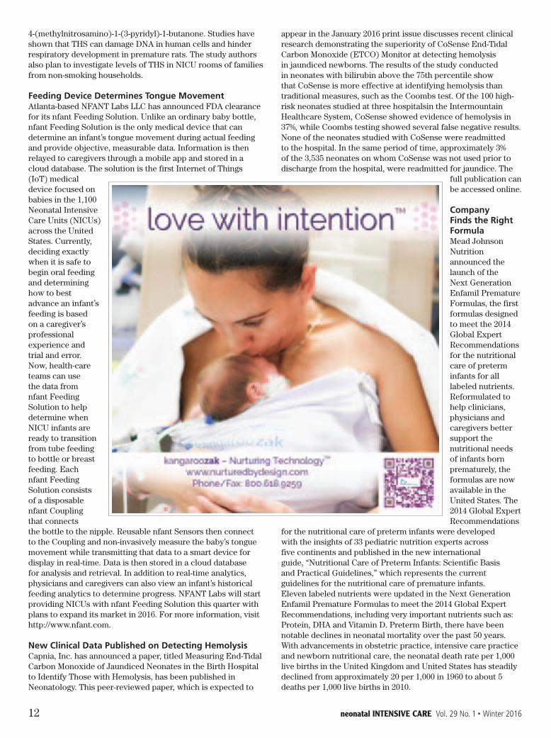
Feeding Device Determines tongue movementAtlanta-based NFANT Labs LLC has announced FDA clearance for its nfant Feeding Solution. Unlike an ordinary baby bottle, nfant Feeding Solution is the only medical device that can determine an infant’s tongue movement during actual feeding and provide objective, measurable data. Information is then relayed to caregivers through a mobile app and stored in a cloud database. The solution is the first Internet of Things (IoT) medical device focused on babies in the 1,100 Neonatal Intensive Care Units (NICUs) across the United States. Currently, deciding exactly when it is safe to begin oral feeding and determining how to best advance an infant’s feeding is based on a caregiver’s professional experience and trial and error. Now, health-care teams can use the data from nfant Feeding Solution to help determine when NICU infants are ready to transition from tube feeding to bottle or breast feeding. Each nfant Feeding Solution consists of a disposable nfant Coupling that connects the bottle to the nipple. Reusable nfant Sensors then connect to the Coupling and non-invasively measure the baby’s tongue movement while transmitting that data to a smart device for display in real-time. Data is then stored in a cloud database for analysis and retrieval. In addition to real-time analytics, physicians and caregivers can also view an infant’s historical feeding analytics to determine progress. NFANT Labs will start providing NICUs with nfant Feeding Solution this quarter with plans to expand its market in 2016. For more information, visit http://www.nfant.com.
new clinical Data published on Detecting HemolysisCapnia, Inc. has announced a paper, titled Measuring End‐Tidal Carbon Monoxide of Jaundiced Neonates in the Birth Hospital to Identify Those with Hemolysis, has been published in Neonatology. This peer‐reviewed paper, which is expected to

Visit childrens.com/excellence to learn more.
Proud to be leading the way in neonatal care.At Children’s Medical Center Dallas, the fl agship hospital of Children’s HealthSM, we offer a full continuum of neonatal care, including a Level IV Neonatal Intensive Care Unit, a comprehensive Fetal Center, and the fi rst TeleNICU program in the state of Texas. Our program also participates in NIH-sponsored research and works with the Children’s Hospitals Neonatal Consortium to collaboratively establish benchmarks and best practices for Level IV NICUs nationwide. With cutting-edge research and a clinical and academic affi liation with UT Southwestern Medical Center, our Fetal Neonatal program continues to earn national recognition.

14 neonatal INTENSIVE CARE Vol. 29 No. 1 n Winter 2016
star power for Brave BeginningsZoe Saldana has been named the 2015 Theatrical Fundraising Spokesperson for Brave Beginnings, a newly renamed program of the Will Rogers Motion Picture Pioneers Foundation (WRMPPF). The program works to improve the lives of premature babies by providing hospitals with grants for purchasing life-saving neonatal equipment and supporting critical care pulmonary services. Premature birth accounts for 35% of all infant deaths in the US, more than any other single cause. Since 2006, Brave Beginnings (formerly the Will Rogers Neonatal Program) has provided essential ventilator equipment to neonatal intensive care units (NICUs) across the country. To date, the neonatal program has contributed $2.9 million in grants to 78 hospitals across 30 states. Each year, the program donates roughly $500,000 in grants — and the demand increases every year. Saldana stars in a thirty-second public service announcement that will appear before movies on more than 4,000 theater screens nationwide. The spot shares the same hand-drawn imagery from Brave Beginnings’ newly launched website, www.bravebeginnings.org, where the spot can be viewed. Playing on the concept that summer at the movies means action, adventure and superheroes, Saldana tells moviegoers, “I want to talk about a different kind of hero. Instead of capes, they wear blankets.” She adds, “Every superhero needs a sidekick. I’d like you to fill that role by giving generously.” For nearly 80 years, movie theaters have honored Will Rogers’ legacy by raising money in movie theaters. Several theater chains will participate in the 2015 campaign, which will benefit Brave Beginnings. Participating theaters will run various types of programs, including concession stand programs that donate a portion of sales to Brave Beginnings, donation pin-ups, collection canisters and lobby monitor displays. The money raised from the 2015 Fundraiser will benefit Brave Beginnings.
Ventilation Device addresses transporting neonatesThe HAMILTON-T1 with neonatal option is a high-end transport ventilator that provides the best possible ventilation therapy for your smallest and most vulnerable patients. During transport, the HAMILTON-T1 delivers the same performance as a fully featured NICU ventilator at the bedside. Its unique features make it one of the best transport ventilators for neonates. Hamilton Medical has specially adapted the HAMILTON-T1 hardware and software to optimally meet the needs of ventilated neonates. Supporting tidal volumes of just 2 ml, the HAMILTON-T1 allows for effective, safe, and lung- protective ventilation for even the smallest preemies. The reliable and robust neonatal flow sensor accurately measures pressure, volume, and flow proximal to the patient. This guarantees the required sensitivity and response time, and prevents dead space ventilation. Therefore, the patient is better synchronized and the work of breathing (WOB) is reduced. The new neonatal expiratory valve can balance even the smallest differences in pressure and offers the neonate the possibility to breathe spontaneously in each phase of a controlled breathing cycle. In addition to all modern neonatal ventilation modes, the HAMILTON-T1 offers a new generation of nCPAP. In the new nCPAP-PC (pressure control) mode, you only define the desired CPAP target value for your patient and the ventilator automatically and continuously adapts the required flow to the patient’s condition and possible leaks. Thanks to the demand flow technology, your patient will receive only as much flow as is necessary to obtain the set CPAP target. This reduces WOB, reduces the need for user interventions and ensures optimal leak compensation. You will also require less oxygen for transport and noise caused by the ventilator decreases
Smallest footprint in the industryONLY 15.9 LBS FULLY OPERATIONAL
Tecotherm Neo utilizes an innovative servo-controlled design
with instant feedback, monitoring the infant’s temperature every 2
seconds and making minute changes to the cooling fluid to ensure
that the infant’s temperature remains stable. Simple-alarms on the
Tecotherm Neo alert staff should the temperature deviate more
than 0.5°C from the set temperature. The user can set the target
temperatures, duration and even the rate of re-warming / cooling,
thereby making it easy and simple to set up and use
Tecotherm NEO®
Total Body Cooling and Warming
Contact Maxtec for an informational booklet e-mail: [email protected]
3 Modes: Provides maximum flexibility during use
TOBY Protocol*: Activated with one, easy step
Adjustable Parameters: Changeable before and during treatment
Data Storage: For simple analysis of temperature profiles
Linear Rewarming: Provides smooth, linear rewarming
Informative references:The Journal of Pediatrics, Vol. 166, Issue 4, p856–861.e2 & Vol. 165, Issue 2, p267–273The New England Journal of Medicine, NEJM,2009;361:1349-58
866.4.Maxtec www.maxtec.com

neonatal INTENSIVE CARE Vol. 29 No. 1 n Winter 2016 15
tubing getting it out of harms way and positioned close the patient’s chest. The second product is the 701VCS/NG offering the same circuitry securement but also adds a second strap used to secure a nasal gastric (or oral gastric) tube keeping it secure and avoiding decannulation thereby reducing these difficult reinsertions. Find out more at www.peppermedical.com.
study consent argued in courtTwo years ago, researchers in a clinical trial involving oxygen levels for the tiniest premature babies were accused by a federal watchdog agency of not properly disclosing the risks to families who participated. What followed was extensive public scrutiny of the trial, called Support, and soul-searching in the research community about how best to obtain informed consent from participants. Some families sued, arguing that their babies suffered serious injuries as a result of their treatment. But a federal judge threw out the suit in 2015, saying the families could not prove that the trial caused the injuries. Last week, the editors of a prestigious medical journal wrote that the decision showed that the trial was solid to begin with. “What the judge was saying was that being in the trial didn’t cause the bad outcomes for these kids,” said Dr. Jeffrey M. Drazen, editor in chief of The New England Journal of Medicine and an author of one of two pieces supporting the study. “And if that’s the case, there’s nothing to complain about in the consent form.” But some bioethicists disagreed. “The consensus in the bioethics community was that the informed consent was not adequate, and that hasn’t changed,” said George J. Annas, director of the Center for Health Law, Ethics and Human Rights at Boston University’s School of Public Health. The lungs of babies born prematurely are typically underdeveloped, and they often need to be given oxygen. But too
distinctively. With approvals and certificates for most types of transport and situations the HAMILTON-T1 is an ideal escort for your tiniest patients, reliable everywhere, both inside and outside the hospital, in the air as well as on the ground. The built-in high-performance turbine makes it completely independent of compressed air, gas cylinders or compressors. This saves weight and space and even noninvasively ventilated neonates can be transported over long distances. The combination of a built-in and an optional hot-swappable battery provides a battery operation of more than 9 hours. This can be extended indefinitely with additional hot-swappable batteries.
Ventilator circuit stabilizer launchedAs many ventilator patients have become more mobile, both in long-term care centers and at home, increased safety has become an issue. One of the areas that is most important is to secure the patients ventilator circuitry and prevent accidental dislodgement. A more mobile patient, moving from bed to wheelchair and through everyday life, presents a unique challenge in not only providing proper ventilation but also in providing a safe method in securing the life sustaining ventilator tubing. In these critical moments of movement, the tubing and circuitry may easily find itself ensnared in bed sheets, on wheelchair railings or other hazards, which can result in serious injury or death from ventilator disconnections. Pepper Medical has introduced two products that will eliminate this issue and provide a safer environment for these patients. The first is the 701VCS (ventilator circuit stabilizer). The 701VCS is a harness style belt made of soft cotton laminate that fits comfortably around a patient’s waist. Incorporated into the harness is a tubing securement strap that reliably secures the ventilator
Neonatal Whole Body Cooling is shown to improve outcomes for newborns meeting the requirements for HIE.1,2 Cincinnati Sub-Zero’s Blanketrol® III with it’s “Gradient Technology” and the Kool-Kit® Neonate provide accurate and safe patient temperature management. This system offers the ability to reach and maintain goal temperature as well as provides controlled re-warming for the patient.
All Therapeutic Hypothermia disposables located in one convenient package
Self sealing/insulated blanket hoses
Mittens/Socks allow more family contact without compromising patient temperature
All products tested and validated by CSZ for CSZ equipment
Therapeutic Temperature Management System
Phone: 513-772-8810Toll Free: 800-989-7373
Fax: 513-772-9119www.cszmedical.com
1. Shankaran, Seetha, et al. “Outcomes of Safety & Effectiveness in a Multicenter Randomized, Controlled Trial of Whole-Body Hypothermia for Neonatal Hypoxic- Ischemic Encephalopathy.” Pediatrics 122 (2008): 790-799.2. Zanelli, S.A., et al. “Implementation of a ‘Hypothermia for HIE’ program: 2-year experience in a single NICU.” Journal of Perinatology 28 (2008): 171-175.

16 neonatal INTENSIVE CARE Vol. 29 No. 1 n Winter 2016
developing any-stage ROP was 0.29 (95% confidence interval [CI], 0.12 - 0.72) compared with infants fed any formula. The risk was also significantly lower for infants exclusively fed human milk compared with those exclusively fed formula (odds ratio [OR], 0.25; 95% CI, 0.13 - 0.49). There were nonsignificant trends for infants fed any human milk vs infants fed exclusive formula (OR, 0.54; 95% CI, 0.15 - 1.96) and those fed mainly milk vs mainly formula (OR, 0.51; 95% CI, 0.26 - 1.03), but these differences were not statistically significant. When the researchers restricted their analysis to the risk for severe ROP, the benefit associated with breast milk appeared larger and reached statistical significance for all comparisons except those fed any milk vs exclusive formula (OR, 0.42; 95% CI, 0.08 - 2.18). Specifically, exclusive human milk vs any formula showed an OR of 0.11 (95% CI, 0.04 - 0.30), mainly human milk vs mainly formula had an OR of 0.16 (95% CI, 0.06 - 0.43), and exclusive human milk vs exclusive formula had an OR of 0.10 (95% CI, 0.04 - 0.29).
infant prep for HiVInfant preexposure prophylaxis (PrEP) for HIV using a pediatric liquid formulation should extend for 12 months in breast-fed babies of HIV-positive mothers, according to a study conducted in Africa through the Université Montpellier in France. The study represents the first assessment beyond 6 months of ways to prevent mother-to-child transmission of HIV-1 while breast-feeding. It included the 6- to 12-month period when breast-feeding decreases and babies are introduced to new foods, and suggests that mothers should be told about continued risk for HIV transmission during this time. Breast-feeding is recommended up to 12 months after birth, especially in developing countries with poor sanitation and high rates of child death resulting from infection. However, breast-feeding can increase the risk for mother-to-child transmission of HIV. Two strategies can reduce this risk: give the mother antiretroviral therapy (ART) to decrease infectiousness, or provide direct protection to the infant, using child formulations of PrEP.
stillbirth rates studiedTwo studies find no correlation between efforts to limit nonmedically indicated deliveries before 39 weeks and the number of stillbirths. There have been multiple efforts in the past decade by professional societies, regulatory bodies, and quality collaboratives to lower rates of early term deliveries that are not medically necessary (beginning of week 37 to end of week 38). However there is some concern that 39-week delivery policies could potentially increase term stillbirths, especially if policies are misused for higher-risk pregnancies. Therefore, the researchers used National Health Statistics data to determine whether those efforts, which lengthened pregnancies overall, were having unintended consequences. Sarah E Little, MD, MPH, from the Division of Maternal-Fetal Medicine at Brigham and Women’s Hospital in Boston, Massachusetts, and colleagues, studied early-term deliveries as a percentage of total term delivery and calculated term stillbirth rates for each state, both overall and for low- and high-risk women, using birth certificate and fetal death data. They found a decline in early-term deliveries across the United States, going from 1.12 million (31.8%) of 3.53 million term, singleton births in the early term in 2005 to 978,000 (28.5%) of 3.43 million in 2011. Reductions varied widely by state “ranging from a 25.5% relative reduction to a 3.9% relative increase,” the authors write in their article. However, they found no change in the term stillbirth rate (123/100,000 births in 2005
much and too little are both bad, so researchers were conducting the study to find the sweet spot, trying certain concentrations on babies born months premature, at just 24 to 27 weeks of gestation. Researchers defended their actions, saying that all of the approximately 1,300 babies in the study had been kept within a band of treatment that was the standard of medical care at the time — oxygen saturation levels of 85 to 95 percent. They argued that doctors did not know which part of the spectrum was better and that until the trial, which was created to try to answer that question, they had really only been guessing. Ultimately, mortality was fairly high in the low-oxygen group: 130 babies out of 654 in the low-oxygen group died. Ninety-one babies out of 509 in the high-oxygen group developed an eye ailment. Researchers disputed the assertion that the babies were worse off for having been in the study; born so early, they said, these children were at high risk to begin with.
Gestational age, Diabetes studied for linkGestational age and type 1 diabetes appear to be linked, independent of familial factors, say researchers who conducted an extensive study on nearly all births in Sweden over 4 decades. The link could be related to insulin resistance developing as a consequence of early-life growth restriction or attributed to altered gut microbiota. Three key findings were highlighted. First, late preterm birth and early-term birth babies are at increased risk of type 1 diabetes. Second, an association seen between large-for-gestational-age babies and increased risk for type 1 diabetes is unlikely to be causal and may be explained by familial factors shared by siblings, he said. Third, and conversely, the researchers found that babies born before 33 gestation weeks (very preterm) and/or with birth weight of less than 1500 g, or small for gestational age, seem to be protected against type 1 diabetes.
educational training Kit now available Accriva Diagnostics is presenting the latest training tool on proper capillary blood sampling when using a heel stick device for PKU neonatal testing. The kit provides samples of Tenderfoot incision devices for nurses to practice while using a life-like foam demo foot. The foot size was accurately based on the median weight of a full term newborn, and has indicated markings outlining the acceptable incision locations. The kit will also include a step by step training video, informational literature and more. With little subcutaneous tissue, it is important to use the correct size and not use a device that compresses the skin further. This can result in the blade striking the bone, potentially causing osteomyelitis. Visit www.tenderfootcares.com for more details.
preventing rOp through Breast milkFeeding very preterm newborns human milk may help prevent retinopathy of prematurity (ROP), according to an analysis through the Children’s Hospital of Fudan University, Shanghai, China. Up to 37% of very prematurely born infants (birth weight <1250 g) develop severe ROP, a vasoproliferative disorder of immature retina and a leading cause of blindness in developed countries, according to the authors. To ascertain the potential protective effect of breast milk compared with formula, the authors searched electronic databases for relevant studies and reviewed 312 studies in full. Five cohort studies conducted from 1992 to 2008 involving 2208 preterm infants met their inclusion criteria. The newborns’ average gestational age ranged from 26 to 30.2 weeks, and their birth weight ranged from 775 to 1376 g. Among infants who were exclusively fed human milk, the risk of

neonatal INTENSIVE CARE Vol. 29 No. 1 n Winter 2016 17
vs 130/100,000 in 2011; P = .189). In another study, Marian F. MacDorman, PhD, from the Maryland Population Research Center, University of Maryland in College Park, and colleagues found that the stillbirth rate was unchanged between 2006 and 2012, although the percentage distribution of live births by gestational age changed considerably in that time: Births at 34 to 38 weeks decreased by 10% to 16%, and births at 39 weeks increased by 17%.
Hearing loss and newbornsNewborns with congenital diaphragmatic hernia (CDH) appear to be at higher risk of sensorineural hearing loss (SNHL), according to a new study. SNHL is either congenital or acquired, but the mechanism for CDH to lead to SNHL is unknown, the researchers say. Possible explanations include severe tissue hypoxia interfering with cochlear oxygenation and function, CDH caused by abnormal neuronal migration at the same time as the cochlea develops, or even postnatal factors. SNHL may develop whether newborn audiograms are negative or positive, so health care providers need to monitor these children closely. Dr Misha Amoils, of Stanford University, Stanford, California and colleagues studied 50 children with CDH born between 1999 and 2008 (26 boys; 24 girls) with a mean gestational age of 37.4 weeks and weighing 3,046 g on average. Thirty-three children had a prenatal diagnosis of CDH, and 40 patients had a left-sided hernia. Eighteen children underwent primary surgical repair; and 32 required more extensive patch repair. Newborn hearing screen results were available for 47 children. Follow-up audiograms were done between ages six months and 10.7 years. Of those with newborn screen results, 40 passed the screen and seven failed. Five of those who failed developed SNHL, as did 20 of those who passed. In all, 28 patients had any degree of SNHL, nine of them with significant disease, defined as having a pure-tone average (PTA) of >40 db. Univariate analysis failed to identify factors associated with overall hearing loss. On multivariate analysis, however, factors associated with significant hearing loss included receipt of extracorporeal membrane oxygenation (ECMO) (p=0.02), nonprimary CDH repair (p=0.01), ventilation of 14 days or longer (p=0.001), and high neonatal furosemide exposure (p=0.03).
Got milk (tested)?Scientists are debating the merits of a new breastmilk testing service — you mail three samples of breast milk to a lab — which claims to offer a full nutritional panel of breast milk, with levels of a wide variety of nutrients from protein to vitamin D. Another test, a strip that changes color when alcohol content is over a certain level, can tell a mother if it’s too soon after an alcoholic drink to feed the baby. “The nutrient value of breast milk will supply all the needs of the baby. There is no need to measure it,” says Arthur Eidelman, co-author of the American Academy of Pediatrics guidelines on breast-feeding, published in 2012. Individualized breast-milk profiles are sold directly to consumers by companies such as Israel’s MyMilk Laboratories Ltd, which hopes to offer its services in the US next year. Happy Vitals Inc, of Washington, DC, began shipping test kits this month at $149.95 for a basic panel and up to $324.95 for a test of 15
different beneficial ingredients as well as for cortisol, a stress hormone that a 2013 study suggests may influence a baby’s temperament. After receiving the test kit, you take three samples of milk, at least two or three days apart, freezing them each time and then shipping them in a cooler to the lab, says Happy Vitals Chief Executive Eric Feigl-Ding, an epidemiologist and research scientist in the department of nutrition at the Harvard TH Chan School of Public Health in Boston. There aren’t published guidelines for ideal composition of breast milk, scientists say. Instead, the
company’s online software multiplies the amount of each nutrient by the amount of milk your baby is drinking daily — and tells you whether your baby is getting enough of each nutrient according to US and European guidelines. For example, the Institute of Medicine recommends an adequate intake of vitamin B12 for an infant is 0.4 micrograms daily. In an information sheet for clients, the company says vitamin B12 can be obtained from meat, fish, eggs and dairy or from supplements for mother or baby. Another option, according to the sheet, is to supplement with formula containing the vitamin. A vegan who isn’t taking supplements or eating foods fortified in B12 is likely to be deficient and should take supplements regardless of breast-milk test results, says Dr Meek, a dietitian, pediatrician and editor in chief of the pediatrics academy’s book, “New Mother’s Guide to
[email protected] • 800-761-7601
Realtime Biofeedback at Bedside The nfant® Feeding Solution is used with standard bottles and nipples and non-invasively measures nipple movement during feeding. Data is streamed to a mobile tablet for display and saved in the cloud nfant® Patient Database.
Patient Progression TrackingFeeding history is aggregated on the nfant® Mobile App and objective measures displayed for clinician review. Progression tracking of measured data allows for evidence based decisions through feeding transition and discharge home.
Safe & E�ective Feeding TransitionDid you know 40-70% of patients experience complications transitioning to oral feeding?

18 neonatal INTENSIVE CARE Vol. 29 No. 1 n Winter 2016
tell us about the latest advances in the area your product serves.Radiologists and physicians in the NICU face a unique set of imaging challenges not always addressed by general purpose mobile imaging systems. NICUs are seeing more Low and Extremely Low Birth Weight Babies than ever before, and these fragile infants often require more, frequent, imaging. When using conventional CR and DR systems, clinicians are often forced to compromise between image quality and dose in order to manage radiation exposure. The KUB 250 System meets these challenges by providing higher resolution to visualize the infants’ delicate anatomy at a lower radiation dose thereby helping reduce the overall risk to the patient.
Discuss your r&D process, including clinical user input.Kubtec’s business is built upon digital X-ray and nothing else. Our institutional knowledge base is deep, and we can truly say that we are the experts in our clearly defined chosen field: neonatal and women’s imaging. Our approach is to identify unmet needs in the market and to move quickly to provide solutions to clinicians, patients and providers. Our concepts are often derived from consultations with our broad customer base and are tested with neonatal physicians, technicians and administrators at tradeshows, hospitals and workshops. Product concepts pass through a stage gate process and are continually tested against user requirements and quality standards prior to release. Workflow and usability studies are provided by physicians/clinicians at every juncture of design and final product release, which includes clinical studies in actual usage.
What new technology do you see as having the greatest impact on your area of expertise?In the future, Kubtec envisions new technology that will greatly impact our area of expertise, such as the creation of new compounds or the expansion of the limits of current technologies in order to take full advantage of their physical properties. These advances will result in new products that can be utilized to improve resolution and/or reduce radiation dose. As a small company, we are able to continually adapt and capitalize on changes or improvements as they become available by utilizing sound physics and engineering.
Breastfeeding.” For vitamin D, tests of breast milk aren’t very useful since nature never intended babies to get their entire supply of the vitamin from feeding, says guideline-author Dr Eidelman, a visiting professor at Albert Einstein College of Medicine in New York. Given that modern babies are protected from the sun, the pediatrics academy recommends routine vitamin D supplementation for all breast-fed infants. The lab tests also measure levels of antibodies present in breast milk.
Home Births Up – But is that Good?With a growing number of American women choosing to give birth at home or in birthing centers, debate is intensifying over an important question: How safe is it to have a baby outside a hospital? A study in the New England Journal of Medicine provides some of the clearest information on the subject to date. The study analyzed nearly 80,000 pregnancies in Oregon, and found that when women had planned out-of-hospital deliveries, the probability of the baby dying during the birth process or in the first month after — though slight — was 2.4 times as likely as women who had planned hospital deliveries. Out-of-hospital births also carried greater risk of neonatal seizures, and increased the chances that newborn babies would need ventilators or mothers would need blood transfusions. On the other hand, out-of-hospital births were far less likely to involve cesarean sections — 5.3 percent compared with 24.7 percent in a hospital. They also involved fewer interventions to augment labor, and mothers had fewer lacerations.
cOmpanY prOFile
KubtecDescribe your product(s) and its unique features.The KUB 250 Neonatal Imaging System is the only portable digital X-ray system designed specifically to address the needs of the Neonatal Intensive Care Unit (NICU). Using CMOS technology and a 96u pixel size to provide the highest resolution images at up to 50% reduction in radiation dose compared to other portable systems, the KUB 250 System delivers image contrast that is specifically configured for enhanced soft tissue visualization, yet allows PICC lines of 1 French diameter to be seen.
The KUB 250 System is compact and portable and fits conveniently between Isolettes and cots. Also, its detector fits directly within the Isolette slot so there is no disturbance for the patient. The system is lightweight and maneuverable with no need for a motor, meaning fewer moving parts, less maintenance and less downtime.
As a system dedicated to the NICU, the KUB 250 System reduces the risk of bringing contaminants from other hospital departments. With no cassettes to transport, images are available instantly to the physician or technologist, enabling rapid review of PICC line placement and other procedures. The system is always ready, always available and runs all day on a single battery charge. Once produced, images can be wirelessly transmitted to PACS with KUB 250 System one touch transmission.

neonatal INTENSIVE CARE Vol. 29 No. 1 n Winter 2016 19
Interview
neonatal intensive care: What is the significance of newborn and pediatric hearing screening today?Diane sabo: According to the National Center for Hearing Assessment and Management (NCHAM) every state and territory in the United States has now established an Early Hearing Detection and Intervention (EHDI) program to ensure that every child born with a permanent hearing loss is identified before three months of age and provided with timely and appropriate intervention services before six months of age.1
randi Winston Gerson: Hearing loss is the most common congenital condition in the US, according to the American Academy of Pediatrics (AAP). AAP estimates that three in 1,000 infants who are born with moderate, severe, or profound hearing loss results in delayed development in language, learning, and speech.2 Timely diagnosis and intervention can help—and it all begins with screening.
Kathleen Hill: Children who are identified with hearing loss and receive intervention early are more likely to demonstrate language development within the normal range by the time they enter school than those who are not identified and served early.3 Hearing loss can occur at any age and hearing screening plays a vital role to ensure that patients can avoid communication roadblocks as they grow.
nic: What are the best practices based on national EHDI guidelines?randi: The 1-3-6 EHDI goals are to screen a newborn prior to one month of age, diagnose hearing loss prior to three months of age and enroll in early intervention programs prior to six months of age. The earlier a child is identified with hearing loss, the sooner the language learning process can begin. In addition, the younger a baby is, the more likely sedation procedures can be avoided and the diagnostic audiological evaluation can be conducted under natural sleep.
nic: What is the earliest you can screen a newborn?randi: Great question. The answer relates to neurological maturation and the screening equipment used—and so this is a significant factor to consider with Auditory Brainstem Response (ABR) screening. I am asked these questions often; Is it ok to screen a 30 week old vs a 36 week old? What is the ideal age to screen a newborn? What is the maximum age?
Should you use equipment on babies beyond six months of age? The answer depends on the specific equipment used, and the response detection method used. It is important to do some investigation with the specific equipment manufacturer to know the answers to these questions.
nic: Beyond the NHS period, do you need to screen babies or is it OK to wait until they enter school?Diane: No, parents shouldn’t wait until the child enters school to have hearing tests conducted. In fact, there is existing research that states hearing loss doubles by the time they enter school.4 Hearing in children needs to be monitored on an ongoing basis because hearing loss can develop at any time, and it is an invisible condition—that is why we need continued screening.
Kathleen: Another reason for continued screening is because there are children born with risk factors. These babies may have a normal hearing screen at birth, but develop a hearing loss after the newborn period; for example, babies born with syndromes, cranio-facial anomalies, a family history of congenital hearing loss, or a NICU (Neonatal Intensive Care unit) stay of five days or more.
nic: Tell us about Otoacoustic emissions (OAE). Should you screen with OAE vs ABR? What population is ideal for OAE? Is it OK to use OAE in the NICU? randi: In their 2007 position statement, the Joint Committee on Infant Hearing recommends OAE screening for well babies, babies without risk factors and babies that have been in the NICU for less than five days.5 Babies who have been in the NICU for five days or more, or who have risk factors should be screened with ABR.
Diane: I agree, but you can also use OAE in the NICU, however it must be in conjunction with ABR screening. Regardless of whether the newborn passes the OAE, the ABR screen must be conducted. If you screen the high-risk population just with OAEs, the potential of missing a baby with auditory neuropathy/dys-synchrony exists—this is a condition more common in babies in the NICU for more than five days and babies with risk factors.
Kathleen: And because auditory neuropathy/dys-synchrony affects the hearing anatomy beyond the level of the cochlea;
OAE Screening: Best Practices for Newborn Hearing Screening and Beyondneonatal intensive care interviews clinicians and healthcare providers about the actual application of specific products and therapies. participating in this interview are audiologists randi Winston Gerson, auD, and Diane sabo, phD, hearing screening program managers at audiology systems, as well as, Kathleen Hill, auD, audiology training and education manager at Gn Otometrics north america.

20 neonatal INTENSIVE CARE Vol. 29 No. 1 n Winter 2016
the eighth nerve to the brainstem it is important to screen with ABR to ensure this condition is not missed.
nic: What about for newborns with any kind of ear malformation or deformity—should they be screened? Or should they go straight to the audiologist? randi: In my opinion, we shouldn’t screen babies with outer ear malformations. Instead, they should go straight to a pediatric audiologist. Unfortunately, I am aware of babies with malformations that were actually screened in the newborn nursery and the malformed ear passed. In addition, if a baby has a unilateral malformation and the normal looking ear is screened and passed, the family may have a false sense of security that since one ear is “normal” they don’t have to get audiological follow up. It’s important that parents or caregivers understand the importance of monitoring the ear that passed the screening and find out the type and severity of the hearing loss in the malformed ear.
Diane: I have a different perspective. Babies in the NICU may have other health conditions that are a higher priority than hearing loss, making it difficult for families to follow-up. Also, for those families that have limited resources or who have socio-economic challenges may have a higher likelihood of falling through the cracks. I’ve seen high no-show rates even when appointments are scheduled. It is important to screen them because you don’t know if they will come back to see an audiologist. They may follow up with an ENT to address the malformation but not always with an audiologist to address hearing. If the baby is from the well-baby nursery, I would still recommend screening when you have access to the baby. That is, while they are in the hospital.
Kathleen: This is a tricky topic. Your readers will simply have varying perspectives to consider. In the two children’s hospitals that I worked at with high intensive care populations, we had families that wouldn’t go to audiology even with appointments. So the theory was that I needed to, at the very least, screen the good ear to verify that something was “good”, and take into consideration that they may never come back.
nic: Since you mentioned equipment earlier, what tools are available for newborn OAE screening and diagnostic follow-up? What features can help screeners and audiologist’s today? randi: For newborn screening, it boils down to accuracy, efficiency, preference and budget. The MADSEN AccuScreen by Otometrics is a compact handheld screener that offers all the capabilities of a cart-based system. Two-step OAE and/or ABR testing is combined into a single hand-held device and will significantly improve your workflow and test time. AccuScreen is great for conducting OAE screening for patients of all ages and the ABR module is intended for babies from 34 weeks to six months of age.
In addition, the device is capable of performing DPOAE and TEOAE testing depending on your screening needs.
On the other hand, the SmartScreener—Plus2 by Intelligent Hearing Systems offers a choice of automated ABR screening, OAE screening, or both. It is a cart-based infant screener that is simple to operate. The battery-operated, ergonomic cart allows for screening flexibility. An integrated data back-up utility allows for seamless results transfer to central databases. The SmartScreener can be used to screen patients of all ages.
Beyond NHS, the MADSEN Alpha OAE screener by Otometrics is another great tool for OAEs. The “child mode” cartoons and touch screen display make hearing screening fun for the child, and fast for the tester. It meets the requirements of CPT code 92587 and makes OAE screening easy to administer in any setting. Alpha screens patients of all ages.
Diane: In addition to the Alpha, PATH Medical Solutions offers several different devices depending on the needs of the clinic. For example, the Sentiero Desktop by PATH Medical Solutions is a unique, flexible OAE device with diagnostic tympanometry, ipsilateral and contralateral acoustic reflex, pure tone audiometry all in one portable desktop device.
Another option is the Sentiero handheld which offers both OAEs and audiometry. It is an ideal solution for professionals who need more than just OAE testing and would like to confirm results with behavioral responses. Having the option to choose between diagnostic, screening or both functionalities is a practical feature to consider for those who plan to add OAE screening to their practice. The Sentiero devices can also test patients of all ages.
nic: Since its foundation in 2012, Audiology Systems has promised to elevate customer care in the US. So in addition to hearing instrumentation, what other services are available to hearing care professionals?randi: Audiology Systems offers everything a professional needs to maximize the use of the equipment—this elevates our service delivery and accessibility. For example, our calibration services (www.audiologysystems.com/calibration) ensure that all screening devices are always up-to-date. Customers can rest assured that they are supported by service professionals who are certified and trained to service any hearing care equipment—not just that of Otometrics. This kind of “one-stop shop” service is a huge time saver.
Kathleen: Another advantage is the ability to shop online for supplies. The Audiology Systems webshop (www.shopASI.com) is our convenient online shopping site featuring commonly used supplies, accessories and disposables, such as the Lilly TM-wick electrode, pediatric foam ear tips, probe tips cables and more. So, screeners and inventory managers can easily restock at their convenience.
We also offer our education page (www.audiologysystems.com/education) which features courses and self-help references for professionals. Between Audiology Systems and Otometrics, we have close to 20 audiologists in the US who provide audiology support and training nationwide. Our audiologists have decades of collective audiologic experience and we are here to be resources to audiologists and hearing care professionals who have purchased instrumentation across the US.
Diane: Randi and I have more than 60 years of combined experience in hearing screening. We joined the company primarily to provide screeners, audiologists and other hearing care professionals with specialized knowledge and consultative support to meet their hearing screening needs, and that of their facility. So all-in-all, Audiology Systems now brings to every customer over 160 years of collective experience in audiology. That experience goes into producing informational and educational material specific to the screening market for Audiology Systems customers.

neonatal INTENSIVE CARE Vol. 29 No. 1 n Winter 2016 21
nic: Thank You for your time Randi, Diane and Kathleen. Learn about Audiology Systems’ screening solutions at www.audiologysystems.com/screening. Reference material, articles, courses and interviews about hearing care techniques and technologies are available at www.audiologysystems.com/education.
Author Biographiesrandi Winston Gerson, auD, is a hearing screening program manager at Audiology Systems. Prior to joining the company, Randi held various program management roles supporting Arizona’s Early Hearing Detection and Intervention Program. She has been in a consulting role as a technical assistance audiologist for the National Center for Hearing Assessment and Management (NCHAM) for over 15 years. Randi has developed various successful training programs for NCHAM with various publications and countless presentations describing those efforts.
Kathleen Hill, auD, is the audiology training and education manager at GN Otometrics. She is responsible for overseeing the education and training program for Audiology Systems employees, distributor, business partners and customers. Dr. Hill joined the company from Advocate Illinois Masonic Medical Center in Chicago, where she worked as an audiologist. Prior to that, she was the audiology manager at Children’s Memorial Hospital (now the Ann & Robert H. Lurie Children’s Hospital), and the audiology supervisor at Cardinal Glennon Children’s Hospital. She has been a program professor at Nova Southeastern University in Fort Lauderdale, Florida; she has also been in private practice. She earned her Bachelor of Science degree in communication disorders/deaf education from the Texas Woman’s University and her doctor of audiology degree from Central Michigan University.
Diane sabo, phD, is a hearing screening program manager at Audiology Systems. Prior to joining the company, has held various managerial positions at Children’s Hospital of Pittsburgh, including that of Coordinator of the Auditory Evoked Potentials and Newborn Hearing Screening Programs and ultimately as Director of Audiology and Speech Pathology. Most recently she has been an Associate Professor at the University of Pittsburgh and a consultant for GN Otometrics. She holds various academic appointments and has numerous publications and presentations to her credit.
References1 Early Childhood Screening and Follow-up. Retrieved from:
http://www.infanthearing.org/earlychildhood/about.html2 Early Hearing Detection and Intervention Program. Retrieved
from: https://www.aap.org/en-us/advocacy-and-policy/aap-health-initiatives/PEHDIC/pages/early-hearing-detection-and-intervention.aspx
3 Moeller, MP. Early intervention and language development in children who are deaf and hard of hearing. Pediatrics. 2000. Retrieved from: http://pediatrics.aappublications.org
4 Eiserman W, Hartel D, Shisler L, Buhrmann J, White K, Foust T. Using otoacoustic emissions to screen for hearing loss in early childhood care settings. Int J Pediatr Otorhinolaryngol 2008; 72:475-82.
5 Executive Summary of Joint Committee on Infant Hearing Year 2007 Position Statement. Retrieved from: http://www.jcih.org/ExecSummFINAL.pdf

22 neonatal INTENSIVE CARE Vol. 29 No. 1 n Winter 2016
Interview
neonatal intensive care: What were the motivating factors that led you to try using the Modified Seldinger Technique in such small patients?tricia Blaine: Our PICC team went to Florida to be trained in using US and MST on infants and children at Jacksonville Children’s hospital through a training session offered by PICC Excellence. We witnessed them using MST with US on babies as small as 500 gms. We hadn’t realized this technique could be used on babies that small.
nic: What did you find were the advantages in using this technique verses the traditional method of an over-the-needle tear-away introducer?tB: Since our population is strictly neonatal with a good percentage of them being micro-preemies, we generally use a transilluminaor for vessel identification. Our first stick success rate using the direct introducer method was 55-65% and we were eager to use the MST technique with better statistics on first-time success rate attempts. We also found that you could use MST on visible vessels without US. Our first stick sucess rate went up to 85% using MST since we started using it. We found that if the wire threaded easily, you could be certain you could thread the PICC line.
nic: What was the size of the smallest infant that you were able to successfully place a PICC in using MST?tB: Great question. The answer relates to neurological maturation and the screening equipment used—and so this is a significant factor to consider with Auditory Brainstem Response (ABR) screening. I am asked these questions often; Is it OK to screen a 30 week old vs a 36 week old? What is the ideal age to screen a newborn? What is the maximum age? Should you use equipment on babies beyond six months of age? 22 week 480 gm baby. This baby was actually the first time we used the Neomagic MST with success.
nic: In the Pediatric and Adult use of Modified Seldinger Introduction it is very common to have to perform a skin nick with a scalpel before placement of the dilator tear-away. What has been your experience?tB: We have found with the smallest babies, their skin is immature and pliable so that the dilator sheath slides in and we have not had to make a nick in the skin. On older larger babies we do have to make a nick.
nic: How frequently do you need to use an alternative method of introduction or how successful is this technique?tB: We have been strictly using this technique now in our NICU since last July. We are using the US more when veins are not visible or with more difficult access situations but are only using MST.
nic: Are there additional advantages to neonatal MST PICC insertion related to improve first-attempt success other that just using fewer introducers?tB: Our comfort level using our regular 24 g angiocaths to access the vein is greater, making first-time attemps more successful.
nic: In looking for a product to use, which one did you find was the most suitable for such small patients?tB: There are very few products on the market for neonatal MST. We wanted to use it on our smallest babies without US when the vein is visible, especially the saphenous vein at the ankle since that is one of our favorite sites. I searched the internet and found the Neomagic. It looked very small so we decided to give it a try.
nic: Why or how often do you still need to use the traditional over-the-needle tear-away introducer for neonatal PICC placement?tB: We are not using that method anymore since we introduced MST last July in our unit.
nic: You mentioned a few questions ago that you used the Neo Magic MST kit. What makes it so special?tB: It is very small and felt comfortable using it on our tiniest babies.
nic: Could any company have produced this good of a product and what would you like to say to the company that did?tB: There needs to be more literature that MST can safely be used on the smallest of babies. It is the standard in the adult and pediatric world and we would like to see that happen in the neonatal world. It is a successful product and we would like more places to be using it on babies! We spoke about our team’s success at AVA and hopefully the word will spread.
Hospital Benefits of Using the Modified Seldinger Technique (MST)in this interview, neonatal intensive care interviews clinicians and healthcare providers about the actual application of specific products and therapies. participating in this interview is tricia Blaine, rn, Bs, rn, picc, neonatal intensive care Unit Brigham and Women’s Hospital.
If you would like to participate in this feature, as a company or healthcare provider, please contact Steve Goldstien at [email protected].

neonatal INTENSIVE CARE Vol. 29 No. 1 n Winter 2016 23
neonatal intensive care: What do you see are the newest trends in neonatal treatment of HIE?Diana Vasil: The newest trends in neonatal treatment of Neonatal Encephalopathy have many hospitals cooling infants < 36 weeks gestational age and cooling infants ≥ 36 weeks gestational age after the initial 6 hours after birth. Currently there is limited research in both of these areas and until any studies are published with sufficient data our centers will not be changing our policies for cooling since we are an academic medical center and our policies are based on evidence-based medicine.
nic: How have your cooling protocols changed in the past 2 years?DV: We have not changed our cooling protocols in the past 2 years, we currently cool infants that are ≥ 36 weeks gestational age who have qualified for cooling with clinical indicators and a neurological examination before 6 hours of life.
nic: What kind of trend are you seeing while extubating during therapy and how do you sedate those babies?DV: We do not sedate infants during therapy.
nic: What is the biggest obstacle you face when cooling a baby?DV: There can be several obstacles when cooling is initiated for an infant. We have a resuscitation team that handles the infant from birth through the first 4 hours of life or longer if necessary. They are trained to set-up cooling protocols and apply the appropriate probes and equipment as necessary. Close monitoring is necessary to ensure that the appropriate temperature is achieved and maintained. The next obstacle can be the re-warming period which has to be a slow process and can be associated with an increase in seizure activity. Infants undergo head MRI at about 7-14 days of age and are required to return for follow-up neurological and developmental exams at about 2 years of age. Follow up is always an important component of their care and is stressed to the parent as part of the child’s care. We keep in touch with the families during the interim period.
nic: What are the biggest fears that you hear about from the parents?DV: Parents are often overwhelmed with their baby’s admission into the NICU. They are stressed and often don’t remember what information they have been given about the baby’s condition or prognosis. They are usually fearful that they will lose their baby and are often very worried about long term neurologic outcome for their child. Fear of the unknown, being surrounded by the equipment and monitors in the NICU, and the lack of understanding of so many processes usually feeds their anxiety.
nic: What site do you prefer to monitor (esophageal or rectal) and what techniques do you use for placing an esophageal probe?DV: We use a esophageal probe. The probe is to be softened before insertion by placing in warm water for a few minutes, placed in the lower third of the esophagus, preferably nasally since it is usually more secure in this position. If the infant is smaller, we can place it in the oropharynx. The measurement is taken by measuring the distance from the nares to the ear to the distal sternum (xiphoid) minus 2 cm and marking with an indelible pen before inserting. It is taped to the nose or side of the face. Usually probe placement is confirmed within 12 hours by X-ray.
nic: What tips can you provide for re-warming difficult babies?DV: The most important tip would be the frequent monitoring of the infant during the entire period of re-warming. Vitals signs should be taken frequently. Overwarming the infant should be avoided and the infant’s temperature should not reach over 37.5°C.
nic: What other diagnoses are you cooling babies for and how is that working for you?DV: Currently we only cool infants for Neonatal Encephalopathy.
Neonatal Treatment of Neonatal Encephalopathyin this feature, neonatal intensive care interviews clinicians and healthcare providers about the actual application of specific products and therapies. participating in the interview from Ut southwestern medical center is Diana m Vasil, Bsn, rnc-nic, senior research nurse-neonatal research network.
If you would like to participate in this feature, as a company or healthcare provider, please contact Steve Goldstien at [email protected].
Interview

24 neonatal INTENSIVE CARE Vol. 29 No. 1 n Winter 2016
Interview
neonatal intensive care: When did you implement the HALO Safer Way to Sleep Program at Christiana Hospital? pamela Jimenez: May 2013.
nic: What was the impetus?pJ: While attending the National SIDS conference in 2011, I met Bill Schmid and learned of the HALO® SleepSack® wearable blankets. Given that Delaware averages about 2-3 unsafe sleep environment related deaths per month, I began to look at how we model safe sleep practice within our own hospital.
nic: Can you also share the steps you took to get the program adopted at the hospital?pJ: I began with tracking NICU sleep environments of all babies who were in cribs and medically stable for a period of 6 months. What I found were babies surrounded by rolls of blankets, propped on their sides, the head of bed elevated and /or on their belly. Although we do teach safe sleep, these behaviors were not modeling safe sleep practice for parents. Following the evaluation of data, over 204 parents previously discharged were contacted to evaluate our safe sleep education. Surprisingly, over 75% were not following safe sleep practices but modeling nursing behaviors. The most common comment noted was “If my nurse in NICU did this, it must be ok.” Updated nursing education reflecting the AAP 2011 safe sleep guidelines and recent data regarding our own practices was shared. This provided an opportunity to approach our Maternal Child service line about joining forces with HALO to integrate their SleepSack wearable blankets and SleepSack Swaddles into our safe sleep practice. It took a great deal of networking between HALO and our leadership team, but with the help of our data reflecting unsafe sleep practices, we forged our relationship with HALO. Mother’s Day 2013 marked our roll out the Safer Way to Sleep program. Data collected after the implementation of the program utilizing HALO SleepSacks, demonstrated a 100% improvement in the NICU within the first month and most importantly, parents began to seek out these safe sleep garments for their baby.
nic: In what parts of the hospital is the program currently used? pJ: Post-Partum, NICU and Pediatrics.
nic: Do you know how many babies are touched by the program each year? pJ: Christiana delivers about 7,000 babies per year. All babies
following their first bath are placed in a HALO SleepSack Swaddle. We also provide a take home program in which parents are provided with a HALO SleepSack Swaddle for home use along with safe sleep education during their stay and prior to discharge.
nic: Specifically how is the program implemented in the NICU and what are the results? pJ: All medically stable babies who are either acclimatizing to a crib or are in cribs are now placed in HALO SleepSack wearable blankets.
nic: How does the program enable the staff to share important safe sleep information with parents, and do you feel that the “demonstration” of safe sleep on babies makes the information easier for new parents to understand and replicate at home? pJ: The HALO SleepSack wearable blankets open the window for education with the families. Parents seek out the HALO SleepSack wearable blankets and like the idea of their baby being comfortable, warm and safe.
nic: Does the program encourage more dialogue between parents and the staff? pJ: It does. However, it is a consistent message by all staff that is important as different cultures or age groups still default to using blankets and need continued education.
nic: Why is it so important for you to have a take home program
Hospital Benefits of Using the HALO® Safer Way to Sleep Programin this feature, neonatal intensive care interviews clinicians and healthcare providers about the actual application of specific products and therapies. participating in the interview from christiana Hospital is pamela Jimenez, rn, msn, Fnp-Bc/pnp-Bc nurse practitioner/coordinator continued care nursery, infant apnea team.
If you would like to participate in this feature, as a company or healthcare provider, please contact Steve Goldstien at [email protected].
Photo courtesy of Christiana Hospital.

neonatal INTENSIVE CARE Vol. 29 No. 1 n Winter 2016 25
economic areas that provide HALO SleepSack wearable blankets and education. We also participate in a variety of community health fairs and partner with Cribs for Kids as a dispensing site both in Maternal Child and the ED. As such, we have attained the highest (gold) designation as a “Certified Safe Sleep Champion” as part of the Cribs for Kids’ National Safe Sleep Hospital Certification Program. In addition, we have a team that sits on the state’s Safe Sleep task force to help bring education and change to Delaware.
nic: Do you feel this program improves the patient experience at the hospital?pJ: Yes and no. When HALO SleepSack wearable blankets are available, staff and families are pleased; however at times it has been difficult to manage pilfering of the safe sleep garments. If we run short, no one is happy. Unfortunately, we are still finding babies in parents beds or sleeping on a nursing pillow so we know this program must continue in order to ensure that all parents are aware of safe sleep for baby.
which provides each new parent with a HALO SleepSack Swaddle to use at home? pJ: I believe this reinforces what we are teaching here and provides the same message for providing safe sleep for baby at home.
nic: Does Christiana Hospital do any safe sleep outreach to the community at large? pJ: Yes, our parent education department offers safe sleep as part of their child birth classes; the NICU provides educational programs as a part of discharge planning that includes safe sleep, and we have community baby showers in our lower socio-
Photo courtesy of Christiana Hospital.
Safe sleep starts with you.
Join the FREE HALO® Safer Way to Sleep® Wearable Blanket Program.*As a healthcare professional, parents look to you for the safest way to care for their baby. When you wrap a newborn in a HALO® SleepSack® Swaddle wearable blanket, you’re teaching parents the proper way to ensure safe sleep for their baby.
The HALO® SleepSack® Swaddle:• The American Academy of Pediatrics suggests the use of wearable blankets.• Designed for in-hospital postpartum and NICU use with easy access to monitor leads.• Designed to meet the special requirements for hospital laundries.• Qualifi es your hospital for the Cribs for Kids® National Safe Sleep Certifi cation Program.
To upgrade your blankets for FREE, call888-999-HALO, visit HaloSleep.com/hospitals or email [email protected].
©2016 Halo Innovations, Inc.*Some restrictions apply.
#1 Choice of Hospitals & Parents
22860 Neonatal Intensive Care Ad_5.indd 1 1/6/16 1:39 PM

26 neonatal INTENSIVE CARE Vol. 29 No. 1 n Winter 2016
No family wants their newborn baby to ever have health complications—of course—but if there ever are complications, they want them identified quickly while still in hospital so they can be treated as soon as possible.
Christensen et al at the University of Utah School of Medicine speculated that the risks associated with an undiagnosed condition and subsequent hospital readmission would go up if a hospital doesn’t have the suitable instrumentation. So the team decided to test out a simple-to-use, non-invasive device for a neonatal condition and see the impact it had and if the results pointed towards larger study as a way to reduce risks associated with readmission for treatment.
In the team’s study—called Measuring End-Tidal Carbon Monoxide of Jaundiced Neonates in the Birth Hospital to Identify Those with Hemolysis-Christensen et al looked at the issue of determining End-tidal breath carbon monoxide (ETCOc) in jaundiced neonates.
A study was conducted at McKay-Dee in July, Intermountain Medical Center in October and Utah Valley in November of 2014 and completed in January 2015. In the study’s introduction, the authors wrote that “End-tidal breath carbon monoxide (ETCOc) levels correlate with the rate of heme catabolism.1 However, methods for identifying high ETCOc levels have not been widely available in practice, because of a lack of suitable instrumentation. In a recent review, Tidmarsh et al1 emphasized the relevance of determining ETCOc in jaundiced neonates, pointing out that hemolytic jaundice is a risk factor for bilirubin-induced neurologic dysfunction and kernicterus. Bilirubin-induced neurologic dysfunction and kernicterus are theoretically preventable,2,3 but cases continue to occur4-6 and many of these are in neonates with hemolytic conditions. Maisels and Kring7 wrote that identifying hemolysis by finding an elevated ETCOc could enable a targeted, rigorous follow-up to prevent extreme hyperbilirubinemia.”
The DeviceSo with a preventable neonatal health issue identified, the team selected a device to test these babies.
“We speculated that if a new device for quantifying hemolysis ETCOc, namely the CoSense monitor (Capnia Inc., Redwood City, Calif., USA) was easy to use in actual practice in the birth
hospital and identified those jaundiced neonates with hemolytic jaundice, it could then be tested as a part of a strategy for preventing kernicterus, bilirubin-induced neurologic dysfunction and hospital readmissions for jaundice treatment,” the authors wrote.
The CoSense ETCO Monitor is composed of a small, battery-powered monitor, connected to a Precision Sampling Set (PSS). The nasal prong of the PSS is inserted into one nostril of a spontaneously-breathing newborn. The non-invasive sampling of a newborn’s end-tidal breath takes approximately 30 seconds to 2 minutes to complete and can be performed while the newborn is being held or at bedside.
Nurses were relied upon to identify potential test subjects as they receive the results from the bilirubin screening protocol. “If the ETCOc was >2.0 ppm (a >95th percentile value reference range in our previous study),8 the study nurse contacted the family and physician and discussed the recommendation for a follow-up determination of the TB <24 h after discharge home. The family was contacted after discharge to determine whether the follow-up had been accomplished as arranged...All 100 ETCOc measurements were performed using a newly developed instrument, the CoSense monitor (Capnia Inc.). The device is portable and utilizes a single-use nasal cannula for quantifying end-tidal breath carbon monoxide. It also samples ambient carbon monoxide, which is subtracted from the breath carbon monoxide value, resulting in a measurement termed ETCOc.8 CoSense has FDA Section 510(k) clearance.
ResultsDuring the study period, many parents were contacted about taking part and 100 babies were approved for the study. Thirty-seven of the 100 jaundiced neonates had an elevated ETCOc. The most common cause (found in 11) was positivity for hemolytic disease with ABO incompatibility, detected with a direct antiglobulin test (DAT; Coombs). The remaining 26 cases had heterogeneous forms of hemolytic disorders: 1 was positive for Rhesus hemolytic disease on DAT, 2 had hereditary spherocytosis, and in 23, no further evaluation was performed as a part of this study. Out of the 100 neonates tested, none was rehospitalized for jaundice treatment.
“In contrast,” the authors wrote, “3,535 neonates not studied with ETCOc during this period at these hospitals had a TB >75th percentile. One hundred and six of these were rehospitalized for jaundice between day of life 3 and 11 (rate; 2.99 per 100 neonates
Identifying Hemolytic Jaundice to Heighten Awareness of Pediatricians and Families Towards Hemolytic RisksChris Campbell
Chris Campbell is the Senior Editor of Neonatal Intensive Care.

neonatal INTENSIVE CARE Vol. 29 No. 1 n Winter 2016 27
with a TB >75th percentile in the birth hospital; p = 0.079 vs. the readmission rate of the ETCOc subjects). Thirty-eight of the 106 readmitted for jaundice had been born in a non-Intermountain Healthcare hospital and did not have a TB determination (that we could identify) prior to the one that prompted their rehospitalization, and 68 born at an Intermountain Healthcare hospital had TB measured during the birth hospitalization. Twenty-six of these 68 had TB values >75th percentile, but none had ETCOc measurements, because they were not part of the study.” Based on these results, the authors say they feel “justified” in two conclusions.
“First, it was feasible to use this method to measure ETCOc. Ten percent of the parents of the jaundiced neonates who were asked about testing refused to participate, but a certain nonacceptance rate is expected when parents are approached about any research study. We found no objectionable aspects to ETCOc testing, which was successful whether the neonate was asleep or awake. In 3 instances, we failed to obtain a measurement, but in retrospect we believe that if the test had been reattempted later, a valid result might have been obtained.”
Secondly, the authors concluded that “when an elevated ETCOc is found during the birth hospitalization, parents are likely to comply with advice to have the TB level rechecked within 24 h of discharge.” As for the issue of whether or not such ETCOc testing will cut the number of hospital readmissions, the authors said the small size of the study means there isn’t a definitive determination on that.
“Since the readmission rate for jaundice treatment is generally <1%,9 it is not likely that with only 100 study subjects we would have had any readmissions and, indeed, we did not. However, our study tested a high-risk subset and therefore perhaps it is indeed relevant that none was readmitted for jaundice treatment.”
The authors urged more study on the readmission issue, adding that “Future studies should evaluate the cost of ETCOc testing (no charge in our study) compared with any offsetting savings if benefits such as reduced readmissions are found to accrue.”
“We suspect, as reported by Wong and Stevenson [10] , that identifying hemolytic jaundice can heighten the awareness of pediatricians and families toward hemolytic risks. We anticipate that larger studies will show that this awareness can enable anticipatory guidance that averts or reduces the risk of hospital readmission for jaundice treatment. Regardless of the limitations in our study, including it being too small to necessitate a change of practice, we maintain that the problem of hospital readmission for jaundice treatment and the risk of neonatal bilirubin neurotoxicity support the need for a practical device for ETCOc determination.”1
References1 Tidmarsh GF, Wong RJ, Stevenson DK: Endtidal carbon
monoxide and hemolysis. J Perinatol 2014; 34: 577-581.2 Bhutani VK, Stevenson DK: The need for technologies to
prevent bilirubin-induced neurologic dysfunction syndrome. Semin Perinatol 2011; 35: 97-100.
3 Watchko JF, Tiribell C: Bilirubin-induced neurologic damage. N Engl J Med 2014; 370: 979.
4 Kaplan M, Bromiker R, Hammerman C: Hyperbilirubinemia, hemolysis, and increased bilirubin neurotoxicity. Semin
Perinatol 2014; 38: 439-437.5 Christensen RD, Yaish HM, Nussenzveig RH, et al:
Acute kernicterus in a neonate with O/B blood group incompatibility and a mutation in SLC4A1. Pediatrics 2013; 132:e531-e534.
6 Christensen RD, Yaish HM, Wiedmeier SE, et al: Neonatal death suspected to be from sepsis was found to be kernicterus with G6PD deficiency. Pediatrics 2013; 132:e1694-e1698.
7 Maisels MJ, Kring E: The contribution of hemolysis to early jaundice in normal newborns. Pediatrics 2006; 118: 276-279.
8 Christensen RD, Lambert DK, Henry E, Yaish HM, Prchal JT: End-tidal carbon monoxide as an indicator of the hemolytic rate. Blood Cells Mol Dis 2015; 54: 292-296.
9 Burgos AE, Schmitt SK, Stevenson DK, Phibbs CS: Readmission for neonatal jaundice in California, 1991-2000: trends and implications. Pediatrics 2008; 121:e864-e869.
10 Wong RJ, Stevenson DK: Neonatal hemolysis and risk of bilirubin-induced neurologic dysfunction. Semin Fetal Neonatal Med 2015; 20: 26-30.

28 neonatal INTENSIVE CARE Vol. 29 No. 1 n Winter 2016
IntroductionGroup B Streptococcus (GBS) Late Onset Disease (LOD) represents less than 20% of Serious Bacterial Infections (SBI) in infants 1 week to 3 months of age with an incidence of about 0.3/1000 live births (1-3). GBS serotypes Ia and III have been isolated in LOD. Most common presentation is sepsis or meningitis (3-16). There is little evidence that GBS causes otitis media (OM) (5,17). In general, only ~1% of infants less than 2 months of age develop otitis media (18). Among 596 infants who were diagnosed with otitis media in Minnesota in 1990s, only 10% were younger than 2 months of age (19). The etiology of OM in that age group is significantly more diverse (Table 1) than in older infants and children, and includes Pseudomonas aeruginosa. Only 2% of young infants with OM had culture-positive proven GBS otitis (20-24). We present a dilemma of management of suppurative OM in a young infant.
Case SummaryA 37-day old African American female who was born out of state via normal vaginal delivery and discharged home after 2 days without any complications is the subject patient. As per the mother she received 3 doses of penicillin prior to delivery. She presented to the ED with greenish right ear discharge of 2 days duration at 35 days of life. There were no complaints of fever, vomiting, or decreased oral intake. On physical examination she was active, normothermic, with heart rate of 153/min, respiratory rate of 44/min, and with normal work of breathing. Copious greenish discharge was noted in the right ear canal. Tympanic membrane was not visualized. The rest of physical examination was unremarkable. Though there were no signs of systemic infection, based on patient’s age and presence of a suppurative process, a decision was made to manage for possible invasive infection. Her WBC count was 9.9 x10⁹/L, C – reactive protein was 9.7 mg/L, and CSF examination was normal. The culture of the purulent ear canal material grew GBS. No serotyping was done. CSF and Blood cultures were negative. Patient was empirically treated with Ceftazidime IV and Ofloxacin topical drops. After 3 days of treatment, the ear discharge resolved. Ten days course of treatment was completed with oral Amoxicillin.
DiscussionGBS is known to cause a wide variety of LOD, from pneumonia, to parotitits, bone/joint or soft tissue infection, to orchitis. OM is among the rarest forms of LOD (Figure 1, Table 2). Most of the data on the etiology of OM in young infants comes from publications prior to1980, when diagnostic middle ear aspiration was a common practice. In one cohort, from Dallas Children’s Medical Center, a single case (2.4%) of GBS middle ear infection was identified (21). We found only three studies on microbiology of suppurative OM from 1980s-2000s (22-24). Streptococcus pneumoniae and Hemophilus influenzae remained the major causative agents (Table 1).
As infants less than 2 months of age may have SBI without fever or leukocytosis, and may even be well-appearing (1,25,26), and due to lack of evidence about complication rate of OM among young infants, we chose to evaluate our patient for invasive infection. The choice of empiric antibiotic treatment was based on the well-established wide range of bacterial causes of OM in that age group.
Late Group B Streptococcus Infection Presenting as OtorrheaEster Silohe Flores Midence, MD, Euripides Roques, MD, Julio David Mendoza, MD, Yekaterina Sitnitskaya, MD
The authors are from the Department of Pediatrics, Lincoln Medical and Mental Health Center, Bronx, New York. Affiliated with Weill Cornell Medical College. Ester can be contacted at Lincoln Medical Center, Department of Pediatrics, 234 E 149th St, Bronx, NY 10451. Email: [email protected] Figure 1. Spectrum of GBS infection in young infants.

neonatal INTENSIVE CARE Vol. 29 No. 1 n Winter 2016 29
ConclusionParenteral antibiotics for the empiric treatment of otorrhea in infants under 2 months of age, regardless of clinical presentation, may be a reasonable approach. Culture results should guide further management.
AcknowledgmentsAuthors sincerely thank Dr. B. K. Rajegowda and Dr. Muhammad Aslam for performing an expert review of this article. Dr. Gowda is a Professor of Pediatrics and Chief of Neonatology at Lincoln
Medical and Mental Health Center, Weill Medical College of Cornell University. Dr. Aslam is an Associate Professor of Pediatrics and Director of Education and Scholarly Activities at University of California, Irvine. Both authors are editorial advisory board members of the journal.
References1 TL Greenhow. The Changing Epidemiology of SBI in young
infants. Pedi Inf Dis Jour 2014; 33(6):595-5992 M. Apostol et al. Trends in perinatal Group B streptococcal
Table 1. Etiology of Otitis Media in infants 3 month old and younger.
Publication Region Institution Years Age N#of patients with + culture
Strep. pneumoN#
Staph. aN#
GBSN#
H. fluN#
ColiformsN#
OtherN#
R. D. Bland, Pediatrics, 1972; 49(2):187-97
US Army General Hospital, Hawaii
1970-71 < 6 wks 21 1 5 0 3 7- E.coli5- Kleb. pn2- Pseud a
-
T. Tetzlaff et al, Pediatrics, 1977; 59(6):827-32
Children’s Med. Center, Dallas, TX
1974-76 < 6 wks 42 13 1 1 11 1- E.coli1- Prot. m.
1- GAS4- “Neisseria” (Morax. c?)
DM Burton et al, Arch Otol Head Neck Surg, 1993; 119:672-75
Children’s Hospital, San Diego, CA
1986-99 < 6 wks 37 4 2 5- E.coli1- Pseud a1- Acinetob.1- Prot. m.
1- GAS1- Enterococ1- Morax. c
Y. Berkun et al, Arch Dis Child, 2008;93:690-94
Jerusalem, Israel 1994-2003 < 2 mo 108 45 19 0 43 7- E.coli10- Kleb. pn6- Pseud a4- Enterobac1- Prot. m.
9- GAS1- Enterococ
P. Sommerfleck et al, Int J Ped Otol, 2013;77:976-80
Argentina 2009-10 < 3 mo 52 26 7 0 18 1- Prot. m.2- Pseud a
3- GAS4- Morax. c
Table 2. Clinical presentations of late GBS infection.
Publication Region Institution Years Age N#of patients
Sepsis± focusN#
Meningitis Pneumonia BoneJointN#
Skin, soft tissueN#
Other
NEJM 2000; 342(1): 15-20
Multiple state US 1993-1998 7-89d 612 63% 24% 2% N/A N/A N/A
Emerg Inf Dis 2013; 19(4): 551-58
Minnesota 2000-2010 7-89d 180 142 37 N/A N/A N/A
New Mexico Epidemiology, 2014; Volume 2014, No 5:1-5
New Mexico, Multicenter
2006-2011 7-89d 75 57% 32% 3% - 5% 3% Otitis1
World J Pediatr, 2014; 10(1): 24-28
Dallas, TX 2006-2012 4-90d 42 43% 55% - 2% - 12% UTI
JCM 2014; 52 95): 1441-1447
Canada 2009-2012 7-89d 59 49 8 0 1 1 1
JCM 2011;49(8):2911-18
Barcelona, Spain multicenter
1992-2009 8-90d 89 64 25 N/A N/A N/A N/A
Pediatr, 2006; 117(6):e1139-45
Germany/Multicenter
2001-2003 7-90d 136 73 34 1 - 2 -
Pediatr, 2006; 117(6):e1139-45
Paris, France 2006-2007 7d-4mo 70 19 46 - 2 - 2- Parotitis1- Orchitis
Pediatrics 2013;131(2):e361–68
Italy Multicenter 2003-2010 7-89 d 98 57 36 6 4 3 -
Clin Inf Dis 2013; 57 (5): 682-8
UK 1991-2010 0-90 d 2498 2254 520 6 12 N/A 25
Emerg Inf Dis 2007; 13 (2): 223-229
Malawi SE Africa 2004-2005 7-90 d 28 10 11 3 0 0 3
Ped and Neonat 2011; 52: 190-5
Taiwan Multicenter
2004-2005 7-90d 79 54 29+30 53+8 0 0 38- UTI
Int J Inf Dis, 2013 17:e379-84
Japan Nationwide 2004-2010 7-88d 162 65 85 3 4 4 -
J Pedi&Child Health, 2015; 1-7
Australia multicenter
2005-2008 7-90 d 61 33 18 2 - - -

30 neonatal INTENSIVE CARE Vol. 29 No. 1 n Winter 2016
Disease. CDC 2009; 58(5):109-1123 S. J. Schra et al. Group B Streptococcal Disease in the Era of
Intrapartum Antibiotic Prophylaxis. New Engl J Med 2000;342 (1):15-20
4 P. Ferrieri. Serotype IV and Invasive Group B Streptococcus Disease in Neonates, Minnesota, USA, 2000-2010. Emerg Inf Dis 2013; 19(4): 551-58
5 K. Watson, J. Bareta, M. Nichols. Late-onset Group B Streptococcus in NM Infants, 2006–2011. New Mexico Epidemiology, 2014; Volume 2014, No 5:1-5
6 7 JB Cantey et al. Late and very late onset group B
Streptococcus sepsis: one and the same? World J Pediatr, 2014; 10(1): 24-28.
8 S. Teatero et al. Characterization of Invasive Group B Streptococcus Strains from the Greater Toronto Area, Canada. JCM 2014; 52 (5): 1441-1447
9 ER Martins et al. Group B Streptococci Causing Neonatal Infections in Barcelona Are a Stable Clonal Population: 18-year Surveillance. JCM 2011;49(8):2911-18
10 K. Fluegge et al. Incidence and Clinical presentation of Invasive Neonatal Group B Streptococcal Infections in Germany. Pediatr, 2006; 117(6):e1139-45
11 C. Poyart et al. invasive Group B Streptococcal Infections in Infants, France. Emerg Inf Dis 2008 14(10):1647-49
12 A. Berardi et al. Group B Streptococcus Late-Onset Disease: 2003–2010. Pediatrics 2013;131(2):e361–68
13 TL Lamagni et al. Emerging Trends in the Epidemiology of the Invasive Group B Streptococcal Disease in England and Wales, 1991-2010. Clin Inf Dis 2013; 57 (5): 682-8
14 KJ Gray et al. Invasive Group B Streptococcal Infection in Infants, Malawi. Emerg Inf Dis 2007;13(2): 223-29
15 H-W Yu et al. Group B Streptococcal Infection in Taiwan: Maternal Colonization and Neonatal Infection. Ped and Neonat 2011; 52: 190-5
16 K. Matsubara al. Early-onset and late-onset group B streptococcal disease in Japan: anationwide surveillance study, 2004-2010. Int J Inf Dis, 2013 17:e379-84
17 D WH Ko et al. Group B streptococcal disease and genotypes in Australian infants. J Pedi&Child Health, 2015; 1-7
18 M. Jevitz Patterson and A. el Batool Hafeez. Group B Streptococci in Human Disease. Bacteriol Rev 1976; 40(3):774-92
19 Jack L. Paradise et al. Otitis Media in 2253 Pittsburgh-Area Infants: Prevalence and Risk Factors During the First Two Years of Life. Pediatrics 1997;99(3): 318 –33
20 K. A. Daly et al. Epidemiology of Otitis Media Onset by Six Months of Age. Pediatrics 1999;103 (6): 1158 –66
21 Richard D. Bland. Otitis media in the first six weeks of life: diagnosis, bacteriology, and Management. Pediatrics, 1972; 49(2): 187-97
22 T. R. Tetzlaff, C. Ashworth, J. D. Nelson. Otitis Media in Children Less Than 12 Weeks of Age. Pediatrics, 1977; 59(6):827-32
23 D.M. Burton et al. Neonatal Otitis Media. Arch Otolar Head Neck Surg, 1993;119:672-75
24 Y. Berkun et al. Acute Otitis Media in the First Two Months of Life: Characteristics and Diagnostic Difficulties. Arch Dis Child, 2008; 93:690-94
25 P. Sommerfleck et al. Acute otitis media in infants younger than three months not vaccinated against Streptococcus pneumonia. Internat J Pedi Otorhinolaryn, 2013;77:976–80
26 G. Vidwan, G. Geis. Evaluation, management, and outcome of focal bacterial infections (FBIs) in nontoxic infants under
two months of age. J Hosp Med. 2010; 5(2):76-82. 27 S. Hernández-Bou, et al. Afebrile very young infants with
urinary tract infection and the risk for bacteremia. Pediatr Infect Dis J. 2014; 33(3):244-7

neonatal INTENSIVE CARE Vol. 29 No. 1 n Winter 2016 31
Positive parental involvement is crucial for proper infant neurodevelopment. The article, Alterations in Brain Structure and Neurodevelopmental Outcome in Preterm Infants Hospitalized in Different Neonatal Intensive Care Unit Environments by Roberta Pineda and colleagues (2013) at Washington St Louis University compares the environmental effects of two NICU layouts with low parental visitation. Results from this study show that a “one size fits all” approach to standardized infant care in the NICU is not effective and many environmental and familial factors need to be considered when selecting the layout of a NICU as it can deeply affect patient outcomes.
research findings by roberta pineda and colleagues at Washington st louis UniversityAdvancements in medical technologies, infrastructure, and care within the Neonatal Intensive Care Unit (NICU) make it possible for infants to survive at increasingly younger gestational ages (the fetal age plus two weeks). The fields of human development and obstetrics classify infants according to gestational age and medical professionals consider a full-term birth as a pregnancy lasting 38 to 42 weeks (VanBeveren, 2014). Births occurring before 37 weeks of gestation are classified as preterm. Many preterm infants born between 24 to 29 weeks of gestation survive, and advancements in medicine make it possible for infants as young as 23 weeks of gestational age to survive (VanBeveren, 2014). With vast improvements in the rate of preterm infant survival, it has now become necessary to study how preterm birth and hospitalization affects infant developmental trajectories (Pineda et al, 2013).
Researchers are currently observing the NICU environment and report that the level of noise and the brightness of overhead lighting are harmful to the growth and development of very preterm infants (Pineda et al, 2013). This finding has been echoed by a number of observational studies in hospitals around the world, enabling organizations like the American Academy of Pediatrics (AAP) to release noise and light recommendations for standard NICU care. These AAP recommendations are a step in the right direction, as more importance is being placed on the overall development of the infant (Pineda et al, 2013).
More and more hospitals around the world are putting in place developmental care protocol to provide supplemental,
sometimes non-medical, care that ameliorates the negative environmental effects of NICU (Pineda et al, 2013). Some hospitals are undergoing construction and transitioning to private room NICU infrastructures to better support family involvement while infants receive medical care. Hospitals undergoing change to a private room layout have reported positive outcomes including more positive staff and caregiver perceptions, decreased noise levels, decreased hospitalization, and lower rates of readmission after discharge. Despite all the positive preliminary data on private room infrastructure,
NICU Open Ward vs Single Room: Do Hospital Room Layouts Affect Infant Brain Development?Yamile Jackson, PhD, PE, PMP and Jasmine LaCoursiere, MS
Yamile Jackson is with nurturedbydesign.com, and Jasmine LaCoursiere is a student at Cornell University.
Figure 1. Open ward layout from www.stph.org/NewFamilyCenter.
Figure 2. Private room layout from www.meriter.com/services/newborn.

32 neonatal INTENSIVE CARE Vol. 29 No. 1 n Winter 2016
urban America and has low parental visitation. They suggest that it may be beneficial for similar hospitals to carry out room assignments on an individual basis, taking note of the child’s medical condition, frequency of parental visitation, and a number of other pertinent factors (Pineda et al, 2013).
References• Pineda, et al, (2014). Alterations in brain structure and
neurodevelopmental outcome in preterm infants hospitalized in different neonatal intensive care unit environments. J Pediatr. 2014 Jan ;164(1):52-60.e2. doi: 10.1016/j.jpeds.2013.08.047. Epub 2013 Oct 17 .
• Pineda, R. G., Tjoeng, T. H., Vavasseur, C., Kidokoro, H., Neil, J. J., & Inder, T. (2013). Patterns of altered neurobehavior in preterm infants within the neonatal intensive care unit. The Journal of Pediatrics, 162(3), 470-476.
• VanBeveren, T. (Presenter). (2014, September). Infant development. Speech presented at The University of Texas at Dallas, Dallas, TX.
little research has been done to study the neurodevelopment outcomes of infants hospitalized in private room environments (Pineda et al, 2013).
Roberta Pineda and colleagues at Washington University School of Medicine in St Louis, MO explored how NICU layout (private rooms versus open ward rooms) affected the neurodevelopmental outcomes of preterm infants. Figure 1 illustrates the open ward NICU design, with multiple isolettes in one large room. Figure 2 illustrates the increasingly popular private room NICU layout, with one isolette per room and ample family space. Pineda and colleagues observed 136 infants born at or later than 30 weeks gestational age, and followed these children though their second birthday. The Washington University School of Medicine was equipped with 36 private rooms and 4 large open ward rooms that housed 8 to 12 beds each. This neurodevelopmental study is profound because it reports findings that contradict the positive preliminary outcomes stated above. As Pineda and colleagues followed the development of their 136 participants, they noted lower language scores, lower motor scores, more externalizing behaviors and altered brain development in infants hospitalized in private rooms even after controlling for parent visitation, time spent holding, and number of siblings in the home.
After finding differences in language, motor, and behavioral outcomes, Pineda and colleagues sought to investigate if there were structural differences in the brain of infants hospitalized in private rooms compared to infants hospitalized in open wards. Through brain imaging techniques and statistical analysis, Pineda and colleagues found differences in the structure of the insular cortex and temporal cortex (illustrated in figure 3). This study finds that there is an association between room type and brain activity, brain structure, and neurodevelopmental outcomes in prematurely born infants (Pineda et al, 2013).
Pineda and colleagues hypothesize that these outcomes may be related to sensory deprivation experienced in a private room with low visitation. They note that their hospital is located in
Figure 3. Differences in the structure of the insular cortex and temporal cortex. “Hemispheric sulcal depth asymmetry (left-right) t-statistic maps for term control (n=12); private room (n=23), and open ward infants (n=20). The superior temporal sulcus asymmetry for the term controls was significant based on the 3D trajectory of the sulcal fundus (illustrated by non-outlined color, as reproduced with permission from Journal of Neuroscience). Note the differences in left-right asymmetry of the superior temporal sulcus (arrows) across groups, with greater hemispheric asymmetry present in the open ward (1647 mm2) compared with the private room infants (1281 mm2). T-maps are scaled to a t-value of ±3.0, which corresponds to p=0.0121 for the term controls; p=0.0074 for the open ward; and p=0.0066 for the private room infants.”

Yet there are no published national guidelines for hyperoxia management.1
We appreciate the complexity of the challenges you face every day. That’s why we’re proud to offer OXYGENISADRUG.com, a comprehensive resource with information about:
• Supplemental oxygen therapy
• Consequences of hyperoxia
• Practice and protocols
• Historical perspectives
Though supplemental oxygen is necessary and often beneficial at appropriate doses, elevated levels can put patients at risk for hyperoxia with the potential for long-term tissue damage.2
©2015 Mallinckrodt. IMK111-01672
References: 1. Agency for Healthcare Research and Quality. National Guideline Clearinghouse website. http://www.guideline.gov/search/search.aspx?term=hyperoxia. Accessed August 18, 2015. 2. Kulkarni AC, Kuppusamy P, Parinandi N. Oxygen, the lead actor in the pathophysiologic drama: enactment of the trinity of normoxia, hypoxia, and hyperoxia in disease and therapy. Antioxid Redox Signal. 2007;9(10):1717-1730.
JOB NUMBER: 15-INO-0861
FILE NAME: 15-INO-0861_UnbrndAd_NIC_Jnry-11-13-15
TRIM SIZE: 8.125 x 10.875
BLEED SIZE: 8.375 x 11.125
LIVE AREA: .5” all sides
COLOR SPACE: CMYK
DATE: 11-13-2015
PUB: Neonatal Intensive Care - January
It is possible to have too much of a good thing. Learn how to manage the challenge at OXYGENISADRUG.com/toxicity
Managing oxygen toxicity can be a challenge

CAP-13734 Cosense Trade Ad-Caution Spread r13.indd 1/22/16
4/0 — 0
7.875" x 10.625" 8.125" x 10.875" +.125"
NEONATAL INTENSIVE CARE MAG / EXPORT AS SINGLE PAGE

REFERENCES: 1. Herschel M, Karrison T, Wen M, Caldarelli L, Baron B. Evaluation of the direct antiglobulin (Coombs’) test for identifying newborns at risk for hemolysis as determined by end-tidal carbon monoxide concentration (ETCOc); and Comparison of the Coombs’ test with ETCOc for detecting significant jaundice. J of Perinatology. 2002; 22:341-347.
The CoSense End Tidal Carbon Monoxide Monitor is not intended to screen or diagnose a specific disease or condition. Rather it is a tool intended for the monitoring of carbon monoxide in medical conditions in which the rate of hemolysis may be relevant.
© 2016 Capnia, Inc. All rights reserved. CoSense is a registered trademark of Capnia, Inc. MM153
-
Insist on knowing more. Visit CoSenseETCO.com.
A known risk factor for adverse outcomes has been hiding from you.
Newborns with a hemolytic condition are at risk for adverse neurodevelopmental
outcomes, and testing bilirubin levels is not enough to identify that risk.
Now, a simple breath test warns of hemolysis with certainty.
The CoSense® End-Tidal Carbon Monoxide (ETCO) Monitor is an easy, rapid, and
proven solution for assessing the rate of hemolysis in newborns. An elevated
ETCO level can alert you to a risk for the dangerous accumulation of bilirubin.
Unlike blood tests such as Coombs (direct antiglobulin test) or reticulocyte count,
an ETCO measurement provides a reliable measure of hemolysis in newborns.1
Insist on a CoSense breath test before discharge to assess hemolysis with
certainty. Send the right babies home.
Spot dangerous hemolysis now.Avoid wishing you’d done more.

36 neonatal INTENSIVE CARE Vol. 29 No. 1 n Winter 2016
Silence is a source of great strength. — Lao Tzu, Chinese poet and philosopher
Quiet Healing: an overlooked considerationIf silence is a source of great strength as Lao Tzu suggests, studies also show silence is a source of healing.
Unfortunately silence is difficult to come by where it’s needed most: the halls and rooms of modern American hospitals. The myriad of technology, resources and personnel we currently bring to bear against disease and illness brings with it a clamor that is anything but healing.
Noise permeates the hospital environment. Beepers, alarms, machines, voices, telephones and mechanical clatter.
In 2014, the Hospital Consumer Assessment of Healthcare Providers and Systems (HCAHPS) responses tallied from more than 3 million hospital patients throughout the country indicated the overall quietness of the hospital environment was unacceptable. In fact, all but one of the HCAHPS categories received less top box scores than the responses to “Quietness of the hospital environment.”
The World Health Organization has found hospital noise levels exceed the organization’s standards by a factor of two for continuous noise in patient rooms. Depending on the patient’s age, condition, hearing acuity and other factors, hospital noise might even be antagonistic, which could worsen an already difficult situation.1 At the very least, noisy hospital wards and rooms are not allowing patients to heal effectively.
A significant body of medical research has established the detrimental effect of high noise levels on patients and staff who are attempting to recover and work in US hospitals and health institutions. Despite those findings, however, hospital noise levels continue to rise and register well above guidelines set by the federal and medical regulators and governing bodies.
The EPA and American Academy of Pediatrics recommend that hospital noise not exceed 45 db during daytime hours and 35 db at night. OSHA recommends protective hearing equipment to prevent hearing loss for continual noise levels at 80 db and above.
Despite those regulations and guidelines, however, current hospital noise levels have been measured as high as 90 to 140 db, adversely affecting the patient healing environment, increasing exposure to hearing loss, increasing stress and cortisol levels, blood pressure, respiratory rates and irritability. The onslaught of hospital noise has been linked to sleep deprivation, sensory overload, and increased pain perception.
More importantly, the problem of too much noise correlates to patients’ decreased confidence in the hospital and its staff, and could impact and impede healing overall.2
The issue of noise in our hospitals is more than a troublesome dilemma that needs to be addressed. Those involved with hospital design state healthcare providers have a responsibility to advocate for healthy healing environments, and while many hospitals are committed to creating a healing environment, the auditory environment, laced with noxious, inappropriate noises, is often not addressed.3
The effect of noise on hospital patients generally has been well documented. However, there exists an even more fragile population in our medical facilities: premature infants.
neonatal intensive care Units: amplifying the problemEvery year in the United States, some 500,000 premature babies are born. Three decades ago, the mortality rate for premature babies was unacceptably high, with few preemies surviving. Fortunately, doctors can currently save most premature babies, with even the most critically received infants now leaving the neonatal intensive care unit, developing and reaching adulthood.
However, research is beginning to show that even with the best outcomes, under the best circumstances, preemies who survive without a medical disability still encounter difficulties socializing and taking risks later in life. Some of the findings include:
• The earlier babies are born, the less likely they are to marry, become parents or earn high salaries—New England Journal of Medicine, 2008, following 1 million former premature babies through the ages of 20 to 36.
• Individuals in their 20s who were former preemies were less likely to leave home, live with a romantic partner, and prone to be more inhibited—Pediatrics, 2008.
• Former preemies are more likely to have symptoms of autism spectrum disorder ranging from mild to severe—Journal of Perinatology, 2003.
Quieting the NICU: How a Sonicu Solution Stabilizes NICU Noise Environments and Calms Babies To Better OutcomesChris Campbell
Chris Campbell is the Senior Editor of Neonatal Intensive Care.

neonatal INTENSIVE CARE Vol. 29 No. 1 n Winter 2016 37
With those findings in mind, consider that during a premature infant’s admission to the NICU, it can undergo an average of 60 procedures, many of them painful and invasive. Moreover, the preterm infant is exposed to intense and persistent stress during a time when its brain is developing at a profound rate. An infant’s brain does not complete its inutero development and neuro-connective wiring until 37 weeks of gestational age. It is at this time, the baby’s brain development is especially vulnerable. It is also at this time that it is undergoing severe physiological stress.
Though genetics and biology play paramount roles in basic brain architecture, environment is also a key component to full and healthy brain development. If an infant’s experiences are abnormal, non-nurturing, traumatic, or chronically stressful during this time of development and growth—especially if final brain development is occurring in a NICU environment—the impact may leave a permanent imprint on brain structure and function.4
the average nicU noise environmentKeeping in mind the EPA’s 45 db daytime hospital noise threshold, the typical NICU acoustic environment fluctuates between 40 and 90 db, with prolonged noise ranging in the 70 to 80 db range. In some cases, sound events in the NICU can peak as high as 140 db.
These unwanted sound events come from a variety of sources:• Respiratory and medical equipment such as cardiac monitors,
oxygen supply, ventilators, infusion pumps, isolettes, suction equipment, and the attendant alarms and operating sounds.
• NICU design: Telephones, overhead paging, doors/entrances, automatic paper towel dispensers, HVAC equipment, and the close proximity of bed spaces.
• Staff and family behaviors such as conversations and laughter, housekeeping services, other infants crying, centralized nursing stations, and tapping of incubators.
While at first blush these acoustic sources might seem minor or inconsequential, from the premature infant’s perspective, they can be deafening.
According to Sound in the NICU, Philbin K, 1997, neonates hear things much differently than adults. Consider the following equivalents to a premature infant’s ears:• Closing both incubator portholes simultaneously is the
equivalent of a jackhammer at 122 db.• Placing infusion pumps on top of an incubator is the
equivalent of a jet plane takeoff at 114 db.• Setting a milk bottle softly on top of an incubator sounds like a
lawnmower at 96 db.• Normal conversation registers at 60 db.• Ambient NICU noise sounds like bustling traffic or a vacuum
cleaner at 70 to 80 db.
As noted above, chronically high noise levels have been shown to have negative effects throughout the hospital environment. In the NICU setting, the problem is exacerbated to the extreme. In the NICU, infants are exposed to excessive noise levels at a time when silence should be a source of strength.
Even brief auditory stimuli in a range greater than 70 to 80 db can cause significant physiological reactions in premature infants. Such sound events have effects that include:• Apnea
• Tachycardia, bradycardia and/or abrupt fluctuations in heart rate
• Abrupt blood pressure fluctuations• Decreased perfusion• Decreased oxygen saturation from exaggerated startle
response• Increased intracranial pressure and hemorrhage risk• Elevated cortisol and stress levels5
Those physiological responses coupled with behavior reactions not only put the premature baby’s health at immediate risk, but studies have shown the links to long-term consequences as well. Inability to rest and heal can translate to longer hospital stays. Adverse changes in cerebral blood flow have been related to noise bursts. Growth and developmental disorders, deficits and delays such as learning disabilities, ADHD, chronic lung disease and feeding problems can also result, along with hearing and speech difficulties.6
If silence can be regarded as a source of strength, it can be argued that noise can be a source of profound difficulty for a premature infant later in life.
the effect of noise on staffPatients and preemies aren’t the only individuals adversely affected by chronically high hospital noise levels. Those who work in noisy environments for prolonged periods of time also display stress-induced behaviors, including:• Exhaustion and burnout• Depression and irritability• Increased medical and nursing errors due to ineffective
communication with co-workers and patients• Decreasing performance with daily tasks and increased
inattention to detail• Increased blood pressure, respiratory rates and cortisol
(stress) levels
The evidence supports the conclusion that not only are hospital patients adversely affected by undue noise in wards and rooms, but the performance of the caregivers is diminished as well.
rethinking the nicU environmentThere is now a move to reduce noise levels throughout the entire hospital environment and especially in the NICU. Patient outcomes and hospital revenues are among the drivers behind this movement, but experts now believe that quieter healthcare is a medical necessity.
“Creating a quieter NICU environment should be a priority. Experts in perinatology agree that the adverse effects (of noise) are severe enough to warrant detailed investigation, arguing that hospitals hold an obligation to measure sound levels in infant nurseries and within incubators. Clinicians need to know the parameters of their own nurseries’ acoustic environment and improve conditions accordingly.”7
However, solving hospital noise problems involves more than incorporating silent technology. It requires changes in knowledge, attitudes, behaviors, and performance. In the end, it is up to the institution to routinely monitor sound levels and conduct ongoing evaluations of their environments.8
Experts recommend altering caregiver behaviors such as decentralizing nursing stations, educating staff, patients and

38 neonatal INTENSIVE CARE Vol. 29 No. 1 n Winter 2016
parents on how their actions contribute to an unacceptably high acoustic environment with laughter and conversation throughout the unit.
Additionally hospitals may consider modifying their existing physical environment by moving toward single-bed intensive care design, implanting lighting reduction measures, using sound-absorbing tiles and building materials, and selecting medical equipment with an eye toward overall noise reduction.
However, where physical and/or financial considerations preclude altering or remodeling care centers and environments, behavior modifications may be the only practical intervention. In this case, the installation of sound pressure monitors and reduced lighting can increase staff and family awareness of unacceptable noise levels to effectively decrease noise levels within the NICU.9
Regardless of the means, experts have come to the conclusion that hospitals should incorporate a regular system of noise assessment in order to comply with the recommended standards for newborn ICU design as well as develop and maintain a program for noise control.10
sonicu is the solution to sound problems in hallways, rooms, and nicUsManaging the acoustic environment involves more than a coherent design, quiet technology and noise-dampening materials. Loud behavior and actions can lay waste to even the best laid plans and noise-reduction strategies. Staff and caregivers must have real-time, first-hand access to actionable sound data that tells them what is happening in their units in order to provide the best care possible
Sonicu employs patented, digital wireless sensors that can measure sound pressure at a variety of locations—rooms, individual incubators, and hallways—to provide immediate awareness of current sound levels.
Additionally, that sound data is sent to a cloud-based software platform that can be accessed from any PC or mobile device and viewed via customized dashboards and reports to understand when and where problems exist.
Weekly summary reports are sent via email, and alerts can be delivered via phone, text or email to plan, initiate, and track noise-reducing initiatives.
By combining real-time monitoring and measuring along with actionable data, Sonicu makes sound visible to help NICUs manage their environments for optimal healing.
Sonicu sound monitoring:• Provides 24/7/365 sound monitoring and access to real-time
and historical data.• Establishes your NICU’s average sound level, sets specific
noise parameters, and sends alerts for “sound events” above those parameters.
• Includes a complete training and education session with NICU staff upon implementation to ensure familiarity with the system.
• Provides quarterly reviews with a SONICU sound consultant to maximize system effectiveness.
In essence, sound monitoring and measuring systems such as Sonicu’s become an additional caregiver in the hospital’s arsenal to provide a constant, consistently quiet healing environment.
real world results for real world problemsSonicu technology is already at work monitoring NICUs and providing healing environments in major hospitals throughout the country. One of these institutions has been recognized for its safety initiatives as a direct result of an installation of a Sonicu noise monitoring solution.
Immediately after Sonicu assessed and installed its monitoring technology, routine noise maintenance in the NICU patient areas dropped below 50 db. More importantly the effect of that noise reduction was observed with a 40 percent decrease in adverse heart rate, oxygen saturation and respiratory rate alarms in the NICU.
With such dramatic success demonstrated by the hospital’s NICU initiatives, administration quickly set in place plans to install noise monitoring systems in hallways outside patient rooms throughout the facility.
Users of Sonicu sound monitoring technology all report significant noise level reductions and improved patient outcomes in their respective units. Staff members reported a calm, improved healing environment after installing Sonicu’s system. Moreover, hospital officials have noted an increased awareness on the part of parents and staff for noise control, improving the efficacy of the NICU environment even further.11
customer testimonials“Sonicu’s monitoring system is unmatched in the industry, with real-time data and historical trends that are valuable tools in protecting our babies.”— Robert White, MD, neonatologist, South Bend Memorial
Hospital
“Premature and sick babies are extremely vulnerable in the NICU and we must do all we can to protect them from adverse stimuli, while they are in our care. Sonicu weekly cloud reports are very useful in helping us monitor how we are doing in creating a quiet healing environment for babies in the NICU. We noticed significant improvements almost from the moment we installed Sonicu. Parents appreciate the sound monitor light indicators because it is easy to see when the noise level is too loud. Even our staff is noticeably quieter. Sonicu has definitely been a positive culture change in our hospital. Thank you Sonicu for helping us to create a quiet healing environment in our NICU.”— Raylene M. Phillips, MD, director of neonatology, Loma Linda
University Medical Center
“With Sonicu we know which areas of our NICU are the quietest, and we place our earliest babies in these environments.”— Stacy Zediker, developmental specialist, NICU at Gulf Coast
Medical Center HCA
citations, sources and recommended reading1 Mazer, S. Patient Safety and Quality Healthcare, 2005.2 Patient Safety and Quality Healthcare, March/April, 2005.3 Ulrich, R. and Zimring, C. “The role of the physical
environment in the hospital of the 21st Century,” The Center for Health Design, 2004.
4 Perry, P. Infant Mental Health, 1995; Teicher, A., Psychiatric

neonatal INTENSIVE CARE Vol. 29 No. 1 n Winter 2016 39
Clinics of North America, 2002; Marony, D. Journal of Perinatology, 2003.
5 Philbin, M.K. and Hall, J.W. “Recommended permissible noise criteria for occupied, newly constructed or renovated hospital nurseries,” Journal of Perinatology,1999.
6 Bremmer,P., Byers, J.F., Kiehl, E. “Noise and the premature infant: Physiological effects and practice implications,” JOGNN 32, pp.447-454, 2003.
7 Darcy, A.E. and Hancock, L.E. “A descriptive study of noise in the Neonatal Intensive Care Unit,” Advances in Neonatal Care 8(5S), pp. S16-S26, 2008.
8 Brandon, D.H., Ryan, D.J. and Barnes, A.H. “Effect of Environmental changes on noise in the NICU,” Neonatal Network, 26(4), pp 213-218, 2007.
9 Darcy, A.E. and Hancock, L.E. “A descriptive study of noise in the Neonatal Intensive Care Unit,” Advances in Neonatal Care 8(5S), pp. S16-S26, 2008.
10 Graven, S.N. “Sound and the developing infant in the NICU: Conclusions and recommendations for care,” Journal of Perinatology,S88-S93, 2000.
11 Medicine on the Net, June 2008.
resources• Philbin, K. “Sound in the Neonatal ICU,” Annual Conference
Developmental Interventions in Neonatal Care, November 13, 1997.
• Walsh-Sukys, M., Reitenback,A., Hudson-Barr, D. and DePompei, P. “Reducing light and sound in the Neonatal Intensive Care Unit: An evaluation of patient safety, staff satisfaction and costs,” Journal of Perinatology 21(4) pp. 230-235, 2001.
• Zahr, L. and Balian, S. “Responses of premature infants to routine nursing interventions and noise in the NICU,” Nursing Research, 44:179-185, 1995.
• Krueger, C., Wall, S., Parker, L. and Nealis, R. “Elevated sound levels with a busy NICU,” Neonatal Network, 24(6), pp. 33-37, 2005.
• Cmiel, C.A. et al. “Noise Control: A nursing team’s approach to sleep promotion,” American Journal of Nursing 104(2), pp. 40-48, 2004.

40 neonatal INTENSIVE CARE Vol. 29 No. 1 n Winter 2016
Abstractintroduction: In 2008, the American Academy of Pediatrics recommended that an infant’s ability to feed independently be considered a primary consideration for discharge home. To date, NICU healthcare teams have relied on clinical experience as well as descriptive scales to determine a high risk infant’s ability to transition safely from tube feeding to oral feeding. The aim of the current case study was to illustrate the clinical utility of nfant® Feeding Solution as a noninvasive and objective instrument for determining a neonate’s readiness to begin and advance oral feeding.
materials and methods: We present the case of AC, a female infant born at 28.6 weeks, with an admitting diagnosis of respiratory distress syndrome. Data collection began when AC was declared medical stable by her attending physician and ready to begin oral feedings. Data was captured during hospitalization and at discharge as well as post discharge for a total of eleven (11) data collections.
results: Results suggested that the subject progressed in all sucking metrics over time. Data analyses revealed three general amplitude and temporal patterns against the nipple: disorganized, hypervigilant, and then organized. Real time data was used to gain immediate feedback on the impact of interventions on sucking performance.
conclusions: Successful infant feeding is a complex process that requires integration of physiologic function and neurobehavioral ability. To model AC’s feeding performance over time, we used nfant Feeding Solution which measures tongue movement against the nipple and streams the data to a mobile tablet so it is displayed in real time. Access to real-time data provides caregivers with objective information that can be used to guide bedside care, help clinicians avoid complications and navigate infants to faster oral feeds and subsequently earlier and safer hospital discharge with lowered expense.
IntroductionFeeding disorders are common among infants born prematurely1
or with chronic medical conditions.2 As many as 25-45% of normally developing children3 and 40-70% of premature infants exhibit both immature and atypical feeding patterns.4,5 Those requiring respiratory support6 and those experiencing delays in beginning oral feeding are most often affected.1 Independent oral feeding requires coordination of sucking, swallowing and breathing (SSB) which requires very precise timing of sequential movements from the lips, tongue, mandible, soft palate, pharynx, larynx and esophagus.7 Clinical decision-making for initiating and advancing oral feedings remains a major challenge8 due to the reliance on descriptive and subjective information regarding an infant’s nonnutritive (eg pacifier) and nutritive (eg breast or bottle) sucking skills.9 Siddell and colleagues report that more than 50% of Neonatal ICUs (NICU) do not have a specific policy for initiating oral feedings and about 75% rely on gestational age and weight.10 NICUs also acknowledge dependence on the opinion and personal experience of physicians and bedside nurses in deciding when to begin oral feeding.10 Timing of the transition to oral feeding has been shown to correlate with the organization of early sucking behaviors with carryover effects to term age11 and feeding infants when they are not ready has been shown to increase stress on the infant, slow the progression of feeding and delay discharge home.12
Tongue strength is believed to be a major contributor to the preterm infant’s oral feeding difficulties13 and a deciding factor in considering when to begin oral feeding.14 The tongue plays a key role in all aspects of neonatal sucking.15,16 The tongue seals the oral cavity to produce the positive and negative pressure required for compression and then expression of fluid from the nipple. The fluid is then carried, via the tongue, to the back of the throat to elicit a swallow. Adequate tongue force is necessary to extract the critical amount of fluid necessary to activate the swallow17 as well as propel the bolus against the posterior pharyngeal wall to initiate the swallow.18
The role of tongue strength in safe, coordinated swallow is well established in adults. Reductions in tongue force that accompany aging result in slower transit times for moving food and liquid, increased time to swallow and a concomitant likelihood for airway penetration and/or aspiration.19 In addition, for adults with acquired conditions such as stroke and amyotrophic lateral sclerosis, tongue weakness has been shown to be associated with diagnosed dysphagia.20 For preterm infants, researchers have speculated that reduced tongue strength may start a cascade of negative events that includes weak or delayed swallow resulting in poor coordination of sucking, swallowing
Objective Assessment of a Preterm Infant’s Nutritive Sucking from Initiation of Feeding Through Hospitalization and Discharge Gilson J Capilouto, PhD, CCC-SLP, Tommy J Cunningham, PhD
Gilson J Capilouto, PhD, CCC-SLP is a salaried professor at the University of Kentucky, a Fellow of the American-Speech-Language-Hearing Association and non-compensated Co-Founder & Chief Clinical Officer for NFANT Labs, LLC. Tommy J Cunningham, PhD is a non-compensated adjunct assistant professor at the University of Kentucky and salaried Co-Founder & Chief Operating Officer for NFANT Labs, LLC.

neonatal INTENSIVE CARE Vol. 29 No. 1 n Winter 2016 41
treated with magnesium sulfate and received steroids at 22 weeks gestation. Delivery was by spontaneous vaginal delivery and AC’s Apgar scores were 6 at 1 minute and 8 at 5 minutes. PPV was given in the delivery room. AC was placed on CPAP on admission, but noted to have increasing FiO2. She was intubated, received one dose of surfactant, and placed on conventional mechanical ventilation. She was extubated to CPAP on day 2 of life but switched to NIPPV due to repeated apnea events. She transitioned to bubble CPAP on DOL 7 and was weaned to room air at 33.1 weeks gestation. Caffeine for apnea of prematurity was started on admission and AC was weaned off at 34.4 weeks gestation. She received Vitamin A for BPD prophylaxis. AC was initially NPO (TPN) and received intralipids as feeds were advanced to goal. Then she was placed on trophic feeds that were gradually advanced. Oral intake began at 34.5 weeks gestation and it was noted in the discharge summary that “this was something with which the patient struggled”. AC reached full oral feeds at 39.5 weeks gestation and was discharged home at 40 weeks gestation.
To be included in the larger study, AC met the following inclusion/exclusion criteria:
Inclusion criteria:• Preterm infant (defined as <37 weeks gestational age)• No anomalies or diseases known to interfere with feeding
(eg cleft lip/palate)• A diagnosis of respiratory distress syndrome, but not been
ventilated for a prolonged periodExclusion criteria:• A genetic or congenital disorders, chromosomal
abnormalities, or major congenital anomalies• Any disorder secondary to exposure to toxic substances • History of intraventricular hemorrhage greater than
Grade II.
proceduresFollowing consent, measures of interest were collected to coincide with regularly scheduled feedings. Table 1 describes the age and weight of AC at each exam, as well as feeder’s notes per session and the Infant Feeding Scale (IFS) score per session.25 Once the subject was declared medically stable and
and breathing and increased chance of fluid aspiration into the lungs.21
The current approach for determining adequate tongue strength and coordination adequacy for oral feeding is via visual feedback of the jaw and cheeks, along with the tactile feedback of rhythmicity of sucking and direction of tongue movement using a gloved finger.9 Based on subjective judgments of nonnutritive sucking strength and coordination, nutritive sucking is initiated. Likewise, the infant’s feeding performance during nutritive sucking is also evaluated using visual assessment of cheeks, jaw and lips as well as visual assessment of the infant’s ability to coordinate sucking, swallowing and breathing.22 The information gleaned from such assessments is limited and based on the relative experience of each individual performing the assessment.23,24 Consequently, this approach has the potential to be detrimental to infant health and development, increase length of stay and increase medical costs.23
To address the absence of objective, evidence-based criteria for determining when a baby is ready to begin oral feeding, we developed nfant Feeding Solution, the first FDA cleared medical device to measure tongue movement against the nipple for clinical interpretation of strength and coordination. The nfant Feeding Solution is intended to provide quantitative, objective data, currently unavailable to the NICU healthcare team, that would assist clinical decision-making relative to safe and efficient oral feeding transition. The aim of the following case study is to illustrate the clinical utility of nfant Feeding Solution as a noninvasive and objective tool for determining a neonate’s readiness to begin and advance oral feeding.
MethodsThe case presented here includes an infant who participated in a larger, ongoing study conducted at the Kentucky Children’s Hospital Neonatal Intensive Care Unit in Lexington, Kentucky (with support from NIH CTSA UL1TR000117; UK CHS Office of Research Grant 1012003440). At the time of this report, there were 30 neonates enrolled in the study and over 175 data points collected from initiation of oral feeding to discharge and post discharge. The study was approved by the human subjects review board of the institution where the work was carried out.
subjectAC was born at 28 6/7 weeks gestation with a birthweight of 1080 grams and admitting diagnosis of respiratory failure of the newborn. AC’s mother had a history of asthma and migraines as well as mitral valve prolapse. The pregnancy was complicated by intermittent bleeding and premature labor. Mother was
Figure 1. nfant Feeding Solution being used on the subject during the 11th feeding session. Tracings on the mobile app are nipple movement during nutritive suck.
Figure 2. Amplitude traces of nipple movement for sessions 2 to 11 (top to bottom) normalized to the session maximum on a scale from 0 to 1.

42 neonatal INTENSIVE CARE Vol. 29 No. 1 n Winter 2016
amplitude tracings used for data analysis of feeding sessions 2 to 11 is shown in Figure 1. Review of tracings by the authors revealed three (3) general amplitude and temporal patterns against the nipple: disorganized, hypervigilant, organized. Representative tracings from feeding session 4 (Figure 3) and 11 (Figure 4) are given as examples of the different amplitude and temporal patterns observed.
Suck parameters from each session are reported in Table 2. Visually distinguishable sections within applicable feedings are also provided and general temporal patterns labeled. After visual observation and review of notes by the authors, Session 1 was omitted as the subject did not undergo nutritive suck. Sessions 9 and 11 each had two feedings due to a bottle change and only data from the 1st bottle were reported. Session 3 also had a 2nd feeding file of 6.5 minutes but with minimal suck tracings so it was omitted from analysis. It should be noted that for all omitted sessions where data was present, the tracings from the 2nd feeding were observed to be similar to the 1st.
DiscussionThe purpose of this case study was to illustrate how sucking performance data collected at bedside can assist the healthcare team in supporting an infant’s transition from tube feeding to full oral feeding. The validity of current subjective measures for determining readiness for oral feeding has been called into question by a number of researchers.27,28 At the same time, the critical need for a simple tool that objectively measures fundamental components of sucking and coordination with swallowing and breathing has been indicated.16,29,30
Historically, researchers have used a variety of instruments to document and describe the developmental progression of nutritive sucking ability in preterm infants and healthy neonates.11,31,32 Typically, the development of sucking has been characterized by the number and duration of sucking bursts, the frequency of sucks within a burst, the length of inter-suck interval and sucking amplitude.31-33 Results from these studies have been useful in providing clinicians with information about changes in sucking that occur with maturation but has had limited applicability to direct patient care. There are a number of possible reasons for this. One limitation is the lack
ready to begin oral feedings by the attending physician, the nurse researchers checked daily notes to determine readiness to begin data collection. Data collection began within 24-36 hours of being notified that the infant was showing evidence of sustained bottle feeding (ie 1-2 feedings per day).26 Specific feeding instructions as ordered by the attending physician were maintained (eg calorie count for formula, feeding position, nipple flow rate) and recorded. AC received 24 calorie formula and the nipple used varied and is identified per each feeder note. Each session began with one minute of nonnutritive sucking followed by nutritive sucking. Infant cue-based feeding procedures22 were followed so that the feeding was stopped according to the cues of the infant and/or after 30 minutes. Any remaining volume was gavage fed.
instrumentation and measuresThe nfant Feeding Solution consists of a disposable nfant Coupling that connects a bottle to a standard nipple or pacier. The reusable nfant® SSB Sensor connects to the coupling and non-invasively measures nipple movement. Data is streamed from the sensor to a mobile tablet and nipple movement is displayed in real time on the nfant Mobile App. Following a feeding, data is then stored in the nfant Cloud Database for analysis.
Nipple movement signal data from each feeding session were filtered with a 4th order low-pass Butterworth filter at 3 Hz to remove high frequency artifacts. Custom algorithms were used to correct for baseline offsets, artifacts caused by pacing and other unwanted movement. Afterwards, sucking events were identified. Amplitude of nipple movement was normalized to the observed maximum for each feeding session. Means and standard deviations of nipple movement were determined for the entire feeding sessions. Upon review of the amplitude traces, distinct amplitude and coordination patterns were observed within several feeding sessions. These locations were identified and nipple movement metrics reported for these sections.
ResultsAC’s exam age and weight along with feeder notes and IFS score from each feeding session are reported in Table I. The subject progressed in all metrics over time. An example of nipple
Table 1. Exam age, exam weight, feeder’s notes, and IFS* score, by nutritive sucking session
Session Number
Age at Exam (weeks/days)
Weight at Exam (Grams) Feeder Notes IFS
Score
1 34.5 1930 Not interested; refused; finger splaying; Mom holding in ESU# position; try again later same day; SSFβ 3D
2 34.5 1930 Rhythmic pacifier sucking; RN hold in ESL^; periods of increased work of breathing; some stress cues; SSF 2B
3 35.6 2200 Mom holding in FF#; Nipple change to DBUPλ due to anterior loss-; external pacing q3-4 sucks 2B
4 36.1 2275 Mom holding in ESU- very weak tongue movement on nipple (flat trace); switched to ESL & saw amplitude; referred to SLP for feeding consult; DBUP 2B
5 36.6 2465 RN hold in ESL; very disorganized sucking in the beginning; improved over time; DBUP switched to SSF at Mom request
2C 2B
6 37.6 2605 RN hold in ESL; disorganized sucking in the beginning; improved over time
7 39.0 2790 Mom holding in ESL; some internal pacing; SSF 2B
8 40.0 2915 RN holding in ESL; ‘frantic’ beginning; became more organized over time; some internal pacing; discharged 1B
9 47.3 3678 Refused NNS; playing with nipple in beginning; improved with time; SSF; Mom fed in cradled position 1A
10 52.3 4536 Mom cradled; SRF£ (AC uses DBL1€ at home); initially played with nipple, slow to get organized 1B
11 55.5 5030 Mom cradled and reported feeding fussiness with biting and gagging and weight loss; slow to organize; frantic beginning; internal pacing but noisy 1B
*Infant Feeding Scale; #Semi-elevated supine; ^Semi-elevated side-lying; βSimilac Slow Flow Nipple; λDr. Brown’s Ultra Preemie Nipple; £Similac Regular Flow Nipple; €Dr. Brown’s Level 1 Nipple
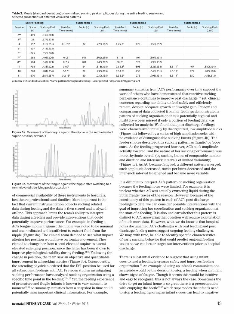
neonatal INTENSIVE CARE Vol. 29 No. 1 n Winter 2016 43
summary statistics from AC’s performance over time support the work of others who have demonstrated that nutritive sucking performance continues to improve past discharge.33 Yet, clinical concerns regarding her ability to feed safely and efficiently remain, despite adequate growth and weight gain. Review and comparison of data collected from her feedings demonstrated a pattern of sucking organization that is potentially atypical and might have been missed if only a portion of feeding data was collected for analysis. We found that post discharge feedings were characterized initially by disorganized, low amplitude sucks (Figure 4a) followed by a series of high amplitude sucks with no evidence of distinguishable sucking bursts (Figure 4b). The feeder’s notes described this sucking pattern as ‘frantic’ or ‘poor start’. As the feeding progressed however, AC’s suck amplitude actually lowered, and the nature of her sucking performance was more rhythmic overall (eg sucking bursts of comparable number and duration and inter-suck intervals of limited variability)(Figure 4c). As AC became fatigued, a different pattern emerged; suck amplitude decreased, sucks per burst decreased and the inter-suck interval lengthened and became more variable.
It is difficult to interpret AC’s pattern of sucking organization because the feeding notes were limited. For example, it is unclear whether AC was actually extracting liquid during the initial frantic traces of the session. However, because of the consistency of this pattern in each of AC’s post discharge feedings to date, we can consider possible interventions with the goal of improving her coordination and sucking organization at the start of a feeding. It is also unclear whether this pattern is distinct to AC. Answering that question will require examination of much more data. However, what we do know is that hospital notes documented AC’s challenges with oral feeding and post discharge feeding notes suggest ongoing feeding challenges. We may, with time, be able to identify specific characteristics of early sucking behavior that could predict ongoing feeding issues so we can better target our interventions prior to hospital discharge.
There is substantial evidence to suggest that using infant cues to lead a feeding increases safety and improves feeding organization.22 An example of using an infant’s communications as a guide would be the decision to stop a feeding when an infant shows signs of fatigue. Though it seems this would be intuitive and easy to recognize, this is not always the case. Sometimes the drive to get an infant home is so great there is a preoccupation with emptying the bottle22,37 which supersedes the infant’s need to stop a feeding. Ignoring an infant’s cues can lead to negative
of commercial availability of these instruments to hospitals, healthcare professionals and families. More important is the fact that current instrumentation collects sucking related data during feeding and the data is then stored and analyzed off-line. This approach limits the team’s ability to interpret data during a feeding and provide interventions that could potentially improve performance. For example, in feeding 4, AC’s tongue moment against the nipple was noted to be minimal and uncoordinated and insufficient to extract fluid from the nipple (Figure 3a). The clinical team decided to see what impact altering her position would have on tongue movement. They elected to change her from a semi-elevated supine to a semi-elevated side-lying position, since the latter has been shown to improve physiological stability during feeding.34,35 Following the change in position, the team saw an objective and quantifiable improvement in all sucking metrics (Figure 3b). Consequently, the attending physician ordered that the ESL position be used for all subsequent feedings with AC. Previous studies investigating sucking performance have analyzed sucking organization using a specific time point in the feeding.7,24,27 Yet, the feeding experience of premature and fragile infants is known to vary moment to moment22,36 so summary statistics from a snapshot in time could potentially miss important clinical information. For example,
Table 2. Means (standard deviations) of normalized sucking peak amplitudes during the entire feeding session and selected subsections of different visualized patterns
Entire Feeding Subsection 1 Subsection 2 Subsection 3
Session Sucks (n)
Sucking Peakµ(σ)
Start-End Time (mins)
Sucks (n) Sucking Peakµ(σ)
Start-End Time (mins)
Sucks (n) Sucking Peakµ(σ)
Start-End Time (mins)
Sucks (n) Sucking Peak(µ(σ))
2*# 419 .339(.203)
3*# 23 .377(.278)
4 157 .418(.251) 0-1.75# 32 .275(.167) 1.75-7^ 125 .455(.257)
5* 207 .411(.235)
6* 225 .356(.228)
7*^ 268 .405(.226) 0-05 141 .502(.250) 11-15 104 .307(.131)
8*^ 904 .344(.173) 0-7.5 281 .446(.207) 08-23 623 .298(.132)
9 798 .433(.222) 0-02# 24 .313(.193) 02-5.5β 303 .526(.238) 5.5-14^ 467 .380(.191)
10 770 .481(.236) 0-1.5# 22 .235(.085) 02-4.5β 258 .648(.231) 4.5-12^ 472 .403(.190)
11 670 .584(.257) 0-2.15# 30 .239(.135) 2.2-5.3β 275 .798(.131) 5.3-11^ 350 .455(.213)
µ=Mean; σ=Standard Deviation; *Same pattern throughout feeding; #Disorganized; ^Organized; βHypervigilant
Figure 3a. Movement of the tongue against the nipple in the semi-elevated supine position, session 4
Figure 3b. Movement of the tongue against the nipple after switching to a semi-elevated side-lying position, session 4

44 neonatal INTENSIVE CARE Vol. 29 No. 1 n Winter 2016
sucking behaviors as a predictor of later neurodevelopmental outcomes in infants with neonatal brain injury. We also are in planning stages for studies designed to gauge the impact of various interventions (eg feeding position, nipple flow rate, pacing etc) on decreasing risk for aspiration using nfant Feeding Solution metrics. We believe a number of these interventions have the potential to reduce the likelihood of later feeding related aversions which hamper adequate growth and nutrition and increase family stress.38
summary & conclusionsSuccessful infant feeding is a complex process that requires integration of physiologic function and neurobehavioral ability. To model AC’s feeding performance over time, we used nfant Feeding Solution which captures variables traditionally reported by researchers (eg suck burst frequency and duration, etc.) and important to the development of oral feeding. For the purposes of this paper, we chose to highlight features exclusive to the instrumentation that translates immediately to decision-making during bedside clinical care. These data can then be used for clinical interpretation of strength and coordination during any given feeding. It is our theory that a medical device such as nfant Feeding Solution will improve the current standard of care for initiation and progression of oral feeding as it goes beyond a ‘trial & error’ approach to feeding based on subjective observations. Objective information will help clinicians avoid complications and navigate infants to faster oral feeds and subsequently earlier and safer hospital discharge with lowered expense.
References1 Hawdon, JM, et al, Identification of neonates at risk of
developing feeding problems in infancy. Dev Med Child Neurol, 2000. 42: p235-239.
2 Thommessen, M, A Heiberg, and BF Kase, Feeding problems in children with congenital heart disease: the impact on energy intake and growth outcome. Eur J Clin Nutr, 1992. 46(7): p457-64.
3 Bryant-Waugh, R, et al, Feeding and eating disorders in childhood Int J Eat Disord, 2010. 43(2): p98-111.
4 Hawdon, JM, et al, Identification of neonates at risk of developing feeding problems in infancy. Dev Med Child Neurol, 2000. 42(4): p235-9.
5 Cerro, N, et al, Eating behaviour of children 15-35 years born preterm: parents’ perceptions. J Paediatr Child Health, 2002. 38(1): p72-8.
6 Burklow, KA, et al, Relationship between feeding difficulties, medical complexity, and gestational age. Nutr Clin Pract, 2002. 17(6): p373-8.
7 White-Traut, R, et al, Lack of feeding progression in a preterm infant: a case study. Adv Neonatal Care, 2013. 13(3): p175-80.
8 Horner, S, et al, Setting the stage for successful oral feeding: the impact of implementing the SOFFI feeding program with medically fragile NICU infants. J Perinat Neonatal Nurs, 2014. 28(1): p59-68.
9 Lau, C and I Kusnierczyk, Quantitative evaluation of infant’s nonnutritive and nutritive sucking. Dysphagia, 2001. 16(1): p58-67.
10 Siddell, EP and RD Froman, A national survey of neonatal intensive-care units: Criteria used to determine readiness for oral feedings. J Obstet Gynecol Neonatal Nurs, 1994. 23(9): p783-789.
11 Bromiker, R, et al, Comparison of sucking patterns at introduction of oral feeding and at term in Israeli and
feeding experiences and later feeding aversion.22 We have found that the addition of objective sucking metrics aids feeders in recognizing a pattern of fatigue spontaneously and they will proceed to stop the feeding without question. Though this finding is only anecdotal, it suggests that objective real-time feedback could positively impact a shift away from volume-driven feeding to infant-driven feeding.
limitations & Future DirectionsInitial results are promising but there were limitations. Comparisons of inter feeding suck amplitude were difficult to make due to the need to normalize the data on a scale from 0 to 1 to the session maximum amplitude achieved. Despite this limitation, we were able to detect intra feeding fatigue and loss of coordination. Going forward, we have implemented a calibration procedure prior to each feeding which eliminates the problem of normalization. Calibration procedures also insure reliability to within manufacturer reliability specifications, 1.2% full scale range. All sucking metrics reported (eg number of bursts, burst length etc.) were determined with custom algorithms, however, identification and categorization of feeding sections and trace patterns was done manually by researchers. Plans to automate this process is ongoing and will improve the validity of reported metrics for clinical interpretation.
We have a number of projects currently underway that include modelling sucking parameters pre- and post- surgery in infants with ankyloglossia, modeling tongue movement post-surgery in infants with congenital heart disease and investigating early
Figure 4a. Movement of the tongue against the nipple at the beginning of session 11
Figure 4b. Movement of the tongue against the nipple following 4a, session 11
Figure 4c. Movement of the tongue against the nipple following 4b, session 11

neonatal INTENSIVE CARE Vol. 29 No. 1 n Winter 2016 45
conceptual age. J Dev Behav Pediatr 2002. 23(4): p231-236.34 Clark, L, et al, Improving bottle feeding in preterm infants:
Investigating the elevated side-lying position. Infant, 2007. 3(4): p154-158.
35 Park, J, et al, Efficacy of semielevated side-lying positioning during bottle-feeding of very preterm infants: A pilot study. J Perinat Neonatal Nurs, 2014. 28(1): p69-79.
36 Shaker, C, Cue-based co-regulated feeding in the Neonatal Intensive Care Unit: Supporting parents in learning to feed their preterm infant. Newborn Infant Nurs Rev, 2013. 13: p51-55.
37 Swift, MC and I Scholten, Not feeding, not coming home: parental experiences of infant feeding difficulties and family relationships in a neonatal unit. J Clin Nurs, 2010. 19(1-2): p249-58.
38 Ross, ES and JV Browne, Feeding outcomes in preterm children after discharge from the neonatal intensive care unit (NICU): A systematic review. Newborn & Infant Nursing Reviews, 2013. 13: p87-93.
American preterm infants. Acta Paediatr, 2005. 94: p201-204.12 Ludwig, S and K Waitzman, Changing feeding documentation
to reflect infant-driven practice. Newborn Infant Nurs Rev, 2007. 7(3): p155-160.
13 Bosma, JF, Form and function in the infant’s mouth and pharynx, in Third Symposium on Oral Sensation and Perception, J.F. Bosma, Editor. 1972, Charles C. Thomas: Springfield, IL. p3-29.
14 Bauer, MA, et al, The oral motor capacity and feeding performance of preterm newborns at the time of transition to oral feeding. Braz J Med Biol Res, 2008. 41(10): p904-7.
15 Geddes, DT, et al, Tongue movement and intra-oral vacuum in breastfeeding infants. Early Hum Dev, 2008. 84(7): p471-7.
16 da Costa, SP, L van den Engel-Hoek, and AF Bos, Sucking and swallowing in infants and diagnostic tools. J Perinatol, 2008. 28(4): p247-57.
17 Bu’Lock, F, M Woolridge, and J Baum, Development of co-ordination of sucking, swallowing and breathing: Ultrasound study of term and preterm infants. Dev Med Child Neurol, 1990. 32: p669-678.
18 Wolf, LS and RP Glass, Feeding and swallowing disorders in infancy: Assessment and management 1992, Tucson: Therapy Skill Builders.
19 McCain, G, Behavioral state activity during nipple feedings for preterm infants. Neonatal Network, 1997. 16: p43-47.
20 Robbins, J, et al, The effects of lingual exercise on swallowing in older adults. J Am Geriatr Soc, 2005. 53(9): p1483-9.
21 Capilouto, GJ, et al, Comparison of tongue muscle characteristics of preterm and full term infants during nutritive and nonnutritive sucking. Infant Behavior & Development, 2014. 37(3): p435-445.
22 Shaker, CS, Cue-based feeding in the NICU: using the infant’s communication as a guide. Neonatal Netw, 2013. 32(6): p404-8.
23 Breton, S and S Steinwender, Timing introduction and transition to oral feeding in preterm infants: Current trends and practices. Newborn Infant Nurs Rev, 2008. 8(3): p153-159.
24 Lau, C and EO Smith, A novel approach to assess oral feeding skills of preterm infants. Neonatology, 2011. 100: p64-70.
25 Capilouto, G and A Lattin, The Infant Feeding Scale. 2011, University of Kentucky: Lexington.
26 Scheel, CE, RJ Schanler, and C Lau, Does the choice of bottle nipple affect the oral feeding performance of very low-birthweight (VLBW) infants? Acta Paediatri, 2005. 94: p1266-1272.
27 White-Traut, R, et al, Evaluating sucking maturation using two pressure thresholds. Early Hum Dev, 2013. 89(10): p833-7.
28 Mizuno, K and A Ueda, The maturation and coordination of sucking, swallowing, and respiration in preterm infants. J Pediatr, 2003. 142(1): p36-40.
29 Lang, WC, et al, Quantification of intraoral pressures during nutritive sucking: methods with normal infants. Dysphagia, 2011. 26(3): p277-86.
30 Miller, JL and SM Kang, Preliminary ultrasound observation of lingual movement patterns during nutritive versus non-nutritive sucking in a premature infant. Dysphagia, 2007. 22(2): p150-60.
31 Medoff-Cooper, B, WB Bilker, and JM Kaplan, Suckling behavior as a function of gestational age: A cross-sectional study. Infant Behav Dev, 2001. 24: p83-94.
32 Lau, C, et al, Characterization of the developmental stages of sucking in preterm infants during bottle feeding. Acta Paediatri, 2000. 89(7): p846-52.
33 Medoff-Cooper, B, JM McGrath, and J Shults, Feeding patterns of full-term and preterm infants at forty weeks post-

46 neonatal INTENSIVE CARE Vol. 29 No. 1 n Winter 2016
Supporting breastfeeding and the provision of breast milk is an important task in the NICU. Dozens of studies now show improved outcomes specific to extremely premature infants. Babies who receive breast milk have fewer infections, sepsis, necrotizing enterocolitis, less chronic lung disease, retinopathy of prematurity, reduced time to full enteral feeds, and have been linked with better cognitive outcomes.1-7 Over the long term, breast milk provides unique advantages into adulthood that influence such things as cardiovascular health, bone health and cognitive function.8,9 Breastfeeding may also reduce risk of obesity and diabetes and has been linked with lower cholesterol levels in later life.9,10 In addition to these medical health benefits, there are also psychological benefits to breastfeeding. The skin-to-skin contact during breastfeeding helps in physiologic stabilization of the infant and increased maternal infant bonding in a situation where this is more difficult — the NICU.
Most NICU nurses today agree that breast milk is at least important in the immediate period and particularly as related to prevention of necrotizing enterocolitis. It may sometimes be difficult to understand why the promotion of breastfeeding is such a challenge in the NICU. I have recently come across a study by Cricco-Lizza11 that puts this into perspective. This is an ethnographic study that reports the results of interviews with nurses in a Level IV or highest level NICU. This NICU had recently had some breastfeeding education but not all staff attended this education. The purpose of the study was to examine everyday practice values and explore how breastfeeding promotion fits into this.
I recently wrote a blog about the cultural influence of breastfeeding, focusing on the culture of the mother. However, there is a culture that exists in the NICU as well. Culture may be defined as “a learned social behavior of a particular group of people.” This unique culture in the NICU is described by Cricco-Lizza11 as focusing on tight control of actions, reliance on technology and maximal efficiency in use of time with the overall themes of uncertainty and teamwork or “sisterhood.” The reports of nurses who were interviewed for this study are very familiar to me! As a staff nurse, getting the routine, once-a-shift “chores” taken care of early in the shift was always important because you never knew when a sick baby might come through the doors,
taking away any extra time you might have thought you would have or one of the babies on your assignment list would take a turn for the worse, become unstable and require far more care than at first anticipated. This theme of uncertainty is extremely common in the intensive care setting. The tight control of actions refers to things like the careful and precise measurement of medications, intravenous fluids, oxygen and recognizing small changes in infants early enough to respond quickly.
Reliance on technology becomes so important in the NICU to almost be distracting from attention to the patient! More and more technology provides the tight control desired but also creates more dependence on the technology and brings another level of challenge to the nurse at the bedside. The length of orientation, particularly to high level NICU care, is necessarily longer today because there are so many more machines that must be learned. Those already experienced in NICU care learn new technology as it comes along but still can feel overwhelmed in order to get skilled at the use and interpretation of an onslaught of information!
The final theme Cricco-Lizza11 describes is maximal efficiency in use of time. Talk to any NICU nurse and she/he will tell you about how busy the day can be! Not usually every day but many days, particularly in high-acuity, high-census units with active birthing units where babies come into the world with different problems and in need of NICU care without notice can be very, very busy. While nurse-patient ratios may help, extra nurses are not always available and certainly for unexpected admissions or changes in patient condition.
So, how does this impact the promotion of breastfeeding? One can see that the importance of efficiency, tight control and reliance on technology fly in the face of the absolutely “tech-less” task of breastfeeding, where nurses have no control other than perhaps when it will be done. Infants who are learning to eat are less than efficient whether breastfeeding or bottle feeding but with bottle feeding, one can measure the progress at least by watching milk disappear.
The unit described by Cricco-Lizza11 was working on improving breastfeeding and many of the nurses interviewed could see the benefit and need for the change. There are many aspects of the NICU that are needed to create a culture change that fosters this move toward breastfeeding friendliness. Bonet, Forcell, Blondel, Draper, Agostino, Cuttini and Zeitlin12 describe some measures in units that had higher breastfeeding rates versus those with low
Breastfeeding in the NICU: Is it Valued Enough?Sandy Sundquist Beauman, MSN, RNC-NIC, CNS
Sandra Sundquist Beauman is a research nurse coordinator at the University of New Mexico. She is also an independent consultation with Medela, Inc. and provides neonatal consultation and continuing education through CNS Consulting.


48 neonatal INTENSIVE CARE Vol. 29 No. 1 n Winter 2016
3 Meinzen-Derr J, Poindexter B, Wrage L, Morrow AL, Stoll B, Donovan EF. Role of human milk in extremely low birth weight infants’ risk of necrotizing enterocolitis or death. J Perinatol. Jan 2009;29(1):57-62.
4 Sisk PM, Lovelady CA, Dillard RG, Gruber KJ, O’Shea TM. Early human milk feeding is associated with a lower risk of necrotizing enterocolitis in very low birth weight infants. J Perinatol. Jul 2007;27(7):428-433.
5 Vohr BR, Poindexter BB, Dusick AM, et al. Persistent beneficial effects of breast milk ingested in the neonatal intensive care unit on outcomes of extremely low birth weight infants at 30 months of age. Pediatrics. Oct 2007;120(4):e953-959.
6 Hylander MA, Strobino DM, Pezzullo JC, Dhanireddy R. Association of human milk feedings with a reduction in retinopathy of prematurity among very low birthweight infants. Journal of perinatology. Sep 2001;21(6):356-362.
7 Patel AL, Johnson TJ, Engstrom JL, et al. Impact of early human milk on sepsis and health-care costs in very low birth weight infants. J Perinatol. Jan 31 2013.
8 Lucas A. Long-term programming effects of early nutrition – Implications for the preterm infant. J Perinatology. 2005;25(S2-S6).
9 Ip S et al. Breastfeeding and maternal and infant health outcomes in developed countries. Evid Rep Tech Assess 2007.
10 Owen CG, Whincup PH, Kaye SJ, Martin RM, Davey Smith G, Cook DG et al. Does initial breastfeeding lead to lower cholesteral in adult life? A quantitative review of the evidence. American Journal of Clinical Nutrition. 2008;88:305-314.
11 Cricco-Lizza, Roberta. “Everyday nursing practice values in the NICU and their reflection on breastfeeding promotion.” Qualitative health research. 2010;21(3):399-409.
12 Bonet, Mercedes, et al. “Approaches to supporting lactation and breastfeeding for very preterm infants in the NICU: a qualitative study in three European regions.” BMJ open6 (2015): e006973.
rates. Units with higher rates encouraged parental presence at all times, provided space for pumping and direct breastfeeding, and talked to all mothers about breastfeeding and pumping early after delivery. One important point to make here is that the benefit of provision of breast milk is not just during the initial and critical period the infant spends in the NICU. The benefit extends beyond discharge. Many nurses feel that by encouraging the mother to pump early after birth and provide milk for the first few weeks or even during the NICU stay is success. While this is better than nothing, the benefits of breast milk extend far beyond the NICU stay and should be encouraged.
Mothers faced with caring for a medically complex/fragile infant may be willing to give up breast feeding unless the importance of continuing is stressed. Also, assisting the mother with direct breast feeding before discharge where she can be assured that the infant is receiving enough milk and tolerating it well will help her to be more confident in continuing to breast feed. One of the units described in the Bonet et al12 study did not place importance on direct breast feeding prior to discharge. An interviewee in this unit said that it would be better to have the mother put the infant to breast after discharge where she had more time and the baby could latch on gradually. Another unit was concerned about the bacterial content of the freshly expressed milk and tested all milk or pasteurized all milk, including mother’s own milk prior to feeding. This meant that the infant had little of the mother’s milk early on, leading to mothers thinking that their milk was unclean and not worth the effort to pump, also creating worry that it may not be good for the infant.
Bonet et al12 found that units with policies and procedures, particularly encouraging open and extensive visitation of mothers, advocating for early pumping, early use of mother’s own milk and starting direct breast feeding well before discharge were most helpful in sustained breastfeeding during and after discharge. Nurses who either had their own positive experience with breast feeding or had breast feeding education were more likely to be supportive and encourage mothers to provide breast milk and breastfeed. NICU nurses, in this study, were observed to focus more on the scientific evidence of breast feeding when talking to parents about it rather than the psychologic/psychosocial aspects. This appears to reflect the focus on technology and the science of infant care that was a high priority in this high level NICU. The findings in a NICU caring for less critical infants may be different and may show a different type of support for breast feeding mothers. In order to change breast feeding support, several aspects of unit culture must be considered. This may include taking into account the time needed for breast feeding support when calculating acuity and staffing levels. Placing value on the time required to support breastfeeding in this manner will provide the necessary time for it to be done. Often, the lower technologic measures like proper positioning, sound control and yes, encouraging breast feeding, make as much difference in outcome as the latest electronic equipment used in the NICU.
References1 Johnston M, Landers S, Noble L, Szucs K, Viehmann L.
Breastfeeding and the use of human milk. Mar 2012;129(3):e827-841.
2 Schanler RJ, Lau C, Hurst NM, Smith EO. Randomized trial of donor human milk versus preterm formula as substitutes for mothers’ own milk in the feeding of extremely premature infants. Pediatrics. Aug 2005;116(2):400-406.
neonatal INTENSIVE CARE Vol. 29 No. 1 n Winter 2016 49
Abstractintroduction: Mechanical ventilation of neonates requires close attention to volume and pressure delivery. The high frequency oscillator creates a challenge because delivered volumes and peak inspiratory pressures are not provided. Aerosol medication delivery during high frequency ventilation further complicates the scenario. There is currently little research regarding the effects of nebulizer use during high frequency oscillatory ventilated neonates. This lack of knowledge could increase the risk of lung damage.
methods: This bench top study used an experimental design that did not involve human subjects. Seven different nebulizers were inserted into the circuit. The tidal volumes and peak inspiratory pressures were recorded twice using a one-way valve and twice without the one-way valve. Pressures and volumes were recorded using the RespiTrainer Infant (IngMar Medical, Pittsburgh, PA). Nebulizer liter flow was adjusted per recommendation of particular modality. An analysis of variance (ANOVA) was used to analyze the data and a Tukey procedure for post hoc analysis. A p value of 0.05 was used to determine statistical analysis.
results: Our one-way ANOVA results demonstrated a significant difference between nebulizer type and volume change (p=.0001) and pressure change (p=.002). On post-hoc analysis there was significant differences (p<.05) between the AeroEclipse and the NebuTech HDN, Hudson MicroMist, Mini-HEART, Uni-HEART, and EZflow MAX in terms of volume change and pressure change.
conclusion: The study findings demonstrated that nebulizer type produced very similar results, with the exception of the AeroEclipse nebulizer. Nebulizer type does not appear to produce a major advantage or disadvantage when considering pressure and volume changes with HFOV. These findings support a variety of nebulizer brands when considering aerosol delivery with HFOV.
IntroductionHigh frequency oscillatory ventilation (HFOV) is used to ventilate and oxygenate patients in an effort to protect the lungs. HFOV is most often used in the NICU to provide ventilatory support and to prevent lung injury that may result from conventional positive pressure mechanical ventilation. HFOV accomplishes this by using very small tidal volumes—smaller than anatomical dead space—and supraphysiologic respiratory rates.1 Despite very high respiratory rates and very low tidal volumes, HFOV can achieve stable mean airway pressure and uniformed lung inflation, potentially leading to lower FiO2 use, improved oxygenation and equal survival rates to conventional mechanical ventilation.2-5 HFOV began in the Neonatal Intensive Care Unit (NICU) to provide rescue support and reduce lung injury that may result from conventional mechanical ventilation. However, the need to deliver nebulized medications did not cease when transitioning to this type of ventilation. Currently there is very little research investigating the impact of nebulizer use on delivered volumes and pressures. The use of nebulization in HFOV is likely to be as effective as nebulization with traditional mechanical ventilation, but the extent to which tidal volumes and inspiratory pressures are altered due to several different nebulizer brands is unknown. This has been unchartered primarily because of the delicateness of the neonatal population being tested.6,7
The research question for this project was does nebulizer brand create significantly different changes in delivered tidal volume and inspiratory pressures when using the HFOV and a neonatal test lung? The null hypothesis states there will be no difference in delivered tidal volumes and pressures when different nebulizers are used in-line with HFOV and a test lung. While the alternative hypothesis states that there will be a difference in delivered gas volume and pressures with different nebulizers placed in-line with HFOV. These vulnerable patients require respiratory therapists to provide adequate support while still preventing long-term lung damage.
A Comparison of Nebulizer Brand On Delivered Tidal Volumes and Peak Inspiratory Pressure During High Frequency Oscillatory VentilationChristopher J. Russian, Ph.D., RRT-NPS, RPSGT, Joshua F. Gonzales, MHA, RRT-NPS, RRT-SDS, Hanah Schelde, Lauren A. Terry, Shireen Albanna, Renee Adams
Chris Russian is an Associate Professor and Director of Clinical Education in the Department of Respiratory Care at Texas State University. Dr. Russian has research and grant activity on respiratory muscle training, mechanical ventilation, suction catheters, and inter-rater reliability. Joshua Gonzales is an Associate Professor in the Department of Respiratory Care at Texas State University. Mr. Gonzales has research and grant activity on high flow nasal cannula, mechanical ventilation, suction devices, and learning styles. Hanah Schelde, Lauren Terry, Shireen Albanna and Renee Adams were students in the Department of Respiratory Care at Texas State University at the time this project was conducted. There are no conflicts of interest reported by any of the authors. Prior abstract publication/presentation: Chris Russian, Hanah Schelde, Shireen Albanna, Renee Adams, Lauren Terry completed an oral and poster presentation of this research at the Texas State University Respiratory Care Research Forum on April 17, 2015 at Texas State University, San Marcos, TX.

50 neonatal INTENSIVE CARE Vol. 29 No. 1 n Winter 2016
There was no significant difference on post hoc analysis between the PariLCplus, NebuTech, MicroMist, Mini-HEART, Uni-HEART, and EZFlow. There was no significant difference between the PariLCplus and the Aeroclipse for volumes or pressures. All volume and pressure data were generated with and without a one-way valve between the nebulizer and the HFOV circuit to remain consistent with clinical practice around the country. There was no significant difference, per one-way ANOVA, in volume change or pressure change when using and not using the one-way valve.
Table 1: Nebulizer Volume and Pressure Mean Values
NebulizerVolume (mL)
± SDPeak Pressure (cmH2O)
± SD
Pari LCplus 65.35 ± 4.06 18.60 ± 1.04
Nebutech HDN 70.00 ± 0.57 19.80 ± 0.00
Hudson MicroMist 70.00 ± 0.57 19.80 ± 0.00
Mini-HEART 69.00 ± 0.57 19.40 ± 0.00
Uni-HEART 68.75 ± 0.96 19.40 ± 0.00
AeroEclipse 58.00 ± 7.51 16.90 ± 2.19
EZFlow MAX 69.50 ± 0.00 19.80 ± 0.00
DiscussionThe study findings demonstrated that nebulizer type produced very similar results, with the exception of the AeroEclipse nebulizer. We also discovered a negative change in volumes and pressures compared to baseline values for the Aeroclipse and the PariLCplus. The Aeroclipse had a lower reduction in peak pressure and volume compared to the PariLCplus. The most likely explanation for a negative change in data variables is a leak in the system. All nebulizers used similar connections between the nebulizer and the circuit. However, it is possible that a leak existed between the top of the each nebulizer and the body of the nebulizer. However, we did not investigate if a leak was present or the source of any leak. Another possibility is that the pressure in the HFOV circuit produced backpressure within the two nebulizers. In this case there would be a reduction in aerosol entering the HFOV circuit. We did not investigate aerosol output with this study.
Nebulizer type does not appear to produce a major advantage or disadvantage when considering pressure and volume changes with HFOV. These findings support a variety of nebulizer brands when considering aerosol delivery with HFOV. The one exception, the AeroEclipse, produced a significantly lower volume and pressure compared to the other brands included in this study. We don’t believe nebulizer flow rate setting caused this significant difference because several of the other nebulizers were set at the same rate, eg 6 Lpm. In addition, a couple of nebulizers operated on lower flow rates, eg 2 Lpm. One possibility is the AeroEclipse put out a smaller amount of aerosol compared to the other nebulizers. A smaller amount of aerosol output could produce a lower volume and pressure delivery. However, there is no literature to support this conclusion. Equally, aerosol output was not investigated in this study. Another explanation is the possibility of a small leak within the manufactured components of the device. However, we did not notice any changes in baseline HFOV settings, ie mean airway pressure reduction or amplitude reduction. Additional research is needed to determine why AeroEclipse produced lower volumes and pressures compared to the other nebulizers used for this study. Based on these findings we rejected the null hypothesis.
MethodsThe experimental design allowed for a benchtop study that did not require human subjects. The use of a test lung versus actual cadaver lungs or human subjects allowed us to control for potential complications, ie air leaks, associated with HFOV in neonates.8,9 The project used the High Frequency Oscillatory Ventilator (HFOV) 3100A mechanical ventilator (CareFusion, San Diego, CA). Baseline settings for the HFOV were: mean airway pressure 21 cmH2O, amplitude 36 cmH2O, inspiratory time percent 0.33, hertz 15, bias flow 20 L/min. The experiment included seven different nebulizers that are commonly used in hospital settings. All nebulizers were placed on the inspiratory limb distal to the humidifier. These nebulizers included: PARI LCplus (PARI Respiratory Equipment, Inc, Midlothian, VA), NebuTech HDN (Salter Labs, Alvin, CA), Hudson MicroMist (Teleflex, Morrisville, NC), Mini-HEART Lo-Flo (Westmed Inc, Tucson, AZ), Uni-HEART (Westmed Inc, Tucson, AZ), AeroEclipse (Monaghan Medical, Plattsburgh, NY), and EZflow MAX (Mercury Medical, Clearwater, FL). AeroEclipse was operated in continuous nebulization mode. The RespiTrainer Infant (IngMar Medical, Pittsburg, PA) was used to measure the volume and pressure produced by the HFOV and nebulizers. Firstly, we intubated the RespiTrainer Infant with a size 3 mm endotracheal tube (SunMed, Largo, FL), and calibrated the RespiTrainer infant unit. Mean baseline values generated for the HFOV without a nebulizer inline were tidal volume of 67.5mL and peak inspiratory pressure of 19.4 cmH2O. Next, nebulizers were placed, one at a time, in the same position in the inspiratory limb of the HFOV circuit, distal to the humidifier. We performed two trials, without using a one-way valve between the nebulizer and circuit, to assess the effect of the nebulizer output on volume and pressure changes delivered to the RespiTrainer. Then a one-way valve was added between the nebulizer and the HFOV circuit and two more trials were performed, ultimately testing each nebulizer four times. Using and not using a one-way valve was implemented to remain consistent with clinical practice. The liter flow was adjusted according to the manufacturers’ recommendations for each nebulizer using an air flow meter. The PARI LC plus was operated on 6 L/min, Nebutech HDN on 6 L/min, Hudson MicroMist on 6 L/min, Mini-HEART on 2 L/min, Uni-HEART on 2 L/min, AeroEclipse on 6 L/min, and the EZflow MAX on 6 L/min. Normal saline was added to the chamber of each nebulizer to mimic actual nebulizer function and output. A blue filter was added between the HFOV circuit and the ETT to limit aerosol delivery to the infant trainer. An analysis of variance (ANOVA) was used to analyze the data and a Tukey procedure for the post hoc analysis. A p value of 0.05 was used to determine statistical analysis.
ResultsOur one-way ANOVA analysis demonstrated a significant difference between the nebulizer type and the volume change (p=.0001) and pressure change (p=.002). On post-hoc analysis there was a significant difference for volume change between the AeroEclipse nebulizer and NebuTech HDN (p=.001), between the AeroEclipse and Hudson MicroMist (p=.001), between the AeroEclipse and Mini-HEART (p=.002), between the AeroEclipse and Uni-HEART (p=.002), between the AeroEclipse and EZFlow MAX (p=.001). On post-hoc analysis there was a significant difference for pressure change between AeroEclipse and Nebutech HDN (p=.003), between AeroEclipse and Hudson MicroMist (p=.003), between AeroEclipse and Mini-HEART (p=.014), between AeroEclipse and Uni-HEART (p=.014), between AeroEclipse and EZFlow MAX (p=.003). See Table 1.

neonatal INTENSIVE CARE Vol. 29 No. 1 n Winter 2016 51
clinical practice: methodology, recommendations, and future developments. Eur Respir J. 2003;22(1):1026-1041.
5 Rettwitz-Volk W, Veldman A. A prospective, randomized, multicenter trial of high-frequency oscillatory ventilation compared with conventional ventilation in preterm infants with respiratory distress syndrome receiving surfactant. J Pediatr. 1998;132(2):249-254.
6 Ari A, Atalay OT. Influence of nebulizer type, position, and bias flow on aerosol drug delivery in stimulated pediatric and adult lung models during mechanical ventilation. Respir Care. 2010;55(7):845-851.
7 Sedeek KA, Takeuchi M, Suschodolski K, Kacmarek RM. Determinants of tidal volume during high frequency oscillation. Crit Care Med. 2003;31(1):227-31.
8 Garner S, Wierst D. Albuterol delivery by metered-dose inhaler in a pediatric high-frequency oscillatory ventilation model. Crit Care Med. 2000;28(6):2086-9.
9 Hager, DN, Fessler HE, Kaczka DW, et al. Tidal Volume delivery during high frequency oscillatory ventilation in adults with acute respiratory distress syndrome. Crit Care Med. 2007;35(6):1522-1529.
10 Cameron D, Clay M. Evaluation of nebulizers for use in neonatal ventilatory circuits. Crit Care Med. 1990;18(8):866-70.
11 Fink JB. Aerosol delivery to ventilated infant and pediatric patients. Respir Care. 2004;49(6):653-665.
12 Clay MM, Pavia D. Factors influencing the size distribution of aerosols from jet nebulizers. Thorax. 1983;38(1):755-759.
This study has several implications for the medical community. First, the results contributed specific and novel information on the effects that nebulizers have on HFOV delivered ventilator parameters. It is important to the safety of the neonate to appropriately choose a nebulizer type that will allow for safe administration of aerosolized medications. It is known that this area of study is relatively new and uncharted.10 Second, our research could ultimately lead to advancement in equipment design, nebulizer set-up and policy development in regards to nebulizer use for neonatal patients on HFOV. Further investigation is needed to understand the reason for the negative change in peak pressures and delivered volumes. If backpressure or a leak are the cause then nebulizer design may need to be addressed. Lastly, respiratory departments can use this information when deciding on which nebulizer to purchase and use with HFOV. Other studies have focused on aerosol deposition amongst different nebulizer types for neonates, but not the delivered pressures and tidal volume.11,12
These vulnerable patients require respiratory therapists and equipment to provide adequate support while still preventing long-term lung damage. Due to the limited amount of patient monitored variables with the HFOV 3100A there is uncertainty in the impact of aerosol administration on volume and pressure changes. This study provides an analysis of two important parameters during mechanical ventilation, ie delivered volumes and inspiratory pressures. Although amplitude is provided on the 3100A this pressure does not represent the peak inspiratory pressure.
LimitationsThere are several limitations of this study. First, we did not investigate aerosol output for each device. Placing a “catch” filter distal to the nebulizer and then weighing the filter at the end of each trial would provide aerosol output data. Second, we did not investigate if similar pressure and volume changes would occur with different baseline HFOV settings. We do not know if the same pressure and volume changes would occur with lower mean airway pressure and amplitude settings. Third, we did not investigate all available nebulizer brands. Specifically, we did not include any vibrating mesh nebulizers. At the time of this project we did not have access to an Aerogen (Galway, Ireland). We attempted to use the Omron MicroAir (Omron Healthcare, Lake Forest, IL); however, the design of the nebulizer created a leak in the set-up circuit. The final limitation of this study involves the bench top design we selected. Although the data collected was reliable we do not know if similar findings will occur in human subjects. For this reason our results must be interpreted with caution when making clinical decisions about nebulizer selection with the HFOV 3100A.
References1 Notter RH, Finkelstein JN, Holm BA, eds. Lung injury:
mechanisms, pathophysiologic, and therapy. Boca Raton, FL: Taylor and Francis Group;2005.
2 Cools F, Askie LM, Offringa M, et al. Elective high-frequency oscillatory frequency oscillatory versus conventional ventilation in preterm infants: a systemic review and meta-analysis of individual patient’s data. Lancet. 2010;375(9731):2082-2091.
3 Sood BG, Shen Y. Effective aerosol delivery during high-frequency ventilation in neonatal pigs. Respirology. 2010;15(3):551-555.
4 Oostveen E, Macleod D. The forced oscillation technique in

52 neonatal INTENSIVE CARE Vol. 29 No. 1 n Winter 2016
The goal of this presentation is to introduce a new concept — a wearable technology — to obstetrical practice. The original purpose was to minimize the risks of stillbirth. However, as the technology evolved, other benefits became evident, e.g. monitoring fetuses in early labor without the patient being admitted to a hospital. This new technological feature will be appreciated by patients, doctors, and hospitals. Patients will be allowed to stay home longer, provided that monitoring results are normal. Hospitals and doctors will be relieved from unnecessary in-house monitoring, admissions, and check-ups.
In industrialized countries, there is an expectation that every pregnancy will end with the birth of a healthy child. Yet, one fetus out of 200 is stillborn. Sudden fetal death syndrome, also known as stillbirth, continues to be an emotionally devastating outcome for both parents and caregivers. The global burden of stillbirth is estimated at 3.2 million fetal deaths every year. In the USA, the mortality rate from stillbirths is equal to that from prematurity and sudden infant death combined.1
probable causes of stillbirthAbnormalities of the placenta and umbilical cord are the probable underlying causes of stillbirth in 25% of cases.2,3 Placental abruption is the cause of approximately 4% of stillbirths.4
The relationship between intrauterine infection and the stillbirth rate is influenced by gestational age. While infection is blamed for 20% of stillbirths in the second trimester it’s contribution to the rate of term stillbirth is far smaller.6 Recently evidence suggests that a patient’s history of cesarean delivery increases the risk of placental abruption in subsequent pregnancies.6 The incidence of placental abruption in the USA has increased by 23% in the last 20 years.7 It is known that even with a complete diagnostic workup, a significant proportion of stillbirths remains unexplained. This is a frustrating outcome for both the parents, who are no wiser as to why they have lost a fully-grown baby, and the caregivers who have the difficult task of counseling the couple with no clear idea as to the risk of recurrent stillbirth in future pregnancies.1 The literature points out that increased risk of stillbirth is detected in mothers over the age of 35, those with complicated pregnancies, and those with histories of previous stillbirths. However, an overwhelming number of stillbirths are occurring in women with no high-risk factors whatsoever.
review of warning signs of fetal demise in early pregnancyDespite the shattering effect of stillbirth on patients’ lives and the enormity of the problem, there are very few published studies analyzing trends in the causes of stillbirth. A PubMed search failed to find any prospective studies on the signs, precautions, and practical ways of preventing stillbirth. Most research papers on warning signs of impending fetal loss are limited to early pregnancy. Thus, Laboda et al,5 reported that normal first trimester fetal heart rates rise from an average of 100 bpm at 5 to 6 weeks to 140 bpm at 8 to 9 weeks. The heart rates of 65 consecutive first trimester fetuses between 5 and 8 weeks were measured to determine whether an unusually slow fetal heartbeat is associated with a poor outcome. All five pregnancies in which heart rates were below 85 ended in spontaneous miscarriages. This study suggests that first trimester bradycardia may be associated with a poor prognosis for the pregnancy.
These observations were confirmed by a number of prospective studies.9,10 Although first trimester fetal physiology is different from that of viable fetuses, fetal heart monitoring remains the most important tool to assess fetal wellbeing.
What do we learn from sudden infant death syndrome (siDs)? In developed countries SIDS is the most common cause of death in children aged between one and 12 months. In 1989, an American panel of experts defined SIDS as the sudden death of an infant less than one year of age which remains unexplained after a thorough case investigation including performance of an autopsy. Early research suggested that apnea attacks were precursors of SIDS, which led to the provision of home apnea monitors for infants considered to be at risk.11
Recently, the American Academy of Pediatrics put together a policy statement on SIDS. It states that after case investigation, SIDS cases can be attributed to asphyxia, infection, metabolic diseases, and arrhythmias.12 Cardiorespiratory monitors can be used at home to detect apnea, and bradycardia.
Stillbirth is chronologically situated between early fetal demise and SIDS. Approximately 65% of unexplained stillbirths occur after 35 completed weeks with risk increasing as the gestation advances.13 The most significant risk factor for unexplained stillbirth, besides female gender and pregnancy, is advanced maternal age. Among women younger than age 35, the risk of
Fetal Heart Vestment Monitor research in progress
BM Petrikovsky, MD, PhD
BM Petrikovsky, MD, PhD, Professor of Obstetrics & Gynecology is an editorial board member for Neonatal Intensive Care.

neonatal INTENSIVE CARE Vol. 29 No. 1 n Winter 2016 53
resulted when women used the simple device. The 24 pregnancy complications were actually picked up by the device, which prompted doctors to perform emergency C-sections. Results from the study have been submitted for publication in the European Journal of Obstetrics & Gynecology and Reproductive Biology, but have yet to be published.
Another similar product on the market is made by NURO group, under the name of RITMO, which is a pregnancy belt equipped with speakers to play music to the fetus. NURO group is planning to eliminate the speakers and upgrade the system by combining EKG and Doppler sensors. Recently, an Italian researcher introduced a pregnancy belt device named telefetalcare, which is EKG based.
A recent study funded by the Eunice Kennedy Shriver National Institute of Child Health and Human Development and Neoventa Medical; ClinicalTrials.gov number, NCT01131260, which included a total of 11,108 patients, concluded that fetal EKG ST-segment analysis, used as an adjunct to conventional intrapartum electronic fetal heart-rate monitoring, did not improve perinatal outcomes.19
Fetal heart monitor vestment If weekly fetal testing undoubtedly contributes to the reduction of stillbirths, then continuous monitoring should significantly reduce stillbirths. We propose a special pregnancy-adjusted garment or pregnancy belt that will allow us to monitor fetal heart rate continuously and be alerted if fetal heart rate patterns become non-reassuring (Figure A). Similar monitors have been inserted into T-shirts to alert its owner on the possible signs of a heart attack. We realize that to date, there is no guarantee that such monitoring will prevent stillbirth in all cases. However, it appears to be the way to monitor fetuses, accumulate research data, and provide the best chances to avoid stillbirth. Distant fetal monitoring has been used before, first by Corometrics, and recently by an Israeli group of researchers and engineers. Our vestment for fetal monitoring is far superior to existing systems.
A monitor and control unit integrated with a garment that constantly analyzes EKG signals in real time for cardiac events such as arrhythmias and bradycardias/decelerations can automatically generate a local alert. The alert can be forwarded via Bluetooth and WiFi to a smart phone, and simultaneously to a medical professional and/or remote monitoring service for further analysis and for a plan of action. A scrolling graph display of fetal heart rate history is easy to obtain and kept on a cloud-based server as a subscription service. Anyone, mother or professional, could sign on and look at the last 30 seconds or 5 minutes, with a single zoom knob or screen button to expand the “paper” roll. The silver-threaded elastic Health Watch material with 3 sewn-in electrodes is harmless, and picks up signal like a radio.
Another option is having no wires at all. In or on the garment would be both the thumb-sized wireless transmitters and thumb-sized microcomputer capable of transmitting to a local WiFi network the most recent 30 seconds of the beat-to-beat heart rate.
The digital transmitters have channel codes so there could be any number of them in a maternity floor without the signals colliding. Prospective mothers could continue walking around
stillbirth is relatively low, (1.1 per 1000 births). For women aged 35 years and older, the rate is 3.6 per 1000 births, and for women 40 years of age or older, 4 per 1000.13 Although stillbirth is relatively rare, even in older women, it occurs six times more than death by SIDS, which was reported to occur at a rate of 0.6 per 1000 births.14
Fretts, et al14 conducted a study to compare three strategies for the prevention of stillbirth in women aged 35 years and older, who were provided standard care; namely, no antepartum testing or induction before 41 weeks, weekly testing at 37 weeks with induction after a positive test, and no testing with induction at 41 weeks.
The study showed that without a strategy of antepartum surveillance between 37 and 41 weeks, women would experience 5.2 unexplained stillbirths per 1000 pregnancies. For nulliparous women, weekly antepartum testing initiated at 37 weeks would avert 3.9 stillbirths per 1000 pregnancies.14,15
Recently, Waldenstrom, et al16 reported the results of a population-based registry including all women aged 25 years and older with singleton pregnancies at 28 weeks of gestation in Sweden from 1990 to 2011; a total of 1,804,442 pregnancies were analyzed. Stillbirth rates increased by maternal age: 25-29 years 0.27%; 30-34 years 0.31%; 35-39 years 0.40%; and 40 years or older 0.53%. Compared with age 25-29 years, this increase was approximately 25% at 30-34 years and doubled at the age of 35. The authors postulated that placental constraints may be the reason for the association between advanced maternal age and stillbirth.
Compared with no testing policy, a policy of weekly antepartum testing beginning at 37 weeks of gestation in otherwise low risk women would result in a reduction in unexplained stillbirths.
History of fetal monitoringFetal heart tones were first described in the seventeenth century. Then in 1917, Dr. Hillis reported on the use of a head stethoscope. Later, DeLee published a report regarding the use of a similar instrument to auscultate the fetal heart. Hon in 1958 published a report on continuous fetal monitoring from the maternal abdomen. Caldeyro-Barcia and Hammacher reported their observations of FHR patterns associated with fetal distress in 1966 and 1967.17 In 1996, we reported our experience with long term continuous fetal monitoring in a hospital setting.18 We were able to identify signs of fetal compromise and alter patient management accordingly. We concluded that continuous in-hospital monitoring may become a life-saving procedure although very cumbersome, expensive, and limiting a patient’s ability to ambulate. In response to these deficiencies, a number of new proposals for wireless monitoring have been reported recently.
The Remotaid monitor can be strapped around a pregnant woman’s abdomen during the later stages of pregnancy — after 32 weeks — so as to provide continuous home surveillance of a fetus’ health. “Continuous,” however, means that the mother is recommended to wear the device for about 20 minutes per day.
The company said its monitor has been tested for safety in about 1,250 Hungarian women, with data showing that using the product daily was easy and safe. Despite the large sample size, which included 24 high-risk pregnancies, zero stillbirths
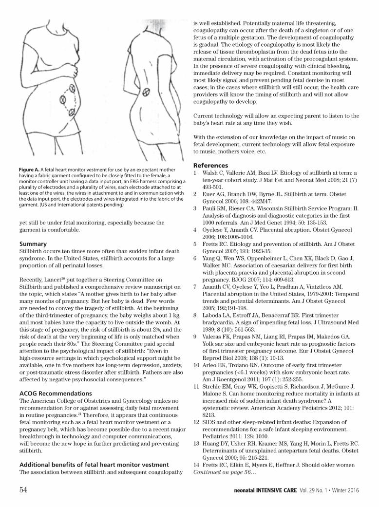
54 neonatal INTENSIVE CARE Vol. 29 No. 1 n Winter 2016
is well established. Potentially maternal life threatening, coagulopathy can occur after the death of a singleton or of one fetus of a multiple gestation. The development of coagulopathy is gradual. The etiology of coagulopathy is most likely the release of tissue thromboplastin from the dead fetus into the maternal circulation, with activation of the procoagulant system. In the presence of severe coagulopathy with clinical bleeding, immediate delivery may be required. Constant monitoring will most likely signal and prevent pending fetal demise in most cases; in the cases where stillbirth will still occur, the health care providers will know the timing of stillbirth and will not allow coagulopathy to develop.
Current technology will allow an expecting parent to listen to the baby’s heart rate at any time they wish.
With the extension of our knowledge on the impact of music on fetal development, current technology will allow fetal exposure to music, mothers voice, etc.
References1 Walsh C, Vallerie AM, Baxi LV. Etiology of stillbirth at term: a
ten-year cohort study. J Mat Fet and Neonat Med 2008; 21 (7) 493-501.
2 Euer AG, Branch DW, Byrne JL. Stillbirth at term. Obstet Gynecol 2006; 108: 442M47.
3 Pauli RM, Rieser CA. Wisconsin Stillbirth Service Program: II. Analysis of diagnosis and diagnostic categories in the first 1000 referrals. Am J Med Genet 1994; 50: 135-153.
4 Oyelese Y, Ananth CV. Placental abruption. Obstet Gynecol 2006; 108:1005-1016.
5 Fretts RC. Etiology and prevention of stillbirth. Am J Obstet Gynecol 2005; 193: 1923-35.
6 Yang Q, Wen WS, Oppenheimer L, Chen XK, Black D, Gao J, Walker MC. Association of caesarian delivery for first birth with placenta praevia and placental abruption in second pregnancy. BJOG 2007; 114: 609-613.
7 Ananth CV, Oyelese Y, Yeo L, Pradhan A, Vintztleos AM. Placental abruption in the United States, 1979-2001: Temporal trends and potential determinants. Am J Obstet Gynecol 2005; 192;191-198.
8 Laboda LA, Estroff JA, Benacerraf BR. First trimester bradycardia. A sign of impending fetal loss. J Ultrasound Med 1989; 8 (10): 561-563.
9 Valeras FK, Prapas NM, Liang RI, Prapas IM, Makedos GA. Yolk sac size and embryonic heart rate as prognostic factors of first trimester pregnancy outcome. Eur J Obstet Gynecol Reprod Biol 2008; 138 (1): 10-13.
10 Arleo EK, Troiano RN. Outcome of early first trimester pregnancies (<6.1 weeks) with slow embryonic heart rate. Am J Roentgenol 2011; 197 (1): 252-255.
11 Strehle EM, Gray WK, Gopisetti S, Richardson J, McGurre J, Malone S. Can home monitoring reduce mortality in infants at increased risk of sudden infant death syndrome? A systematic review. American Academy Pediatrics 2012; 101: 8213.
12 SIDS and other sleep-related infant deaths: Expansion of recommendations for a safe infant sleeping environment. Pediatrics 2011: 128: 1030.
13 Huang DY, Usher RH, Kramer MS, Yang H, Morin L, Fretts RC. Determinants of unexplained antepartum fetal deaths. Obstet Gynecol 2000; 95: 215-221.
14 Fretts RC, Elkin E, Myers E, Heffner J. Should older women
yet still be under fetal monitoring, especially because the garment is comfortable.
summaryStillbirth occurs ten times more often than sudden infant death syndrome. In the United States, stillbirth accounts for a large proportion of all perinatal losses.
Recently, Lancet20 put together a Steering Committee on Stillbirth and published a comprehensive review manuscript on the topic, which states “A mother gives birth to her baby after many months of pregnancy. But her baby is dead. Few words are needed to convey the tragedy of stillbirth. At the beginning of the third-trimester of pregnancy, the baby weighs about 1 kg, and most babies have the capacity to live outside the womb. At this stage of pregnancy, the risk of stillbirth is about 2%, and the risk of death at the very beginning of life is only matched when people reach their 80s.” The Steering Committee paid special attention to the psychological impact of stillbirth: “Even in high-resource settings in which psychological support might be available, one in five mothers has long-term depression, anxiety, or post-traumatic stress disorder after stillbirth. Fathers are also affected by negative psychosocial consequences.”
acOG recommendationsThe American College of Obstetrics and Gynecology makes no recommendation for or against assessing daily fetal movement in routine pregnancies.21 Therefore, it appears that continuous fetal monitoring such as a fetal heart monitor vestment or a pregnancy belt, which has become possible due to a recent major breakthrough in technology and computer communications, will become the new hope in further predicting and preventing stillbirth.
additional benefits of fetal heart monitor vestmentThe association between stillbirth and subsequent coagulopathy
Figure A. A fetal heart monitor vestment for use by an expectant mother having a fabric garment configured to be closely fitted to the female, a monitor controller unit having a data input port, an EKG harness comprising a plurality of electrodes and a plurality of wires, each electrode attached to at least one of the wires, the wires in attachment to and in communication with the data input port, the electrodes and wires integrated into the fabric of the garment. (US and International patents pending)
Continued on page 56…

(800) 332-6326www.codanuscorp.com
All-In-One Sets Decreases CLABSI Rates Bifurcated and Trifurcated Increased Infection Control Microbore & Minibore Tubing Complete & Sterile Burette Sets 0.2 Micron Air Eliminating Filter
Needlefree Adapters Available to fit all Syringe Pumps Latex Free and Non-DEHP Tubing Custom Sets available in 4 Weeks Meets CDC & FDA Requirements Quality Tubing Sets made in the
United States
Protect What Is Most PreciousCODAN infection prevention sets are designed to effectively reduce CLABSI risks in the NICU and PICU. Specialty neonate/pediatric sets are fully customizable to specific customer needs. We offer completely sterile burettes for truly sterile procedures. Eliminate the need to assemble multiple sets and increase efficiencies of line change outs.
Infusion Therapy made Clean, Safe + Simple
Follow Us on Facebook
CODAN_NICfull_7-1-15.indd 1 7/1/15 2:11 PM

56 neonatal INTENSIVE CARE Vol. 29 No. 1 n Winter 2016
have antepartum testing to prevent unexplained stillbirth? Obstet Gynecol 2004; 104: 56-64.
15 Fretts RC. Etiology and prevention of stillbirth. Am J Obstet Gynecol 2005; 193: 1923-1935.
16 Waldenstrom U, Chattingius S, Norman M, Schytt E. Advanced maternal age and stillbirth risk in nulliparous and parous women. Obstet Gynecol 2015; 126: 355-62
17 Tucker SM. Pocket Guide to Fetal Monitoring and Assessment. 4th edition Mosby 2000.
18 Petrikovsky BM. Long-term continuous FHR monitoring in pregnancy complicated by oligohydramnios - pros & cons. Neonat Intens Care 1996; 7, 31-4
19 Belford MA, Saade GR, Thorn E. A randomized trial of intrapartum fetal ECG ST-segment analysis. N Engl J Med 2015; 373: 632-41.
20 Froen JF, Cacciatore J, McClure EM, Kuti O, Jokhio AH, Islam JM, Shiftman J, for The Lancet’s Stillbirth Series Steering Committee. Stillbirth: why they matter, Lancet, vol 377, April 16 2011.
21 ACOG. Antepartum Fetal Surveillance. ACOG Practice Bulletin, Number 9. Washington, DC: American College of Obstetrics and Gynecology; October 1999.
addendum to the reportOngoing research on an ultrasound alternative is being conducted by M. Bogart, an MIT-educated electrical engineer and computer scientist, CTO of Rampage Digital and former Vice President of Research and Development for Rampage Systems, Inc.
The use of continuous ultrasound presents significant sonic energy impact on the fetus, the long-term and certainly continuous use ramifications of which are not precisely known. Furthermore the logistics of constant use and patient acceptance present further disadvantages.
Abdominal surface skin electrodes are used to pick up the fetal heartbeat signal or signals. Current technology of miniature wearable electronics, micro-controllers, and wireless links enable signal pickup and transmission in a form that will be acceptable to the pregnant patient for continuous use.
Since, as is known, such signals include a mix of both the maternal and the fetal heartbeats, a unique method, based on proven digital signal processing techniques, is used to provide the needed fetal-only heart-rate log and time history. The details are the subject of ongoing patent processes.
Continued from page 54

neonatal INTENSIVE CARE Vol. 29 No. 1 n Winter 2016 57
Background: Pulse oximeters display pulse rate (PR) and pulse oxygenation (SpO2). These vital signs are important components of screening and diagnostic algorithms in newborns,1,2 making it necessary to establish pulse oximeter performance in the neonatal population. Inaccurate reporting of PR or SpO2 values could lead to false alarms and inappropriate interventions.
method: This study compared PR performance of the Nellcor N-600x pulse oximeter and the Masimo SET module of a Philips IntelliVue MX800 in a neonatal intensive care unit (NICU). Data were collected from 30 subjects during wakefulness and sleep. PR readings were compared to a heart rate (HR) reference obtained by ECG.
results: The Nellcor N-600x pulse oximeter more accurately reported PR than did the Masimo SET module (RMSD 3.93 beats per minute (bpm) vs. 5.07 bpm, P < .001). Also, a significantly smaller proportion of PR measurements reported by the Nellcor monitor differed from the reference heart rate by more than 40 beats per minute (0.07% vs. 0.23%, P < .001). For three subjects, the Masimo SET module exhibited a clinically significant error (CSE), a PR that differed from the reference by at least 40 bpm for more than 30 seconds continuously. No CSEs occurred with the Nellcor N-600x monitor. SpO2 readings reported by the two monitors were similar during periods when both instruments accurately reported PR. However, during periods when the Masimo SET module exhibited CSEs, the SpO2 readings of the two pulse oximeters differed.
conclusion: The Nellcor N-600x pulse oximeter more accurately reported PR compared to an ECG reference than did the Masimo SET module in this population of NICU patients. Only the Masimo instrument exhibited errors in PR that were large enough to potentially result in inappropriate intervention (CSEs).
IntroductionPulse oximeters are commonly used to estimate SpO2 in newborns, a recommended component of critical congenital heart disease (CCHD) screening in newborns.1 Pulse oximeters also report pulse rate (PR). Heart rate is a factor in the American Heart Association’s algorithm for neonatal resuscitation.2 However, the methods recommended by the AHA to collect neonatal HR (intermittent auscultation of the praecordium and palpation of the umbilical cord) have been shown to be imprecise and inaccurate3 while pulse oximetry has been suggested to accurately report PR in infants in the delivery
room.4,5 The accuracy of reported vital signs impacts clinician intervention. Inaccurate information can lead to unnecessary medical intervention due to false positive alarms. Therefore, it is essential to establish the accuracy of the data provided by pulse oximeters in a neonatal population. The present study compared the PR performance of two modern pulse oximeters relative to an ECG reference and assessed the SpO2 performance of the instruments.
MethodThe study was conducted at British Columbia Children’s Hospital between February and June, 2013. After Institutional Review Board approval and receipt of informed parental consent, 30 neonatal patients in the intensive care unit were enrolled in the study. Twenty-nine of these subjects were included in the analysis. One subject was excluded after data loss due to a failure of the third-party data acquisition software. Subjects were closely monitored by caregivers and registered nurses at all times.
All sensors and monitors used in the study were cleared by the FDA and Health Canada. A Nellcor SpO2 MAX-N sensor and a Masimo LNCS, LNCS Neo, or LNCS NeoPt sensor were applied to the wrist, or the foot if the wrist was unavailable, of each subject, and connected with the respective pulse oximeter monitors. All sensors were applied at post-ductal sites (ie left hand, feet). Sensors were covered with probe wraps to secure them against movement and to reduce ambient light and prevent optical crosstalk. ECG reference heart rate (HR) values were acquired using either a PHILIPS IntelliVue MP70 or a MX800 monitor. Signals were recorded using a combination of proprietary data acquisition software and commercially available software (Rugloop, Demed Inc, Temse, Belgium).
After sensors were placed on each subject, electronic data were collected for approximately four hours. Data were collected for each subject during both wakefulness and sleep. Data were excluded if the ECG or oximeter reported zero or if no data were available, eg, due to sensor disconnect, sensor turning off, etc.
To analyze PR accuracy, measurements from each test instrument were extracted once per second and compared to simultaneous data from the ECG monitor. Standard measures of bias, precision (1 SD), and accuracy (RMSD, the root mean square of differences) were calculated as recommended by the pulse oximetry industry standard.6
Pulse Rate Performance of Two Pulse Oximeters in the NICUKeith Batchelder, PhD, Rakesh Sethi, BSc, BEng, and Yu Jung Pinto, MSc

58 neonatal INTENSIVE CARE Vol. 29 No. 1 n Winter 2016
pulse rateTo assess the accuracy of the PR reported by each device, the RMSD between the pulse oximetry–derived PR and ECG-derived HR (ECG HR) was calculated. RMSD for the Nellcor N-600x pulse oximeter was significantly less than that for the Masimo SET module was (4.14 bpm vs. 10.48 bpm, P < .001) (Table 2). The Nellcor N-600x oximeter performance was within its device specifications of ± 5 bpm under motion.
The percentage of time during which the absolute PR error was greater than 40 bpm (E40) for the Nellcor N-600x oximeter was 0.08%, compared to 0.89% for the Masimo SET module, across all subjects (Table 2). The difference between instruments was statistically significant (P < .001). Figure 1 shows pulse oximeter–reported PR vs. ECG HR for both the Nellcor N-600x oximeter and the Masimo SET module for all subjects.
One subject was identified as an outlier, with a PR RMSD outside of the range of (3 x SD + mean PR RMSD). For this subject, the overall PR RMSD was 50 bpm for the Masimo SET module and 8 bpm for the Nellcor N-600x oximeter (Table 2). Data from the outlier subject is highlighted in Figure 1. The E40 results for this subject were 0.54% for the Nellcor N-600x oximeter and 22.46% for the Masimo SET module (Table 2). Figure 2 shows an interval of data from this subject during which the PR error for the Masimo SET module met the criteria of CSE.
Excluding the outlier subject, the PR RMSD for the Masimo SET module was greater than 5 bpm, not within specifications. E40, excluding the outlier subject, was still significantly smaller for the Nellcor N-600x oximeter compared to the Masimo SET module (0.07% vs. 0.23%, P < .001).
spO2
The Nellcor N-600x pulse oximeter reported SpO2 values between 42-100%, and Masimo SET module reported SpO2 values between 30-100%. The pooled SpO2 difference for all collected data (ΔSpO2 RMSD) was 1.66 %. During intervals when absolute PR error relative to ECG for both devices was less than 5 bpm, SpO2 RMSD was 1.5%. During continuous intervals during which the Masimo SET module exhibited a CSE, ΔSpO2 RMSD was significantly greater, 3.8% (P < .001). The increased disagreement between SpO2 values during CSEs is also seen in Figure 2. No equivalent intervals of CSE were observed for the Nellcor N-600x pulse oximeter. The SpO2 data are summarized in Table 3.
Discussion Accurate data about neonatal vital signs are required to facilitate appropriate medical intervention, for example, as part of the pediatric resuscitation algorithm, or in CCHD screening. Therefore, it is important to characterize pulse oximeter performance in the NICU environment, particularly as it pertains to the usefulness of pulse oximetry–reported data to clinicians.
The PR reported by the Nellcor N-600x pulse oximeter was significantly closer to the reference HR value than that reported by the Masimo SET module, even after the data from an outlier subject was removed from the analysis (RMSD 3.93 bpm vs. 5.07 bpm, P < .001). The Nellcor N-600x pulse oximeter also had a significantly smaller proportion of PR measurements that differed from the reference HR by at least 40 bpm (0.07% vs. 0.23%, P < .001).
Additionally, the percentage of time that the PR reported by each monitor differed from the reference HR value by at least 40 beats per minute (bpm) was measured (E40). Forty bpm was chosen because an error of this size might result in an inappropriate or missed intervention when following published neonatal resuscitation guidelines.7 The Chi-square test was used to assess whether E40 differed significantly between monitors. A P-value less than 0.05 was considered significant.
Clinically significant errors (CSE) were defined as differences between PR and HR greater than 40 bpm and continuously sustained for more than 30 seconds. This interval of time was chosen because it is sufficiently long to trigger an inappropriate clinical intervention in response to a sustained erroneous PR value.5 Additionally, according to the ISO oximetry standard 80601-2-61, 30 seconds is the maximum time an oximeter is allowed to “hold” without indicating that it is holding.6
The Wilcoxon rank-sum test was used to determine the statistical significance of differences between devices. This non-parametric version of a paired samples t-test was chosen because the errors were not normally distributed. A P-value less than 0.05 was considered significant.
No convenience arterial blood gas sample was obtained during the study. Without a reference from co-oximetry, the accuracy of SpO2 readings taken from the two pulse oximetry monitors were compared for agreement. The RMSD between the SpO2 values from the two monitors was calculated during intervals of “good” agreement (< 5 bpm) between ECG-determined HR and pulse oximetry–determined PR, and during intervals where there was a CSE between the PR derived from either oximeter and the HR reference signal derived from the ECG.
ResultsData were collected from 29 subjects. Demographics and patient characteristics are summarized in Table 1. The majority of enrolled subjects (77%) were born prematurely. A majority of subjects were diagnosed with respiratory distress syndrome. A total of 114 hours of pulse oximetry data were collected with ECG HR reference values available for comparison. ECG HR values among the subjects ranged between 46 and 235 bpm. Transient bradycardia was observed in 7 subjects during the trial; medical interventions including stimulation and supplemental oxygen were provided for recovery.
3
Table 1. Patient Demographics and Baseline Characteristics
Category n (%)
Male 18 (60)
Race/ethnicity
Asian 9 (30)
African American 0 (0)
Caucasian 20 (67)
Hispanic 1 (3)
Median (standard deviation)
Conceptional age, weeks 35.7 (13.6)
Height, cm 44.0 (21.0)
Weight, kg 2.3 (3.2)
Pulse rate
To assess the accuracy of the PR reported by each device, the RMSD between the pulse oximetry–derived PR and ECG-derived HR (ECG HR) was calculated. RMSD for the Nellcor N-600x pulse oximeter was significantly less than that for the Masimo SET module was (4.14 bpm vs. 10.48 bpm, P < .001) (Table 2). The Nellcor N-600x oximeter performance was within its device specifications of ± 5 bpm under motion.
The percentage of time during which the absolute PR error was greater than 40 bpm (E40) for the Nellcor N-600x oximeter was 0.08%, compared to 0.89% for the Masimo SET module, across all subjects (Table 2). The difference between instruments was statistically significant (P < .001). Figure 1 shows pulse oximeter–reported PR vs. ECG HR for both the Nellcor N-600x oximeter and the Masimo SET module for all subjects.
One subject was identified as an outlier, with a PR RMSD outside of the range of (3 x SD + mean PR RMSD). For this subject, the overall PR RMSD was 50 bpm for the Masimo SET module and 8 bpm for the Nellcor N-600x oximeter (Table 2). Data from the outlier subject is highlighted in Figure 1. The E40 results for this subject were 0.54% for the Nellcor N-600x oximeter and 22.46% for the Masimo SET module (Table 2). Figure 2 shows an interval of data from this subject during which the PR error for the Masimo SET module met the criteria of CSE.
Excluding the outlier subject, the PR RMSD for the Masimo SET module was greater than 5 bpm, not within specifications. E40, excluding the outlier subject, was still significantly smaller for the Nellcor N-600x oximeter compared to the Masimo SET module (0.07% vs. 0.23%, P < .001).
SpO2
The Nellcor N-600x pulse oximeter reported SpO2 values between 42-100%, and Masimo SET module reported SpO2 values between 30-100%. The pooled SpO2 difference for all collected data (ΔSpO2 RMSD) was 1.66 %. During intervals when absolute PR error relative to ECG for both devices was less than 5 bpm, SpO2 RMSD was 1.5%. During continuous intervals during which the Masimo SET® module exhibited a CSE, ΔSpO2 RMSD was significantly greater, 3.8% (P < .001). The increased disagreement between SpO2 values during CSEs is also seen in Figure 2. No equivalent intervals of CSE were observed for the Nellcor N-600x pulse oximeter. The SpO2 data are summarized in Table 3.
Table 2. Pulse Oximeter Performance in Reporting Pulse Rate
Nellcor N-600x monitor Masimo SET moduleDevices significantly
different
All subjects RMSD (mean ± SD) (bpm) 4.14 (0.16 ± 4.14) 10.48 (-1.26 ± 10.41) P < .001a
Outlier subject RMSD (mean ± SD) (bpm) 8.32 (0.21 ± 8.32) 49.88 (-24.94 ± 43.19) P < .001a
All subjects, omitting outlier RMSD (mean ± SD) (bpm)
3.93 (0.15 ± 3.93) 5.07 (-0.44 ± 5.05) P < .001a
All subjects E40 0.08% 0.89% P < .001b
Outlier subject E40 0.54% 22.46% P < .001b
All subjects, omitting outlier E40 0.07% 0.23% P < .001b
a Wilcoxon rank-sum testb Chi-square test
Table 1. Patient Demographics and Baseline Characteristics.

© 2016 Medtronic. All rights reserved. Medtronic, Medtronic logo and Further, Together are trademarks of Medtronic. All other brands are trademarks of a Medtronic company. 16-PM-0017
UNSURPASSED ACCURACY.DEPENDABLE PERFORMANCE.Critical Congenital Heart Defects (CCHD) and Pulse Oximetry Screening
Our Nellcor™ pulse oximetry portfolio facilitates quick, noninvasive screenings for CCHD.
By relying largely on cardiac-based signals, Nellcor™ pulse oximetry generates highly accurate readings closely tied to neonate physiology.
The result is consistent performance you can rely on during challenging conditions, including:
∙Patient motion
∙Noise
∙Low perfusion
Since1993,morethan33,000newborns—spanningfiveseparateclinical studies on the use of pulse oximetry as a tool in CCHD screening — have been evaluated with Nellcor™ technology.
Learn more atmedtronic.com/covidien/cchd

60 neonatal INTENSIVE CARE Vol. 29 No. 1 n Winter 2016
this signal quality is good.7 In this study, the agreement of the SpO2 readings reported by the two oximeters was characterized during periods of good and poor agreement between PR and HR. The SpO2 values reported by the two pulse oximeters were very similar during intervals where reported PRs were within 5 bpm of the reference HR. However, differences between reported SpO2 values were close to 4 saturation points during intervals where the Masimo SET monitor exhibited a CSE, significantly greater than the difference in SpO2 readings over the entire experiment. Assuming that PR agreement with HR can be used as a surrogate for confidence in SpO2 readings, it is probable that SpO2 readings from the Masimo instrument were responsible for the observed differences in SpO2 readings during intervals when the Masimo instrument exhibited CSE. Further experiments with comparative SpO2 values can investigate this possibility.
ConclusionsThe Nellcor N-600x pulse oximeter more accurately reported PR compared to an ECG reference, as measured by RMSD, than the Masimo SET module in this population of NICU patients. Only the Masimo monitor exhibited errors in PR that were large enough to potentially result in inappropriate intervention (CSEs). During periods when the Masimo monitor exhibited CSE, the SpO2 values reported by the two oximeters also differed. There is a high probability that the Masimo instrument is responsible for the observed poor agreement between reported SpO2 values during periods when it exhibited CSE. However, further investigation is necessary to investigate this possibility.
In this analysis, data from 29 subjects was pooled. Extreme deviations in PR performance in individual subjects who present a unique set of challenging conditions may be missed when analyzing pooled data. Therefore, pulse oximeter performance was assessed during periods defined as clinically significant errors (CSEs); periods during which the absolute error in PR relative to HR was equal to or greater than 40 bpm for at least 30 seconds. No CSEs occurred with the Nellcor N-600x pulse oximeter. CSEs occurred with 3 subjects with the Masimo SET technology. For one of these 3 subjects, the PR reported by the Masimo monitor fell below 60 bpm for large intervals of time. Such a deviation from the correct HR value could result in inappropriate medical intervention.
These results are consistent with other observations in the literature. Kamlin et al5 also reported one outlier subject, monitored with the Masimo SET technology, for whom a clinically significant difference between reported PR and the ECG reference “may have led to interventions that were not indicated.” Additionally, Du and Gorensky reported a case study using the Masimo SET technology in which a persistent PR error resulted in an inappropriate intervention.9
Due to limitations associated with collecting blood samples from neonates, co-oximetry reference data was not obtained in this study. Often, clinicians use agreement between pulse oximetry-reported PR and ECG-reported HR as a signal quality metric, and place greater confidence in SpO2 readings when
3
Table 1. Patient Demographics and Baseline Characteristics
Category n (%)
Male 18 (60)
Race/ethnicity
Asian 9 (30)
African American 0 (0)
Caucasian 20 (67)
Hispanic 1 (3)
Median (standard deviation)
Conceptional age, weeks 35.7 (13.6)
Height, cm 44.0 (21.0)
Weight, kg 2.3 (3.2)
Pulse rate
To assess the accuracy of the PR reported by each device, the RMSD between the pulse oximetry–derived PR and ECG-derived HR (ECG HR) was calculated. RMSD for the Nellcor N-600x pulse oximeter was significantly less than that for the Masimo SET module was (4.14 bpm vs. 10.48 bpm, P < .001) (Table 2). The Nellcor N-600x oximeter performance was within its device specifications of ± 5 bpm under motion.
The percentage of time during which the absolute PR error was greater than 40 bpm (E40) for the Nellcor N-600x oximeter was 0.08%, compared to 0.89% for the Masimo SET module, across all subjects (Table 2). The difference between instruments was statistically significant (P < .001). Figure 1 shows pulse oximeter–reported PR vs. ECG HR for both the Nellcor N-600x oximeter and the Masimo SET module for all subjects.
One subject was identified as an outlier, with a PR RMSD outside of the range of (3 x SD + mean PR RMSD). For this subject, the overall PR RMSD was 50 bpm for the Masimo SET module and 8 bpm for the Nellcor N-600x oximeter (Table 2). Data from the outlier subject is highlighted in Figure 1. The E40 results for this subject were 0.54% for the Nellcor N-600x oximeter and 22.46% for the Masimo SET module (Table 2). Figure 2 shows an interval of data from this subject during which the PR error for the Masimo SET module met the criteria of CSE.
Excluding the outlier subject, the PR RMSD for the Masimo SET module was greater than 5 bpm, not within specifications. E40, excluding the outlier subject, was still significantly smaller for the Nellcor N-600x oximeter compared to the Masimo SET module (0.07% vs. 0.23%, P < .001).
SpO2
The Nellcor N-600x pulse oximeter reported SpO2 values between 42-100%, and Masimo SET module reported SpO2 values between 30-100%. The pooled SpO2 difference for all collected data (ΔSpO2 RMSD) was 1.66 %. During intervals when absolute PR error relative to ECG for both devices was less than 5 bpm, SpO2 RMSD was 1.5%. During continuous intervals during which the Masimo SET® module exhibited a CSE, ΔSpO2 RMSD was significantly greater, 3.8% (P < .001). The increased disagreement between SpO2 values during CSEs is also seen in Figure 2. No equivalent intervals of CSE were observed for the Nellcor N-600x pulse oximeter. The SpO2 data are summarized in Table 3.
Table 2. Pulse Oximeter Performance in Reporting Pulse Rate
Nellcor N-600x monitor Masimo SET moduleDevices significantly
different
All subjects RMSD (mean ± SD) (bpm) 4.14 (0.16 ± 4.14) 10.48 (-1.26 ± 10.41) P < .001a
Outlier subject RMSD (mean ± SD) (bpm) 8.32 (0.21 ± 8.32) 49.88 (-24.94 ± 43.19) P < .001a
All subjects, omitting outlier RMSD (mean ± SD) (bpm)
3.93 (0.15 ± 3.93) 5.07 (-0.44 ± 5.05) P < .001a
All subjects E40 0.08% 0.89% P < .001b
Outlier subject E40 0.54% 22.46% P < .001b
All subjects, omitting outlier E40 0.07% 0.23% P < .001b
a Wilcoxon rank-sum testb Chi-square test
Table 2. Pulse Oximeter Performance in Reporting Pulse Rate.
4
Figure 1: Pooled data from 29 subjects (outlier plotted in red) showing PR vs. ECG HR for Nellcor N-600x oximeter (left) and Masimo SET® module (right). The dashed lines in each graph indicate errors from ECG HR by ± 40 bpm.
Figure 2: SpO2 and pulse rate reported by two pulse oximeters for outlier subject, during a long period of disagreement between the Masimo SET module and ECG. When the Masimo SET module PR is different from the ECG HR, the Masimo SET module and Nellcor N-600x oximeter SpO2 values also disagree.
Figure 1: Pooled data from 29 subjects (outlier plotted in red) showing PR vs. ECG HR for Nellcor N-600x oximeter (left) and Masimo SET module (right). The dashed lines in each graph indicate errors from ECG HR by ± 40 bpm.

neonatal INTENSIVE CARE Vol. 29 No. 1 n Winter 2016 61
5 Kamlin COF, Dawson JA, O’Donnell CPF, et al. Accuracy of pulse oximetry measurement of heart rate of newborn infants in the delivery room. J Pediatr 2008;152:756-760.
6 International Standards Organisation. Medical electrical equipment Part 2-61: Particular requirements for basic safety and essential performance of pulse oximeter equipment. 2011. ISO 80601-2-61:2011.
7 Sahni R, Gupta A, Ohira-Kist K, Rosen TS. Motion resistant pulse oximetry in neonates. Arch Dis Child Fetal Neonatal Ed 2003;88:F505–F508.
8 Tobin RS, Polage JA, Batchelder PB. A Characterization of Motion Affecting Pulse Oximetry in 350 Patients. Anesth Analg 2002;94(1 Suppl):S54–61.
9 Du EH, Goresky GV. Disparate motion-resistant SpO2 and pulse rate parameters. Anesth Analg. 2011;112(Suppl):S-226
References1 Knapp AA, Metterville DR, Kemper AR, Prosser L, Perrin
JM. Evidence review: Critical congenital cyanotic heart disease. Health Resources and Services Administration, Maternal and Child Health Bureau. http://www.hrsa.gov/advisorycommittees/mchbadvisory/heritabledisorders/nominatecondition/reviews/cyanoticheart.pdf Published September 3, 2010. Accessed February 25, 2015.
2 Kattwinkel J, Perlman JM, Aziz K, et al. Part 15: neonatal resuscitation: 2010 American Heart Association Guidelines for Cardiopulmonary Resuscitation and Emergency Cardiovascular Care. Circulation. 122:S909-S919.
3 Kamlin CO, O’Donnell CP, Everest NJ, Davis PG, Morley CJ. Accuracy of clinical assessment of infant heart rate in the delivery room. Resuscitation 2006;71: 319-321.
4 Dawson JA, Saraswat A, Simionato L, et al. Comparison of heart rate and oxygen saturation measurements from Masimo and Nellcor pulse oximeters in newly born term infants. Acta Paediatr 2013;102:955-960.
4
Figure 1: Pooled data from 29 subjects (outlier plotted in red) showing PR vs. ECG HR for Nellcor N-600x oximeter (left) and Masimo SET® module (right). The dashed lines in each graph indicate errors from ECG HR by ± 40 bpm.
Figure 2: SpO2 and pulse rate reported by two pulse oximeters for outlier subject, during a long period of disagreement between the Masimo SET module and ECG. When the Masimo SET module PR is different from the ECG HR, the Masimo SET module and Nellcor N-600x oximeter SpO2 values also disagree.
Figure 2. SpO2 and pulse rate reported by two pulse oximeters for outlier subject, during a long period of disagreement between the Masimo SET module and ECG. When the Masimo SET module PR is different from the ECG HR, the Masimo SET module and Nellcor N-600x oximeter SpO2 values also disagree.
Table 3. Pulse Oximeter Performance in Reporting SpO2.
5
Table 3. Pulse Oximeter Performance in Reporting SpO2
ΔSpO2 RMSD (%, mean ± SD)
Compared to overall
ΔSpO2 RMSDOverall 1.66 (-0.61 ± 1.55)
While absolute PR error is < 5 bpm
1.41 (-0.58 ± 1.29)
N.S.§
During periods of CSE for Nellcor N-600x
NA* NA*
During periods of CSE for Masimo SET module
3.81 (1.12 ± 3.64) P < .001§
Outlier during periods of CSE of Masimo SET module
5.16 (-3.57 ± 3.72) P < .001
§Wilcoxon rank sum test
* No CSE was observed using Nellcor N-600x pulse oximeter during the trial
Discussion
Accurate data about neonatal vital signs are required to facilitate appropriate medical intervention, for example, as part of the pediatric resuscitation algorithm, or in CCHD screening. Therefore, it is important to characterize pulse oximeter performance in the NICU environment, particularly as it pertains to the usefulness of pulse oximetry–reported data to clinicians.
The PR reported by the Nellcor N-600x pulse oximeter was significantly closer to the reference HR value than that reported by the Masimo SET module, even after the data from an outlier subject was removed from the analysis (RMSD 3.93 bpm vs. 5.07 bpm, P < .001). The Nellcor N-600x pulse oximeter also had a significantly smaller proportion of PR measurements that differed from the reference HR by at least 40 bpm (0.07% vs. 0.23%, P < .001).
In this analysis, data from 29 subjects was pooled. Extreme deviations in PR performance in individual subjects who present a unique set of challenging conditions may be missed when analyzing pooled data. Therefore, pulse oximeter performance was assessed during periods defined as clinically significant errors (CSEs); periods during which the absolute error in PR relative to HR was equal to or greater than 40 bpm for at least 30 seconds. No CSEs occurred with the Nellcor N-600x pulse oximeter. CSEs occurred with 3 subjects with the Masimo SET technology. For one of these 3 subjects, the PR reported by the Masimo monitor fell below 60 bpm for large intervals of time. Such a deviation from the correct HR value could result in inappropriate medical intervention.
These results are consistent with other observations in the literature. Kamlin et al 5 also reported one outlier subject, monitored with the Masimo SET technology, for whom a clinically significant difference between reported PR and the ECG reference “may have led to interventions that were not indicated.” Additionally, Du and Gorensky reported a case study using the Masimo SET technology in which a persistent PR error resulted in an inappropriate intervention.9
Due to limitations associated with collecting blood samples from neonates, co-oximetry reference data was not obtained in this study. Often, clinicians use agreement between pulse oximetry-reported PR and ECG-reported HR as a signal quality metric, and place greater confidence in SpO2 readings when this signal quality is good.7 In this study, the agreement of the SpO2 readings reported by the two oximeters was characterized during periods of good and poor agreement between PR and HR. The SpO2 values reported by the two pulse oximeters were very similar during intervals where reported PRs were within 5 bpm of the reference HR. However, differences between reported SpO2 values were close to 4 saturation points during intervals where the Masimo SET monitor exhibited a CSE, significantly greater than the difference in SpO2 readings over the entire experiment. Assuming that PR agreement with HR can be used as a surrogate for confidence in SpO2 readings, it is probable that SpO2 readings from the Masimo instrument were responsible for the observed differences in SpO2 readings during intervals when the Masimo instrument exhibited CSE. Further experiments with comparative SpO2 values can investigate this possibility.
Conclusions
The Nellcor N-600x pulse oximeter more accurately reported PR compared to an ECG reference, as measured by RMSD, than the Masimo SET module in this population of NICU patients. Only the Masimo monitor exhibited errors in PR that were large enough to potentially result in inappropriate intervention (CSEs). During periods when the Masimo monitor exhibited CSE, the SpO2 values reported by the two oximeters also differed. There is a high probability that the Masimo instrument is responsible for the observed poor agreement between reported SpO2 values during periods when it exhibited CSE. However, further investigation is necessary to investigate this possibility.
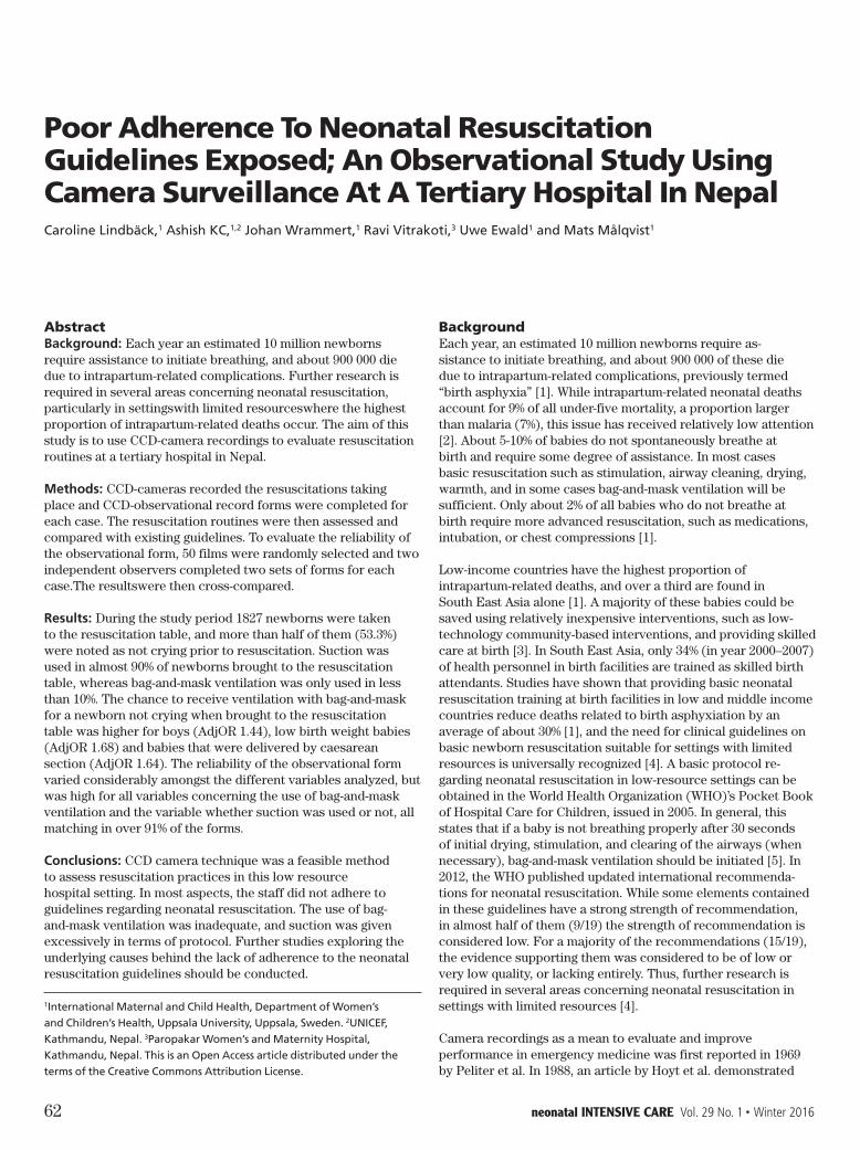
62 neonatal INTENSIVE CARE Vol. 29 No. 1 n Winter 2016
Abstract Background: Each year an estimated 10 million newborns require assistance to initiate breathing, and about 900 000 die due to intrapartum-related complications. Further research is required in several areas concerning neonatal resuscitation, particularly in settingswith limited resourceswhere the highest proportion of intrapartum-related deaths occur. The aim of this study is to use CCD-camera recordings to evaluate resuscitation routines at a tertiary hospital in Nepal.
methods: CCD-cameras recorded the resuscitations taking place and CCD-observational record forms were completed for each case. The resuscitation routines were then assessed and compared with existing guidelines. To evaluate the reliability of the observational form, 50 films were randomly selected and two independent observers completed two sets of forms for each case.The resultswere then cross-compared.
results: During the study period 1827 newborns were taken to the resuscitation table, and more than half of them (53.3%) were noted as not crying prior to resuscitation. Suction was used in almost 90% of newborns brought to the resuscitation table, whereas bag-and-mask ventilation was only used in less than 10%. The chance to receive ventilation with bag-and-mask for a newborn not crying when brought to the resuscitation table was higher for boys (AdjOR 1.44), low birth weight babies (AdjOR 1.68) and babies that were delivered by caesarean section (AdjOR 1.64). The reliability of the observational form varied considerably amongst the different variables analyzed, but was high for all variables concerning the use of bag-and-mask ventilation and the variable whether suction was used or not, all matching in over 91% of the forms.
conclusions: CCD camera technique was a feasible method to assess resuscitation practices in this low resource hospital setting. In most aspects, the staff did not adhere to guidelines regarding neonatal resuscitation. The use of bag-and-mask ventilation was inadequate, and suction was given excessively in terms of protocol. Further studies exploring the underlying causes behind the lack of adherence to the neonatal resuscitation guidelines should be conducted.
Background Each year, an estimated 10 million newborns require as-sistance to initiate breathing, and about 900 000 of these die due to intrapartum-related complications, previously termed “birth asphyxia” [1]. While intrapartum-related neonatal deaths account for 9% of all under-five mortality, a proportion larger than malaria (7%), this issue has re ceived relatively low attention [2]. About 5-10% of babies do not spontaneously breathe at birth and require some degree of assistance. In most cases basic resuscitation such as stimulation, airway cleaning, drying, warmth, and in some cases bag-and-mask ventilation will be sufficient. Only about 2% of all babies who do not breathe at birth require more advanced resuscitation, such as medications, intubation, or chest compressions [1].
Low-income countries have the highest proportion of intrapartum-related deaths, and over a third are found in South East Asia alone [1]. A majority of these babies could be saved using relatively inexpensive interventions, such as low-technology community-based interventions, and providing skilled care at birth [3]. In South East Asia, only 34% (in year 2000–2007) of health personnel in birth facil ities are trained as skilled birth attendants. Studies have shown that providing basic neonatal resuscitation training at birth facilities in low and middle income countries re duce deaths related to birth asphyxiation by an average of about 30% [1], and the need for clinical guidelines on basic newborn resuscitation suitable for settings with limited re sources is universally recognized [4]. A basic protocol re-garding neonatal resuscitation in low-resource settings can be obtained in the World Health Organization (WHO)’s Pocket Book of Hospital Care for Children, is sued in 2005. In general, this states that if a baby is not breathing properly after 30 seconds of initial drying, stimulation, and clearing of the airways (when necessary), bag-and-mask ventilation should be initiated [5]. In 2012, the WHO published updated international recommenda-tions for neonatal resuscitation. While some elements contained in these guidelines have a strong strength of recommendation, in almost half of them (9/19) the strength of recommendation is considered low. For a ma jority of the recommendations (15/19), the evidence sup porting them was considered to be of low or very low quality, or lacking entirely. Thus, further research is re quired in several areas concerning neonatal resuscitation in settings with limited resources [4].
Camera recordings as a mean to evaluate and improve performance in emergency medicine was first reported in 1969 by Peliter et al. In 1988, an article by Hoyt et al. demonstrated
Poor Adherence To Neonatal Resuscitation Guidelines Exposed; An Observational Study Using Camera Surveillance At A Tertiary Hospital In Nepal Caroline Lindbäck,1 Ashish KC,1,2 Johan Wrammert,1 Ravi Vitrakoti,3 Uwe Ewald1 and Mats Målqvist1
1International Maternal and Child Health, Department of Women’s and Children’s Health, Uppsala University, Uppsala, Sweden. 2UNICEF, Kathmandu, Nepal. 3Paropakar Women’s and Maternity Hospital, Kathmandu, Nepal. This is an Open Access article distributed under the terms of the Creative Commons Attribution License.

neonatal INTENSIVE CARE Vol. 29 No. 1 n Winter 2016 63
project between Uppsala University, Paropakar Ma ternity and Women’s Hospital (PMWH) in Kathmandu, and the Ministry of Health and Population in Nepal. The aim of the main project is to evaluate and improve neo natal resuscitation and survival in a tertiary hospital in Kathmandu [12]. This will be achieved by using the Help ing Babies Breathe (HBB) protocol; a neonatal resuscitation guideline developed in association with the American Academy of Paediatrics, designed to train birth attendants in developing countries the essential skill of new born re suscitation [13]. PMWH is a tertiary government hospital and works as a central referral hospital of the country, pro viding gynaecological and obstetric services. The hospital has just over 23 000 deliveries per year, which gives an average delivery rate at about 63 babies per day. The peri natal mortality rate (PMR) at the hospital is currently about 30 per 1,000 live births, with an early neonatal mor tality rate (death in the first 7 days of life) of 9 per 1,000 and a stillbirth rate of 19 per 1,000 pregnancies. Abnormal deliveries account for 26% -28% of the total deliveries, and the caesarean rate is about 17% [14].
Data collection Budget Dome change-coupled device (CCD) Cameras (model no. MTC-505DH) were used to collect data on the hospital’s neonatal resuscitation routines. A camera was placed at each of the 6 resuscitation tables in the hospital, arranged accordingly: one in the Operating Theatre, one in the maternal and newborn service centre (MNSC), one in the emergency admission room and three in the labour rooms. The cameras had a pro gressive scan sensor and excellent low light perform ance. The cameras were
successful use of video recordings when aiming to improve the staff performance in trauma re suscitation [6]. However, the majority of published work dealing with resuscitation issues is based on medical re cords, and most are only regarding the adult population [7]. Before the year 2000 there had been no reports pub lished concerning the use of similar camera recordings when assessing neonatal resuscitation [6]. Since then, only a few articles have been published on the matter, although these have mostly generated positive results when using camera recordings as a way of assessing neonatal resuscitation [6,8-10]. It can provide import ant information used for quality assessment and educa tion as well as improving teamwork, leadership and communication within a resuscitation workgroup. When analyzing resuscitation recordings it has been re ported that in over 50% of these some deviation from current guidelines could be identified. By recognizing reoccurring errors, preventative measures can be imple mented in an effort to correct them [10]. Video record ings have great potential in terms of optimizing newborn resuscitation, and therefore further studies are needed to assess the validity and reliability when used in clinical practice [6,11].
The aim of the present study is to use Dome change-coupled device (CCD) Camera recordings to evaluate re suscitation routines at a tertiary hospital in Nepal.
Methods setting This cross-sectional study is a sub-study within a collab orative
The most frequent action taken was suction of the air-ways, both for crying (85.0%) as well as for non-cryingbabies (92.3%) (Table 1). Ventilation with bag-and-maskwas only performed on 172 (9.4%) of the recorded cases,whereof 162 were noted as not crying when brought to theresuscitation table.The mean birth weight for recorded cases was 2860
grams, with non-crying newborns being lighter (2910 gramvs 2817 grams, p < 0.01). The low birth weight rate in thesample was 16.9% (308/1827) and 56% (1031/1827) ofrecorded cases were boys. The chance to receive ventila-tion with bag-and-mask for a newborn not crying whenbrought to the resuscitation table was higher for boys andlow birth weight babies (AdjOR 1.44 and 1.68 respectively).There was no difference in ventilation if the baby was bornat night-time (8 pm-8 am), but there was an increasedchance for a non-breathing baby to be ventilated if thedelivery was done by caesarean section (AdjOR 1.64)(Table 2).Apgar-scores were recorded one and five minutes after
birth. Median Apgar-score after one minute was 5/10 andafter five minutes 7/10. There were 198 babies (10.8%)
with an Apgar score less than 7/10 at five minutes. Ofthese 198 depressed neonates 90 (45.5%) received ventila-tion with bag-and-mask. In the sample there were 31intra-partum related deaths, where 30 had an Apgar-scoreof 0/10 at both one and five minutes. A minority, 11/31(35.5%), of the intra-partum related deaths received venti-lation with bag-and-mask, whereas suction of the airwayswas applied in 16/31 (51.6%).When cross-comparing the four separate observational
record forms that were completed for each randomizedCCD-camera recording, it was found that the inter- andintra-rater reliability varied considerably amongst thedifferent variables analyzed. The variables that had thehighest correspondence across all forms where Out-come, Bag & Mask total time, Bag-and-mask time inter-vals, Bag-and-mask yes/no, and Suction yes/no, allmatching in over 91% (91.3-97.8%) of the forms(Figure 2).
DiscussionSimilar to other published work on the use of CCD-cameras as a method to evaluate neonatal resuscitation,
Figure 1 Newborns taken to the resuscitation table. The number and % of newborns that were taken to the resuscitation table, crying andnot crying, out of the total number of babies born during the delivery period (6465).
Table 1 Actions taken for newborns brought to the resuscitation table
All newborns brought totable
Newborns crying when brought totable
Newborns not crying when brought totable
n % n % n %
Stimulation
Yes 824 45.1 197 23.1 627 64.4
No 1003 54.9 656 76.9 347 35.6
Bag and mask
Yes 172 9.4 10 1.2 162 16.6
No 1655 90.6 843 98.8 812 83.4
Suction performed
Yes 1624 88.9 725 85.0 899 92.3
No 203 11.1 128 15.0 75 7.7
Oxygen provided
Yes 687 37.6 175 20.5 512 52.6
No 1140 62.4 678 79.5 462 47.4
Lindbäck et al. BMC Pediatrics 2014, 14:233 Page 4 of 7http://www.biomedcentral.com/1471-2431/14/233
Figure 1. Newborns taken to the resuscitation table. The number and % of newborns that were taken to the resuscitation table, crying and not crying, out of the total number of babies born during the delivery period (6465).
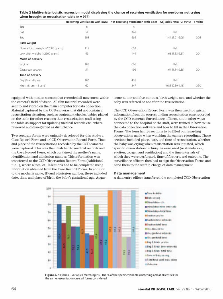
64 neonatal INTENSIVE CARE Vol. 29 No. 1 n Winter 2016
score at one and five mi nutes, birth weight, sex, and whether the baby was re ferred or not after the resuscitation.
The CCD Observation Record Form was then used to register information from the corresponding resuscita tion case recorded by the CCD-cameras. Surveillance of ficers, not in other ways connected to the hospital or the staff, were trained in how to use the data collection soft ware and how to fill in the Observation Forms. The form had 14 sections to be filled out regarding observa tions made when watching the camera recordings. These sections included place, date, and time of resuscitation, whether the baby was crying when resuscitation was ini tiated, which specific resuscitation techniques were used (ie stimulation, suction, oxygen and ventilation) and the time intervals of which they were performed, time of first cry, and outcome. The surveillance officers then had to sign the Observation Forms and hand them to the staff in charge of data management.
Data management A data entry officer transferred the completed CCD Ob servation
equipped with motion sensors that recorded all movement within the camera’s field of vision. All film material recorded were sent to and stored on the main computer for data collection. Ma terial captured by the CCD-cameras that did not con tain a resuscitation situation, such as equipment checks, babies placed on the table for other reasons than resuscitation, staff using the table as support for updating medical records etc., where reviewed and dis regarded as disturbance.
Two separate forms were uniquely developed for this study: a Case Record Form and a CCD Observation Rec ord Form. Time and place of the resuscitations recorded by the CCD-cameras were captured. This was then matched to medical records and the Case Record Form, which contained the mother’s name, identification-and admission number. This information was transferred to the CCD Observation Record Form (Additional file 1), where a total of 12 sections had to be completed using information obtained from the Case Record Forms. In addition to the mother’s name, ID-and admission num ber, these included date, time, and place of birth, the baby’s gestational age, Apgar-
this observational study found this technique to be use-ful for identifying deviations from protocol and areas forimprovement [6,8-10]. It is reasonable to assume that itcould also be applicable to other evaluation situations,not only in low-income countries, but in a variety ofsettings.Assessing the resuscitation routines at Paropakar Ma-
ternity and Women’s Hospital found that suction wasused in almost 90% of newborns brought to the resusci-tation table, whereas bag-and-mask ventilation was onlyused in less than 10%. According to both the nationalguidelines and the hospital’s own protocol, suction andbag-and-mask ventilation should be performed in allcases where the baby is not breathing at birth, which
gives a high adherence to the guidelines in terms of useof suction, but a very low when considering the use ofbag-and-mask ventilation. The guidelines for neonatalresuscitation at PMWH are very similar to the WHO’sinternational guidelines. Both state that after no morethan 30 seconds of initial drying, stimulation, clearing ofairways and evaluation, bag-and-mask ventilation shouldbe performed [5].WHO recommendations on basic newborn resuscita-
tion states that: ‘In neonates born through clear amni-otic fluid who start breathing on their own after birth,suctioning of the mouth and nose should not be per-formed’. Strength of this recommendation is strong andthe quality of evidence is high. They also state that
Table 2 Multivariate logistic regression model displaying the chance of receiving ventilation for newborns not cryingwhen brought to resuscitation table (n = 974)
Receiving ventilation with B&M Not receiving ventilation with B&M Adj odds ratio (CI 95%) p-value
Sex n n
Girl 54 348 Ref
Boy 108 464 1.44 (1.01-2.06) 0.05
Birth weight
Normal birth weight (≥2500 grams) 117 663 Ref
Low birth weight (<2500 grams) 45 149 1.68 (1.13-2.51) 0.01
Mode of delivery
Vaginal 105 616 Ref
Caesarean section 57 196 1.64 (1.14-2.36) 0.01
Time of delivery
Day (8 am-8 pm) 100 465 Ref
Night (8 pm – 8 am) 62 347 0.83 (0.59-1.18) 0.30
Figure 2 All forms – variables matching (%). The % of the specific variables matching across all entries for the same resuscitation case, allforms considered.
Lindbäck et al. BMC Pediatrics 2014, 14:233 Page 5 of 7http://www.biomedcentral.com/1471-2431/14/233
Figure 2. All forms – variables matching (%). The % of the specific variables matching across all entries for the same resuscitation case, all forms considered.
this observational study found this technique to be use-ful for identifying deviations from protocol and areas forimprovement [6,8-10]. It is reasonable to assume that itcould also be applicable to other evaluation situations,not only in low-income countries, but in a variety ofsettings.Assessing the resuscitation routines at Paropakar Ma-
ternity and Women’s Hospital found that suction wasused in almost 90% of newborns brought to the resusci-tation table, whereas bag-and-mask ventilation was onlyused in less than 10%. According to both the nationalguidelines and the hospital’s own protocol, suction andbag-and-mask ventilation should be performed in allcases where the baby is not breathing at birth, which
gives a high adherence to the guidelines in terms of useof suction, but a very low when considering the use ofbag-and-mask ventilation. The guidelines for neonatalresuscitation at PMWH are very similar to the WHO’sinternational guidelines. Both state that after no morethan 30 seconds of initial drying, stimulation, clearing ofairways and evaluation, bag-and-mask ventilation shouldbe performed [5].WHO recommendations on basic newborn resuscita-
tion states that: ‘In neonates born through clear amni-otic fluid who start breathing on their own after birth,suctioning of the mouth and nose should not be per-formed’. Strength of this recommendation is strong andthe quality of evidence is high. They also state that
Table 2 Multivariate logistic regression model displaying the chance of receiving ventilation for newborns not cryingwhen brought to resuscitation table (n = 974)
Receiving ventilation with B&M Not receiving ventilation with B&M Adj odds ratio (CI 95%) p-value
Sex n n
Girl 54 348 Ref
Boy 108 464 1.44 (1.01-2.06) 0.05
Birth weight
Normal birth weight (≥2500 grams) 117 663 Ref
Low birth weight (<2500 grams) 45 149 1.68 (1.13-2.51) 0.01
Mode of delivery
Vaginal 105 616 Ref
Caesarean section 57 196 1.64 (1.14-2.36) 0.01
Time of delivery
Day (8 am-8 pm) 100 465 Ref
Night (8 pm – 8 am) 62 347 0.83 (0.59-1.18) 0.30
Figure 2 All forms – variables matching (%). The % of the specific variables matching across all entries for the same resuscitation case, allforms considered.
Lindbäck et al. BMC Pediatrics 2014, 14:233 Page 5 of 7http://www.biomedcentral.com/1471-2431/14/233

neonatal INTENSIVE CARE Vol. 29 No. 1 n Winter 2016 65
The mean birth weight for recorded cases was 2860 grams, with non-crying newborns being lighter (2910 gram vs 2817 grams, p < 0.01). The low birth weight rate in the sample was 16.9% (308/1827) and 56% (1031/1827) of recorded cases were boys. The chance to receive ventila tion with bag-and-mask for a newborn not crying when brought to the resuscitation table was higher for boys and low birth weight babies (AdjOR 1.44 and 1.68 respectively). There was no difference in ventilation if the baby was born at night-time (8 pm-8 am), but there was an increased chance for a non-breathing baby to be ventilated if the delivery was done by caesarean section (AdjOR 1.64) (Table 2).
Apgar-scores were recorded one and five minutes after birth. Median Apgar-score after one minute was 5/10 and after five minutes 7/10. There were 198 babies (10.8%) with an Apgar score less than 7/10 at five minutes. Of these 198 depressed neonates 90 (45.5%) received ventila tion with bag-and-mask. In the sample there were 31 intra-partum related deaths, where 30 had an Apgar-score of 0/10 at both one and five minutes. A minority, 11/31 (35.5%), of the intra-partum related deaths received venti-lation with bag-and-mask, whereas suction of the airways was applied in 16/31 (51.6%).
When cross-comparing the four separate observational record forms that were completed for each randomized CCD-camera recording, it was found that the inter-and intra-rater reliability varied considerably amongst the different variables analyzed. The variables that had the highest correspondence across all forms where Out come, Bag & Mask total time, Bag-and-mask time inter vals, Bag-and-mask yes/no, and Suction yes/no, all matching in over 91% (91.3-97.8%) of the forms (Figure 2).
Discussion Similar to other published work on the use of CCD-cameras as a method to evaluate neonatal resuscitation, this observational study found this technique to be use ful for identifying deviations from protocol and areas for improvement [6,8-10]. It is reasonable to assume that it could also be applicable to other evaluation situations, not only in low-income countries, but in a variety of settings.
Assessing the resuscitation routines at Paropakar Ma ternity and Women’s Hospital found that suction was used in almost 90% of newborns brought to the resusci tation table, whereas bag-and-mask ventilation was only used in less than 10%. According to both the national guidelines and the hospital’s own protocol, suction and bag-and-mask ventilation should be performed in all cases where the baby is not breathing at birth, which gives a high adherence to the guidelines in terms of use of suction, but a very low when considering the use of bag-and-mask ventilation. The guidelines for neonatal resuscitation at PMWH are very similar to the WHO’s international guidelines. Both state that after no more than 30 seconds of initial drying, stimulation, clearing of airways and evaluation, bag-and-mask ventilation should be performed [5].
WHO recommendations on basic newborn resuscita tion states that: ‘In neonates born through clear amni otic fluid who start breathing on their own after birth, suctioning of the mouth and nose should not be per formed’. Strength of this recommendation is strong and the quality of evidence is high. They also state that suctioning of the mouth and nose of newborns that are not breathing at birth and are born through clear amni otic fluid should not be done routinely before initiating bag-and-mask
Forms into The Census and Survey Processing System (CSPro), a public domain software package de veloped and supported by the US Census Bureau and ICF Macro, which is interfaced with Statistical Package for the Social Sciences (SPSS 12.0).
Data analysis The data collected from resuscitation cases recorded between July 1st and October 31st 2012 were analyzed to assess the resuscitation routines at the hospital. These were then compared with existing guidelines. Relations between the use of bag and mask ventilation and factors such as sex, birth weight, mode, and time of delivery and were also analyzed. In order for future comparison before and after HBB-intervention, Apgar-scores were extracted from medical records and analyzed in terms of resuscitation techniques used. 50 CCD-camera recordings were randomly selected out of 257 recorded in October and November 2012. To evaluate the inter- and intra-rater reliability of the obser vational forms, two independent observers completed two sets of forms for each case, with roughly 6 weeks in-between the two viewings conducted by each observer. Out of the 14 original sections in the Observational form, 12 were used when analyzing the reliability. Place and date of the resuscitation were filled out in order to match the two sets of forms together, and thus were not included in the analysis. Using the time intervals given for each of the four individual resuscitation techniques, the total time each technique was performed was calcu lated and analyzed as four additional variables. The total time from when the baby was placed at the resuscitation table until the first cry was also calculated. In total there were 17 variables individually analyzed and cross-compared in each resuscitation case. Analyses were made in SPSS 12.0. A confidence level of 95% was con sidered significant.
ethical approval CCD cameras were mounted at all resuscitation tables with the permission of hospital management. All babies brought to the tables were recorded. Because of logistic concerns, since it being impossible to predict who needed resuscitation, written informed consent was ob tained from all parents of referent population before dis charge [12]. The video material for referent babies was thereafter edited and stored in a safe location and was only utilized by the researchers. The study was approved by the Nepal Health Research Council (reg. No 37/2012) and the Ethical Review Board of Uppsala University (dnr 2012/267).
Results Between July 1st and October 31st 2012 there were 6465 deliveries taking place at the study site. Out of these, 28 percent of the newborns (1827) were taken to the resus citation table and captured by surveillance cameras. More than half of the newborns (53.3%) brought to the resuscitation table were noted as not crying prior to re suscitation (Figure 1).
Figure 1 Newborns taken to the resuscitation table. The number and % of newborns that were taken to the resuscitation table, crying and not crying, out of the total number of babies born during the delivery period (6465).
The most frequent action taken was suction of the air ways, both for crying (85.0%) as well as for non-crying babies (92.3%) (Table 1). Ventilation with bag-and-mask was only performed on 172 (9.4%) of the recorded cases, whereof 162 were noted as not crying when brought to the resuscitation table.

66 neonatal INTENSIVE CARE Vol. 29 No. 1 n Winter 2016
W, Bhutta ZA, Bang A, Narayanan I, Ariawan I, Lawn JE: Neonatal resuscitation in low-resource settings: what, who and how to overcome challenges to scale up. Int J Gynecol Obstet 2009, 107(Suppl 1):47–S64.
2 Lawn JE, Lee AC, Kinney M, Sibley L, Carlo WA, Paul VK, Pattinson R, Darmstadt GL: Two million intrapartum-related stillbirths and neonatal deaths: where, why and what can be done? Int J Gynecol Obstet 2009, 107(Suppl):5–19.
3 Shiffman J: Issue attention in global health: the case of newborn survival. Lancet 2010, 375(9730):2045–2049.
4 Diaz-Rosello JL, Gisore P, Niermeyer S, Paul VK, Quiroga A, Saugstad OL, Silvestre MA, Singhal N, Sugiura T, Uxa F: Guidelines on Basic Newborn Resuscitation 2012. Geneva: World Health Organization; 2012.
5 World Health Organization: Pocket book of Hospital care for children: Guidelines for the management of common illnesses with limited resources. [http://whqlibdoc.who.int/publications/2005/9241546700.pdf]
6 Carbine DN, Finer NN, Knodel E, Rich W: Video recording as a means of evaluating neonatal resuscitation performance. Pediatrics 2000, 106:654–658.
7 Oakley E, Stocker S, Staubli G, Young S: Using video recording to identify management errors in pediatric trauma resuscitation. Pediatrics 2006, 117:658–664.
8 van der Heide PA, van Toledo-Eppinga L, van der Heide M, van der Lee JH: Assessment of neonatal resuscitation skills: a reliable and valid scoring system. Resuscitation 2006, 71(2):212–221.
9 Layouni I, Danan C, Durrmeyer X, Dassieu G, Azcona B, Decobert F: Video recording of newborn resuscitation in the delivery room: technique and advantages. Arch Pediatr 2011, 18(2):S72–S78.
10 Gelbart B, Hiscock R, Barfield C: Assessment of neonatal resuscitation performance using video recording in a perinatal centre. J Paediatr Child Health 2010, 46(7–8):378–383.
11 Donoghue A, Nishisaki A, Sutton R, Hales R, Boulet J: Reliability and validity of a scoring instrument for clinical performance during pediatric advanced life support stimulation scenarios. Resuscitation 2010, 81(3):331–336.
12 KC A, MŒlqvist M, Wrammert J, Verma S, Aryal DR, Clark R, Naresh PK, Vitrakoti R, Baral K, Ewald U: Implementing a simplified neonatal resuscitation protocol – helping babies breathe at birth (HBB) – at a tertiary level hospital in Nepal for an increased perinatal survival. BMC Pediatr 2012, 12:159.
13 Singhal N, Lockyer J, Fidler H, Keenan W, Little G, Bucher S, Qadir M, Niermeyer S: Helping babies breathe: global neonatal resuscitation program development and formative educational evaluation. Resuscitation 2012, 83(1):90–96.
14 Paropakar Maternity and Women’s Hospital: About Us, Statistics. [http:// prasutigriha.org.np/aboutus.php, http://prasutigriha.org.np/statistics.php]
15 Kattwinkel J, Perlman JM, Aziz K, Colby C, Fairchild K, Gallagher J, Hazinski MF, Halamek LP, Kumar P, Little G, McGowan JE, Nightengale B, Ramirez MM, Ringer S, Simon WM, Weiner GM, Wyckoff M, Zaichkin J: American Heart Association guidelines for cardiopulmonary resuscitation and emergency cardiovascular care science. Circulation 2010, 2010(122):909–919.
ventilation, although the strength of this recommendation is weak due to lack of published evi dence [4]. Several studies recently conducted have shown that routine suctioning is associated with lower five-minute Apgar-scores, lower oxygen saturation levels, and bradycardia [4,15]. The fact that suction was used for a large number of the cases where the baby was breathing satisfactory when placed on the resuscitation table shows that there is a lot of room for improvement in terms of the use of suction at the hospital.
limitations There were several limitations to the technique used in this study that made assessing the variables included in the observational forms difficult, and therefore affecting the reliability of the observational forms in a negative way. We have chosen only to present the variables show ing a good inter-rater reliability. When analysing the re corded resuscitations the baby’s condition was often hard to assess, particularly in the films where the picture quality was far from optimal. The most sensitive indica-tor of resuscitation being successful is an increase in the baby’s heart rate, which could be assessed using pulse oximeters. Assessment of pulse should be done regularly for babies requiring resuscitation [15], and should ac cording to both international guidelines and the hospi tal’s protocol be evaluated within the first 30 seconds. In the cases studied, the baby’s heart rate was rarely checked at any point during the resuscitation. In this study, pulse oximeters where not used due to technical difficulties in setting them up and coordinating them with the camera recordings. Therefore, neither the staff nor the observers had sufficient information about the baby’s heart rate or oxygenation, something that could have been very valuable when assessing the resuscitation efforts and outcome.
As in many other low-resource hospitals, a large por tion of the health personnel working in the birth facility where not qualified skilled birth attendants. Many of the labours stations were run by nursing students, and there were relatively few doctors and supervisors present. This may have contributed to the fact that the neonatal resus citation routines at the hospital to a great extent were not in accordance with the guidelines.
The scope of this study did not allow for the evalu ation of teamwork and communication. This would re quire separate methods of investigation as well as an alternative application of the CCD-camera technique. However, it is an interesting area of potential research using the CCD camera technique, which could be inves tigated at a later date.
Conclusion CCD camera technique was found to be a feasible method to assess resuscitation practices in this low re source hospital setting. The neonatal resuscitation proto col in place at the hospital did not differ much from the international guidelines for limited-resource settings. In most aspects, the staff did not adhere to these guide lines. The use of bag-and-mask ventilation was inad equate, and both suction and oxygen were given excessively in terms of protocol. Further studies explor ing the underlying causes behind the lack of adherence to the neonatal resuscitation guidelines should be con ducted in order to improve compliance and increase the possibility of fulfilling Millennium Development Goal (MDG) 4 by 2015.
References 1 Wall SN, Lee AC, Niermeyer S, English M, Keenan WJ, Carlo

Mercury Med EDIT Winter 2015.indd 1 2014-11-24 8:48 PM

Creche Innovations17745 Metcalf, One Penguin Plaza, Stilwell, KS 66085 USA
(913) 948.6290 [email protected] www.CrecheInnovations.com
NEWThe Penguin® Enteral Warmer
Warming the Gold Standard
to the Gold Standard
FeaturesSafely Warms All Enteral Feedings
No rapid overheating of breastmilk8 built-in safety features
100% of All Feedings Warmed to Mom’s Temperature
Never above or below proper temperature
Proprietary Low Heat TechnologyProtects milk from denaturing
Prevents extractables and leachables
Articulating IV Pole Mounting Arm Conveniently moves warmer away
from Isolette when not in use
Call for a FREE warmer validation! Free CE credits upon demo!
Interchangeable with ALL BRANDS of Enteral Feeding Pumps & Sets
No added expenses due to unnecessary changes to infusion sets
Now available for sale in the US FDA 510(k) clearance granted