Embed Size (px)
Citation preview
ECONOMIC EVALUATION AND ASSESSMENT OF EARLY TOXICITY OFHYPOFRACTIONATED RADIOTHERAPY COMPARED TO
STANDARD FRACTIONATION IN BREAST CANCER
DEPARTMENT OF RADIOTHERAPYCHRISTIAN MEDICAL COLLEGE
VELLORE 632004
DISSERTATION SUBMITTED IN PARTIAL FULFILLMENT OF
MD BRANCH IX RADIOTHERAPYEXAMINATION APRIL 2015
TAMIL NADU DR. M.G.R MEDICAL UNIVERSITY
CHENNAI - 600032.
CHRISTIAN MEDICAL COLLEGE, VELLORE
DEPARTMENT OF RADIOTHERAPY
This is to certify that the dissertation entitled “ECONOMIC EVALUATION AND
ASSESSMENT OF EARLY TOXICITY OF HYPOFRACTIONATED RADIOTHERAPY
COMPARED TO STANDARD FRACTIONATION IN BREAST CANCER” is a bonafide
work done by Dr.K.Chandralekha, Post Graduate Student in the Department of
Radiotherapy, Christian Medical College, Vellore during the period from June 2013 to April
2015 and is being submitted to The Tamil Nadu Dr. M. G. R Medical University in partial
fulfillment of the MD Branch IX Radiotherapy examination conducted in April 2015.
Guide
Dr.Selvamani B
Professor
Department of Radiotherapy
Christian Medical College
Vellore, India – 632004
This is to certify that the dissertation entitled “ECONOMIC EVALUATION AND
ASSESSMENT OF EARLY TOXICITY OF HYPOFRACTIONATED RADIOTHERAPY
COMPARED TO STANDARD FRACTIONATION IN BREAST CANCER” is a
bonafide work done by Dr.K.Chandralekha, Post Graduate Student in the Department of
Radiotherapy, Christian Medical College, Vellore during the period from June 2013 to
April 2015 and is being submitted to The Tamil Nadu Dr. M. G. R Medical University in
partial fulfillment of the MD Branch IX Radiotherapy examination conducted in April
2015.
Head of the Department
Principal Dr.Selvamani B
Christian Medical College Prof and Head of the department
Vellore,India- 632 004 Department of Radiotherapy
Christian Medical College
Vellore, India - 632004
I K.Chandralekha, PG Registrar ,Department of Radiation therapy ,Christian Medical College
Vellore hereby declare that the dissertation titled “Economic Evaluation And Assessment
Of Early Toxicity Of Hypofractionated Radiotherapy Compared To Standard
Fractionation In Breast Cancer ” is a bonafide work done by me for partial fulfilment
towards MD Radiotherapy ( Branch IX) Degree examination of the Tamil Nadu Dr M G R
Medical University to be held in April 2015.
DR.K.CHANDRALEKHA
PG REGISTRAR ,
DEPARTMENT OF RADIOTHERAPY,
CHRISTIAN MEDICAL COLLEGE,
VELLORE


ACKNOWLEDGEMENT
This work in hand would have never been accomplished without an outstanding support
system and is a fruit of efforts of many and I am grateful to God and all who have directly
or indirectly helped me.
Mere words are not enough to thank my Guide Prof. Dr. Selvamani Backianathan, who
has a multitasking capability and sincerity, had led me from initiation to culmination of
this dissertation. She had held hands and led through the journey throughout like a kid
spending immense care and time for making this dissertation into a perfect shape.
I am extremely grateful to Dr. Balukrishna, whose goal is perfection in a prompt
manner, who by his innovative thinking kindles our thought processes. His able guidance
and timely advice has helped in decision making.
I extend my gratitude to Prof. Dr Jasmine Prasad, who had spent long times patiently
and had made valiant efforts for the output and statistical analysis.
I thank Prof. Dr. Subhashini John and the Consultants for their concern, care and
encouragement.
Patient’s patient sharing of information had been the foundation for this study and i thank
them all. I would like to thank the Accounts department, Staff and Technicians in
Department of Radiotherapy for gathering the machine data and cost analysis.
Radiographers had shared their support for assisting in data collection. I also thank Mr
Prakash, from Biostatistics department for helping in analysis.
I extend my warm gratitude to My Mother who has always pampered me in all hardships
and My Father who has always been encouraging. I thank My Husband for his
unwavering support for this accomplishment and well wisher My Grandmom.
I express my gratitude to Arun who is always willing to help and Moses who guides me
and all my friends and seniors of Department of Radiotherapy.

CONTENTS
ACKNOWLEDGEMENT i
CONTENTS ii
1 AIM AND OBJECTIVES 1
2 INTRODUCTION 2
3 REVIEW OF LITERATURE 4
3.1 Breast cancer Statistics 4
3.2 Financial scenario in India 9
3.3 Management of breast cancer- an overview 10
3.4 Role of radiation therapy in breast cancer 11
3.5 Radiotherapy Planning and Simulation 14
3.6 Treatment Volumes 16
3.7 Fractionation in breast cancer radiotherapy 18
3.8 Trials for Hypofractionated Radiotherapy 19
3.9 Extreme Hypofractionation 27
3.10 Controversies with Hypofractionated Radiotherapy 27
3.11 Grade and Hypofractionation 28
3.12 Post Mastectomy Radiotherapy 29
3.13 Acceptance of hypofractionation in India 31
3.14 Economic Burden of cancer Therapy 33
3.15 Economic evaluation 35
3.16 Economic Studies in India 40
3.17 Outcome Parameters 40
3.18 Need for Economic analysis 41

3.19 Radiotherapy Cost structure : Institution Perspective 42
3.20 Non Medical costs of the Patient 43
3.21 Radiation Toxicity 44
4 MATERIALS AND METHODS 48
4.1 Study design 49
4.2 Radiotherapy planning 50
4.3 Radiotherapy techniques 52
4.4 Data collection 58
4.5 Statistical analysis 61
5 RESULTS 62
5.1 Demography and patient characteristics 63
5.2 Disease Characteristics 72
5.3 Treatment details 77
5.4 Machine costing 80
5.5 Acute toxicities 84
5.6 Patient costing and economic analysis 87
6 DISCUSSION 100
6.1 Limitations 109
7 CONCLUSIONS 110
8 BIBLIOGRAPHY 111
9 APPENDIX 117

ABSTRACT
Economic Evaluation And Assessment Of Early Toxicity Of Hypofractionated
Radiotherapy Compared To Standard Fractionation In Breast Cancer
Chandralekha*, Selvamani.B, Balukrishna, Jasmine Prasad, Department of Radiotherapy,
Christian Medical College, Vellore
Introduction: Breast cancer is the most frequent cancer among women with the globalincidence in women to be 25.2% of all reported new cancers. In India, breast cancer is themost common cancer at 27% of all new cancers . Breast cancer is associated with substantialmedical and economic burden and henceforth the management of breast cancer accounts for alarge percentage of health care budget. Radiation therapy as an integral part in the multi-modality management of breast cancer significantly reduces the locoregional recurrence andalso improves the overall survival. To overcome the economic burden related to radiotherapyin breast cancer various hypofractionated schedules like 39 Gy in 13 fractions, 40 Gy in 15fractions were tried and have proven to achieve similar local control rates, survival rates andcosmetic outcome. This study aims to do the economic evaluation and to assess the acutetoxicities associated with 40 Gy in 15 fraction(hypofractionated regimen).
Aims and objectives: To analyse the cost difference in breast cancer radiotherapy betweenconventional fractionation and hypofractionated radiotherapy.
The study also aims at assessing the early toxicities of patients receiving post mastectomyradiotherapy.
Methods and materials: This Prospective study group consisted of 30 consecutive patientsseen in the Radiation therapy department of Christian Medical College, Vellore fromFebruary to August 2014, treated with standard fractionation and hypofractionated postmastectomy radiotherapy by conventional technique. Each patient was interviewed using apilot tested questionnaire to collect data on the health economics. The costs imparted to thepatient were classified as direct and indirect costs. The cost effect for each was assessed atthe end of the treatment. The occurrence of early toxicity in patients treated with standard andhypofractionated radiotherapy was recorded and analysed using RTOG acute skin toxicitycriteria.
Results: Twenty three patients were included in the 40 Gy in 15 fractions arm and 7 patientswere in the 50 Gy in 25 fractions arm. Of the 30 patients 15 were treated in the Cobalt and 15were treated in Linear accelerator. The analysis showed that there was significant reduction incosts in hypofractionation with conventional treatment in Cobalt 60. The difference in Linearaccelerator was not found to be significant.

Conclusion : Adoption of hypofractionated radiotherapy in breast cancer treatment can leadto significant reduction in resource utilisation and is especially pronounced for conventionalradiotherapy settings with high patient loads.
Keywords : Carcinoma Breast PostMastectomy Radiotherapy Hypofractionation

1 . AIMS & OBJECTIVES
AIM:
To analyse the financial benefits of the cost difference in breast cancer radiotherapy
between conventional fractionation and hypofractionated radiotherapy. The study also aims at
assessing the early toxicities of patients receiving post mastectomy radiotherapy.
Primary objective:
Report the difference in mean cost per patient for treatment with standard and
Hypofractionated post mastectomy radiotherapy for carcinoma breast (COST
MINIMISATION).
Secondary objective:
To assess early toxicity in patients treated with standard and hypofractionated
radiotherapy.

2. INTRODUCTION:
Breast cancer is the most frequent cancer among women according to the Globocan
2012 update. The incidence of breast cancer in women worldwide is reported to be 25.2% of
all new cancers and is expected to increase much more by 2020. (1) In India, breast cancer is
the most common cancer contributing to 27% of all new cancers in women (1).
Taking into account cancers worldwide, the total economic burden of this disease was
estimated to be in the range US dollar 300-400 billion in 2001 [about US dollar 100-140
billion as direct costs and the rest as indirect costs (2). There is significant medical and
economic burden associated with breast cancer and it accounts to large expenses on the
public, but the expenses have been difficult to gauge.
Radiation therapy is an integral part of the multi-modality management of breast
cancer. The increase in the incidence of the breast cancer has contributed significantly to the
rising numbers of patients receiving post mastectomy radiotherapy. The conventional dose of
radiotherapy in breast cancer is to deliver 50 Gy in 25 fractions over 5 weeks.
In view of the prolonged duration of treatment and machine availability, various
hypofractionated schedules have been investigated and it is proven to be advantageous by
many randomised trials. (3) This lesser treatment schedule is not only favourable for the
patients by decreasing the number of hospital visits, but also beneficial for the health services
with limited resources, waiting list in the hospital etc., by reducing the machine time and
human resources. This time saved may be adequately utilised for the treatment of another
patient. Moreover, the total treatment time, that is, the daily treatment time multiplied by the
number of fractions attributes to the cost of radiotherapy.

Hence, a reduction in the daily treatment time and /or a decrease in the number of fractions
would in turn reduce the cost of radiotherapy.
These short course treatments make an impact by achieving similar local control rates,
survival rates and cosmetic outcome with the great advantage in reducing the number of visits
to the hospital to the patients and to the health services by improving machine utilisation.
The aim of this study is to do the economic evaluation of hypofractionated radiotherapy along
with assessment of the acute toxicities associated with it.

Review of Literature______________________________________________________________________________________________________________________

3. LITERATURE REVIEW
3.1 BREAST CANCER STATISTICS:
The most frequent cancer among women is breast cancer with the Globocan 2012 update
showing the global incidence of breast cancer in women to be 25.2% of all reported new
cancers (Fig.3.1.1) (1). Also in India, breast cancer has become the most common cancer at
27% of all new cancers in women (1).
Breast cancer is also the most common cause of cancer related mortality in women
(14.7%) worldwide (Fig.3.1.2).
Fig 3.1.1: Cancer Incidence in Women, World - GLOBOCAN 2012
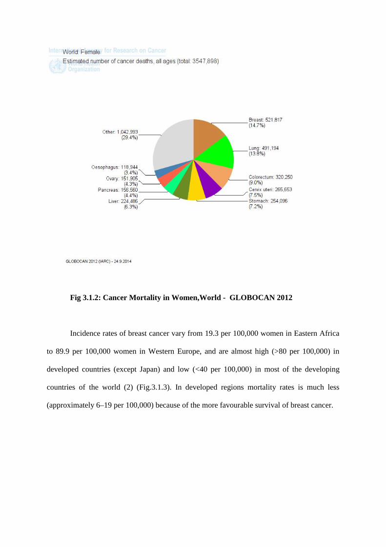
Fig 3.1.2: Cancer Mortality in Women,World - GLOBOCAN 2012
Incidence rates of breast cancer vary from 19.3 per 100,000 women in Eastern Africa
to 89.9 per 100,000 women in Western Europe, and are almost high (>80 per 100,000) in
developed countries (except Japan) and low (<40 per 100,000) in most of the developing
countries of the world (2) (Fig.3.1.3). In developed regions mortality rates is much less
(approximately 6–19 per 100,000) because of the more favourable survival of breast cancer.

Fig 3.1.3: Age standardised incidence of breast cancer across the world
By 2020, 70% of the world’s cancer cases will be in poor countries, with a fifth in
India (2). In 2011, ICMR conducted an analysis of cancer cases among women in Delhi,
Chennai, Bangalore and Mumbai from 1982 to 2005. The study showed that 10 per 100,000
women got breast cancer until about 10 years ago compared with 23 per 100,000 later on (2).
It was predicted in this report that by 2020, breast cancer will overtake cervical cancer as the
most common type of cancer among all women in India.

Fig 3.1.4: Age standardised mortality rates of breast cancer across the world
There is an increasing trend in breast cancer incidence which may be due to increased
awareness and advanced diagnostic modalities, but the corresponding decreasing trend in
cervical cancer suggests that it is a true increase. This increase in incidence of breast cancers
may be due to genetic and environmental factors. Etiologically though women in poor
countries are less prone for breast cancers than those in the west, they are more likely to die
of it because of late presentation. While incidence is about 130 per 100,000 women in the
USA, it is only about 19 per 100,000 in India (4). But the chances of surviving cancer in a
low-income or middle-income country are much worse than in the UK or USA. Fig 3.1.4
shows the age standardised mortality rates of breast cancer across the world.
3.2 FINANCIAL SCENARIO IN INDIA:
The most probable reason for the scenario above said is that women seek medical care
very late. Poor awareness of breast cancer among public, inhibition, reluctance, financial
constraints of the people, staying away from home, inability to access care and following
alternative medicine are the most common reasons for presenting late to medical care.
Financial constraints also prevent patients from having the best treatment, for e.g.
Trastuzumab(5). Women delay seeking medical care and hence often present with large
lumps . Even when patients finally do seek care, they often cannot complete the treatment
due to financial constraints.
Most of the patients are farmers or labourers or daily wage makers, who land in major
Government and Trust hospitals. Though the concessions and insurances are offered by major
Government and Trust hospitals, transport and accommodation costs can hamper patients’
finances.
Even with patients who get comprehensive insurance policies, people are found to
suffer financial hardships. Though insurance is provided to decrease the financial burden, lot
of out of pocket costs are met by the patient and family in addition to the loss of income by
them and the low socio economic status people are the most affected ones. Also, the patients
have to spend quite a large sum for accommodation, food, medicines including the expenses
for the accompanying person which are not covered by insurance.
3.3 MANAGEMENT OF BREAST CANCER: AN OVERVIEW
Management of invasive breast cancer comprises a multimodality approach with
surgery, chemotherapy, radiation therapy hormonal therapy and targeted therapy all playing
integral parts.
The factors that influence the choice of treatment include age, pathological stage of
the cancer especially tumour size and nodal status, biological prognostic factors and
hormonal receptor status. Various combinations and the sequences of the multimodality
treatment in addition to surgery are determined by these factors. Introduction of
multimodality treatment reduced breast cancer mortality by 18% and improved overall
survival (5).
The primary modality of management with surgery for invasive breast carcinoma has
undergone a shift over the years from radical mastectomy to breast conservation surgery and
sentinel lymph node biopsy. Similarly locally advanced breast cancers are made feasible for
breast conservation surgery after neoadjuvant chemotherapy following good response to
chemotherapy. The adjuvant therapy of breast cancer has also improved with the advent of
new chemotherapeutic, hormonal and targeted agents.
Radiation therapy has also advancements and amendments in the technique of
delivery from conventional through 3D conformal techniques to IMRT and recently
accelerated partial breast irradiation. Besides the techniques there has also been a transition in
the dose and fractionation of the radiotherapy delivered.

3.4 ROLE OF RADIATION THERAPY IN BREAST CANCER
Radiation therapy has been a modality in the treatment of the breast cancer ever since
the discovery of X-rays and its tumoricidal properties were discovered. It is an integral part in
the management of both early as well locally advanced breast cancers. It has been established
that post operative radiotherapy significantly reduces the locoregional recurrence and also
improves the local control which indirectly increases the cancer specific and overall survival.
The importance of local control in breast cancer survival cannot be discounted.
In early breast cancers it is used as adjuvant therapy to deliver whole breast radiation
followed by boost to the lumpectomy site. In locally advanced cancers it is use to deliver
radiation to the chest wall after mastectomy. The inclusion of regional lymphatic region is
based on the number of axillary lymph nodes positive for tumour deposits after axillary
clearance or on the basis of use of neoadjuvant chemotherapy.
A total of 9422 patients from 15 randomised trials were included in the analysis by
Vin Hung et al and it was concluded that the relative risk of ipsilateral breast recurrence for
omitting radiotherapy was 3 and relative risk of mortality on omitting radiotherapy was 1.086
with a relative excess of 8.6% in mortality on omitting radiotherapy in breast conservation
therapy (6).
The indications for post mastectomy radiation therapy have been defined from the results of
various randomized trials like the Danish trial and British Columbia trial(7,11). Benefit in
terms of local control and survival was found in stages II / III and node positive breast
cancers. Studies showed that the addition of post mastectomy radiotherapy reduced loco
regional recurrence rate by 2/3rd to3/4th when compared to the groups that did not receive
radiotherapy (7,8). Decrease in local recurrences and improvement in overall survival with
radiation therapy have been established in many randomized trial involving both
premenopausal and postmenopausal breast cancer women (7-11).
Findings from 78 randomized clinical trials were analysed by the Early Breast Cancer
Trialists’ Collaborative Group (EBCTCG) (12), these trials were done for evaluating the
extent of surgery and the use of radiation therapy. The analysis revealed improved local
control at 5 years and significant improvement in survival and overall survival at 15 years.
Moreover the Trialsists’ were established that the absolute reduction in the 5-year rate of
local recurrence was proportional to the absolute reduction in 15-year breast-cancer mortality.
Modality of therapies with minimal or no effect on reducing the 5-year rate of local
recurrence had no benefit in decreasing 15-year cancer related mortality; however, treatments
that resulted in improvement in the 5-year rate of local recurrence also resulted in a reduction
in breast-cancer mortality at 15 years (Fig 3.4.1).
Regardless of the method of achieving the reduction (i.e., by extensive surgery or by
the addition of radiotherapy) the absolute benefit for cancer related mortality was similar for
a given reduction in local recurrence. Among treatments that had more than a 10% reduction
in the 5-year risk of local recurrence, breast-cancer mortality was reduced by 1.6% at 5 years,
3.7% at 10 years, and 4.9% at 15 years.
The trials in the EBCTCG meta-analysis that studied had demonstrated significant
improvement in 15-year absolute overall survival by the addition of radiotherapy after breast-

conservation surgery, by 5.3% (P = 0.005), and after mastectomy in node-positive patients,
by 4.4% (p = 0.001) (10).
On the other hand, the role of post mastectomy radiotherapy in T1/T2 tumour, grade grade 2
with 1-3 lymph nodes positive is still debatable. The indication in this group of breast
cancers is expected from the results of the on-going Supremo trial(13). Various factors such
as size of tumour (>4 cm), close/positive margins, lymph vascular invasion, extra capsular
extension, ER/PR/Her2 neu status, grade of tumour are known to affect the loco regional
recurrence rate, and these factors contribute significantly towards the decision making.
Fig 3.4.1: EBCTCG Meta-Analysis

3.5 RADIOTHERAPY PLANNING AND SIMULATION (19)
Radiotherapy can be delivered in breast cancer by various techniques starting from
conventional to IMRT. The preferred position for the treatment of breast cancer patients is in
supine position with 90 degrees abduction of the arms on a breast board used as
immobilisation. Various commercially available instruments are available to eliminate the
slope of the chest wall and to better immobilise the patient.
The entire breast in the case of breast conservation surgery and the chest wall in case
of mastectomy patients are included in the radiation portal. The upper margin is kept at the
lower edge of the head of the clavicular bone and the lower border is usually kept at a level to
include the entire breast which usually comes to about at two to three cm below the
inframammary fold. The opposite breast is used as a reference for the lower border in the case
of post mastectomy radiation therapy. The midline of the body makes up the medial border
and the mid axillary line makes up the lateral border. The field may be extended to include
the scar and the drain sties.
The regional lymphatics are included as per the clinical indications. The
supraclavicular fossa is included in patients who received neo adjuvant chemotherapy, as the
prognostic information usually obtained by axillary dissection is altered in such setting.
Supraclavicular fossa is also included in cases where ≥ 4 lymph nodes are positive for
metastatic deposits in the axillary dissection.
Indications for axillary radiation therapy include nodal positivity with extracapsular
extension, inadequate axillary dissection and patients with estimated probability of nodal
involvement greater than 10 to 15% with no axillary dissection.
6 MV x-rays are usually used for the treatment however in cases where the field
separation is large, it may result in dose inhomogeneity in which case higher energy X rays
may be used to achieve a better cosmetic outcome as homogeneity has been correlated with
cosmesis. Various techniques like the use of standard wedges, dynamic wedges, MLCs can be
used to achieve dose homogeneity in the breast. Use of bolus is not necessary in cases of
T1-2 tumors whereas it may be necessary in the case of locally advanced cancer with the
intent of achieving additional radiation dose (boost) at the skin at the site of the surgical scar
in mastectomy patients.
It is aimed to keep the amount of lung included in the tangential field at any section to
less than 2-3 cm of length from the chest wall lung junction. The amount of lung involved has
been correlated with the incidence of radiation pneumonitis. Half beam block is used in the
treatment to reduce the incidence of pneumonitis. In cases where the supraclavicular fossa
(SCF) is included special attention has to be paid to the field junctions and appropriate
techniques employed to avoid excess dose at the junction of the supraclavicular field and the
tangential field. The SCF is usually irradiated by an anterior field matched to the tangential
field and the dose is delivered at the d max. There are number of ways for matching the
tangential fields with the supraclavicular field. By angling the foot of the couch away from
the source, the divergence of the tangential fields can be eliminated. The collimator can be
rotated to eliminate the overlap at this junction. The inferior divergence of the supraclavicular
field can be blocked by placing half beam block for the field.
Motion management techniques are used in breast radiotherapy to counter the effects
of breathing on the radiation portals. The technologies available for the same include 4DCT
and gating. Breath hold technique is also used as a simpler solution to motion management.

3.6 TREATMENT VOLUMES
In the Danish and EBCTG randomized trials of Post mastectomy radiation therapy
(PMRT), RT delivered to the chest wall and surgical scar, including the supraclavicular,
infraclavicular, axillary and inframammary lymph nodes demonstrated that radiotherapy
results in improvement of local control as well as improvement in overall survival. On the
basis of these results, it is well acknowledged that the treatment volume for breast cancer
should include the entire chest wall and the scar of the mastectomy surgery. However, with
regards to the inclusion of the regional nodal regions in the treatment volume there is still
quite a bit of controversy existing.
Positive lymph nodes in the axilla entail the inclusion of ipsilateral supraclavicular
fossa in the volume of treatment. But there is wide variation as to whether the Internal
Mammary (IM) lymph nodes should be included. This controversy is partly in view of the
potential toxicity particularly in the case of left sided breast cancers when the IMN is
included in the radiation portals. In addition the added benefit of IMN irradiation is very
uncertain (15).
In 1996, the European Organization for Research and Treatment of Cancer (EORTC)
conducted a trial to evaluate the (protocol 22922/10925) the benefit of including the Internal
Mammary nodes and medial supraclavicular nodes in the radiation field for those patients
who have axillary node positivity. The medial tumors treated by either breast conservation
surgery or by mastectomy were included in this trial.
Another French study also randomized 1334 women of breast cancer who had
undergone mastectomy with axillary nodes positivity or central tumors to chest wall and
supraclavicular filed irradiation with or without the inclusion of the internal mammary nodal
chain in the radiation filed. Preliminary data analysis of this study has not detected any
difference in the overall survival (OS) in the two arms (16).
The results of the recently presented MA-20 National Cancer Institute of Canada
clinical trial shows an improvement in the DFS in node postive and high risk node negative
patients who have been treated by Breast conservation surgery, when the regional nodal
regions, including IM lymph nodes, are included while delivering whole breast radiation
therapy (17).
Another retrospective study from the MGH has shown similar rates of Loco Regional
Relapse (LRR), Disease Free Survival (DFS), and OS in patients with one to three positive
lymph nodes treated with chest wall radiation therapy only as compared to those treated with
chest wall and nodal irradiation, suggesting that PMRT to the chest wall only may be
appropriate for women with tumors <5 cm and one to three positive LNs (18).
Internal mammary chain radiation may be considered in axillary node positivity with
central and medial quadrant tumours.
3.7 FRACTIONATION IN BREAST CANCER RADIOTHERAPY:
The conventional radiotherapy regimen after mastectomy for cancer breast delivers
50Gy in 25 fractions of 2 Gy over 5 weeks. In UK and Canada, several trials on alternate
schedules (hypofractionation) were done years ago on an empirical basis in breast
conservation therapy and based on the results of these trials 40 Gy in 15 fractions over 3

weeks is practiced as standard in these countries. The results of the trials which employed
hypofractionated radiotherapy in the treatment of breast cancer are showing favourable
outcomes in terms of control of the tumor and also with respect to late adverse effects if the
modest increases in size of radiation fractions are adjusted accordingly with appropriate
decrease in the total dose of radiation delivered.
Although the hypofractionated radiotherapy schedules have been in wide use in the
United Kingdom, the schedule of 40Gy delivered in 15 fractions over 3 weeks has never been
tested formally in a randomised trial with the standard fractionation schedules. This lack of
strong trial based evidence for hypofractionated radiation raised concerns regarding the safety
and the effectiveness of such a schedule when compared with the standard schedule of 50 Gy
in 25 fractions. In an effort to address this uncertainty START Trials were initiated by the
then UK Coordinating Committee for Cancer Research to test the effects of hypofractionated
radiotherapy schedules.
3.8 TRIALS FOR HYPOFRACTIONATED RADIOTHERAPY:
Royal Marsden Hospital/Sutton and Gloucestershire Oncology Centre:(20,21)
In a randomized phase III trial conducted from 1986 - 1998, 1410 patients with early
breast cancer were randomized to three arms of different fractionation to assess the effect of
fraction size on late change in breast appearance with tumor control as one of the secondary
endpoints. The study included only patients with stage 1-3 tumors with only one node
positive for metastasis and had undergone only lumpectomy of the tumor site.
The conventional regimen of 50 Gy in 25 fractions over 5 weeks was compared with
39 Gy in 13 fractions, or 42.9 Gy in 13 fractions, both given over 5 weeks. The fraction size
of 3 Gy was calculated on the basis of an alpha-beta ratio of 1.8 Gy while the 3.3 Gy fraction

size was arrived at assuming an alpha-beta ratio of 6.0 Gy. The trial was analysed on an
intention to treat principle. As per clinical indications, the regional lymphatics were included
in the treatment field. Radiation boost was also delivered as indicated in 7 daily fractions to a
total dose of 14 Gy.
The primary end point of photographic assessments of the patients were analysed at a
median follow up of 8.1 years. Statistically significant difference was seen between the 50 Gy
and 39 Gy arm (p=0.01) however the difference between the 50 Gy and 42.9 Gy arm was
borderline (p=0.05). The risk of ipsilateral tumour relapse after 10 years was 12.1% in the 50
Gy arm , 14.8% in the 39 Gy arm and 9.6% in the 42.9 Gy arm at a median follow-up of 9.7
years (difference between 39 Gy and 42.9 Gy groups, chi2 test, p=0.027).
Based on these results it was estimated that the fractionation sensitivity of breast
cancer is around 4 Gy which is similar to that of the late reacting tissues.
UK START A: (22, 23)
START-A trial, the first of hypofractionation study in the UK was done across 17
centres from 1998 to 2002. The trial was designed based on data available from the pilot trial
described above. One of the aims of the trial was to combine the data from this trial and the
pilot study to arrive at a better understanding of the fractionation sensitivity of the breast
tumors.
The trial included patients with pT1-3a and N0-1 disease who had undergone primary
surgery and then required adjuvant radiation. The trial included patients who had breast
conservation surgery and also those who had undergone mastectomy. 2236 eligible patients

were randomized to three different fractionation arms. The conventional regimen of 50 Gy in
25 fractions over 5 weeks was compared with the experimental regimens of 41.6 Gy in 13
fractions over 5 weeks or 39 Gy in 13 fractions over 5 weeks (Fig 3.8.1). All treatment
schedules were delivered over 5 weeks so that treatment time duration will not be a factor in
the final analysis. The regional lymphatics were included in the treatment field as per
indication. The boost dose to the tumor bed was left up to the discretion of the treating centre.
Fig 3.8.1: Trial Design of START A (# - fraction)
The primary end points of the trial were local relapse, normal tissue effects and the
effect on the quality of life (QoL). Patients were followed up every year after radiation for
locoregional relapse and normal tissue effects.
The trial was designed to detect 5% difference in the local relapse rates between the
different radiotherapy schedules. At a median follow up of 9.3 years 6.2% of patients on the
trial had locoregional relapse.
The locoregional relapse rate did not differ significantly between the 41·6 Gy and 50
Gy regimen groups (6·3%, vs 7·4%, hazard ratio [HR] 0·91; p=0·65) or the 39 Gy (8·8%)
and 50 Gy regimen groups (HR 1·18; p=0·41). Breast size reduction and induration of the
breast tissue were the most common side effects observed at the end of 10 years.
The incidence of moderate or marked normal tissue effects were significantly less in
the 39 Gy group, whereas no significant changes were observed among 41.6Gy and 50 Gy
group.
UK START-B: (24)
In the second of the parallel hypofractionated radiotherapy study, START B Trial,
2215 patients were randomized to two regimens comparing 40 Gy in 15 fractions of 2.67 Gy
in 3 weeks with a control group of 50 Gy in 25 fractions of 2 Gy over 5 weeks.
The trial included patients with pT1-3a pN0-1 tumor after primary surgery requiring
adjuvant radiotherapy (Fig 3.8.2). The trial was carried in 21 centres in the United Kingdom
between 1999 and 2001. The inclusion of regional lymphatics in the irradiation field was
based on clinical indications.
The delivery of boost radiotherapy to breast conserved patients was left to the
discretion of the treating centre. Electron boost of appropriate energy delivering 10 Gy in 5
fractions was used for boost field radiation therapy. The primary end points in this trial were
locoregional control, normal tissue toxicity and quality of life.

Fig 3.8.2: Trial Design of START B
1105 women were assigned to the 50 Gy group and 1110 to the 40 Gy group. The
proportion of patients with locoregional relapse at 10 years did not differ significantly
between the 40 Gy group (4·3%, 95% CI 3·2–5·9) and the 50 Gy group (5·5%, 95% CI 4·2–
7·2; HR 0·77, 95% CI 0·51–1·16; p=0·21). At 10 years follow up the most common of the
late effects were breast shrinkage and induration similar to the START A trial. All of the
moderate to marked late normal tissue effects were significantly less common in the 40 Gy
group than in the 50 Gy group.
In a combined post hoc analysis of the two START trials together and the pilot trial
showed that the hypofractionation arms combined together did not vary significantly from the
conventional fractionation with respect to control rates regardless of factors like age, type of
surgery, stage of the tumor, grade of the tumor and this was also the same with respect to
effects on the normal tissues.

CANADIAN TRIAL: (25)
Ontario Clinical Oncology Group carried out a randomized trial to find out the
optimal fractionation regimen in adjuvant whole breast radiotherapy. 1234 patients who had
undergone breast conservation surgery with negative margins and negative nodes on axillary
clearance were randomized to two regimens. 612 patients were randomized to the
conventional regimen of 50 Gy in 25 fractions over 5 weeks and 622 patients were
randomized to 42.5 Gy in 16 fractions over 22 days.
The patients were stratified according to tumor size, systemic therapy, age and centre
of treatment. The radiation therapy was delivered using tangential beams from Monday to
Friday. The regional nodal regions (the axilla, supraclavicular fossa and the internal
mammary nodes) were not included in the radiation portals. The patients in the trial did not
get any tumor bed boost.
The primary endpoint in this trial was local recurrence and the secondary endpoints
were regional and distant recurrence, late toxicities and cosmetic outcomes of the treatment
and survival. After completion of the radiotherapy patients were followed up every 6 months.
The first mammography was obtained 6 months after completion of the radiotherapy and then
yearly during follow up. Late toxicities and the cosmetic outcomes of the treatment were
assessed at 3, 5 and 10 years after completion of the treatment. The RTOG Late morbidity
scoring criteria were used to assess the late skin toxicities and EORTC scale was used for
assessing the cosmetic outcomes of breast conservation.
The cumulative incidence of local recurrence was 6.7% in the control arm compared
to the 6.2% in the hypofractionated arm at a median follow up of 12 years. This is an absolute
difference of 0.5% with 95% confidence interval of -2.5 to 3.5. Hence the null hypothesis that
the hypofractionated arm would be more than 5% worse than conventional radiotherapy was
rejected on the basis of non inferiority (p<0.001).
Subgroup analysis also showed there was no effect of prognostic factors like receptor
status, age, tumour size and the use of chemotherapy on the fractionation regimen. However
high grade seemed to fare worse with hypofractionated therapy with local recurrence in
control being only 4.7% compared to the 15.6% in the hypofractionated regimen in this
subgroup. This was statistically significant (p=0.01). The probability of survival at 10 years
follow up is 84.6 % vs 84.4 % favouring the hypofractionated regimen. However this
difference was not statistically significant, p=0.56.
META ANALYSIS:
A meta-analysis from Cochrane Reviews published in 2010 (26) included four
randomised trials mentioned above, which they described to be of low to medium quality. It
analysed the effect of hypofractionation on local recurrence risk, breast appearance and
survival at five years. The risk ratio (RR) for local recurrence was 0.97 (95% CI 0.76 to 1.22,
p=0.78) and for survival at five years RR was 0.89 (95% CI 0.77 to 1.04, p=0.16). With
regards to toxicity and cosmetic outcomes of the breast, the analysis showed that acute skin
toxicity was significantly lesser with conventional fractionation (p = 0.007). As for the
appearance of breast after radiation, the RR of 1.17 (95% CI 0.98 to 1.39, p=0.09) also
confirms the superiority of hypofractionation regimens.
The results of the above described hypofractionated radiotherapy breast cancer trials
are summarized in the following table (Table 3.8.1)
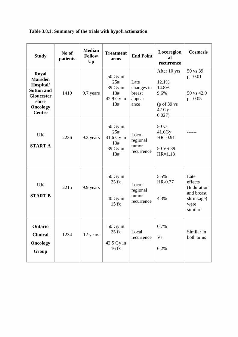
Table 3.8.1: Summary of the trials with hypofractionation
StudyNo of
patients
MedianFollow
Up
Treatmentarms End Point
Locoregional
recurrence
Cosmesis
RoyalMarsdenHospital/
Sutton andGloucester
shireOncology
Centre
1410 9.7 years
50 Gy in25#
39 Gy in13#
42.9 Gy in13#
Latechanges inbreastappearance
After 10 yrs
12.1%14.8%9.6%
(p of 39 vs42 Gy =0.027)
50 vs 39p =0.01
50 vs 42.9p =0.05
UK
START A
2236 9.3 years
50 Gy in25#
41.6 Gy in13#
39 Gy in13#
Loco-regionaltumorrecurrence
50 vs41.6GyHR=0.91
50 VS 39HR=1.18
------
UK
START B
2215 9.9 years
50 Gy in25 fx
40 Gy in15 fx
Loco-regionaltumorrecurrence
5.5%HR-0.77
4.3%
Lateeffects(Indurationand breastshrinkage)weresimilar
Ontario
Clinical
Oncology
Group
1234 12 years
50 Gy in25 fx
42.5 Gy in16 fx
Localrecurrence
6.7%
Vs
6.2%
Similar inboth arms

3.9 EXTREME HYPOFRACTIONATION:
The encouraging results of the previously described hypofractionated radiotherapy
trials, inspired people to experiment with further hypofractionation regimens.
The UK FAST trial is one such trial. It randomized 729 eligible patients to three arms
comparing 50 Gy in 25 fractions and 28.5 or 30 Gy in 5 once-weekly fractions of 5.7 or 6.0
Gy, respectively, was given to the whole breast. The objective was to assess the 2-year
change in photographic breast appearance. The early results published concluded that at the
end of 3 years of median follow up, it was found that 28.5 Gy is comparable to the
conventional 50 Gy and it is significantly milder with regard to adverse effects than 30 Gy
(27). This trial shows the extreme hypofractionation may be possible to an extent.
3.10 CONTROVERSIES WITH HYPOFRACTIONATED RADIOTHERAPY
ALPHA/BETA RATIO:
The alpha beta ratio of any particular tumour is a numerical representation of the
fractionation sensitivity to radiotherapy. Outcome data from several clinical trials of breast
cancer were used by Qi et al, to develop a model which was based on linear quadratic (LQ)
model and Poisson model to calculate the alpha beta ratio of breast cancers. The linear
quadratic parameters were used to propose hypofractionated regimens. The analysis of the
available data helped to arrive at a conclusion of an alpha/beta ratio of 3.89+/-6.25 Gy. With
this low alpha beta ratio the following regimens are equivalent to the conventional regimen of
2.0Gyx25 in 5weeks: 2.26Gyx20, 3.34Gyx10 and 4.93Gyx5 (28).

Further in the analysis of the locoregional relapse from the START A trial, the alpha
beta ratio was derived to be around 4 Gy with 95% CI from 0 - 8.9 after adjusting for all the
prognostic factors. Also a meta-analysis of the START trials and the pilot study also
confirmed a low alpha beta ratio of around 3.5 Gy with 95% CI of 1.2–5.7 (23). This
confirms the fact that though hypofractionation in breast cancer treatment was started off
more empirically than based on real evidence, it does have strong radiobiological basis.
3.11 GRADE AND HYPOFRACTIONATION:
In the Canadian trial it was noted on subgroup analysis that the grade of the tumor
was a significant factor for the fractionation sensitivity of breast cancers. In an author reply to
this analysis by Whelan et al (25), Yarnold et al (29) did a small meta-analysis of three trials
including START A, START B, and the Canadian study to test this hypothesis. The
locoregional relapse rates was 4.9% in the patients on the conventional regimen compared to
the 5.2% on the combined hypofractionation arms. The three trials combined had 4833
patients on whom details of the tumor grade was available for analysis.
The hazard ratio for locoregional relapse of the hypofractionated regimens combined
together was 1.28 for grade 1 and 2 tumors and was 0.83 for grade 3 tumors. This result was
not statistically significant, p=0.12. The α/β values were estimated to be 3.6 Gy (95% CI, 0 to
7.4) for grade 1 and 2 tumours and α/β was 2.2 Gy (95% CI, 0 to 5.5) for grade 3 tumors.
Although the difference in α/β values seems to be large the CI is overlapping for all grades of
tumor. This may be accounting for the non significance of the tumor grade on fraction size.

3.12 POST MASTECTOMY RADIATION THERAPY:
All of the randomised trials discussed above have not specifically looked at post
mastectomy patients getting hypofractionated radiotherapy. In a trial from Egypt, 107
patients who had mastectomy and adjuvant radiotherapy were randomised to three arms of 50
Gy in 25 fractions, 45 Gy in 17 fractions versus 40 Gy in 15 fractions. Although grade 2-3
erythema was significantly more in case of hypofractionated arms, there was significant
difference in the local control rates (30). Similarly pain, fibrosis, arm oedema and
pigmentation were also not significantly different in the three arms.
Another randomised trial from Pakistan compared three different fractionation
regimens in locally advanced breast cancer patients following mastectomy. The three arms
were 40 Gy in 15 fractions, 27 Gy in 5 fractions and 35 Gy in 10 fractions. The study
assessed differences in local control, toxicity and workload between the three arms. Except
for the difference in the skin toxicities, all the other factors did not show any statistically
significant difference in the three arms (31).
GUIDELINES ON POST MASTECTOMY RADIOTHERAPY:
The recommendations for PMRT are T3N1,T4N1,T4N2 and T1-2 tumours with 4 or
more positive nodes
In patients with T1-2 disease with 1-3 lymph nodes, there is some controversy
regarding the benefit of PMRT.
There is significant locoregional control in patients with pT3N0, though there is
questionable benefit regarding the survival.
Patients who received neoadjuvant chemotherapy followed by mastectomy, it is
mandatory to deliver PMRT if the initially stage was III or residual nodal
involvement.
The American society for radiation oncology (ASTRO) constituted a task force in
2011 to devise recommendations for hypofractionated radiotherapy in early breast cancers
(32). They concluded that there was enough evidence to state that hypofractionated
radiotherapy is equivalent to conventional radiotherapy albeit with certain caveats. The
committee recommended certain criteria to consider patients for hypofractionation.
Also it was suggested that 42.5 Gy in 16 fractions is a favourable dose schedule and
that heart should be excluded from the treatment because there is a lack of mature data on the
safety of cardiac tissues in hypofractionated radiotherapy. The committee felt for the patients
who do not meet all the aforementioned criteria the evidence is not good enough to
recommend hypofractionation, as these kind of patients are not represented well enough in
the randomised trials or are not mentioned in the subgroup analysis of these trials.
The ESMO guidelines for the management of invasive breast cancer published in
2012 (33) suggests that hypofractionated radiation therapy is an alternative to the standard
conventional fractionation. Their favoured dose schedule in that case is 42.6 Gy in 16
fractions. They advise caution in the case of grade 3 tumors, young patients, post mastectomy
patients and in those patients in whom regional radiotherapy is warranted.
NCCN guidelines of 2013 also state the use of 42.6 Gy in 16 fractions as equivalent to
50 Gy in 25 fractions in the setting of whole breast radiotherapy. But the dose guidelines for
the regional lymphatics mention only conventional fractions of radiotherapy to 50 Gy.
Interestingly there is no mention of dose fractionation in the section on post mastectomy
radiation therapy.

3.13 ACCEPTANCE OF HYPOFRACTIONATION IN INDIA
Indian literature on hypofractionated post mastectomy radiotherapy is limited. In a
study conducted between 1989 to 1992 by Goel et al (36) compared two radiotherapy
schedules, 40 Gy in 17 fractions (2.35 Gy per fraction) over 3.2 weeks and 45 Gy in 20
fractions (2.25 Gy per fraction) over 4 weeks in patients who have undergone modified
radical mastectomy. Cobalt 60 unit was used for the treatment. Chest wall failure was noted
in 10% and 5.6 % of patients in the first and second treatment groups respectively. Skin
reactions, which were reversible, were the commonest side effect in both the groups. This
study concluded that, both these shorter fractionation schedules are equally efficacious and
tolerable for the Indian women.
Another retrospective study from Post Graduate Institute Chandigarh(35),
published in 2007, assessed 688 patients who had post mastectomy radiotherapy between
1995 and 2000. The schedule used was 40 Gy in 15 fractions using Co 60. The five year local
control was 94.4 % and frequency of locoregional recurrence was 8.5%. The incidence of
WHO Grade III dermatitis was 7.1% and acute pneumonitis was seen in 3% of patients.
A recent practice survey which looked into patterns of locoregional treatment (2006 -
2008) in breast cancer conducted by Tata Memorial Hospital, published in 2010, reported that
67% of Radiation Oncologists approved the standard 50 Gy in 25 fractions schedule for
patients with early breast cancer, after breast conservation surgery. Another 23% of doctors
preferred 45 Gy in 25 fractions and surprisingly none of them approved hypofractionated
radiotherapy. The questionnaire in that survey gave five different schedules and the most
common schedule (82 %) was 50 Gy in 25 fractions (34).

These studies suggest that eventhough hypofractionated radiotherapy was being
practiced in our country from as early as 1989; there is still paucity in whole hearted
acceptance of this shorter radiotherapy schedule. One of the reasons might be the lack of
availability of Three Dimensional Conformal Radiotherapy facilities across the country,
which is safer in delivering this higher dose per fraction.
Another hurdle in applying this regimen in our country is the limited finances of our
patients which precludes 3DCRT for them. Then, among the affordable patients, there is a
tendency to assume that the longer treatment schedule would benefit them more in terms of
recurrence of cancer. When informed about the higher dose per fraction, there is a fear among
some patients regarding higher chance of side effects.
Breast cancer patients form a major proportion of patients being treated in our
Institution and many of them are able to afford 3DCRT. Even though there is robust
evidence for safety and efficacy of hypofractionated radiotherapy, our Institution was
continuing the longer (46-50 Gy in 23-25 fractions) schedule. With the increase in breast
cancer patients, the load on the Linear accelerator also increased and hence we proposed this
study to look into the feasibility of changing over to the shorter regimen for eligible patients.
3.14 ECONOMIC BURDEN OF CANCER THERAPY:
HIGHLIGHTS OF AMERICAN CANCER SOCIETY:
Disability from cancer and the total economic impact of premature death was
estimated in 2008 to be $895 billion. The direct treatment costs which was not
included in this analysis was around 1.5% of the world’s GDP.
The global burden of cancer was estimated using a formula accepted by the public
health researchers and economists and was found that 83 million years ( $ 188
billion), colorectal cancers( $99 billion) followed by breast cancers ($ 88 billion).
Across the world, cancer causes thge highest economic loss among the top 15 leading
causes of death.
Cancer is responsible for the highest economic toll (20 % more than for heart disease)
followed by heart disease which is the second cause.
“Financial toxicity” is an adverse event of any type of cancer therapy. More and more
attention is being paid to the financial toxicity of cancer treatment. An adverse effect of
financial toxicity is that a significant number of patients take less than the prescribed amount
of medication or avoid medication altogether (37). In a country like India, these people may
be pushed towards cheaper alternatives like traditional medicine, the efficacy of which in
cancer care is not really known. Standard treatment at affordable cost may prevent these
patients moving towards cheaper alternatives.
Further the reduction in duration also helps in better utilisation of available resources.
This is particularly imperative in a country like ours where the cancer treatment centres are
woefully under resourced to cater to the huge volume of patients.

Economic analyses can be applied to a common clinical condition where two
alternative management can be used with substantial financial benefit. Economic evaluation
assesses the expenses for a treatment that can achieve a measurable health outcome, like
number of years of life gained. Economic analysis can be useful for planning and developing
a breast cancer control policy, to guide budget development and to allocate scarce resources
to National Cancer Control Programs.
However there is a dearth of economic evaluations of breast cancer radiotherapy in
literature. Barbieri et al (38) in a study in UK searched the literature for studies assessing the
economics of radiotherapy in breast cancer. They concluded that there is a lack of evidence
on the cost effectiveness radiotherapy in cancer to support decision making in the United
Kingdom.
Few of the studies assessing the cost effectiveness in breast cancer treatment with
radiotherapy are elucidated below.
Sen et al (39) analysed the cost effectiveness of radiation therapy in elderly female
patients with favourable risk breast cancer. They concluded that EBRT is cost effective for
elderly women but did not have any susbstantial benefit in patients with shorter expected
survival. Alvarado et al (40) analysed the cost effectiveness of intraoperative radiotherapy in
breast cancer and concluded that apart from the need for finances in the initial process of
setting up the unit, the practice of IORT is cost effective in early breast cancer treatment.
Cost comparison analysis for various radiation modalities after lumpectomy for early
breast cancer is available. Greenup et al (41) analysed the radiation modalities needed for the
patients by a cost minimization strategy. It was shown that by receiving the cheapest
modality that the patient was eligible, the radiation cost was reduced by US $ 5.69 million per

1000 patients treated which amounted to a 43 percent reduction from what would have been
spent had all patients received the same treatment of whole breast irradiation.
The cost effectiveness of fractionation in breast cancer has been analysed by a few
(42,43). Dwyer et al assessed the impact of hypofractionated radiotherapy in breast cancer on
cost and hospital waiting time in a hospital in Australia where it is not the standard regimen.
He concluded that had all patients been treated this way, extra 14 patients would be treated
each month and also the cost would have been reduced by 24% (41).
3.15 ECONOMIC EVALUATION :
Evaluation of the economics of two alternative intervention is the formal comparison,
in aspects of resources as well as clinical outcome generated. The results of these economic
evaluation are expressed in terms of incremental cost effectiveness ratios and cost per quality
adjusted life year or per life year gained.
Economic evaluation deals with costs and effectiveness of an intervention. Well
documented clinical evidence for the intervention is essential. Most important step in
economic evaluation is that alternatives to be compared are defined. Economic evaluation
should take into considerations the costs and consequences of alternatives.
Economic evaluations are distinguished by the outcomes of the comparable
alternatives are measured. Economic evaluation are classified into four categories namely :
cost minimisation, cost effectiveness, cost utility and cost benefit analysis (Table 3.15).
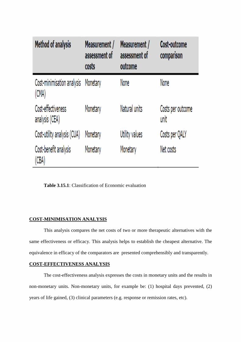
Table 3.15.1: Classification of Economic evaluation
COST-MINIMISATION ANALYSIS
This analysis compares the net costs of two or more therapeutic alternatives with the
same effectiveness or efficacy. This analysis helps to establish the cheapest alternative. The
equivalence in efficacy of the comparators are presented comprehensibly and transparently.
COST-EFFECTIVENESS ANALYSIS
The cost-effectiveness analysis expresses the costs in monetary units and the results in
non-monetary units. Non-monetary units, for example be: (1) hospital days prevented, (2)
years of life gained, (3) clinical parameters (e.g. response or remission rates, etc).
COST-UTILITY ANALYSIS
The same principle of cost-effectiveness analysis holds for cost-utility analysis. This
analysis expresses costs in monetary units and the benefit as a non-monetary unit. This
concept merges quality of life and life expectancy. This measure of analysis should be chosen
when quality of life is considered as an important aspect of therapy.
COST-BENEFIT ANALYSIS
Assessesment of all effects, especially health effects, in monetary units are done by
cost benefit analysis. The limitation in this analysis is that a monetary assessment of clinical
results must be made even though methodologically this is difficult to perform. This method
of analysis is not used due to these methodological limitations.
COST DETERMINATION
Basically, all costs pertaining to the chosen study must be assessed and included in
the analysis. In health economics, costs are defined in terms of the economic means and
understood as the financially quantified consumption of resources.
Direct costs attribute to all utilization of resources as a result of therapy and directly
contribute to this. Direct costs comprise direct medical and non-medical costs. Those
expenses that arise directly from the treatment are direct medical costs (e.g. diagnosis, drug
therapy, medical care, in-patient treatment, etc). Non-medical costs are those that arise from
the effects of the disease or treatment (e.g. transport costs, care services, etc.).

Consumption of resources occurring not in direct relation to the treatment of the
disease are quantified as Indirect costs. This comprises loss of productivity resulting from
premature death and illness. Measurement of Indirect costs is essential if impairment of
capacity to work and the absence from the workplace is to be considered together.
Losses of productivity are expressed by the human capital approach, i.e. the time
duration related income of the concerned patient group. Mean values can be used from
official statistics when no specific data are available for the patient group.
Loss of productivity = Incapacity for work x Wage costs
Dependent employees x 365 days

Table 3.15.2: Table showing the different types of cost/components of out of pocket costs(Financial Burden of cancer : Estimates from a study of women with breast cancer-Arozullah et al)

3.16 ECONOMIC STUDIES IN INDIA:
A recent study conducted at the All India Institute of Medical Sciences (AIIMS),
New Delhi (2011analysed about the mean cost of radiotherapy. 59% of this spent on the
direct non medical costs like food, transportation and accommodation and 41% is spent on
the direct medical costs that is treatment specific costs.(44)
Mahal et al. (2010) showed that almost 50% of households having a member
with cancer experiences catastrophic spending and 25% are driven to poverty by health care
costs.(45)
3.17 OUTCOME PARAMETERS :
The following outcome parameters can be chosen:
Economically directed outcome measures like number of days of hospitalization, days of
incapacity for work, etc.
Clinical outcome parameters – This includes biochemical or physiological, morbidity- or
mortality-related parameters. Three end points are used as a measure of outcome. They are :
- Final endpoints
- intermediate endpoints and
- surrogate endpoints
Health-related quality of life is considered as the outcome indicator in specific indications -
particularly where the medical treatment does not contribute to the prospect of either a cure
or a significant prolongation of life.
3.18 NEED FOR ECONOMIC ANALYSIS:
Health policy of a nation especially should address on the access, quality and cost of
the health care. A ‘free market’ of medical care relies on the notions of “perfect information”
and “perfect competition”. Yet, the awareness of financial impact of the disease does not
prevails among the people. Cancer is one such disease that brings greater stress for patients
and families due to the financial burden of treatment. Substantial section of income and
family budget are lost due to the out-of –pocket costs incurred. However, a good estimate of
the expenses involved is lacking in part of patients and their family. This is the reason of
concern about health policies, even in developed countries like UK and USA.
Alternative management strategies may have feasible financial effects that make
economic analyses appropriate. People may consider radiation therapy to be expensive,
especially in the post mastectomy setting, where complex treatments essential to deliver
multiple fields administered.
The expense of such modality of therapy may be justified by robust cost-effectiveness
analysis. The 58th World Health Assembly in May 2005 (46), has acknowledged the
increasing burden of breast cancer in its resolution on cancer prevention and control. Therein,
member states are intended to reinforce or to develop comprehensive cancer control programs
to reduce cancer related mortality and to enhance quality of life for patients and their
families.
Information on reinforcement of planning or developing a breast cancer control policy
is best provided by economic analysis. This economic analysis can guide budget
development, justify allocation of scarce resources to national cancer control programs. This
analysis can contribute to identify the efficient ways of delivering diagnostic and treatment
services. Earlier most of such economic analysis involving the costs and health effects of
breast cancer control interventions were performed in developed countries. But information
to guide decisions in resource allocation is lacking in developing countries. Moreover, studies
have largely ignored interactions among interventions, rather they focused on individual
interventions. Moreover, majority of studies have been performed on places where breast
cancer care was already existed, instead focusing on situations where they barely needed it.
This limitation precludes comparisons with interventions in settings where care systems have
not been established or with interventions that might be more relevant to other regions of the
world.
Earlier studies have shown that modest increase in fraction size when combined with
appropriate downward adjustments to total dose in post mastectomy setting has satisfactory
outcomes in terms of tumor control and late adverse effects. Reduction in duration of therapy
lessens the financial burden on the family and indirectly helps in delivering care to more
individuals. It also helps in better utilization of available resources.
3.19 RADIOTHERAPY COST STRUCTURE: INSTITUTION PERSPECTIVE
The main reason for the rise in health care expenses is the rapid evolution in medical
technology and its developments. Radiation oncology is highly technological discipline which
includes many developments in the treatment delivery.
But the cost consequences of the technical advances remain is yet to be explored and
evaluated. The most crucial determinant of radiation oncology is the equipment costs which is
highly expensive that also demands sound infrastructure and high level maintenance. The
advanced treatment planning and delivery systems in radiotherapy is also labour intensive.
The third determinant is that as the complexity of treatment increases, the resources utilized in
terms of person, place and time will be more and hence adds to the total cost(42).

The cost of radiotherapy is calculated by the treatment time which is the daily
treatment time multiplied by the number of fractions. Inversely, we can conclude that
decrease in the daily treatment time or decrease in the number of fractions can result in cost
containment.
3.20 NON MEDICAL COSTS OF THE PATIENT:
In a broader perspective, to reflect the societal aspect, an American analysis calculated
the non medical costs which accounted for 8- 25% of the total costs. It includes the expenses
for transportation and time spent on the treatment which was multiplied with the average
hourly wages of the breast cancer patients. It was obvious that there was link between the
number of fraction and nonmedical costs in terms of expenses towards travel, time and loss of
productivity.
3.21 RADIATION TOXICITY
The benefits of local control and overall survival provided by post mastectomy
radiotherapy are associated with certain side effects. The organs at risk (OAR) in post
mastectomy radiotherapy are the skin, subcutaneous tissue, ribs, lungs, heart, spinal cord and
the contralateral breast.
Radiation induced damage can be influenced by certain patient and treatment related
factors. Patient related factors like obesity being associated with higher risk of skin toxicity,
co morbidities like diabetes mellitus, connective tissue disorders, cardiac diseases and
previous history of smoking also could have a detrimental effect on the toxicity profile. The
treatment related factors like the energy chosen, the technique applied, dose prescribed and

the treatment plan also can have an impact on the radiation induced damage. The dose
delivered per fraction and the total duration of therapy will influence the toxicity of therapy
Toxicities can be classified as early and late effects. Early effects occur during the
course of radiation therapy and upto six months post treatment. Late effects may occur from
six months to years after the treatment. The acute side effect which is most commonly
encountered is fatigue and irritation of skin. Fatigue is usually mild and does not affect the
activities of daily living. Some form of radiation dermatitis occurs in most of the patients
(90%) receiving post mastectomy radiotherapy. Radiation induces injury in the basal stem
cells that are responsible for replenishing the superficial cornified layer of the epidermis (Fig:
8). As a result of the insult to the basal stem cells, there is shedding of the cornified layer
eventually, which is termed as dry desquamation.
Fig: 3.21.1: Layers of skin

Radiation also causes dilatation of capillaries, increased vascular permeability,
enhanced inflammatory response leading to erythema and oedema. Hyperpigmentation,
epilation, loss of sebaceous glands and sweat glands are all part of radiation dermatitis,
resulting in dry and pruritic skin. Migration of the melanocytes from the basal layer to the
superficial layers causes hyper-pigmentation. Moist desquamation occurs with continued loss
of basal layer which exposes the dermis. Moist desquamation can lead to frank ulceration.
A study from Egypt, which looked into radiation dermatitis in conventional
radiotherapy and hypo-fractionated radiotherapy in conserved breasts, reported that the peak
incidence of severe skin reaction occurred during the fifth week of treatment in the
conventional group and in the third week in the hypo-fractionated group. The study also
reported that these reactions lasted for about three weeks in the conventional fractionation
group and for five weeks in the hypofractionated arm (45).
The explanation for the early incidence of reactions in the hypofractionated group
may be the dependence of timing and magnitude of inflammatory response on the rate of
accumulation of dose. Inflammatory response does not clear up in hours like the sublethal
damage do and hence the inflammatory response accumulates quickly (47). START B trial
analysed patient self-assesments of five key normal tissue effects on the breast and chest.
This analysis showed that rates of moderate or marked changes were lower in the
hypofractionated radiotherapy group compared to the conventional arm (48).
The various normal tissue effects like breast shrinkage, hardness, change in skin
appearance, swelling in the area of affected breast at five years were all consistently in favour
of the 40 Gy in 15 fractions regimen.

An unusually marked acute skin reaction occurred in 16 (0.7%) patients in the
START B trial. Of these 16 patients, 13 (1.2%) were in the conventional fractionation group
and 3 (0.3 %) were from the study arm. Radiation dermatitis is graded based on the RTOG
Acute Radiation morbidity scoring criteria (49).
Table 3.21.1: RTOG Acute Radiation Morbidity Scoring Criteria
Radiation pneumonitis typically occurs as a late effect and may present with low grade
fever and dry cough. Interstitial inflammation is the hallmark of radiation pneumonitis.
Several patient and treatment related factors are associated with radiation pneumonitis.
GRADE 0 1 2 3 4
SKIN
Nochangeoverbaseline
Follicular/faint/dull erythemaepilation, drydesquamationdecreasedsweating
Tender/bright/brighterythema,patchy moistdesquamation,moderateedema
Confluentmoistdesquamation,other than inskin folds,pitting edema
Ulceration,hemorrhage,necrosis

Cardiac toxicity can also occur as late toxicity and is there is likelihood of its incidence in
left sided breast cancers. Lymphoedema is the abnormal swelling of the arm which may
occur after modified radical mastectomy or more commonly as a sequel of both surgery and
adjuvant radiotherapy to the axilla. The highest rate of lymphoedema is seen in patients who
undergo complete axillary dissection (levels I-III) followed by axillary irradiation.

Materials and Methods______________________________________________________________________________________________________________________
4. MATERIALS AND METHODS
This is a prospective study conducted in Dept of Radiotherapy, Christian Medical
College (CMC). CMC provides services to a large number of patients from in and around
Vellore and surplus patients from the Eastern parts of our country. Patients who required
Radiotherapy after Modified Radical Mastectomy or adjuvant chemotherapy were included in
the study.
Study design:
This is an economic evaluation study. A cost minimisation analysis was done for the
two groups as the outcome for the two groups would be the same as known from the
randomised trials of Hypofractionated radiotherapy.
Inclusion and Exclusion criteria
INCLUSION CRITERIA:
Age 18 to 70 years
All patients receiving conventional post mastectomy radiation therapy
EXCLUSION CRITERIA:
Patients not willing to maintain a diary for this study
Patients who had breast conservation surgery
Patients receiving conformal radiotherapy
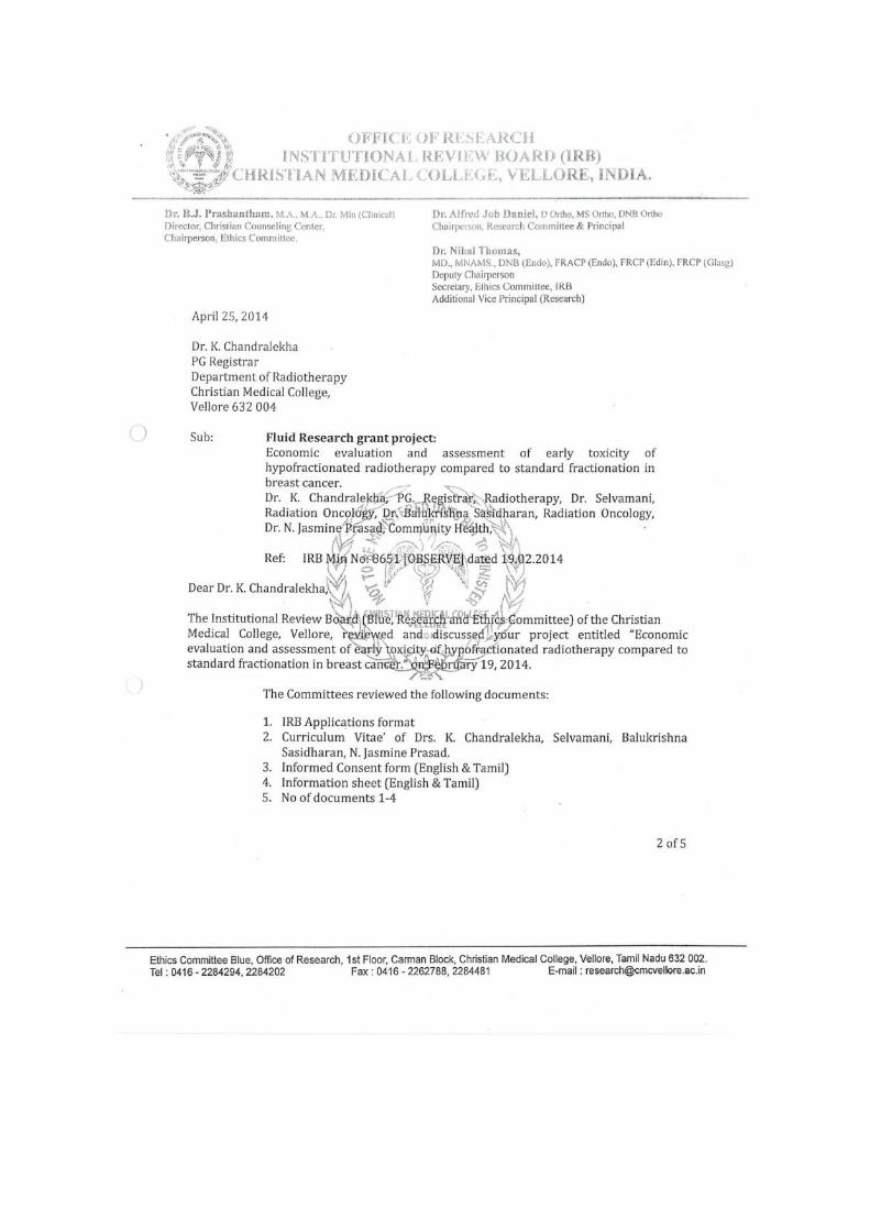
The proposed study was presented in the Institutional Review Board (IRB) which
includes Research committee and Ethics Committee and approval was obtained (copy
enclosed). The patients were selected based on the inclusion and exclusion criteria
from among the invasive breast cancer patients after mastectomy into standard or
hypo fractionated radiation therapy.

4.1 STUDY DESIGN:
This is an economic evaluation study. A cost minimisation analysis was done for the
two groups as the outcome for the two groups would be the same as known from the
randomised trials of Hypofractionated radiotherapy
Detailed diagrammatic Algorithm of the study
TREATMENT AS PER PHYSCIAN’S CHOICE
CONSENT
SAMPLE SIZE:
This study group consisted of 30 patients seen in the Radiation therapy department
from February to August 2014, treated with standard fractionation and hypofractionated post
mastectomy radiotherapy by conventional technique meeting the eligibility criteria were
included in each arm.
Post mastectomy patientsplanned for radiotherapy
Standard fractionation Hypo fractionation
Data collection of costs andexpenditureAssessment of acute toxicityand grading
Data collection of costs andexpenditure
Assessment of acute toxicityand grading
Data entry and analysis Data entry and analysis

Post mastectomy patients who came to the Dept of Radiotherapy after adjuvant chemotherapy
were assessed. Some of the patients might have been already started on hormonal therapy.
Those patients who had indications for PMRT were discussed about the need, costs, duration,
benefits and side effects of radiotherapy. Those patients who opted for conventional
radiotherapy are included in the study.
They were advised to review with basic blood investigations like complete blood
counts, Serum creatinine, and liver enzymes. Ultrasound abdomen screening was also done
for metastatic work up before initiation of radiotherapy in locally advanced carcinoma breast
patients.
4.2 RADIOTHERAPY PLANNING:
The patients were planned for external radiation therapy as follows-
1. Immobilisation:
The patient were positioned on the simulation couch in supine position with the arm
abducted (90 degree or greater) and head turned to the contralateral side on a breast board in
stable and reproducible manner. Unilateral arm pole was used for immobilisation. A
triangular wedge was inserted under the shoulder to correct the slope of the chest wall and to
ensure that the sternum was parallel to the couch.
These measures are done to decrease problems arising with field matching between the
tangential and supraclavicular fields. It was made sure that the arm was not in the path of the
tangential beams and nodal regions could be treated without any difficulty.

Fig 4.2.1 : Immobisation of the patient
2.Planning Process:
After the patient was setup in the treatment position, the laser alignment system was
used make sure that the patient was in straight position and the patient’s midline was marked.
4.3 RADIATION THERAPY TECHNIQUES
The volume of irradiation includes the chest wall and the drainage lymph nodes when
indicated.
Radiation Portals:
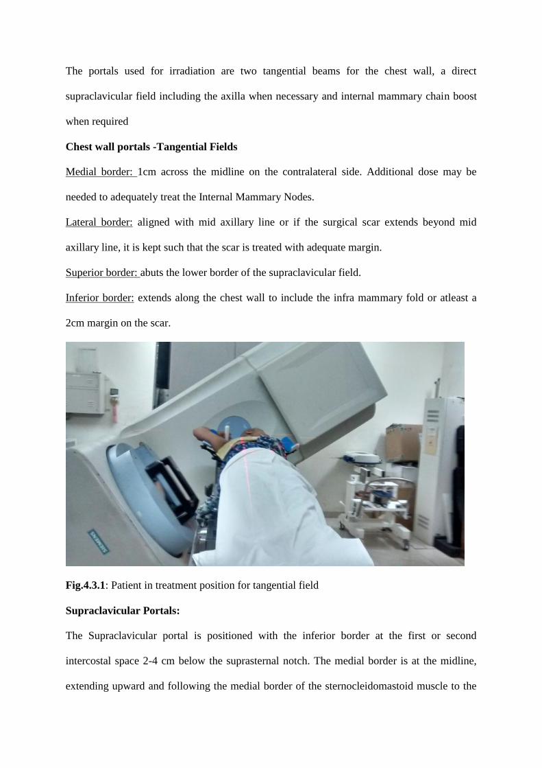
The portals used for irradiation are two tangential beams for the chest wall, a direct
supraclavicular field including the axilla when necessary and internal mammary chain boost
when required
Chest wall portals -Tangential Fields
Medial border: 1cm across the midline on the contralateral side. Additional dose may be
needed to adequately treat the Internal Mammary Nodes.
Lateral border: aligned with mid axillary line or if the surgical scar extends beyond mid
axillary line, it is kept such that the scar is treated with adequate margin.
Superior border: abuts the lower border of the supraclavicular field.
Inferior border: extends along the chest wall to include the infra mammary fold or atleast a
2cm margin on the scar.
Fig.4.3.1: Patient in treatment position for tangential field
Supraclavicular Portals:
The Supraclavicular portal is positioned with the inferior border at the first or second
intercostal space 2-4 cm below the suprasternal notch. The medial border is at the midline,
extending upward and following the medial border of the sternocleidomastoid muscle to the

thyrocricoid groove. The lateral border is at the medial end of head of Humerus and the
superior border is at the level of the thyroid notch. The larynx is shielded.
Fig 4.3.2: Patient with external markings for supraclvicular and tangential fields
Supraclavicular and Axillary Portals:
Direct Anterior field:
Direct Anteroposterior Field
The Supraclavicular portal is positioned with the inferior border at the first or second
intercostal space 2-4 cm below the suprasternal notch. The medial border is at the midline,
extending upward and following the medial border of the sternocleidomastoid muscle to the
thyrocricoid groove. The lateral border appears as a vertical line at the level of the anterior
axillary fold. The superior border is at the level of the thyroid notch. The larynx and the head
of humerus are shielded.
Direct Postero Anterior Axillary Field
Medial border is along the convex lateral wall of the bony thorax and 1 to 1.5cm of peripheral
lung.

Medial Superior border is along the spine of scapula.
Lateral Superior border bisects the head of humerus.
Lower lateral border is medial to the border of Lattissmus dorsi.
Inferior border matches the lower border of the supraclavicular field anteriorly with not more
than 1cm overlap as seen in treatment position.
Fig 4.2.3 : simulation film during planning to measure the lung distance
After simulation, planning is done in the treatment planning system and appropriate plan is
finalised by the oncologist

Fig 4.2.4 : Image showing isodose distribution in treatment planning system

Region treated Energy used Dose prescription Duration
Chest wall Cobalt/ 6 MV 50 Gy in 25 fractions
Or
40 Gy in 15 fractions
to tumour volume
5 days a week for
3weeks/ 5 weeks
Supraclavicular
region
Cobalt/ 6 MV 50 Gy in 25 fractions
Or
40 Gy in 15 fractions
to 3 cm depth or D
max
5 days a week for
3weeks/ 5 weeks
Axilla Cobalt/ 6 MV 50 Gy in 25 fractions
Or
40 Gy in 15 fractions
to midplane
5 days a week for
3weeks/ 5 weeks
Table 4.2.1: dose, energy and duration of different portals in breast treatment
During planning and simulation, the patients were advised radiation therapy on the
basis of dose to the Organs at Risk into 15 days or 25 days. Those patients who were planned
for standard fractionation or hypofractionation were included in the study after explaining the
purpose of the study and getting informed consent.
4.4 DATA COLLECTION:
This constituted the baseline data about the patient including demographics and
disease characteristics of the patient were entered in the data sheet (Appendix number
3). Details regarding the initial stage, neoadjuvant chemotherapy, date of surgery,
pathological staging, adjuvant chemotherapy, hormone receptor status and hormonal
therapy were collected from the Medical Records in the Clinical Workstation.
Radiotherapy details were also entered in the data sheet including the dose,
fractionation, regions treated, energy, Machine, date of starting radiotherapy and date
of completion of radiotherapy.
The patients were classified into two groups either those receiving standard
fractionation or those receiving hypofractionated radiotherapy over a period of 5
weeks or 3 weeks respectively. During this period the patient along with the care taker
came to the hospital on all weekdays from Monday to Friday for the treatment.
The patients received treatment either in Cobalt machine or Linear accelerator.
Collection of Health Economics data from patients:
Patients were explained about the purpose of the economic analysis and a informed
consent was obtained. Each patient was interviewed using a pilot tested questionnaire
(Appendix no 4) to collect data on the health economics initially and on a regular
basis. Maximum of 30 minutes was required for data collection in each patient.
Baseline data was collected which included demographic and social profile of the
women. The demographic data regarding their hometown, occupation, marital status,
parity, occupation of the children and husband. Money spent on individual’s health
pertaining to radiotherapy (either direct or indirect) were obtained. The patients were
also asked to maintain a diary to note their daily expenses which were then reviewed

for collection of the data. They were also discussed about the mode of travel to their
hometown, local travel, food and accommodation. Information was also collected
about their monthly income, loss of wages due to their absence from work and loss of
wages for the accompanying person. They were also asked about the source of money
for the radiotherapy treatment.
The cost for the patients included
1. The direct medical cost – treatment cost, bed charges, treatment for side effects,
medications and investigations charges
2. The direct non-medical cost – food, travel and accommodation for the patient and
the accompanying person.
3. Cost due to productive loss – wage loss for patient and care taker
The questionnaire comprised of four parts:
1. Socio demographic profile
2. Disease characteristics - staging surgery and chemotherapy
3. Radiotherapy details
4. Costs incurred by the subjects (direct, indirect and productivity costs)
The cost effect for each were assessed at the end of the treatment.
MACHINE COSTS:
The health care costs were derived using a format which is used for cost accounting
for any procedure from the accounts department in CMC Vellore (Appendix No:5).
The list of P.G. Registrars, Consultants, Physicists and Radiographers were collected
and the average time spent by each of them for the various steps involved in the breast
planning and treatment were calculated. The average salary for each cadre was also
calculated.
The machine details including the date and cost of purchase, annual maintenance
charges for cobalt, Linear accelerator and the Simulator and other equipments in the
treatment room were collected separately.
With the help of the Accounts department staff, we calculated and derived a cost
value which was the actual procedural cost. Values were separately derived for
Cobalt and Linear Accelerator for duration of 15 and 25 days of breast treatment.
Assesssment of early toxicity in patients treated with standard and hypofractionated
radiotherapy:
The occurrence of early toxicity in patients treated with standard and hypo
fractionated radiotherapy were recorded and analysed using RTOG acute skin toxicity
criteria.
The acute toxicities of the patient were recorded while on treatment and upto 6 weeks
after the completion of the radiotherapy.
WEEKLY ASSESMENT:
The patients on hypofractionation and standard fractionation arm included in the
study were monitored on a weekly basis for any side effects. Clinical examination was
done for these patients to monitor any skin reactions (dermatitis) over the chest wall
weekly and at the end of the end treatment. Grading of Dermatitis was done according
to the Acute Radiation Morbidity Scoring Criteria.
POST TREATMENT FOLLOW-UP:
Patients were kept on follow up and were reviewed after 6 weeks of radiation therapy
to assess for radiation dermatitis.
4.5 STATISTICAL ANALYSIS:
Data entry was done in Epidata and was analysed using SPSS 16.0 (Statistical
Package for Social Sciences). Frequencies and percentage were calculated for discrete
variables like demographic details and disease characteristics. The association between the
outcome variables was tested using Independent t test. The data was represented graphically
using bar diagrams and pie diagrams. Correlation between the variables was studied. The
average cost of treating one patient in both arms was determined. Cost minimisation by
adopting hypofractionated radiotherapy in breast cancer was determined by comparing the
average costs of the two arms.
The demographic variables were analysed and mean and percentage was documented.
Similar analysis was done for patient characteristics,treatment details.

Results and Discussion______________________________________________________________________________________________________________________
5. RESULTS
From February to August 2014, 30 patients seen in the Dept of Radiotherapy were
treated with standard fractionated and hypofractionated post mastectomy radiotherapy
by conventional technique meeting the eligibility criteria were enrolled in the study
after an informed consent. Patients were included in either arm based on the clinical
indication and choice of fractionation by the physician.
The patients receiving standard fractionation or those receiving hypo fractionated
radiotherapy received treatment over a period of 5 weeks or 3 weeks respectively.
During this period the patient along with the care taker came to the hospital on all
weekdays from Monday to Friday for the treatment. Patients received treatment either
in Cobalt machine or Linear accelerator.
Baseline data was collected which included demographic and social profile of the
women. The demographic data included the place of origin, marital status, obstetric
history, occupation of the subject, number of children, children’s occupation and other
dependents in the family. These variables were obtained from the subjects by a direct
interview.

5.1: DEMOGRAPHY AND PATIENT CHARACTERISTICS:
Variables Groups Frequency PercentageAge <35 4 13
35-45 12 40
46-55 8 27
>55 6 20
Place Localites (Vellore
and within 100 km
from Vellore)
5 16
Other (other places
of Tamil Nadu and
other states)
25 84
Employmemt House wife 26 87
Skilled worker 1 3
Unskilled worker 3 10
Marital status Married 29 97
Unmarried 1 3
Children 0 1 3
1 11 39
2 13 45
3 3 10
4 1 3
Children Occupation Patients with children
being students
11 36
Patients with children
having an
employment
18 61
Other Dependents 0 24 83
1 4 14
4 1 3
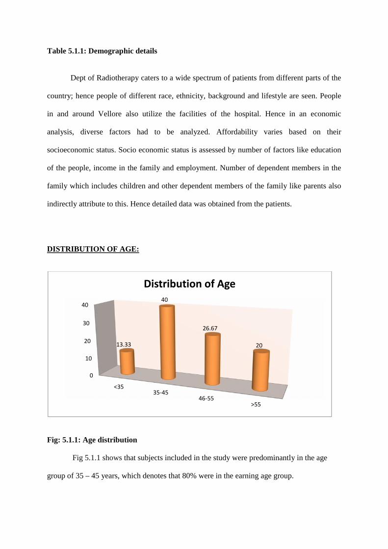
Table 5.1.1: Demographic details
Dept of Radiotherapy caters to a wide spectrum of patients from different parts of the
country; hence people of different race, ethnicity, background and lifestyle are seen. People
in and around Vellore also utilize the facilities of the hospital. Hence in an economic
analysis, diverse factors had to be analyzed. Affordability varies based on their
socioeconomic status. Socio economic status is assessed by number of factors like education
of the people, income in the family and employment. Number of dependent members in the
family which includes children and other dependent members of the family like parents also
indirectly attribute to this. Hence detailed data was obtained from the patients.
DISTRIBUTION OF AGE:
Fig: 5.1.1: Age distribution
Fig 5.1.1 shows that subjects included in the study were predominantly in the age
group of 35 – 45 years, which denotes that 80% were in the earning age group.
0
10
20
30
40
<3535-45
46-55>55
13.33
40
26.67
20
Distribution of Age

Variable Mean SD Range(Min, Max)
Age 46 9.71 28, 67
The mean age of the patients in the study group was 46. The age group ranged from 28 and
67. The standard deviation was 9.71.
HOMETOWN OF THE PATIENTS: LOCALITE (Vellore and within 100 km fromVellore) vs other places in TAMIL NADU and other STATES
Other Places of Tamil Nadu Dharmapuri 1
Namakkal 1
Tiruchendur 1
Thiruppur 1
Other States Assam 3
Andhra Pradesh 3
Kerala 1
Karnataka 1
Bihar 2
West Bengal 10
Localites In and around Vellore 6
Table 5.1.2: Hometown of the patients
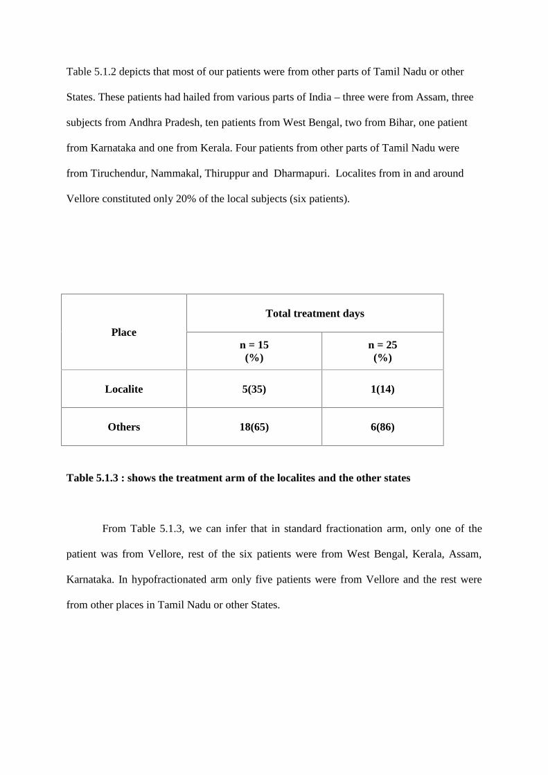
Table 5.1.2 depicts that most of our patients were from other parts of Tamil Nadu or other
States. These patients had hailed from various parts of India – three were from Assam, three
subjects from Andhra Pradesh, ten patients from West Bengal, two from Bihar, one patient
from Karnataka and one from Kerala. Four patients from other parts of Tamil Nadu were
from Tiruchendur, Nammakal, Thiruppur and Dharmapuri. Localites from in and around
Vellore constituted only 20% of the local subjects (six patients).
Place
Total treatment days
n = 15(%)
n = 25(%)
Localite 5(35) 1(14)
Others 18(65) 6(86)
Table 5.1.3 : shows the treatment arm of the localites and the other states
From Table 5.1.3, we can infer that in standard fractionation arm, only one of the
patient was from Vellore, rest of the six patients were from West Bengal, Kerala, Assam,
Karnataka. In hypofractionated arm only five patients were from Vellore and the rest were
from other places in Tamil Nadu or other States.
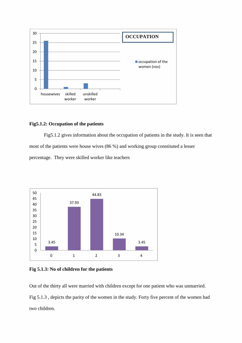
Fig5.1.2: Occupation of the patients
Fig5.1.2 gives information about the occupation of patients in the study. It is seen that
most of the patients were house wives (86 %) and working group constituted a lesser
percentage. They were skilled worker like teachers
Fig 5.1.3: No of children for the patients
Out of the thirty all were married with children except for one patient who was unmarried.
Fig 5.1.3 , depicts the parity of the women in the study. Forty five percent of the women had
two children.
0
5
10
15
20
25
30
housewives skilledworker
unskilledworker
occupation of thewomen (nos)
OCCUPATION
3.45
37.93
44.83
10.34
3.45
05
101520253035404550
0 1 2 3 4

Fig 5.1.4: Children Occupation
Fig 5.1.4 depicts that 61% of the patient’s children were employed and only 39 % were
students
39%
61%
Student EmployedCHILDREN OCCUPATION

.
Fig 5.1.5: Other dependents in their familyFig 5.1.5, shows that more than 82 % of the
patients didn’t have other dependent family members in their house
0
10
20
30
40
50
60
70
80
90
01
4
82.76
13.79
3.45
OTHER DEPENDENTS
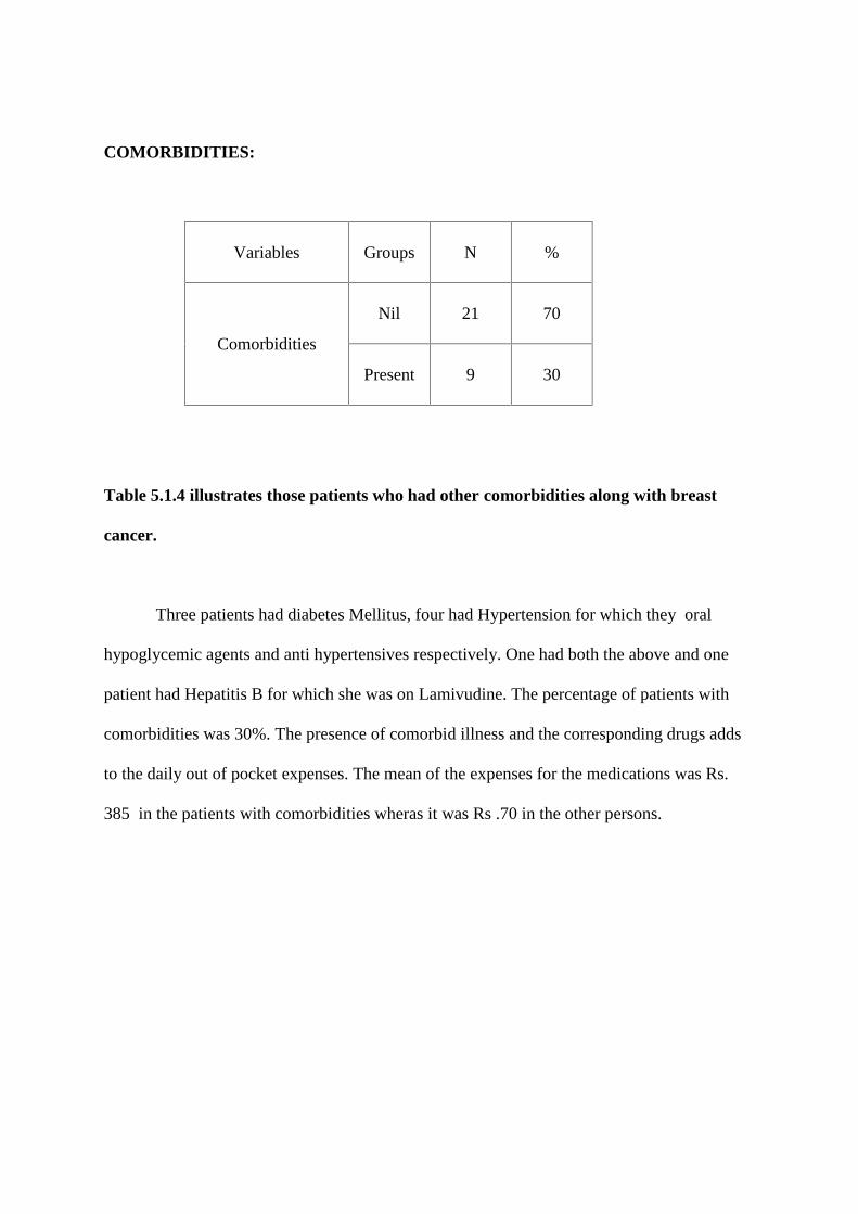
COMORBIDITIES:
Variables Groups N %
Comorbidities
Nil 21 70
Present 9 30
Table 5.1.4 illustrates those patients who had other comorbidities along with breast
cancer.
Three patients had diabetes Mellitus, four had Hypertension for which they oral
hypoglycemic agents and anti hypertensives respectively. One had both the above and one
patient had Hepatitis B for which she was on Lamivudine. The percentage of patients with
comorbidities was 30%. The presence of comorbid illness and the corresponding drugs adds
to the daily out of pocket expenses. The mean of the expenses for the medications was Rs.
385 in the patients with comorbidities wheras it was Rs .70 in the other persons.

5.2 DISEASE CHARACTERISTICS:
Variables Groups Frequency Percentage
Laterality Right 18 60
Left 12 40
Regions treated with
radiation
Supraclavicular region and chest
wall
28 93.33
Supraclavicular region, axilla and
chest wall
2 6.67
Type of
chemotherapy
Adjuvant 4 13.33
Neoadjuvant & Adjuvant 26 86.67
ER Positive 15 50
Negative 15 50
PR Positive 12 40
Negative 18 60
Her2neu Positive 10 33.33
Negative 20 66.67
Molecular Subtype Luminal A 10 33
Luminal B 5 17
Her 2 type 5 17
Basal type 10 33
Table 5.2.1 :enumerates the patient characteristics with frequency and percentage

INITIAL STAGE OF THE TUMOUR:
Fig 5.2.1:. depicts the stages of the patients included in the study
48% of the patients presented with stage 3 and locally advanced disease and 27% in the
stage 2. Some patient who had underwent initial lumpectomy or Mastectomy with no
information about the preoperative staging were classified as TxNx.(Fig 5.2.1
Fig 5.2.2: depicts the number of receptor positive and negative patients
Fig 5.2.2 depicts of ER, PR, Her 2 neu positivity separately. Based on the molecular
classification- ten patients were Luminal A subtype, five patients were Luminal B subtype,
five patients were Her 2 type and ten were triple negative or basal subtype.
0
10
20
30
40
50
60
STAGE 3 STAGE 2 TXNX
PERCENTAGE OFPATIENTS
1512
10
1518
20
0
5
10
15
20
25
ER PR HER
ER, PR, Her2neu
Positive Negative
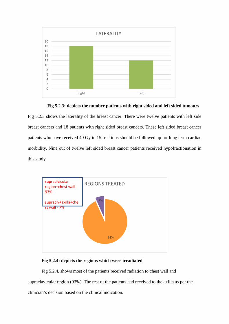
Fig 5.2.3: depicts the number patients with right sided and left sided tumours
Fig 5.2.3 shows the laterality of the breast cancer. There were twelve patients with left side
breast cancers and 18 patients with right sided breast cancers. These left sided breast cancer
patients who have received 40 Gy in 15 fractions should be followed up for long term cardiac
morbidity. Nine out of twelve left sided breast cancer patients received hypofractionation in
this study.
Fig 5.2.4: depicts the regions which were irradiated
Fig 5.2.4, shows most of the patients received radiation to chest wall and
supraclavicular region (93%). The rest of the patients had received to the axilla as per the
clinician’s decision based on the clinical indication.
02468
101214161820
Right Left
LATERALITY
93%
7%
REGIONS TREATEDsupraclvicularregion+chest wall-93%
supraclv+axilla+chest wall - 7%
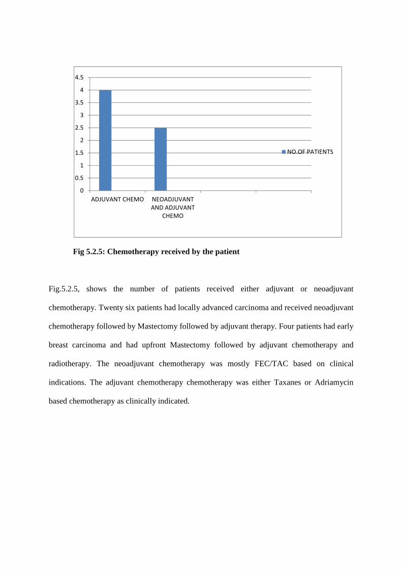
Fig 5.2.5: Chemotherapy received by the patient
Fig.5.2.5, shows the number of patients received either adjuvant or neoadjuvant
chemotherapy. Twenty six patients had locally advanced carcinoma and received neoadjuvant
chemotherapy followed by Mastectomy followed by adjuvant therapy. Four patients had early
breast carcinoma and had upfront Mastectomy followed by adjuvant chemotherapy and
radiotherapy. The neoadjuvant chemotherapy was mostly FEC/TAC based on clinical
indications. The adjuvant chemotherapy chemotherapy was either Taxanes or Adriamycin
based chemotherapy as clinically indicated.
0
0.5
1
1.5
2
2.5
3
3.5
4
4.5
ADJUVANT CHEMO NEOADJUVANTAND ADJUVANT
CHEMO
NO.OF PATIENTS
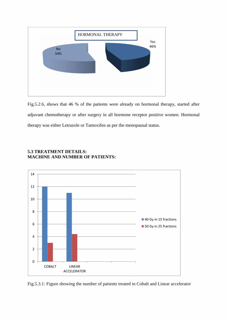
Fig.5.2.6, shows that 46 % of the patients were already on hormonal therapy, started after
adjuvant chemotherapy or after surgery in all hormone receptor positive women. Hormonal
therapy was either Letrazole or Tamoxifen as per the menopausal status.
5.3 TREATMENT DETAILS:MACHINE AND NUMBER OF PATIENTS:
Fig.5.3.1: Figure showing the number of patients treated in Cobalt and Linear accelerator
Yes46%
No54%
HORMONAL THERAPY
0
2
4
6
8
10
12
14
COBALT LINEARACCELERATOR
40 Gy in 15 fractions
50 Gy in 25 fractions

Variables Groups n %
DOSE
40Gy in 15 Fractions 23 23%
50Gy in 25 Fractions 7 77%
MACHINE
Cobalt 15 50%
Linear Accelerator 15 50%
Table 5.3.1: Number of patients in Cobalt and LINAC and dose fractionation
DOSE FRACTIONATION AND NUMBER OF PATIENTS:
Machine
Number of patients in each group
40Gy in 15 fractionsn (%)
50Gy in 25 fractionsn (%)
COBALT 12(52) 3(43)
PRIMUS 11(48) 4(57)
Table 5.3.1: 2 X 2 table depicting the number of patients in each arm (standard versus
hypofractionation) and each machine(Cobalt and Linear Accelerator)
Fifteen patients received radiotherapy using TeleCobalt and fifteen patients received
radiotherapy in Linear accelerator.
In Cobalt twelve out of fifteen were treated with hypofractionation and three with
standard fractionation.
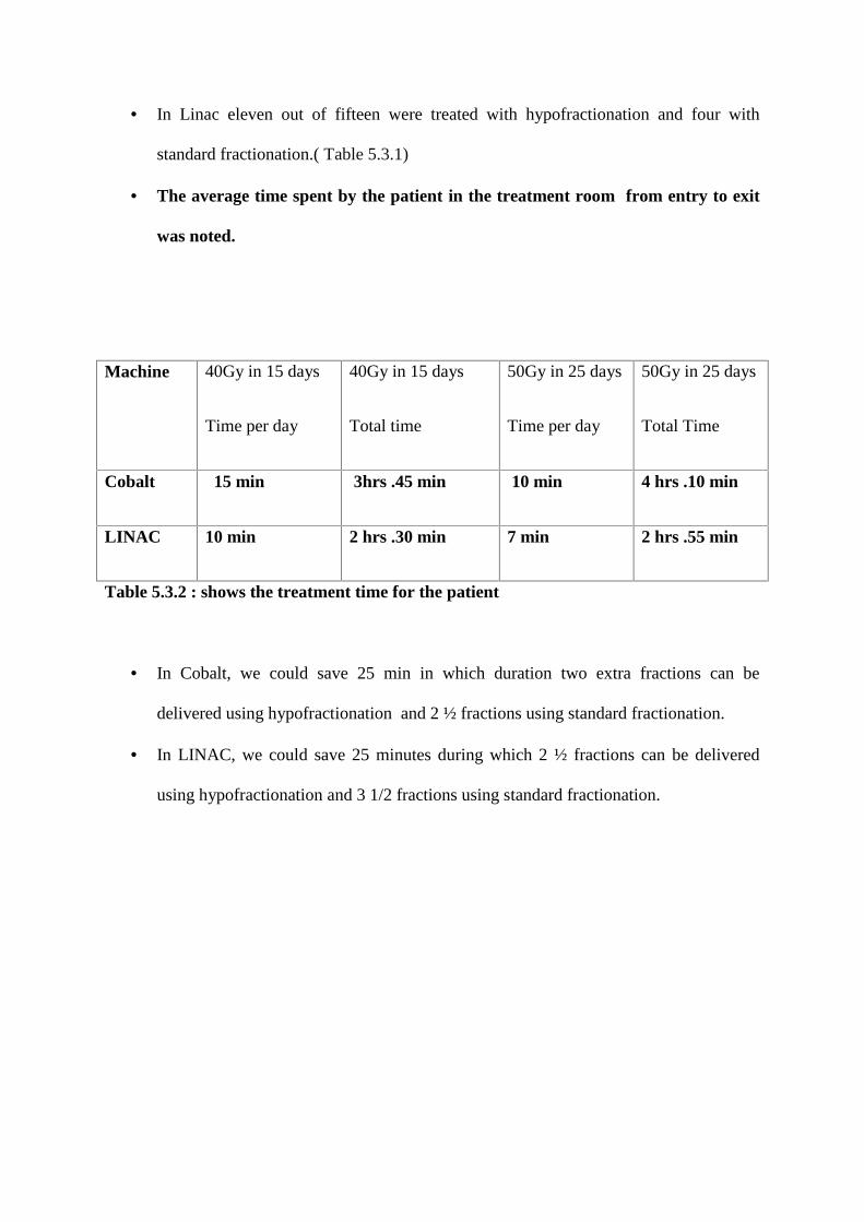
In Linac eleven out of fifteen were treated with hypofractionation and four with
standard fractionation.( Table 5.3.1)
The average time spent by the patient in the treatment room from entry to exit
was noted.
Machine 40Gy in 15 days
Time per day
40Gy in 15 days
Total time
50Gy in 25 days
Time per day
50Gy in 25 days
Total Time
Cobalt 15 min 3hrs .45 min 10 min 4 hrs .10 min
LINAC 10 min 2 hrs .30 min 7 min 2 hrs .55 min
Table 5.3.2 : shows the treatment time for the patient
In Cobalt, we could save 25 min in which duration two extra fractions can be
delivered using hypofractionation and 2 ½ fractions using standard fractionation.
In LINAC, we could save 25 minutes during which 2 ½ fractions can be delivered
using hypofractionation and 3 1/2 fractions using standard fractionation.

5.4 MACHINE COSTING :
Table 5.4.1: COSTING OF RADIOTHERAPY PLANNING AND SIMULATION
PARTICULARS AMOUNT in Rs
PERSONNEL -Consultant, PG Registrar, RT Technician,Physicist
852.68
ELECTRICITY MAINTENANCEMedical and General Equipments
72.07
EQUIPMENT MAINTENANCEMedical Equipment, Repairs and AMC
171.47
DEPRECIATIONMedical and General equipments
1193.08
INFRASTRCUTURE FACILITIESWater-1%, Building 1%, Education andResearch 2% each Contingencies on expenses
87.70
INSTITUTION OVERHEAD@on expenses excludes depreciation
142.51
CONCESSION 1% 10.96
TOTAL COST PER PROCEDURE (rounded off) Rs.2510 .00

Table 5.4.2: COSTING OF RADIOTHERAPY TREATMENT DELIVERY IN COBALT 60
PARTICULARS AMOUNT in Rs (Per Patient/PerFraction)
PERSONNEL-Consultant, PG Registrar, RT Technician,Physicist
42.79
ELECTRICITY MAINTENANCEMedical and General Equipments
9.86
EQUIPMENT MAINTENANCEMedical Equipment, Repairs and AMC
17.08
DEPRECIATIONMedical and General equipments
275.65
INFRASTRCUTURE FACILITIESWater-1%, Building 1% , Education andResearch 2% each Contingencies on expenses
5.58
INSTITUTION OVERHEAD@on expenses excludes depreciation
9.06
CONCESSION 1% 0.70
TOTAL COST (after concession) rounded off Rs. 360.00 per patient /per fractionFRACTION 40 Gy in 15 fractions 50 Gy in 25 fractions
Rs 5400.00 Rs 9000.00
Weekly Check Up Rs 101.63 Rs 169.38
Total Cost / Entire Treatment Rs 5501.63 Rs 9169.38
ROUNDED OFF Rs 5500.00 Rs 9200.00
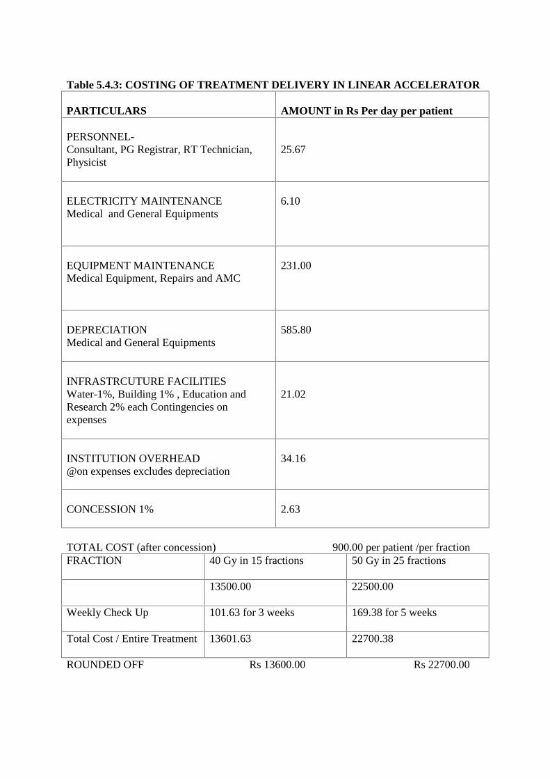
Table 5.4.3: COSTING OF TREATMENT DELIVERY IN LINEAR ACCELERATOR
PARTICULARS AMOUNT in Rs Per day per patient
PERSONNEL-Consultant, PG Registrar, RT Technician,Physicist
25.67
ELECTRICITY MAINTENANCEMedical and General Equipments
6.10
EQUIPMENT MAINTENANCEMedical Equipment, Repairs and AMC
231.00
DEPRECIATIONMedical and General Equipments
585.80
INFRASTRCUTURE FACILITIESWater-1%, Building 1% , Education andResearch 2% each Contingencies onexpenses
21.02
INSTITUTION OVERHEAD@on expenses excludes depreciation
34.16
CONCESSION 1% 2.63
TOTAL COST (after concession) 900.00 per patient /per fractionFRACTION 40 Gy in 15 fractions 50 Gy in 25 fractions
13500.00 22500.00
Weekly Check Up 101.63 for 3 weeks 169.38 for 5 weeks
Total Cost / Entire Treatment 13601.63 22700.38
ROUNDED OFF Rs 13600.00 Rs 22700.00
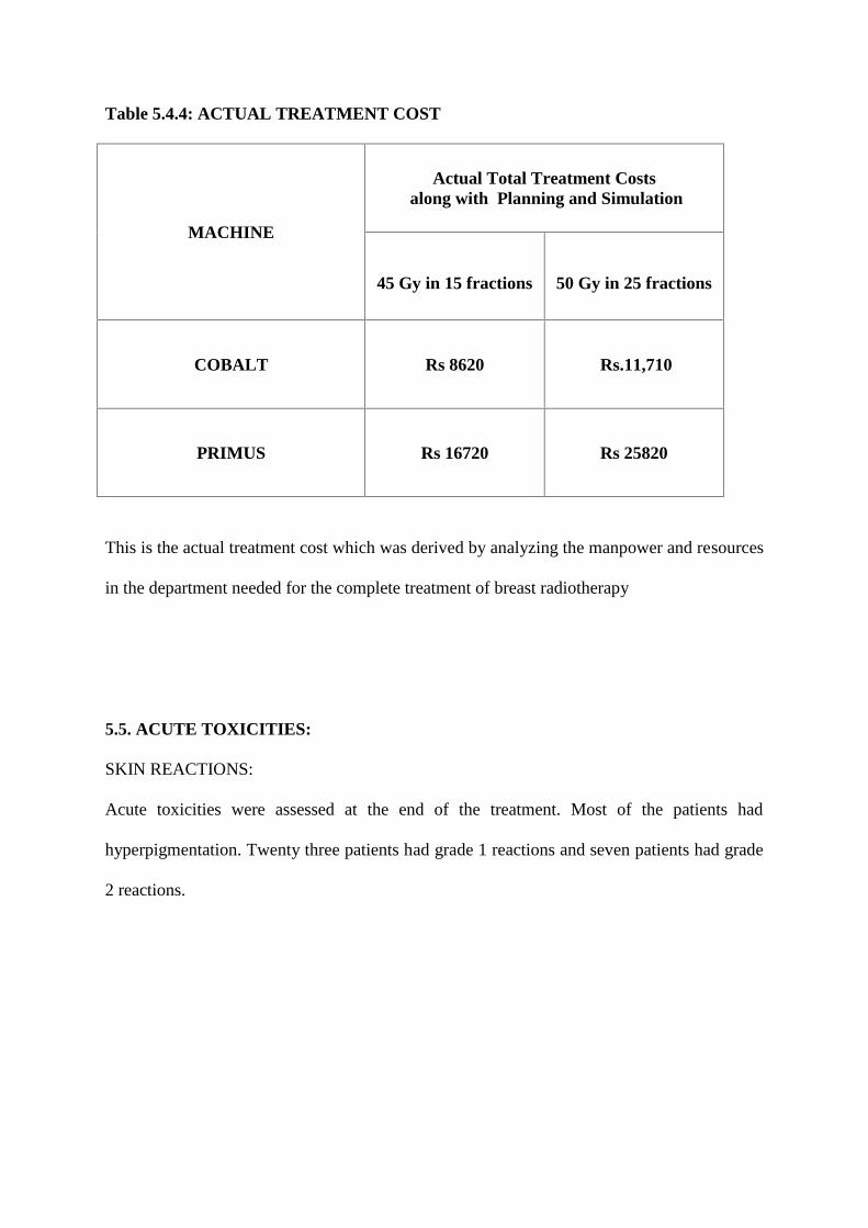
Table 5.4.4: ACTUAL TREATMENT COST
MACHINE
Actual Total Treatment Costsalong with Planning and Simulation
45 Gy in 15 fractions 50 Gy in 25 fractions
COBALT Rs 8620 Rs.11,710
PRIMUS Rs 16720 Rs 25820
This is the actual treatment cost which was derived by analyzing the manpower and resources
in the department needed for the complete treatment of breast radiotherapy
5.5. ACUTE TOXICITIES:
SKIN REACTIONS:
Acute toxicities were assessed at the end of the treatment. Most of the patients had
hyperpigmentation. Twenty three patients had grade 1 reactions and seven patients had grade
2 reactions.

Fig 5.5.1: Picture showing hyperpigmentation in a patient who had recievd 40 Gy in 15
fraction at the end of treatment
Fig 5.5.2: shows the overall incidence of grades of skin reactions
23
7
Skin reactions
Grade 1 Grade 2

Skin reactions
Total treatment days
40 Gy in 15 fractionsn (%)
50 Gy in 25 fractionsn (%)
Grade 1 17(73.91) 6(85.71)
Grade 2 6(26.09) 1(14.29)
Table 5.5.1: incidence of skin reactions among patients treated with hypofractionation andstandard fractionation.
Table 5.5.2: Table showing details and grade of skin reactions in both the arms ( standard
versus hypofractionation)
Other Symptoms Week No of Patients in 40 Gy
in 15 Fractions
No of Patients in 50 Gy
in 25 Fractions
COUGH Week 2 and 3 7 out of 23 patients 3 out of 7 patients
FATIGUE Weed 2 and 3 8 out of 23 patients 3 out of 7 patients
INSURANCE:
Insurance Yes 7 27
No 19 73
Seven out of thirty patients had treatment under the cover of insurance. Investigations were
not included in the insurance. Those patients who had insurance cover were from Vellore or
from Tamil Nadu. The out of pocket costs were still there for these patients which was spent
for patient and the relative’s food and accommodation, travel from their home and daily
travel to the hospital. They also spent specific amount ranging from Rs.200 to Rs.500 for
applying and sanction of the insurance.
Three patients received concession for the investigations and treatment (in view of the poor
socio-economic status and affordability). The expenditure incurred by the patients was
5.6 PATIENT COSTING AND EOCNOMIC ANALYSIS:
STATISTICS FOR THE COSTS -- 15 DAYS VS 25 DAYS INTERPRETATION:
Direct medical costs:
Direct medical costs were calculated including the investigations, medicines and the
actual treatment cost. The mean is Rs. 13587, 95% C.I (11661 to 15513) for 15 days
compared to Rs. 213386, 95% C.I (14450 to 28272) for 25 days treatment. Table 5.6.1 shows
the mean and 95% C.I for each cost separately for 15 days vs 25 days.

Fig 5.6.1: Stacked bar diagram for the direct medical costs for treatment in 15 days vs
25 days
11884
19424
1479
1747
224
214
0
2500
5000
7500
10000
12500
15000
17500
20000
22500
15 days 25 days
Direct medical cost
Medicine cost
Investigation cost
Actual trt cost

TYPE OF COST STATISTICS COST FOR 15DAYS IN INR
COST FOR 25 DAYSIN INR
Actual Cost Mean(95% CI)
11884(10095 to 13673)
19424(12750 to26098)
Investigation Cost Mean(95% CI)
1479(1165 to 1794)
1747(-38 to 3533)
Medicine Cost Mean(95% CI)
224(113 to 335)
214(42 to 387)
Direct Medical Cost Mean(95% CI)
13587(11661 to 15513)
21386(14450 to 28272)
Table 5.6.1 shows the mean and 95% C.I for each cost separately for 15 days vs 25 days.
Direct non medical costs:
Table 5.6.2, shows the mean and the 95% C.I for each cost separately for 15 days vs 25 days.
Indirect medical costs were calculated including the patient and the relative’s food,
accommodation daily local travel expenses, travel expense from their hometown and care
giver’s expenses. Mean and the 95% C.I was calculated each of the nonmedical costs. The
mean of direct nonmedical costs were significantly lower for the 15 days arm compared to
the 25 days arm. Figure 5.6.2 and 5.6.3 illustrates the the non medical costs for the patient
and the relatives respectively.

Fig 5.6.2: Stacked graph showing the patients’ non medical costs in both the arms
0
1000
2000
3000
4000
5000
6000
7000
8000
9000
10000
11000
12000
15 DAYS 25 DAYS
Mea
n co
st
Total treatment days
OTHER COST
PT DAILY LOCAL TRAVEL
PT TRAVEL FROM OTHER STATE
PT FOOD
PT ACCOMODATION

Fig 5.6.3: Stacked graph illustrating the expenditure for the relatives (non medical
costs)
1410
2714
1674
2786
861
1643
44
28
208
443
0
750
1500
2250
3000
3750
4500
5250
6000
6750
7500
8250
15 DAYS 25 DAYS
Chart Title
CARE GIVER EXTRA
RELATIVE LOCAL TRAVEL
RELATIVE TRAVEL FROM OTHERSTATE
RELATIVE FOOD
RELATIVE ACCOMODATION

NON MEDICAL COSTS:
COST STATISTICS 15 DAYS 25 DAYS
PATIENTACCOMODATION
MEAN(95% CI)
17581258 to 2260
35711657 to 5486
PATIENT FOOD MEAN(95% CI)
27132050 to 3376
40002489 to 5510
PATIENT TRAVELFROM OTHER
STATES
MEAN(95% CI)
542223 to 860 1928
109 to 3748
PATIENT DAILYLOCAL TRAVEL
MEAN(95% CI)
593198 to 990 696
71 to 1322
OTHER COST MEAN(95% CI)
865398 to 1333
614261 to 867
RELATIVEACCOMODATION
MEAN(95% CI)
1410994 to 1827
27141555 to 3873
RELATIVE FOOD MEAN(95% CI)
16741307 to 2040
27861866 to 3705
RELATIVETRAVEL FROMOTHER STATE
MEAN(95% CI)
861493 to 1229
1643-220 to 3506
RELATIVELOCAL TRAVEL
MEAN(95% CI)
4412 to 77
283 to 53
CARE GIVEREXTRA
MEAN(95% CI) 208
96 to 321443
37 to 849
DIRECT NONMEDICAL COSTS
MEAN(95% CI)
106718699 to 12642
1842410711 to 26137
Table 5.6.2: Table gives information of all the direct non medical costs separately for 15
dats versus 25 days

PRODUCTIVITY LOSS:
Fig 5.6.4: Figure showing the productivity loss for the patient and the care giver for 15 days
versus 25 days
1174286
5739
10857
0
2000
4000
6000
8000
10000
12000
15 DAYS 25 DAYS
Mea
n co
st
Total treatment days
WAGE LOSS FOR CAREGIVERS
WAGE LOSS PT

COST STATISTICS 15 DAYS 25 DAYS
Wages Lost Patient Mean(95% CI)
1174-11 to 2360
286-413 to 985
Wages LostCare Giver
Mean(95% CI)
57394571 to 6907
108577679 to 14035
Productivity Loss Mean(95% CI)
69135164 to 8662
111438346 to 13939
Table 5.6.3 gives the productivity loss for the patient and the care giver for 15 days versus 25
days
Table 5.6.3 shows the productivity loss, which was calculated summing the loss of
wages for the patient and the relative. The mean was Rs.6913, 95% C.I (5164-8662) for 15
days compared to Rs.11143. 95% C.I (8346 – 13939) for 25 days treatment. The mean of the
productivity loss was lower for the 15 days arm than the 25 days.

ANALYSIS:
Independent t- test was done for the analysis.
Table 5.6.4: COSTS FOR COBALT VS PRIMUS
COSTS MACHINE STD.ERROR DIFFERENCEIN MEANS
t- TESTp- VALUE
Direct MedicalCosts
COBALT
PRIMUS
475
1137
-10171 0.006
Direct NonMedical Costs
COBALT
PRIMUS
1730
1644
352 0.082
Productivity Loss COBALT
PRIMUS
696
1365
-1533 0.326
TOTAL COST COBALT
PRIMUS
2662
3275
-11353 0.012
T- test was done for the direct medical, direct nonmedical costs, productivity loss separately
for the Cobalt and the Primus (Linear Accelerator). The p value was significant for the direct
medical and nonmedical costs except for the productivity loss. (Table 5.6.4)

Table 5.6.5:ANALYSIS FOR COBALT FOR 15 DAYS VERSUS 25 DAYS
COSTS FORCOBALT
DAYS MEAN STD.ERROROF MEAN
DIFFERENCEIN MEANS
t- TESTp- VALUE
Direct MedicalCosts
15
25
9471
13718
157.8
393.2
-4246 0.003
Direct NonMedical Costs
15
25
10750
20280
1558.1
3875.5
-9529 0.021
Productivity Loss 15
25
6250
10666
616.9
881.9-4416
0.005
TOTAL COST 15
25
26471
44665
2064.8
4674.4
-18193 0.002
T test was done for Cobalt for 15 versus 25 days. The p value was significant favouring 15
days for the direct medical costs, nonmedical costs and productivity loss. The p value for the
direct medical costs was 0.003 that of the non medical costs is 0.021, productivity loss is
0.005 and p value for the total costs is 0.002, which is statistically significant (Table 5.6.5).

Fig 5.6.5: Bar diagram showing the types of cost and their mean cost for cobalt for 15 daysversus 25 days
0
2500
5000
7500
10000
12500
15000
17500
20000
22500
DIRECT MEDICAL COSTS DIRECT NON MEDICAL COSTS PRODUCTIVITY LOSS
Mea
n co
st
Type of costs
COBALT
15 days
25 days
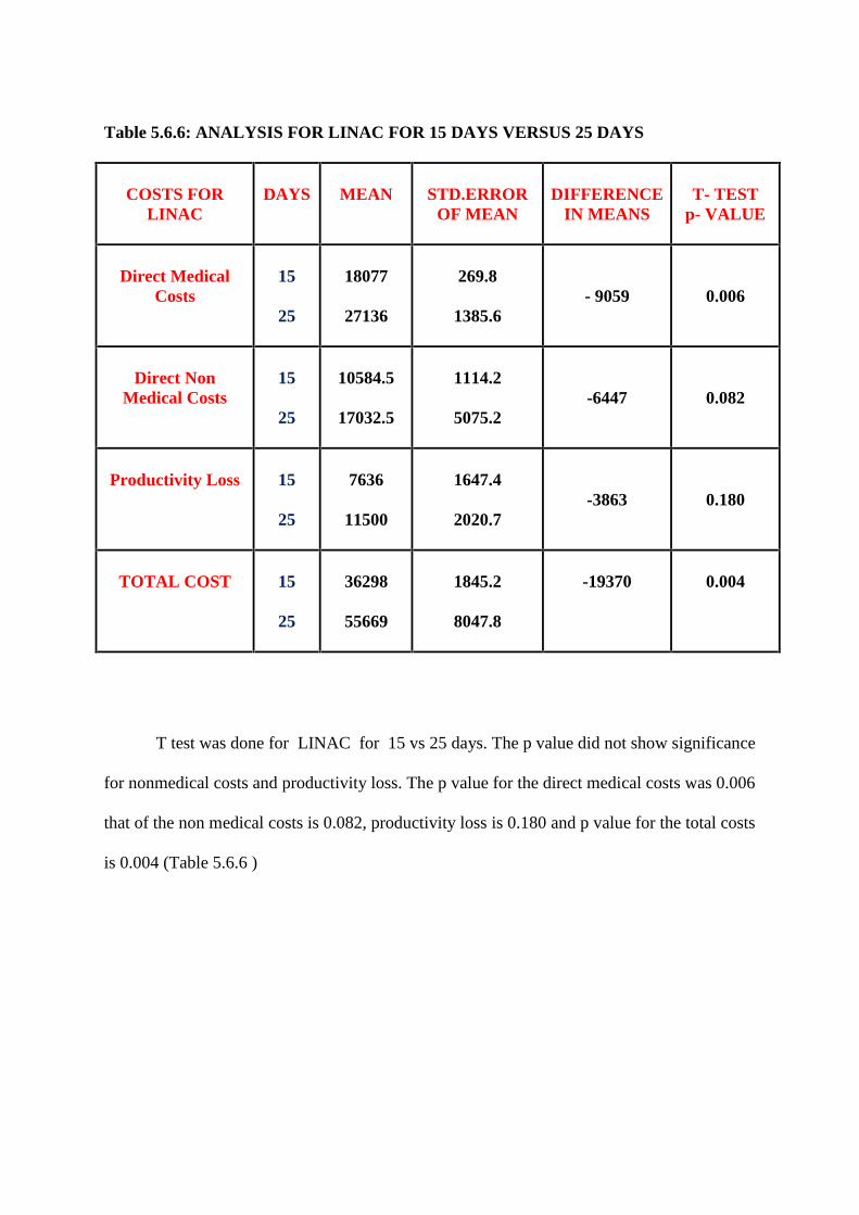
Table 5.6.6: ANALYSIS FOR LINAC FOR 15 DAYS VERSUS 25 DAYS
COSTS FORLINAC
DAYS MEAN STD.ERROROF MEAN
DIFFERENCEIN MEANS
T- TESTp- VALUE
Direct MedicalCosts
15
25
18077
27136
269.8
1385.6- 9059 0.006
Direct NonMedical Costs
15
25
10584.5
17032.5
1114.2
5075.2-6447 0.082
Productivity Loss 15
25
7636
11500
1647.4
2020.7-3863 0.180
TOTAL COST 15
25
36298
55669
1845.2
8047.8
-19370 0.004
T test was done for LINAC for 15 vs 25 days. The p value did not show significance
for nonmedical costs and productivity loss. The p value for the direct medical costs was 0.006
that of the non medical costs is 0.082, productivity loss is 0.180 and p value for the total costs
is 0.004 (Table 5.6.6 )

Fig 5.6.6: Bar diagram showing the types of cost and their mean cost for LINAC for 15 days
versus 25 days
0
2500
5000
7500
10000
12500
15000
17500
20000
22500
25000
27500
30000
DIRECT MEDICAL COSTS DIRECT NON MEDICALCOSTS
PRODUCTIVITY LOSS
Mea
n co
st
Type of cost
LINAC
15 days
25 days

6. DISCUSSION:
Financial hardships in families of cancer patients:
Nearly one-half of patients are reporting to private health facilities as the first point of
contact for cancer related diseases. More than three-fourths of the cancer patients faced
financial constraints in management. Due to financial problem, poor families delay their
treatment decision. (51)
The SUPPORT study (Study to Understand Prognoses and Preferences for
Outcomes and Risks of Treatment) was done to improve outcomes for seriously ill
hospitalized adults by improving information and decision-making. They enrolled 2652
patients in the intervention arm and 2152 in the control arm of a block-randomized trial of
enhanced information, counseling, and support. They had analysed that around one third of
the families had lost most of their savings following treatment for cancer and in one fifth of
the family, a member quit work or had changed their lifestyle to provide care for the patients.
Because of limited resources, poor families had financial hardships.(52)
The poor become poorer due to the expensive cost of diagnosis and treatment. Modern
technology though proved to be effective in curing the diseases the costs of diagnosis and
treatment, have consequently increased due to the technology costs.
Increase in out of pocket costs: (56)
With regard to high out-of-pocket payments, it is essential to reduce the financial
burden to the poor patients, thereby enabling them to access treatment services. Patient has to
pay for transportation and child-care especially in Indian scenario where patient has to travel
farther to the tertiary care facilities. While hospitalization do requires an accompanying
person, most of the time they are adults and that person’s time is a direct cost. Friends and
family also provide informal assistance to the care of patient. Patients requiring multimodal

form of treatment including surgery and chemotherapy incur a higher expenditure. Most
often, depending on their type and stage of cancer, patients would require more than one form
of treatment. All the three modalities of treatment are highly expensive and when it is
combined modality of treatment, patients are financially knocked down. Moreover, the
duration of the entire treatment taking more than six months and throughout the period of
treatment they are dependent on others for finances or self care. Hence apart from the direct
medical causes by the treatment, nonmedical causes also shoot up. This pushes the families to
deep financial crisis when cancer treatment is expensive and is also long term with no surety
of cure.
Minimising the duration of radiotherapy
Radiation therapy as an integral part in the multi-modality management of breast
cancer significantly reduces the loco regional recurrence and also improves the overall
survival. The conventional way of radiotherapy in breast cancer imparts larger expenses to
the patients. To overcome this economic burden and to minimise the acute toxicities of
conventional Radiotherapy, various Hypo fractionated schedules were tried and had proven
beneficial in terms of local control and overall survival.
But there is more to it for society than just costs. In the context of societal objective of
deriving the maximum health benefits from invested money, it is inevitably essential to
identify which health outcomes can be expected from a given treatment. The optimal
allocation of resources is better facilitated when the information on both the efficacy of
medical interventions and their costs must be balanced against each other in a systematic
fashion. To do so, economic evaluations (e.g. cost-effectiveness) and cost-utility analyses are
being done.

The shorter fractionation schedules are equally efficacious and tolerable for the Indian
women. The out-of –pocket costs incurred during radiotherapy is reduced by minimizing the
treatment duration. The reduction in duration helps in better utilization of available resources.
The landmark trials are the UK standardization of breast radiotherapy (START) Trials which
have laid evidence for hypofractionated radiotherapy for patients with breast cancer. START
B trial had compared conventional standard schedule with hypofractionated regimen. (40 Gy
in 15 fractions).Most of the patients in the trial had breast conservation surgery and only 8%
of the patients had undergone Mastectomy.
The economic impact of hypofractionated schedules can be evaluated by weighing the costs
to effectiveness(42). This was analysed in a Markov model- that is cost effectiveness data
was modeled with factors like impact of recurrence, cosmetic outcome and late side effects
by Hayman et al(53). Many of the radiotherapy centers in UK and Canada have already
adopted hypofractionated regimens routinely (42).
But late cardiac toxicity makes other nation to hesitate to adopt the same. But
recently study of 7000 Canadian women, where the impact of cardiac mortality was analysed
shows deviation of curves for cardiac death(54). After the START trial there has been
acceptance of the hypofractionated approach and many leading radiotherapy centres in India
have adopted the same. In this study the cost minimization that may be experienced by
introducing a hypofractionated approach in breast cancer radiotherapy has been studied
This was a single arm prospective study design to assess the cost minimization
between standard and hypofractionated arm.

Demographics :- how do they influence the costs?
The demographic data included the place of origin, marital status, obstetric history,
occupation of the subject, number of children, children’s occupation and other dependents in
the family . The mean age was 46 in this study which is similar to the data from most of the
similar studies in India, herein the mean age at presentation was 47(55)
Occupation of the patients:
In this study, around 90% were housewives. A similar study by MAMSI (Moulana
Azad Medical college, New Delhi) depicted their study population had > 94% as housewives
in the breast cancer group. (55) Housewives do contribute to immense hardwork in their
houses which indirectly contribute to their income of their spouses. The work done by
housewives is often not accounted to the loss of wages or productivity losses. The patients
who are basically housewives, when they are away from their home for treatment, they
require extra hands/ care givers help to look after their children and their work to compensate
for their absence.
Hence, the fact is that housewives indirectly require help from other people , thus
increasing their total expenses . This is included in the questionnaire, as ” care givers” who
are required , to look after the children and house hold work in the absence of these patients
at their home.
The term caregiver means as defined in public health as a family member, friend, or
neighbor who takes care of a person who has chronic ailments and needs help to manage a
variety of tasks. Some people also required an extra person apart from the relative
accompanying for the treatment, to assist them for their daily activities. These person had to
travel daily or come whenever necessary. Hence as the duration of stay increases the

expense for the care giver also increases. The mean expense for the care giver was Rs 208
versus Rs 445 in the two arms, 40 Gy and 50 Gy respectively.
Children’s occupation:
17 out of 30 patients had their children who were employed and because of that fact
their children were able to support their family financially. It was also seen that eight of the
patients were accompanied by their children who were employed. So there is also a problem
of loss of productivity for these patients when the working groups of children accompany
them for treatment taking leave from their concerned working places.
Other dependents:
The data on dependent members of the family and the number of children indirectly
gives information about the scattering of finances of the monthly income which might be
indirectly affected due the present expenses for treatment of breast radiotherapy. Other
dependents , as informed by the patients were either their parents or their in laws who were
staying with the patients’ family in their hometown.
Co morbid illness:
The presence of comorbid illness and the corresponding drugs adds to the daily out of
pocket expenses. The mean of the expenses for the medications was Rs. 385 in the patients
with comorbidities wheras it was Rs.70 in the other persons.
Local lodgings:
The hometown of the patients is of importance in this economic assessment studies,
because the travel and the stay expenses made by the patients who are from other places and
other states contributes a sum of amount which are the significant out of pocket expenses and
predominantly contributes to the non medical costs of the patient. They are bound to stay
outside the hospital either in lodges or house for rent for the entire treatment of breast
radiotherapy for the known fact that radiotherapy is offered over a period of 3- 5 weeks on

daily fractionated basis. People who came to our hospital predominantly stayed in nearby
lodges whose daily rent varied from Rs 60 per day to Rs 500 per day. Some people had
rented a house in the adjacent areas whose monthly rent ranged from Rs 3000 per month to
7500 per month. 94% of the patients had stayed in lodges whose rent is calculated on daily
basis. Hence decreasing the treatment duration from 5 weeks to 3 weeks will bring down the
total accommodation costs of the patient. The mean of the accommodation costs for the
patient and the relative was Rs.3510 and Rs.6285 in the 40 Gy arm and 50 Gy arm
respectively.
Transport:
The lodges and the houses were located either near to the hospital or at a distance of
3- 5 kms from the hospital. The patients who stayed and houses at 3- 5 kms had to use local
transport facilities for reaching the hospital daily. The localite patients, who were residing in
and around Vellore, also used local transport facilities daily to reach the hospital. All of our
patients had received Radiotherapy on OPD basis and none of them were admitted in bed for
the same or for any complications.
Source of income for radiotherapy treatment:
The patients had to borrow, sell their assets or utilize from their saving for the
radiotherapy treatment. Among our patients sixteen out of thirty had borrowed from known
person, seven patients had utilized the Chief Ministers health Insurance (patients from Tamil
Nadu), four had sold their assets (land) and three had utilized their savings.

Direct medical and non medical costs:
The direct medical costs (mean) was Rs. 13587 and Rs. 21386 for 15 days and 25
days respectively irrespective of the machine used for treatment. This accounts for 56% and
53% of the total expenses for the two arms respectively.
The direct non medical costs (mean) was Rs. 10671 and Rs. 18424 for 15 days and 25
days respectively irrespective of the machine used for treatment. This accounts for 44% and
47% of the total expenses for the two arms respectively.
In a study done in All India Instititute of Medical Sciences (44) to compute the cost
during the course of radiotherapy, it was found that 59% of the patients’ expenses were spent
for food, lodging and transportation, whereas 41 % was only utilized for the direct medical
costs.
Cost computation Cobalt and Linac:
Comparing the cost of treatment in Cobalt machine between the hypofractionated and
the conventionally fractionated arm there was significant difference in the direct medical
costs with a mean of Rs.9471 versus Rs.13718 respectively (p= 0.003).
The non medical costs was also associated with significant difference with mean of
Rs.10750 versus Rs.20280 for 40 and 50 Gy (p=0.021).The loss of productivity was also
significant favoring hypofractionated arm with the mean costs of Rs. 6250 and Rs.10666 (p=
0.005) in the two arms.
On the whole, the total costs were lower with 40 in 15 fractions arms compared to 50
Gy in 25 fractions (p value =0.002) which is statistically significant.
In Linear accelerator, the costs were analyzed and compared. Direct costs and the non
medical costs were significant was (p=0.006 and 0.082 respectively). The productivity loss
didn’t show any significance with the mean loss of Rs 7636 versus Rs.11000 (p value of
0.180). But the total costs were lesser in the 40 Gy arm implying its significance.
The difference in the productivity loss was more in the 15 days arm because the
average income of the patient who was working was more in this arm compared to 25 days
arm. This can also be attributed to the reason that mixture of patients of different socio
economic status were in the study group which may be the confounding factors for this
difference.
This can be explained by the fact that all the localites and the patients from other
states were analyzed together. There was also difference in the amount spent for the
accommodation based on the affordability even among the same arm of 15 days, ranging
from 60 Rs per day to Rs 400 per day. Hence the mean value of 15 days versus 25 days
becomes different to correlate as more patients who received radiotherapy for 15 days stayed
in costlier lodges and so the lodging cost were significantly low for 25 days group.
Hospital perspective:
Medical and personnel resources were also analysed which showed significant
reduction in resource utilization and time consumption in the 15 days group. In Cobalt, the
average treatment time was 3 hour and 45 minutes versus 4 hours and 10 minutes in 40 and
50 Gy respectively for the entire duration of treatment. So we could save 25 min in which
duration two extra fractions can be delivered using hypofractionation and 2 ½ fractions using
standard fractionation.
In LINAC, the average treatment time was 2 and half hours versus 2 hours and 55 min
for the entire treatment in 40 and 50 Gy respectively. Here also we could save 25 minutes
during which 2 ½ fractions can be delivered using hypofractionation and 3 1/2 fractions using
standard fractionation. This may be advantageous for the radiotherapy departments since it
may reduce the patients waiting time especially in hospitals which have large waiting list.
Acute toxicity:
Only two patients had received radiation to axilla also based on the clinical and
pathological indications. When axilla was also included, intense skin reactions may be
anticipated, especially in obese patients due to the axillary folds. Those patients who had
received radiation to axilla were treated with 50 Gy.
The patients tolerated the treatment with 7 patients developing not more than Grade
2 reactions. 23 manifested with Grade 1 reactions. All patients had hyperpigmentation which
was slightly more in patients treated with Cobalt. Grade 2 reactions were manifested in 6 out
of 7 patients treated with 40 Gy in 15 fractions with only 1 patient in 50 in 25 fractions arm.
But none of the patients developed severe reactions like ulceration. Overall 40 Gy in 15
fractions was tolerated well with respect to the skin reactions.
6.1 LIMITATIONS:
This study is a single institutional study which includes diverse demographic details
and subjects of spectrum of socioeconomic status.
Patients treated with Cobalt and Linear accelerator were analysed together.
Patients hailing from in and around Vellore and those from other parts of Tamil Nadu
and other States were not separately analysed.
Long term analysis of locoregional control and overall survival is needed to follow up
the costs which may occur in case of recurrence/ late complications.
General attitude of an individual in India is not to reveal the cost correctly, and some
of them did not report clearly, thus, cost estimation may not be accurate.
Hence, precise estimation of non medical cost and loss of productivity of patient and
relative could not be determined, thus, study of cost estimates do not cover costs of
many individuals correctly.
In order to formally validate this therapeutic approach from a societal perspective,
cost effectiveness evaluations weighing long-term outcome against the societal costs incurred
until many years after treatment are needed.
7. CONCLUSION
To conclude, our study estimates the direct and indirect medical costs for a range of
cancer subjects on the basis of socio demographic characteristics, place of residence and
cancer stages. The study showed that there is a significant reduction in total costs when
hypofractionated radiotherapy is adopted in breast cancer treatment. This was more
pronounced when patients are treated in telecobalt machine. This throws light on significance
of adopting hypofractionated approaches as a national policy to reduce resource utilization
while maintaining the efficacy.

Bibliography______________________________________________________________________________________________________________________

REFERENCES
1. Ferlay J SI, Ervik M, Dikshit R, Eser S, Mathers C, Rebelo M, Parkin DM, Forman D,
Bray, F. GLOBOCAN 2012 v1.0, Cancer Incidence and Mortality Worldwide: IARC
CancerBase No. 11 [Internet]. Lyon, France: http://globocan.iarc.fr2012 [cited 2014 15th
August
2. Radice D, Redaelli A. Breast cancer management: Quality-of-life and cost considerations.
Pharmacoeconomics 2003;21:383-96.
3. Whelan TJ, Pignol JP, Levine MN, Julian JA, MacKenzie R, Parpia S, et al. Long-term
results of hypofractionated radiation therapy for breast cancer. The New England journal
of medicine. 2010 Feb 11;362(6):513-20. PubMed PMID: 20147717. Epub 2010/02/12.
eng.
4. Ferlay J, Shin HR, Bray F, Forman D, Mathers C, Parkin DM. Estimates of worldwide
burden of cancer in 2008: GLOBOCAN 2008. International journal of cancer Journal
international du cancer. 2010 Dec 15;127(12):2893-917. PubMed PMID: 21351269.
Epub 2011/02/26. eng.
5. March 17–23, 2012. The Lancet. //;379(9820):i.
6. Vinh-Hung V, Verschraegen C. Breast-conserving surgery with or without radiotherapy:
pooled-analysis for risks of ipsilateral breast tumor recurrence and mortality. Journal of
the National Cancer Institute. 2004 Jan 21;96(2):115-21. PubMed PMID: 14734701.
Epub 2004/01/22. eng.
7. Overgaard M, Hansen PS, Overgaard J, Rose C, Andersson M, Bach F, et al.
Postoperative radiotherapy in high-risk premenopausal women with breast cancer who
receive adjuvant chemotherapy. Danish Breast Cancer Cooperative Group 82b Trial. The
New England journal of medicine. 1997 Oct 2;337(14):949-55. PubMed PMID: 9395428.
Epub 1998/02/12. eng.
8. Overgaard M, Jensen MB, Overgaard J, Hansen PS, Rose C, Andersson M, et al.
Postoperative radiotherapy in high-risk postmenopausal breast-cancer patients given
adjuvant tamoxifen: Danish Breast Cancer Cooperative Group DBCG 82c randomised
trial. Lancet. 1999 May 15;353(9165):1641-8. PubMed PMID: 10335782. Epub
1999/05/21. eng.
9. Nielsen HM, Overgaard M, Grau C, Jensen AR, Overgaard J. Loco-regional recurrence
after mastectomy in high-risk breast cancer--risk and prognosis. An analysis of patients
from the DBCG 82 b&c randomization trials. Radiotherapy and oncology : journal of the
European Society for Therapeutic Radiology and Oncology. 2006 May;79(2):147-55.
PubMed PMID: 16647152. Epub 2006/05/02. eng.
10. Ragaz J, Jackson SM, Le N, Plenderleith IH, Spinelli JJ, Basco VE, et al. Adjuvant
radiotherapy and chemotherapy in node-positive premenopausal women with breast
cancer. The New England journal of medicine. 1997 Oct 2;337(14):956-62. PubMed
PMID: 9309100. Epub 1997/10/02. eng.
11. Ragaz J, Olivotto IA, Spinelli JJ, Phillips N, Jackson SM, Wilson KS, et al. Locoregional
radiation therapy in patients with high-risk breast cancer receiving adjuvant
chemotherapy: 20-year results of the British Columbia randomized trial. Journal of the
National Cancer Institute. 2005 Jan 19;97(2):116-26. PubMed PMID: 15657341. Epub
2005/01/20. eng.
12. Early breastCancer Trialists’ Colloborative Group.effect of radiotherapy and of the
differences in extent of surgery for early breast cancer: Lancet 2005:366(9503):2087-
2106
13. Kunkler IH, Canney P, van Tienhoven G, Russell NS. Elucidating the role of chest wall
irradiation in 'intermediate-risk' breast cancer: the MRC/EORTC SUPREMO trial. Clin
Oncol (R Coll Radiol). 2008;20(1):31-34.
14. Clarke M, Collins R, Darby S, Davies C, Elphinstone P, Evans E, et al. Effects of
radiotherapy and of differences in the extent of surgery for early breast cancer on local
recurrence and 15-year survival: an overview of the randomised trials. Lancet. 2005 Dec
17;366(9503):2087-106. PubMed PMID: 16360786. Epub 2005/12/20. eng.
15. American collge of Radiology: ACR appropriateness criteria, Post Mastectomy
Radiotherapy:Treatment and volumes: Kathleen C. Horst, M Bruce G. Haffty, MD;
Sharad Goyal, MD

16. Romestaing P, Belot A, Hennequin C, et al. Ten-yearResults of a Randomized Trial of
Internal Mammary
17. Chain Irradiation after Mastectomy. International Journal of Radiation
Oncology*Biology*Physics.2009;75(3, Supplement 1):S1-S1.
18. 59. Whelan TJ, Olivotto I, Ackerman I, et al. NCIC-CTG MA.20: An intergroup trial of
regional nodal irradiation
19. Perez and Brady : Textbook of Radiation Oncology,Planning and techniques:
page-1107
20. Owen JR, Ashton A, Bliss JM, Homewood J, Harper C, Hanson J, et al. Effect of
radiotherapy fraction size on tumour control in patients with early-stage breast cancer
after local tumour excision: long-term results of a randomised trial. Lancet Oncol. 2006
Jun;7(6):467-71. PubMed PMID: 16750496. Epub 2006/06/06. eng.
21. Yarnold J, Ashton A, Bliss J, Homewood J, Harper C, Hanson J, et al. Fractionation
sensitivity and dose response of late adverse effects in the breast after radiotherapy for
early breast cancer: long-term results of a randomised trial. Radiotherapy and oncology :
journal of the European Society for Therapeutic Radiology and Oncology. 2005
Apr;75(1):9-17. PubMed PMID: 15878095. Epub 2005/05/10. eng.
22. Bentzen SM, Agrawal RK, Aird EG, Barrett JM, Barrett-Lee PJ, Bliss JM, et al. The UK
Standardisation of Breast Radiotherapy (START) Trial A of radiotherapy
hypofractionation for treatment of early breast cancer: a randomised trial. Lancet Oncol.
2008 Apr;9(4):331-41. PubMed PMID: 18356109. Pubmed Central PMCID:
Pmc2323709. Epub 2008/03/22. eng.
23. Haviland JS, Owen JR, Dewar JA, Agrawal RK, Barrett J, Barrett-Lee PJ, et al. The UK
Standardisation of Breast Radiotherapy (START) trials of radiotherapy hypofractionation
for treatment of early breast cancer: 10-year follow-up results of two randomised
controlled trials. The Lancet Oncology. 2013;14(11):1086-94.
24. Bentzen SM, Agrawal RK, Aird EG, Barrett JM, Barrett-Lee PJ, Bentzen SM, et al. The
UK Standardisation of Breast Radiotherapy (START) Trial B of radiotherapy
hypofractionation for treatment of early breast cancer: a randomised trial. Lancet. 2008
Mar 29;371(9618):1098-107. PubMed PMID: 18355913. Pubmed Central PMCID:
Pmc2277488. Epub 2008/03/22. eng.
25. Whelan T, MacKenzie R, Julian J, Levine M, Shelley W, Grimard L, et al. Randomized
trial of breast irradiation schedules after lumpectomy for women with lymph node-

negative breast cancer. Journal of the National Cancer Institute. 2002 Aug 7;94(15):1143-
50. PubMed PMID: 12165639. Epub 2002/08/08. eng.
26. James ML, Lehman M, Hider PN, Jeffery M, Hickey BE, Francis DP. Fraction size in
radiation treatment for breast conservation in early breast cancer. The Cochrane database
of systematic reviews. 2010 (11):CD003860. PubMed PMID: 21069678. Epub
2010/11/12. eng.
27. Agrawal RK, Alhasso A, Barrett-Lee PJ, Bliss JM, Bliss P, Bloomfield D, et al. First
results of the randomised UK FAST Trial of radiotherapy hypofractionation for treatment
of early breast cancer (CRUKE/04/015). Radiotherapy and oncology : journal of the
European Society for Therapeutic Radiology and Oncology. 2011 Jul;100(1):93-100.
PubMed PMID: 21752481. Epub 2011/07/15. eng.
28. Qi XS, White J, Li XA. Is alpha/beta for breast cancer really low? Radiotherapy and
oncology : journal of the European Society for Therapeutic Radiology and Oncology.
2011 Aug;100(2):282-8. PubMed PMID: 21367477. Epub 2011/03/04. eng.
29. Haviland JS, Yarnold JR, Bentzen SM. Hypofractionated radiotherapy for breast cancer.
The New England journal of medicine. 2010 May 13;362(19):1843; author reply -4.
PubMed PMID: 20463347. Epub 2010/05/14. eng.
30. Eldeeb H, Awad I, Elhanafy O. Hypofractionation in post-mastectomy breast cancer
patients: seven-year follow-up. Medical oncology (Northwood, London, England). 2012
Dec;29(4):2570-6. PubMed PMID: 22354766. Epub 2012/02/23. eng.
31. Shahid A, Athar MA, Asghar S, Zubairi T, Murad S, Yunas N. Post mastectomy adjuvant
radiotherapy in breast cancer: a comparision of three hypofractionated protocols. JPMA
The Journal of the Pakistan Medical Association. 2009 May;59(5):282-7. PubMed PMID:
19438129. Epub 2009/05/15. eng.
32. Smith BD, Bentzen SM, Correa CR, Hahn CA, Hardenbergh PH, Ibbott GS, et al.
Fractionation for whole breast irradiation: an American Society for Radiation Oncology
(ASTRO) evidence-based guideline. International journal of radiation oncology, biology,
physics. 2011 Sep 1;81(1):59-68. PubMed PMID: 20638191. Epub 2010/07/20. eng.
33. Aebi S, Davidson T, Gruber G, Cardoso F, Group ObotEGW. Primary breast cancer:
ESMO Clinical Practice Guidelines for diagnosis, treatment and follow-up. Annals of
Oncology. 2011 September 1, 2011;22(suppl 6):vi12-vi24.
34. Patterns of locoregional treatment of breast cancer among radiation oncologists in India:
A practice survey Budrukkar A, Tiwana M, Jalali R, Munshi A, Sarin R - J Can Res Ther
[Internet]. [cited 2013 Dec 10]. Available from:

http://www.cancerjournal.net/article.asp?issn=09731482;year=2010;volume=6;issue=4;p
age=530;epage=536;aulast=Budrukkar
35. PGI Chandigarh- Retrospective study in PMRT-Annals of Oncology
36. Goel et al Potential of intraoperative radiotherapy Technology Surg clinincs of North
America 2003
37. Zafar SY, Peppercorn JM, Schrag D, Taylor DH, Goetzinger AM, Zhong X, et al. The
financial toxicity of cancer treatment: a pilot study assessing out-of-pocket expenses and
the insured cancer patient's experience. The oncologist. 2013;18(4):381-90. PubMed
PMID: 23442307. Pubmed Central PMCID: Pmc3639525. Epub 2013/02/28. eng.
38. Barbieri M, Weatherly HL, Ara R, Basarir H, Sculpher M, Adams R, et al. What is the
Quality of Economic Evaluations of Non-Drug Therapies? A Systematic Review and
Critical Appraisal of Economic Evaluations of Radiotherapy for Cancer. Applied health
economics and health policy. 2014 Jul 25. PubMed PMID: 25060829. Epub dxdsw
39. Alvarado MD, Mohan AJ, Esserman LJ, Park CC, Harrison BL, Howe RJ, et al. Cost-
effectiveness analysis of intraoperative radiation therapy for early-stage breast cancer.
Annals of surgical oncology. 2013 Sep;20(9):2873-80. PubMed PMID: 23812769. Epub
2013/07/03. eng.
40. Greenup RA, Camp MS, Taghian AG, Buckley J, Coopey SB, Gadd M, et al. Cost
comparison of radiation treatment options after lumpectomy for breast cancer. Annals of
surgical oncology. 2012 Oct;19(10):3275-81. PubMed PMID: 22851048. Epub
2012/08/02. eng.
41. Dwyer P, Hickey B, Burmeister E, Burmeister B. Hypofractionated whole-breast
radiotherapy: impact on departmental waiting times and cost. Journal of medical imaging
and radiation oncology. 2010 Jun;54(3):229-34. PubMed PMID: 20598011. Epub
2010/07/06. eng.
42. Lievens Y. Hypofractionated breast radiotherapy: financial and economic consequences.
Breast (Edinburgh, Scotland). 2010 Jun;19(3):192-7. PubMed PMID: 20511069. Epub
2010/06/01. eng.
43. Mannino M, Yarnold JR. Shorter fractionation schedules in breast cancer radiotherapy:
clinical and economic implications. European journal of cancer (Oxford, England : 1990).
2009 Mar;45(5):730-1. PubMed PMID: 19243933. Epub 2009/02/27. eng.
44. Estimating the Economic Burden of Cancer at a Tertiary Public Hospital: A Study at the
All India Institute of Medical Sciences,Bidhu Kalyan Mohanti,July 2011

45. Mahal A, Karan A, Engelgau M (2010). The economic implications of non-
communicable diseases for India.
46. Fifty-eighth World Health Assembly, resolution WHA58.22, Cancer Prevention and
Control. . In: Organization. WH, editor. Geneva, Switzerland: World Health
Organization.
47. Denham JW, Hauer-Jensen M. The radiotherapeutic injury--a complex
“wound.”Radiother Oncol J Eur Soc Ther Radiol Oncol. 2002 May;63(2):129–45.
48. Bentzen SM, Agrawal RK, Aird EGA, Barrett JM, Barrett-Lee PJ, Bentzen SM, et al. The
UK Standardisation of Breast Radiotherapy (START) Trial B of radiotherapy
hypofractionation for treatment of early breast cancer: a randomised trial. Lancet. 2008
Mar 29;371(9618):1098–107.
49. Acute Radiation Morbidity Scoring Criteria [Internet]. 2012 [cited 2012 Sep
file:///C:/Users/Dr.Balu%20George/Downloads/RT/THESIS/AcuteRadiationMorbidity
Scoring Criteria.aspx.htm
50. Costs of radiotherapy: Sweden.Acta Oncology 2003.42:411-415
51. Covinsky KE, Goldman L, Cook F, et al: The impact of serious illness on patients’
families. JAMA 1994;272:1839–1844.
52. Costs of treatment for cancer: experiences of patients in public hospitals in India:
Kesavan Nair, Asian Pacific journal of cancer prevention Vol 14 2013.
53. Hayman JA. A cost comparison analysis of partial versus whole-breast irradiation after
breast-conserving surgery for early-stage breast cancer.Int J Radiat Oncol Biol Phys
2005;62(3):790e6
54. Marhin W, Wai E, Tyldesley S. Impact of fraction size on cardiac mortality in women
treated with tangential radiotherapy for localized breast cancer.Int J Radiat Oncol Biol
Phys2007;69(2):483e9.
55. Expenditure audit of women with breast cancer in tertiary care hospital in Delhi:
Pakseresht,Maulan Azad Medical college: Indian Journal of cancer, volume 48, issue
4,Oct- Dec 2011.
56. Financial Burden of cancer.:Ahsan M Arozullah: VA Chicago health care systemsJournal
of supportive oncology: volume 2 no.3may/June 2004

Appendices______________________________________________________________________________________________________________________

ANNEXURE – IINFORMED CONSENT FORM FOR SUBJECTS
Study Title: ECONOMIC EVALUATION AND ASSESSMENT OF EARLY TOXICITYOF HYPOFRACTIONATED RADIOTHERAPY COMPARED TOSTANDARD FRACTIONATION IN BREAST CANCER
Study Number: ____________
Subject’s Initials: __________________ Subject’s Name:_________________________
Date of Birth / Age: ___________________________
(i) I confirm that I have read and understood the information sheet dated………..for
the above study and have had the opportunity to ask questions
(ii) I understand that my participation in the study is voluntary and that I am free to
withdraw at any time, without giving any reason, without my medical care or legal
rights being affected.
(iii) I understand that the Investigator of the clinical trial, others working on the trial,
the Ethics Committee and the regulatory authorities will not need my permission
to look at my health records both in respect of the current study and any further
research that may be conducted in relation to it, even if I withdraw from the trial. I
agree to this access. However, I understand that my identity will not be revealed in
any information released to third parties or published
(iv) I agree not to restrict the use of any data or results that arise from this study
provided such a use is only for scientific purposes.
(v) I agree to take part in the above study.
Signature or Thumb impression of the Subject / Legally acceptable
Date: _____/_____/______

Signatory’s Name: _________________________________ Signature:
or
Representative: _________________
Date: _____/_____/______
Signatory’s Name: _________________________________
Signature of the Investigator: ________________________
Date: _____/_____/______
Study Investigator’s Name: _________________________
Signature of the Witness: ___________________________
Date: _____/_____/_______
Name & Address of the Witness: ______________________________

ANNEXURE – IIINFORMATION SHEET
Title of Research :ECONOMICEVALUATIONANDASSESSMENTOFEARLYTOXICITY
OFHYPOFRACTIONATEDRADIOTHERAPYCOMPAREDTOSTANDARD
FRACTIONATIONINBREASTCANCER
Person carrying out the research: Dr. Chandralekha K
Part I: - Information sheet
Introduction- I am Dr.Chandralekha, post graduate student in the department of
Radiotherapy. I am doing a research onEconomic evaluation and assessment of early toxicity
of short duration radiotherapy compared to standard radiotherapy in breast cancer. I am going
to give you the information regarding my study and invite you to be a part of my study. At
any point of time if there is any doubt or if you are not clear with the study protocol please
feel free to ask.
Purpose of the research: Radiotherapy is essential for patients with early and
advancedbreast cancer for reducing chances of disease recurrence. This study is done to
analyse the expenditure and early toxicity of the patients undergoing two different
radiotherapy schedules after mastectomy.
Participant Selection: You have been invited to participate in this study because you have
been diagnosed to have carcinoma breast and you will be treated with one of the radiotherapy
schedules. We will provide a questionnaire for economic analysis of your radiotherapy
regimen.
Voluntary participation: Your participation in this research is entirely voluntary. It isyour
choice whether to participate or not.Whether you choose to participate or not, your
managementdoes not change at all. You may even change your mind and withdraw even if
you had agreed earlier.
Department of RadiotherapyCMC Hospital Vellore, Tamil Nadu Informed Consent Sheet No..............

Information on the Research study:
Patients with breast cancer treated with Radiotherapy to reduce recurrence. We provide a
validated questionnaire related to economic evaluation of radiotherapy schedules. The costs
and expenditure of the two different radiotherapy schedules are analysed.Early toxicity
occurring in both these schedules also analysed.
Side effects: This is a cross sectional study based on questionnaire posing no harm to the
patients.
Risks:The study has no risks to the patients.
Confidentiality: Your name will not be mentioned anywhere in the data sheet or the final
published study. Your data will bear a study number and the same number will be used till
analysis.
Sharing of the result: The result of research is the property of Christian Medical College
and . I may publish it (Q-K , Statement modififed) in a journal or at a conference
Right to refuse or withdraw: You do not have to participate in this research if you do not
wish to. It is your choice and all your rights will be respected.
This study has been reviewed by [IRB, Christian Medical College], which is a committeewhose task is to make sure that research participants are protected from harm. It has alsobeen reviewed by the Ethics Review Committee CMC Vellore, which is supporting the study.
Contact
Dr.Chandralekha
PG registrar, Radiotherapy,
CMC Vellore
Contact number: 9841001766

ANNEXURE – III
DATA SHEET
Name of the patient:
Age:
Address and Phone number:
Employment:
Marital status:
Guardian / Husband’s occupation:
Children:
Number and education:
Other dependents:
Stage of tumour:
Comorbiditites:
Surgery: Date of surgery:
Radiation :
Date of starting radiotherapy:
Date of completion of radiotherapy:
Regions treated: Dose of radiotherapy:
Any side effects/complications due to treatment-
Stay in hospital for treatment of complications

THE PURPOSE OF THE QUESTIONNAIRE
1. In this questionnaire we are trying to find out about the costs to you, the patient and
also the costs to your family of the radiotherapy treatment you have been receiving for
carcinoma breast. Your answers are important because they will give persons who
make decisions about patient treatment within the National Health Service an idea of
how much the treatment costs you.
2. Please answer every question. If you are not sure or cannot remember the exact
details, pleasegive the best answer you can. If you have a problem in answering any
question, please write that problem beside the question.
3. The information that you provide will be completely confidential. Your answers
will be combined with the answers of other patients involved in the study and reported
in such a way that it will not identify you or influence your pattern of treatment.
4. If you would like any further information about this study please contact
Dr Chandralekha at Dept of Radiation Therapy, Christian Medical College on 0416
2283145 or 0416 2282465.

ANNEXURE -IV
QUESTIONNAIRE
DIRECT MEDICAL COSTS:
1. How much did you pay during treatment? Please indicate what were they for, in the tablebelow. Write the purpose and the amount of money spent.
Purpose Amount spent
Investigation Rs.
Treatment Rs.
Bed Charge Rs.
Food Rs.
Medicines Rs.
Miscellaneous Rs.
2. How much do you pay for your accommodation per day?
COSTS AFTER TREATMENT UPTO 6 WEEKS:
3. During treatment how many times have you visited the hospital (regular treatment andtreatment of side effects if any)? Please writethe number of times in the box below.
Number of times
4. During treatment did you visit a health care professional other than for your normalhospital appointments related to breast cancer and for which you made some payment?Please circle the appropriate answer (use ‘response’ if preferred).
Yes ……………………………………....................................1
No……………………………………………………...............2
5. In the table below please write the number of visits to each type of professional listed andthe amount of any payments made. Please write zero if there were no visits to each of theprofessionals listed.

Care professional Number of visits If you had to pay for any ofthese services please indicatehow much for each visit
Hospital doctor or consultant Rs.
Nurse Rs.
Physiotherapist Rs.
Others (please specify) Rs.
DIRECT NON-MEDICAL COSTS:
6. When you visited the hospital, how much did you spend to travel there from your home?Please write the total amount including the amount for return journey.
Cost Rs: (including return journey)
7. When you visited the hospital, if anyone (friend/relative) accompanied with you? If yesand If your main companion normally travelled with you mention amount spent onjourney for companion in the box below.
Cost of journey Rs: (including return journey)
8. In case your from another state ,how much did you spend for the travel?(including return)
Did anybody accompany you, If so mention the expenditure for them
9. How much did you spend for your companion/care taker accommodation per day?
10. How much amount did you spend on food per day for companion/care taker?

HOME HELP:
11. During treatment have you been assisted and/or cared for by a home help in your homebecause of your breast cancer? Please circle the appropriate answer.
Yes …………………………………….....................................1
No ……………………………………………………...............2
12. During treatment how often has a home help assisted and/or cared for you in your homebecause of your breast cancer? Please write the number of visits or care episodes in thebox below.
Number of visits
13. What was the average cost of a visit? Please write the average cost of a visit in the boxbelow.
Average cost of a visit: Rs
14. Have you applied for insurance? if yes money spent to get insurance assistance?
PRODUCTIVITY COSTS:
15. What is your main occupation?…………………………………………………………………………………………
16. If you took time off from paid work (or business activity if self-employed) to come to thehospital approximately how much days didyou take off work (or business activity if self-employed)? Please write the number of days in the box below.
Number of days:With pay:Without pay:
17. Did you lose earnings as a result? Please circle the appropriate answer and mention howmuch is the loss of pay?
Yes ............................................................................ 1No ............................................................................. 2Loss of pay: Rs
18. Did your time off from paid work affect your increment/promotion?
19. How many days did your companion/care taker take off from work because of yourcurrent treatment?
20. Loss of wages for your companion/care taker for taking off from work – Rs.
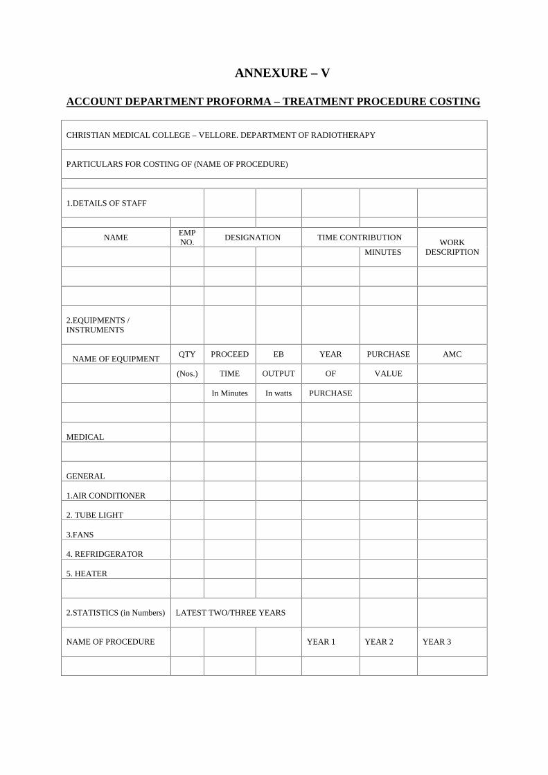
ANNEXURE – V
ACCOUNT DEPARTMENT PROFORMA – TREATMENT PROCEDURE COSTING
CHRISTIAN MEDICAL COLLEGE – VELLORE. DEPARTMENT OF RADIOTHERAPY
PARTICULARS FOR COSTING OF (NAME OF PROCEDURE)
1.DETAILS OF STAFF
NAMEEMPNO.
DESIGNATION TIME CONTRIBUTIONWORK
DESCRIPTIONMINUTES
2.EQUIPMENTS /INSTRUMENTS
NAME OF EQUIPMENTQTY PROCEED EB YEAR PURCHASE AMC
(Nos.) TIME OUTPUT OF VALUE
In Minutes In watts PURCHASE
MEDICAL
GENERAL
1.AIR CONDITIONER
2. TUBE LIGHT
3.FANS
4. REFRIDGERATOR
5. HEATER
2.STATISTICS (in Numbers) LATEST TWO/THREE YEARS
NAME OF PROCEDURE YEAR 1 YEAR 2 YEAR 3
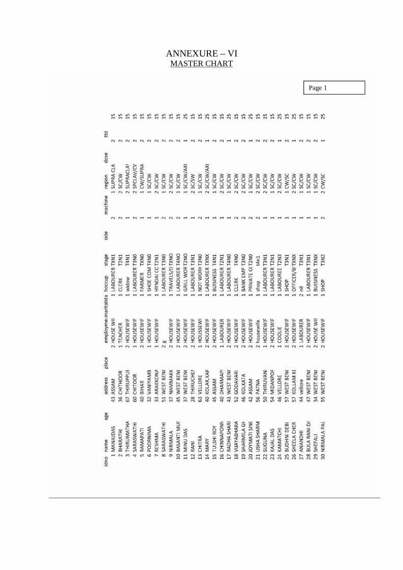
ANNEXURE – VIMASTER CHART
Page 1
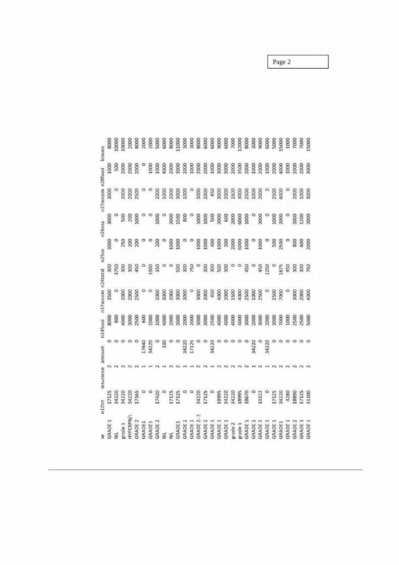
Page 2