Embed Size (px)
Citation preview

“Eczema Coxsackium” and Unusual CutaneousFindings in an Enterovirus Outbreak
WHAT’S KNOWN ON THIS SUBJECT: Coxsackievirus A6 (CVA6) wasidentified as an important cause of “severe” hand, foot, andmouth disease (HFMD) during the 2011–2012 outbreak in NorthAmerica. The atypical cutaneous features in this outbreak havenot been well documented.
WHAT THIS STUDY ADDS: The cutaneous manifestations of CVA6-associated HFMD may be more extensive and variable than classicHFMD. Four distinct morphologies characterize this exanthem:(1) widespread vesiculobullous and erosive lesions, (2) “eczemacoxsackium,” (3) an eruption similar to Gianotti-Crosti, and (4)purpuric lesions.
abstractOBJECTIVE: To characterize the atypical cutaneous presentations inthe coxsackievirus A6 (CVA6)–associated North American enterovirusoutbreak of 2011–2012.
METHODS: We performed a retrospective case series of pediatricpatients who presented with atypical cases of hand, foot, and mouthdisease (HFMD) from July 2011 to June 2012 at 7 academic pediatricdermatology centers. Patients were included if they tested positive forCVA6 or if they met clinical criteria for atypical HFMD (an enanthem orexanthem characteristic of HFMD with unusual morphology or extent ofcutaneous findings). We collected demographic, epidemiologic, andclinical data including history of skin conditions, morphology and ex-tent of exanthem, systemic symptoms, and diagnostic test results.
RESULTS: Eighty patients were included in this study (median age 1.5years, range 4 months–16 years). Seventeen patients were CVA6-positive, and 63 met clinical inclusion criteria. Ninety-nine percent ofpatients exhibited a vesiculobullous and erosive eruption; 61% ofpatients had rash involving .10% body surface area. The exanthemhad a perioral, extremity, and truncal distribution in addition toinvolving classic HFMD areas such as palms, soles, and buttocks. In55% of patients, the eruption was accentuated in areas of eczematousdermatitis, termed “eczema coxsackium.” Other morphologies includedGianotti-Crosti–like (37%), petechial/purpuric (17%) eruptions, anddelayed onychomadesis and palm and sole desquamation. Therewere no patients with serious systemic complications.
CONCLUSIONS: The CVA6-associated enterovirus outbreak wasresponsible for an exanthem potentially more widespread, severe, andvaried than classic HFMD that could be confused with bullousimpetigo, eczema herpeticum, vasculitis, and primary immunobullousdisease. Pediatrics 2013;132:e149–e157
AUTHORS: Erin F. Mathes, MD,a,b Vikash Oza, MD,a Ilona J.Frieden, MD,a,b Kelly M. Cordoro, MD,a,b Shigeo Yagi, PhD,c
Renee Howard, MD,a Leonard Kristal, MD,d Christine C.Ginocchio, PhD,e Julie Schaffer, MD,f,g Sheilagh Maguiness,MD,h Susan Bayliss, MD,i Irene Lara-Corrales, MD,j MariaTeresa Garcia-Romero, MD,j Dan Kelly, MD,k Maria Salas,MPH,c M. Steven Oberste, PhD,l W. Allan Nix, BS,l CarolGlaser, DVM, MPVM, MD,c and Richard Antaya, MDm
Departments of aDermatology, and bPediatrics, University ofCalifornia, San Francisco, San Francisco, California; cState ofCalifornia, Department of Public Health, Richmond, California;dDepartment of Dermatology, Stony Brook University, StonyBrook, New York; eDepartment of Pathology and LaboratoryMedicine, North Shore-Long Island Jewish Health SystemLaboratories, Lake Success, New York; Departments of fDermatology,and gPediatrics, New York University School of Medicine, NewYork, New York; hPediatric Dermatology, Children’s HospitalBoston, Boston, Massachusetts; iDivisions of Dermatology,Pediatrics and Internal Medicine, Washington University Schoolof Medicine, St. Louis Children’s Hospital, St. Louis, Missouri; jDeptof Pediatrics, Division of Dermatology, The Hospital for SickChildren, Toronto, Ontario, Canada; kPediatrics Private Practice,San Francisco, California; lDivision of Viral Diseases, Centers forDisease Control and Prevention, Atlanta, Georgia; and mDermatologyand Pediatrics, Yale University School of Medicine, New Haven,Connecticut
KEY WORDShand, foot, and mouth disease, coxsackievirus, atopic dermatitis,exanthem
ABBREVIATIONSAD—atopic dermatitisBSA—body surface areaCDC—Centers for Disease Control and PreventionCVA6—coxsackievirus A6CVA16—coxsackievirus A16HFMD—hand, foot, and mouth diseaseHSV—herpes simplex virusMRSA—methicillin resistant staphylococcus aureusMSSA—methicillin senstitive staphylococcus aureusPCR—polymerase chain reactionUCSF—University of California, San Francisco
(Continued on last page)
PEDIATRICS Volume 132, Number 1, July 2013 e149
ARTICLE
by guest on May 4, 2018http://pediatrics.aappublications.org/Downloaded from

In March 2012, the Centers for DiseaseControl and Prevention (CDC) reporteda growing number of “severe and ex-tensive” cases of hand, foot, and mouthdisease (HFMD) attributed to coxsack-ievirus A6 (CVA6).1 Over the past 5years, CVA6 has also been implicated inHFMD outbreaks in Finland, France,Spain, India, Taiwan, Japan, Singapore,China, and Boston, Massachusetts.2–11
The atypical cutaneous features char-acterizing the 2011–2012 North Ameri-can CVA6-associated HFMD outbreakhave not been clearly defined.
HFMD was first described by C.R.Robinsonasa self-limited febrile illnesscharacterized by “pharyngeal lesionsand vesicular exanthem” affectingyoung children in a Toronto suburbin the summer of 1957.12 Since thatreport, HFMD has become a widelyrecognized childhood exanthem, rep-resenting a frequent and characteris-tic manifestation of enterovirusinfections. Classic HFMD consists offever, oral erosions, and gray-white,oval vesicles on the hands, feet, andbuttocks.13,14 Children aged ,5 yearsare most often affected with trans-mission occurring via fecal-oral route,vesicle fluid, or respiratory secretions.15
Coxsackievirus A16 (CVA16) and en-terovirus 71 are the 2 most commonlyreported causes of HFMD.16,17 CVA16 hastraditionally been considered the mostfrequent cause of HFMD in the UnitedStates.17 Enterovirus 71 has been re-sponsible for a number of outbreaks ofHFMD associated with severe neuro-logic complications in Asia.18,19
The purpose of this multi-institutional,retrospective study was to better de-fine the more severe, varied, and ex-tensive dermatologic manifestations ofthe CVA6-associated outbreak in NorthAmerica.
METHODS
We performed a multi-institutional re-trospective case series of pediatric
patients between the ages of 0 and 18years evaluated by pediatric derma-tologists at 7 institutions (Universityof California, San Francisco [UCSF];New York University; WashingtonUniversity in St Louis; The Hospital forSick Children, Toronto; Children’sHospital Boston; Stony Brook Univer-sity in collaboration with NorthShore-Long Island Jewish HealthSystem Laboratories; and Yale Uni-versity) from July 2011 to June 2012with atypical presentations of HFMD.Patients were identified througha search of electronic health recordsand photo archives at each in-stitution. Patients were included ifinfection with CVA6 was confirmed bynucleotide sequencing of samplesfrom oropharynx, skin, blood, orstool or if they met clinical case cri-teria for atypical or severe HFMD. Theclinical inclusion criteria were pre-determined by the lead authorsbased on initial experience with thisoutbreak. Two categories of clinicalcase criteria needed to be fulfilled forinclusion: (1) features suggestive ofHFMD and (2) unusual extent ormorphology. Features suggestive ofHFMD were defined as an enanthemcharacteristic of HFMD (small vesi-cles and erosions on the oral mu-cosa), exanthem with featuressuggestive of HFMD (gray-white, ovalvesicles on the hands, feet, and but-tocks), or history of exposure toHFMD 2 to 14 days before diseaseonset. To meet the criteria for un-usual morphology or extent of der-matologic findings, patients neededto have at least 1 of the following:HFMD exanthem involving.5% of thebody surface area (BSA); erosions,vesicles (fluid filled blister ,1 cm)and/or bullae (fluid filled blister.1cm) with acrofacial accentuation;purpuric, petechial, or hemorrhagiclesions; coalescing papules symmet-rically distributed on the face, arms,and legs with relative sparing of the
trunk similar to Gianotti-Crosti; orlarge bullae (.2 cm). Patients wereexcluded if the presentation could beexplained by another specific illnesssuch as, but not limited to, varicella orherpes simplex virus (HSV) infection,bullous impetigo, uncomplicated atopicdermatitis (AD), contact dermatitis, orother bullous diseases, or if the exan-them and enanthem were consistentwith classic HFMD with ,5% BSA in-volved. Two investigators (EM, IJF)reviewed data collection forms andavailable photographs for all cases. In-stitutional review board approval wasobtained from UCSF as the coordinatingboard and from each participating in-stitution as required.
A standardized data collection formwas used to record epidemiologic andclinical data including age, gender,ethnicity, race, school or day-care par-ticipation, sick contacts, travel history,history of skin conditions, disease on-set and duration, systemic symptoms,relevant laboratory results, treatmentreceived, and whether they weremanaged as an inpatient or outpatient.The morphology and distribution of theeruption were assessed in detail in-cluding primary skin lesion types andlocation, extent (BSA), secondary fea-tures, nail changes, and oropharyngealinvolvement. We also recordedwhetherthe eruption was concentrated in areaspreviously or currently affected by AD(similar to eczema herpeticum), local-ized toanareaofpreviousskin injury, orappeared similar to Gianotti-Crosti.Enterovirus detection was done by ei-ther 1-step real-time reverse tran-scriptase polymerase chain reaction(PCR) or nucleic acid sequence–basedamplification using the NucliSensEasyQ EV assay (bioMerieux, Durham,NC). For the enterovirus-positive sam-ples, when available, enterovirus typewas determined by sequencing at theCDC or the California Department ofPublic Health.20
e150 MATHES et al
by guest on May 4, 2018http://pediatrics.aappublications.org/Downloaded from
Data were compiled and analyzed byusing Excel and SAS version 9.2 (SASInstitute, Cary, NC). Characteristicswere examined for 3 subsets ofpatients: (1) patients with and with-out AD; (2) patients PCR-positive forCVA6 versus patients not tested forCVA6; and (3) patients classified into 3age groups (,1, 1–5,.5 years). TheMann-Whitney test was used tocompare differences in medians ofa continuous characteristic betweengroups, and the Fisher’s exact testwas used to compare differences inpercentages of a categorical char-acteristic between groups.
RESULTS
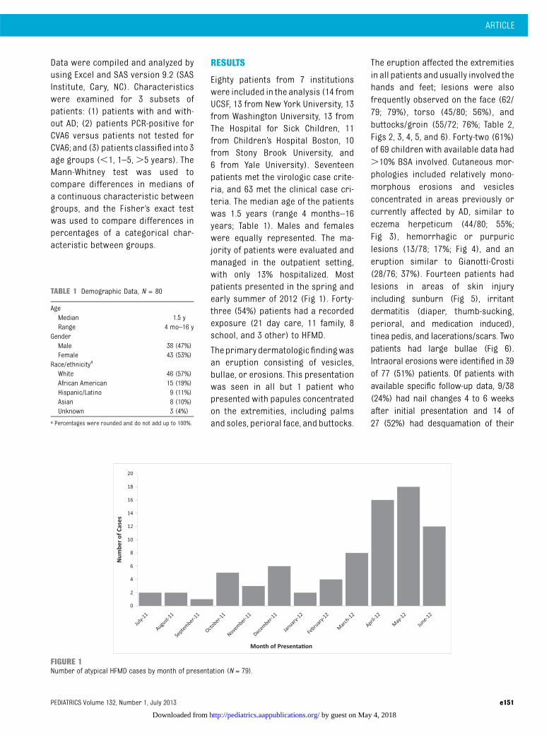
Eighty patients from 7 institutionswere included in the analysis (14 fromUCSF, 13 from New York University, 13from Washington University, 13 fromThe Hospital for Sick Children, 11from Children’s Hospital Boston, 10from Stony Brook University, and6 from Yale University). Seventeenpatients met the virologic case crite-ria, and 63 met the clinical case cri-teria. The median age of the patientswas 1.5 years (range 4 months–16years; Table 1). Males and femaleswere equally represented. The ma-jority of patients were evaluated andmanaged in the outpatient setting,with only 13% hospitalized. Mostpatients presented in the spring andearly summer of 2012 (Fig 1). Forty-three (54%) patients had a recordedexposure (21 day care, 11 family, 8school, and 3 other) to HFMD.
The primary dermatologic findingwasan eruption consisting of vesicles,bullae, or erosions. This presentationwas seen in all but 1 patient whopresented with papules concentratedon the extremities, including palmsand soles, perioral face, and buttocks.
The eruption affected the extremitiesin all patients and usually involved thehands and feet; lesions were alsofrequently observed on the face (62/79; 79%), torso (45/80; 56%), andbuttocks/groin (55/72; 76%; Table 2,Figs 2, 3, 4, 5, and 6). Forty-two (61%)of 69 children with available data had.10% BSA involved. Cutaneous mor-phologies included relatively mono-morphous erosions and vesiclesconcentrated in areas previously orcurrently affected by AD, similar toeczema herpeticum (44/80; 55%;Fig 3), hemorrhagic or purpuriclesions (13/78; 17%; Fig 4), and aneruption similar to Gianotti-Crosti(28/76; 37%). Fourteen patients hadlesions in areas of skin injuryincluding sunburn (Fig 5), irritantdermatitis (diaper, thumb-sucking,perioral, and medication induced),tinea pedis, and lacerations/scars. Twopatients had large bullae (Fig 6).Intraoral erosions were identified in 39of 77 (51%) patients. Of patients withavailable specific follow-up data, 9/38(24%) had nail changes 4 to 6 weeksafter initial presentation and 14 of27 (52%) had desquamation of their
TABLE 1 Demographic Data, N = 80
AgeMedian 1.5 yRange 4 mo–16 y
GenderMale 38 (47%)Female 43 (53%)
Race/ethnicitya
White 46 (57%)African American 15 (19%)Hispanic/Latino 9 (11%)Asian 8 (10%)Unknown 3 (4%)
a Percentages were rounded and do not add up to 100%.
FIGURE 1Number of atypical HFMD cases by month of presentation (N = 79).
ARTICLE
PEDIATRICS Volume 132, Number 1, July 2013 e151
by guest on May 4, 2018http://pediatrics.aappublications.org/Downloaded from

palms or soles 1 to 3 weeks after initialpresentation.
Infants ,1 year old were significantlymore likely to have bullae (Fig 6) thanolder children (38% of patients aged,1 year vs 7% of patients aged 1–5years vs 18% of patients aged .5
years; P = .039). Older children weremore likely to have hemorrhagic orpurpuric lesions (Fig 4; 8%, 1 year vs12% 1–5 years vs 43% .5 years; P =.021) and nail changes (33%,1 year vs14% 1–5 years vs 75% .5 years, P =.022). There were no other significantmorphologic differences between agegroups.
Forty-nine of 79 (62%) patients hada preexisting skin condition, 40 (82%)of whom had a history of AD. Patientswith AD were significantly more likelyto have an eczema herpeticum-likepresentation (81% of patientswith AD,versus 24% of patients without AD; P, .0001) (Figure 3). There were noother significant differences betweenpatients with and without AD.
Table 3 lists the results of the mostcommon diagnostic tests that wereperformed. Seventeen patients wereconfirmed to have CVA6 by nucleotidesequencing. Confirmed CVA6 caseswere compared with cases withoutconfirmation of CVA6 infection (Ta-ble 2). The 2 groups were similar withregard to demographics, morphology,and distribution of skin lesions, andsystemic symptoms. Four patients hadskin biopsies of representative pap-ulovesicles, vesicles, or bullae withvariable findings including spongioticdermatitis, focal interface dermatitiswith areas of subepidermal separa-tion, papillary dermal edema, anddermal inflammation.
The most commonly reported symp-tomswere fever (75%) and sore throat/mouth (36%). Other symptoms such ascough, vomiting, diarrhea, or headachewere reported in #10 patients. Nopatients had serious systemic or neu-rologic complications. Total illness du-ration (onset of first symptom toclearance of rash and all other symp-toms) ranged from 3 to 35 days (meandays 12.2, SD 7.1).
DISCUSSION
HFMD is classically defined as anenterovirus-associated exanthem char-acterized by fever; stomatitis of the oralmucosa; and a vesicular rash affectingthe hands, feet, and occasionally thebuttocks.12,13,21 This study helps char-acterize the wide-ranging and severecutaneous features observed in theCVA6-associated HFMD outbreak firstreported by the Centers for DiseaseControl and Prevention in March 2012.1
We identified 4 morphologies thatcharacterize the severe end of thespectrum of disease associated withthis atypical exanthemanddistinguish itfrom classic HFMD: (1) widespreadvesiculobullous and erosive lesionsextending beyond the palms and soles,(2) an eczema herpeticum-like eruption
TABLE 2 Comparison of CVA6-Positive Cases to Cases Without CVA6 Confirmation, No. Positive /Total No. Reported (%)a
All Cases CVA6+ Cases Cases Without CVA6Confirmation
P
(n = 80) (n = 17) (n = 63)
Age, y, median (range) 1.5 (0.33–16) 1.25 (0.33–16) 1.58 (0.33–16) .59SexFemale 42/80 (53%) 12/17 (71%) 30/63 (48%) .11Male 38/80 (48%) 5/17 (30%) 33/63 (52%)
Body surface area,10% 27/69 (39%) 6/12 (50%) 21/57 (37%) .3411%–25% 18/69 (26%) 4/12 (33%) 14/57 (25%).26% 25/69 (35%) 2/12 (17%) 23/57 (39%)
DistributionPalm/soles 67/80 (84%) 15/17 (88%) 52/63 (83%) .72Extremities 79/79 (100%) 17/17 (100%) 62/62 (100%) —
Face 62/79 (79%) 12/17 (71%) 50/62 (81%) .51Torso 45/80 (56%) 12/17 (70%) 33/63 (52%) .27Buttocks, groin, perineum 55/72 (76%) 12/17 (71%) 43/55 (78%) .53
MorphologyVesicle, bullae, or erosions 79/80 (99%) 17/17 (100%) 62/63 (98%) 1.00Eczema herpeticum-like 44/80 (55%) 6/17 (35%) 38/63 (60%) .10Gianotti-Crosti-like 28/76 (37%) 8/16 (50%) 20/60 (33%) .25Purpuric/petechial 13/78 (17%) 3/17(18%) 10/61 (16%) 1.00Accentuation in areas
of skin injury14/76 (18%) 4/17 (24%) 10/59 (17%) .50
Oral erosions/ulcerations 39/77 (51%) 9/17 (53%) 30/60 (50%) 1.00Nail changes 9/38 (24%) 2/8 (25.0%) 7/30 (23%) 1.00
Hospitalized 10/80 (13%) 2/17 (12%) 8/63 (13%) 1.00Fever 56/75 (75.0%) 11/14 (79%) 45/61 (74%) 1.00Illness duration,d; (mean 6 SD)
12.2 6 7.0 (n = 39) 8.8 6 3.6 (n = 12) 13.7 6 7.7 (n = 27) .07
a Not all featureswere known for each patient. Patients could be reported as havingmultiple morphologies and distributions.Percentages were rounded and do not add up to 100%. —, indicates P value can not be calculated when the condition ispresent in 100% of patients.
FIGURE 2A 13-year-old with facial erosions and vesicleswho had confirmed CVA6 infection.
e152 MATHES et al
by guest on May 4, 2018http://pediatrics.aappublications.org/Downloaded from

termed “eczema coxsackium,” (3) aneruption similar to Gianotti-Crosti, and(4) a petechial or purpuric eruption(Table 4).
The most common dermatologicpresentation in our sample waswidespread vesicles, bullae, and/orerosions. Classically, the vesicularexanthem of HFMD is restricted to thehands, feet, and occasionally thebuttocks.10,13,14,19,21,22 The accompa-nying enanthem consists of smallvesicles and erosions on the oralmucosa.12,13 The exanthem in our se-ries differed by commonly involvingthe perioral area, extremities, andtorso in addition to more classicHFMD locations. Other recent studies
have also emphasized the perioraldistribution of CVA6-associated dis-ease.10,23 Vesicles, variably sizedbullae, and/or erosions involved.10% BSA in the majority of ourpatients. To the best of our knowl-edge, widespread vesiculobullousexanthems have not been reported inprevious HFMD outbreaks. Intraoralerosions were less common than inclassic HFMD where the rate ofintraoral erosions ranges from 75%to 100%.9,12,24–26
A large percentage of patients withunderlying AD presented with vesiclesand erosions within areas affected byAD that we term “eczema coxsackium.”This morphology was strikingly similarto eczema herpeticum caused by HSV1.CVA16 is the only enterovirus known tocause a similar eruption, with only 3cases reported in the literature (1 caseinvolving an adult patient with Darier’sdisease27 and 2 in children with AD28).Why only certain viruses such as HSV1,vaccinia, and now CVA6 more com-monly lead to vesicles and erosions inareas of dermatitis is not understood.Enteroviral infections, particularlyCVA6, should now be considered in thedifferential diagnosis of patients pre-senting with new-onset vesicles and
extensive erosions in preexisting areasof eczema.
In addition to localizing to areas of AD,the eruption in this recent outbreakdemonstratedapredilection forareasof previous trauma or inflammation.Examples of this phenomenon in ourseries included vesicles, bullae, anderosions that developed in areas ofpreexisting sunburn, diaper derma-titis, irritant dermatitis, healing lac-erations, and tinea pedis. Thispredilection for areas of trauma orinjury may explain in part why classicHFMD and this more severe eruptionare commonly seen on the buttocks,palms and soles, all ofwhichare areasof increased trauma and friction inchildren. Other viral exanthems, suchas varicella, have also been reportedto occur in areas of sunburn and di-aper dermatitis.28,29
A distribution similar to Gianotti-Crostiwas documented in one-third of thepatients in our study, with lesions in-volving the cheeks, extensor surfaces ofthe extremities, and buttocks, butsparing the trunk.30 Classic Gianotti-Crosti is characterized by mono-morphous lichenoid papules and/orpapulovesicles, whereas the eruptionassociated with this outbreak wasmore often papulovesicular withprominent erosions. Epstein-Barr virusand hepatitis B virus are the mostcommonly reported causes of Gianotti-Crosti,30 but enteroviruses such ascoxsackieviruses A16, B4, and B5 havealso been implicated.31
A petechial and purpuric rash wasdocumented in 17% of our patients,most often in those aged.5 years andmost frequently found on acral sites.Petechial and purpuric eruptions area known cutaneous manifestation ofenteroviral infections,32,33 and CVA6 in-fection should be added to the differ-ential diagnosis of acral purpura,particularly in the setting of a commu-nity outbreak.
FIGURE 3Eczema coxsackium: A toddler with confirmed CVA6 infection who had erosions localized to areas of AD.
FIGURE 4A 16-year-old with confirmed CVA6 infectionwho had purpuric papules and vesicles on thefeet.
ARTICLE
PEDIATRICS Volume 132, Number 1, July 2013 e153
by guest on May 4, 2018http://pediatrics.aappublications.org/Downloaded from

Delayed cutaneous features of this ex-anthem include nail changes and des-quamation of the palms and soles,which typically occurred weeks afterthe resolution of the vesiculobullous
eruption. Onychomadesis (separationof the proximal nail plate from the nailmatrix and nail bed), and Beau’s lines(horizontal ridging of the nail plate)were a common feature of CVA6 in-
fection in previous reports23,34 andwere documented in 9 of 38 cases withavailable follow-up data in this series.However, this may be an underestimatebecause only aminority of patients wasfollowed beyond their acute illness.Onychomadesis associated with HFMDmost often occurs 3 to 8 weeks afterHFMD is diagnosed.35,36 It is generallyasymptomatic, and the nails typicallyregrow normally within severalmonths.
The demographics and systemic man-ifestations observed in our patients arerepresentative of previous reports ofHFMDoutbreaks, with a predilection forpreschool-age children of either gen-der, occurring primarily during the latespring to early summer.3,6 Fever andoropharyngeal pain were the 2 mostcommonly reported symptoms. Thelow rate of gastrointestinal and re-spiratory symptoms is also consistentwith past reports of CVA6 HFMD.3,6
Overall, the extracutaneous features ofthe 2011–2012 HFMD outbreak appearsimilar to those of the most commoncause of HFMD, CVA16, rather thanmore virulent strains such as en-terovirus 71.37 Only 10 patients in oursample were hospitalized, primarilybecause of the unusual nature of theskin disease. These children oftenreceived empriric antivirals andantibiotics and a diagnostic evalua-tion for extensive vesicles and bul-lae. Although a few patients didhave dehydration in the setting oforal ulcerations, no patients in ourstudy developed serious systemiccomplications that sometimes occurwith enterovirus infections, such asmyocarditis, pneumonia/pneumonitis,aseptic meningitis, or meningoenceph-alitis. Our report suggests that the ex-tensive and varied cutaneous featuresseen in this outbreak do not portend anincreased risk for severe systemic ill-ness.
FIGURE 5A toddler with confirmed CVA6 infection who had vesicles localized to areas of sunburn (a) in additionperianal and buttocks erosions (b).
FIGURE 6A 4-month-old (a) and 15-month-old (b) with confirmed CVA6 infection who presented with acralbullae.
TABLE 3 Outcomes of Diagnostic Tests
Diagnostic test No. Positive No. Performed
Coxsackievirus A6 nucleotide sequencing 17 17a
Enterovirus PCR and/or NucliSens nucleic acidsequence based amplification
24 27b
Enteroviral culture 1 13HSV (DFA, culture, or PCR) 0 27VZV (DFA or PCR) 0 3Bacterial culture 2 (MSSA, MRSA) 14Skin biopsy —
c 4
DFA, direct fluorescent antibody; MRSA, methicillin resistant staphylococcus aureus; MSSA, methicillin senstitive staphylo-coccus aureus; VZV, varicella zoster virus.a Coxsackievirus A6 nucleotide sequencing was not available to all patients who had enterovirus PCR and/or NucliSensnucleic acid sequence–based amplification. For specimens tested at the CDC or the California Department of Public Health,enterovirus PCR was performed first: if positive, CVA6 nucleotide sequencing was performed; if negative, no additionaltesting was done.b The 3 cases with negative enterovirus PCR were included based on clinical criteria. One specimen was collected 10 d afterthe acute phase of the illness. Two of the 3 specimens were skin swabs, which are less sensitive and more dependent oncollection technique.c For skin histopathology, see discussion in text.
e154 MATHES et al
by guest on May 4, 2018http://pediatrics.aappublications.org/Downloaded from

The phenotypic variability and un-usual nature of the skin eruptionsdocumented in this HFMD outbreakmimic andmaybe confusedwith otherinfectious and inflammatory skindiseases, emphasizing the need foraccurate diagnostic testing. Entero-virus PCR testing requirements varyby laboratory, but testing can beperformed from swabs of skin vesiclefluid, oropharynx, perirectal skin,stool, or blood. Viral culture forCVA6 isnot recommended because CVA6 doesnot grow well in culture.38 However, HSVculture and/or direct fluorescent anti-body testing should be considered torule out herpesvirus infections inpatients presenting with vesiculobullousdisease.
Our study has several limitations. Theretrospective nature of this studyresulted in incomplete data capture.CVA6 was confirmed in only 17 of our
cases, raising the possibility that notall of the patients in this report hadCVA6 and that other enterovirusstrains may have coexisted duringthis outbreak. However, no sub-stantial differences in clinical pre-sentation were noted when theconfirmed CVA6 patients were com-pared with the patients meetingclinical inclusion criteria. Threepatients had negative enterovirusPCR and were included based onclinical criteria. It is possible thatthese patientswere not CVA6 infected,but it is also possible that their testswere negative because of inadequateskin specimen collection technique orbecause they were collected after theacute phase of the illness. Not allpatients were tested for HSV or bac-terial infections; therefore, we cannotrule out the possibility that other viralor bacterial infections could have
contributed to some of the eruptions.Lastly, because all patients includedin our sample were referred to anacademic pediatric dermatologycenter, a referral bias toward moresevere manifestations likely exists.Approximately half of the patients inthis series had exposure to contactswith clinical HFMD who were nottested for enterovirus, or, more spe-cifically, for CVA6. Therefore, it ispossible that some patients infectedwith CVA6 demonstrate a more typicalHFMD course. However, our studydoes not attempt to characterize allcutaneous eruptions associated withCVA6. Instead, our goal was to capturethe more severe and unusual der-matologic features seen with thisoutbreak. Larger, prospective studiesare needed to provide comprehen-sive epidemiologic data on the fullclinical spectrum of disease and de-mographic risk factors of CVA6infections.
Despite these limitations, this reporthighlights several important clinicalfindings not previously reported inoutbreaks of HFMD. Awareness of thepotential extent and variability of thiscondition should help to avoid confu-sion with other skin conditions such aseczema herpeticum, vasculitis, impe-tigo, and primary immunobullous dis-ease, as well as to avoid errors indiagnosis and management in futureoutbreaks. We recommend enterovirusPCR testing in cases in which diagnosisis in doubt.
ACKNOWLEDGMENTSThe authors thank the following peo-ple for technical support: ShannonRogers, Centers for Disease Controland Prevention, Atlanta, GA; TashaPadilla and the staff of the Viraland Rickettsial Disease Laboratory,California Department of PublicHealth, Richmond, CA.
TABLE 4 Clinical Features and Differential Diagnosis of Severe CVA6-Associated HFMD
Findings Suggestive of HFMD a: 1) Fever, 2) Oral erosions, 3) Mild gastrointestinal symptoms, 4) Oval vesicles onhands and feet, 5) Known sick contacts
Atypical Cutaneous Morphology Clinical Differential Diagnosis
Vesiculobullous anderosive eruption
• Widespread (.5% BSA distribution) • Bullous impetigo• Perioral, acral, buttock predilection • Varicella• Bullae more common aged ,1year • Primary immunobullous
disorders
Eczema coxsackium • Vesicles and erosions in areas ofeczematous dermatitis
• Eczema herpeticum• Secondary bacterial infectionin setting of AD
Gianotti Crosti-likeeruption
• Acrofacial papulovesicles anderosions with relative sparingof the trunk similar toGianotti-Crosti syndrome
• Gianotti Crosti syndrome• Other viral exanthems• Urticaria multiforme
Petechial and purpuricrash
• Most often seen in patients. 5 years of age
• Leukocytoclastic vasculitis
• Often acral• Glove and stocking purpura(parvovirus infection)
Delayed cutaneousfindings
• Onychomadesis (nail shedding)and Beau’s lines (tranversegrooves)
• Onychomadesis: Medicationinduced (tetracyclines), aftersevere systemic illness
• Acral desquamation • Acral Desquamation: after toxin orsuperantigen-mediated disease(Group A Streptococcus infection,Kawasaki disease, or toxic shocksyndrome)
Diagnosis can be confirmed by enterovirus PCR (serum; oropharyngeal and skin swab as available). As indicated, rule outother entities with viral and bacterial cultures, viral DFA or PCR, and skin biopsy.a May be variably present.
ARTICLE
PEDIATRICS Volume 132, Number 1, July 2013 e155
by guest on May 4, 2018http://pediatrics.aappublications.org/Downloaded from

REFERENCES
1. Centers for Disease Control and Pre-vention (CDC). Notes from the field: se-vere hand, foot, and mouth diseaseassociated with coxsackievirus A6—Alabama, Connecticut, California, andNevada, November 2011–February 2012.MMWR Morb Mortal Wkly Rep. 2012;61(12):213–214
2. Osterback R, Vuorinen T, Linna M, Susi P,Hyypiä T, Waris M. Coxsackievirus A6 andhand, foot, and mouth disease, Finland.Emerg Infect Dis. 2009;15(9):1485–1488
3. Mirand A, Henquell C, Archimbaud C, et al.Outbreak of hand, foot and mouth disease/herpangina associated with coxsackievirusA6 and A10 infections in 2010, France:a large citywide, prospective observationalstudy. Clin Microbiol Infect. 2012;18(5):E110–E118
4. Gopalkrishna V, Patil PR, Patil GP,Chitambar SD. Circulation of multiple en-terovirus serotypes causing hand, foot andmouth disease in India. J Med Microbiol.2012;61(pt 3):420–425
5. Bracho MA, González-Candelas F, ValeroA, Córdoba J, Salazar A. Enterovirus co-infections and onychomadesis afterhand, foot, and mouth disease, Spain,2008. Emerg Infect Dis. 2011;17(12):2223–2231
6. Lo SH, Huang YC, Huang CG, et al. Clinicaland epidemiologic features of Coxsack-ievirus A6 infection in children in northernTaiwan between 2004 and 2009. J MicrobiolImmunol Infect. 2011;44(4):252–257
7. Blomqvist S, Klemola P, Kaijalainen S, et al.Co-circulation of coxsackieviruses A6 andA10 in hand, foot and mouth disease out-break in Finland. J Clin Virol. 2010;48(1):49–54
8. Fujimoto T, Iizuka S, Enomoto M, et al. Hand,foot, and mouth disease caused by cox-sackievirus A6, Japan, 2011. Emerg InfectDis. 2012;18(2):337–339
9. Wu Y, Yeo A, Phoon MC, et al. The largestoutbreak of hand; foot and mouth dis-ease in Singapore in 2008: the role ofenterovirus 71 and coxsackievirus Astrains. Int J Infect Dis. 2010;14(12):e1076–e1081
10. Flett K, Youngster I, Huang J, et al. Hand,foot, and mouth disease caused by cox-sackievirus a6. Emerg Infect Dis. 2012;18(10):1702–1704
11. Lu QB, Zhang XA, Wo Y, et al. Circulation ofCoxsackievirus A10 and A6 in hand-foot-mouth disease in China, 2009-2011. PLoSONE. 2012;7(12):e52073
12. Robinson CR, Doane FW, Rhodes AJ. Reportof an outbreak of febrile illness with pha-ryngeal lesions and exanthem: Toronto,summer 1957; isolation of group A Cox-sackie virus. Can Med Assoc J. 1958;79(8):615–621
13. Cherry JD, Jahn CL. Hand, foot, and mouthsyndrome. Report of six cases due to Cox-sackie virus, group A, type 16. Pediatrics.1966;37(4):637–643
14. Lerner AM, Klein JO, Cherry JD, Finland M.New viral exanthems (concluded). N Engl JMed. 1963;269:736–740 [CONCL.]
15. Ruan F, Yang T, Ma H, et al. Risk factors forhand, foot, and mouth disease and herpan-gina and the preventive effect of hand-washing. Pediatrics. 2011;127(4). Availableat: www.pediatrics.org/cgi/content/full/127/4/e898
16. Ho M, Chen ER, Hsu KH, et al; Taiwan En-terovirus Epidemic Working Group. Anepidemic of enterovirus 71 infection inTaiwan. N Engl J Med. 1999;341(13):929–935
17. Khetsuriani N, Lamonte-Fowlkes A, Oberst S,Pallansch MA; Centers for Disease Controland Prevention. Enterovirus surveillance—United States, 1970–2005. MMWR SurveillSumm. 2006;55(8):1–20
18. Lee TC, Guo HR, Su HJ, Yang YC, Chang HL,Chen KT. Diseases caused by enterovirus 71infection. Pediatr Infect Dis J. 2009;28(10):904–910
19. Solomon T, Lewthwaite P, Perera D, CardosaMJ, McMinn P, Ooi MH. Virology, epidemi-ology, pathogenesis, and control of en-terovirus 71. Lancet Infect Dis. 2010;10(11):778–790
20. Nix WA, Oberste MS, Pallansch MA. Sensi-tive, seminested PCR amplification of VP1sequences for direct identification of allenterovirus serotypes from original clinicalspecimens. J Clin Microbiol. 2006;44(8):2698–2704
21. Alsop J, Flewett TH, Foster JR. “Hand-foot-and-mouth disease” in Birmingham in1959. BMJ. 1960;2(5214):1708–1711
22. Adler JL, Mostow SR, Mellin H, Janney JH,Joseph JM. Epidemiologic investigation ofhand, foot, and mouth disease. Infectioncaused by coxsackievirus A 16 in Baltimore,June through September 1968. Am J DisChild. 1970;120(4):309–314
23. Wei SH, Huang YP, Liu MC, et al. An out-break of coxsackievirus A6 hand, foot,and mouth disease associated with ony-chomadesis in Taiwan, 2010. BMC InfectDis. 2011;11:346
24. Yang F, Zhang T, Hu Y, et al. Survey of en-terovirus infections from hand, foot andmouth disease outbreak in China, 2009.Virol J. 2011;8:508
25. Higgins PG, Warin RP. Hand, foot, andmouth disease. A clinically recogniz-able virus infection seen mainly in chil-dren. Clin Pediatr (Phila). 1967;6(6):373–376
26. Liu MY, Liu W, Luo J, et al. Characterizationof an outbreak of hand, foot, and mouthdisease in Nanchang, China in 2010. PLoSONE. 2011;6(9):e25287
27. Higgins PG, Crow KD. Recurrent Kaposi’svaricelliform eruption in Darier’s disease.Br J Dermatol. 1973;88(4):391–394
28. Messner J, Miller JJ, James WD, Honig PJ.Accentuated viral exanthems in areas ofinflammation. J Am Acad Dermatol. 1999;40(2 pt 2):345–346
29. Belhorn TH, Lucky AW. Atypical varicellaexanthems associated with skin injury.Pediatr Dermatol. 1994;11(2):129–132
30. Brandt O, Abeck D, Gianotti R, Burgdorf W.Gianotti-Crosti syndrome. J Am Acad Der-matol. 2006;54(1):136–145
31. James WD, Odom RB, Hatch MH. Gianotti-Crosti-like eruption associated with cox-sackievirus A-16 infection. J Am AcadDermatol. 1982;6(5):862–866
32. Fretzayas A, Douros K, Moustaki M, Nic-olaidou P. Papular-purpuric gloves andsocks syndrome in children and adoles-cents. Pediatr Infect Dis J. 2009;28(3):250–252
33. Nielsen HE, Andersen EA, Andersen J, et al.Diagnostic assessment of haemorrhagicrash and fever. Arch Dis Child. 2001;85(2):160–165
34. Davia JL, Bel PH, Ninet VZ, et al. Onycho-madesis outbreak in Valencia, Spain asso-ciated with hand, foot, and mouth diseasecaused by enteroviruses. Pediatr Dermatol.2011;28(1):1–5
35. Bernier V, Labrèze C, Bury F, Taïeb A. Nailmatrix arrest in the course of hand, footand mouth disease. Eur J Pediatr. 2001;160(11):649–651
36. Clementz GC, Mancini AJ. Nail matrix arrestfollowing hand-foot-mouth disease: a re-port of five children. Pediatr Dermatol.2000;17(1):7–11
37. Chang LY, Lin TY, Huang YC, et al. Com-parison of enterovirus 71 and coxsackie-virus A16 clinical illnesses during theTaiwan enterovirus epidemic, 1998.Pediatr Infect Dis J. 1999;18(12):1092–1096
e156 MATHES et al
by guest on May 4, 2018http://pediatrics.aappublications.org/Downloaded from

38. Vuorinen T, Vainionpää R, Hyypiä T. Fiveyears’ experience of reverse-transcriptase
polymerase chain reaction in daily di-agnosis of enterovirus and rhinovirus
infections. Clin Infect Dis. 2003;37(3):452–455
(Continued from first page)
Dr Mathes conceptualized and designed the study, designed the data collection instruments, coordinated the 7 sites, supervised data collection at 1 site, carriedout the initial analyses, interpreted the data, and reviewed and revised the initial manuscript; Dr Oza drafted the initial manuscript and supervised the initialanalysis; Dr Frieden conceptualized and designed the study, designed the data collection instruments, and reviewed and revised the initial manuscript; Drs Yagiand Oberste coordinated acquisition of data, assisted in interpretation of data, and critically revised the initial manuscript; Dr Cordoro critically reviewed andrevised the data collection instruments, and reviewed and revised the initial manuscript; Dr Howard conceptualized and designed the study, and reviewed andrevised the initial manuscript; Dr Kristal conceptualized and designed the study, coordinated and supervised data collection at 1 site, and critically revised theinitial manuscript; Dr Ginocchio coordinated acquisition of data at 1 site and critically revised the initial manuscript; Drs Schaffer, Maguiness, Bayliss, Lara-Corrales, and Garcia-Romero coordinated and supervised data collection at 1 site, and critically revised the initial manuscript; Dr Kelly conceptualized the studyand critically revised the initial manuscript; Ms Salas conceptualized the study, coordinated acquisition of data at 1 site, and critically revised the initialmanuscript; Mr Nix coordinated acquisition of data, assisted in interpretation of data, and critically revised the initial manuscript; Dr Glaser conceptualized anddesigned the study, designed the data collection instruments, assisted with collection and interpretation of data, and reviewed and revised the initial manuscript;Dr Antaya conceptualized and designed the study, designed the data collection instruments, coordinated collection of data at 1 site and interpretation of data, andreviewed and revised the initial manuscript; and all authors approved the final manuscript as submitted.
www.pediatrics.org/cgi/doi/10.1542/peds.2012-3175
doi:10.1542/peds.2012-3175
Accepted for publication Apr 12, 2013
This work was presented in preliminary form as a poster at the Society for Pediatric Dermatology Annual Meeting; July 11–14, 2012; Monterey, CA. It was presentedas an abstract at the American Academy of Dermatology Annual Meeting; March 2, 2013; Miami, FL.
Address correspondence to Erin Mathes, MD, Departments of Dermatology and Pediatrics, University of California, San Francisco, 1701 Divisadero St, Box 0316, SanFrancisco, CA 94143. E-mail: [email protected]
PEDIATRICS (ISSN Numbers: Print, 0031-4005; Online, 1098-4275).
Copyright © 2013 by the American Academy of Pediatrics
FINANCIAL DISCLOSURE: Christine C. Ginocchio is a consultant, received payment for lectures, and has a grant/pending grant from bioMerieux. bioMerieux makesthe enterovirus assay that was used in the diagnosis of enterovirus at North Shore-Long Island Jewish Health System Laboratories. The other authors haveindicated they have no financial relationships relevant to this article to disclose.
FUNDING: The data analysis for this publication was supported by the National Center for Advancing Translational Sciences, National Institutes of Health, throughUniversity of California, San Francisco-Clinical and Transitional Science Institute grant UL1 TR000004. Its contents are solely the responsibility of the authors and donot necessarily represent the official views of the National Institutes of Health. No additional external funding was secured for this study.
ARTICLE
PEDIATRICS Volume 132, Number 1, July 2013 e157
by guest on May 4, 2018http://pediatrics.aappublications.org/Downloaded from

DOI: 10.1542/peds.2012-3175 originally published online June 17, 2013; 2013;132;e149Pediatrics
Salas, M. Steven Oberste, W. Allan Nix, Carol Glaser and Richard AntayaSusan Bayliss, Irene Lara-Corrales, Maria Teresa Garcia-Romero, Dan Kelly, Maria
Maguiness,Howard, Leonard Kristal, Christine C. Ginocchio, Julie Schaffer, Sheilagh Erin F. Mathes, Vikash Oza, Ilona J. Frieden, Kelly M. Cordoro, Shigeo Yagi, Renee
Outbreak''Eczema Coxsackium'' and Unusual Cutaneous Findings in an Enterovirus
ServicesUpdated Information &
http://pediatrics.aappublications.org/content/132/1/e149including high resolution figures, can be found at:
References
1http://pediatrics.aappublications.org/content/132/1/e149.full#ref-list-This article cites 38 articles, 5 of which you can access for free at:
Subspecialty Collections
ogy_subhttp://classic.pediatrics.aappublications.org/cgi/collection/epidemiolEpidemiologydiseases_subhttp://classic.pediatrics.aappublications.org/cgi/collection/infectious_Infectious Diseasegy_subhttp://classic.pediatrics.aappublications.org/cgi/collection/dermatoloDermatologyfollowing collection(s): This article, along with others on similar topics, appears in the
Permissions & Licensing
https://shop.aap.org/licensing-permissions/in its entirety can be found online at: Information about reproducing this article in parts (figures, tables) or
Reprintshttp://classic.pediatrics.aappublications.org/content/reprintsInformation about ordering reprints can be found online:
ISSN: . 60007. Copyright © 2013 by the American Academy of Pediatrics. All rights reserved. Print American Academy of Pediatrics, 141 Northwest Point Boulevard, Elk Grove Village, Illinois,has been published continuously since . Pediatrics is owned, published, and trademarked by the Pediatrics is the official journal of the American Academy of Pediatrics. A monthly publication, it
by guest on May 4, 2018http://pediatrics.aappublications.org/Downloaded from

DOI: 10.1542/peds.2012-3175 originally published online June 17, 2013; 2013;132;e149Pediatrics
Salas, M. Steven Oberste, W. Allan Nix, Carol Glaser and Richard AntayaSusan Bayliss, Irene Lara-Corrales, Maria Teresa Garcia-Romero, Dan Kelly, Maria
Maguiness,Howard, Leonard Kristal, Christine C. Ginocchio, Julie Schaffer, Sheilagh Erin F. Mathes, Vikash Oza, Ilona J. Frieden, Kelly M. Cordoro, Shigeo Yagi, Renee
Outbreak''Eczema Coxsackium'' and Unusual Cutaneous Findings in an Enterovirus
http://pediatrics.aappublications.org/content/132/1/e149located on the World Wide Web at:
The online version of this article, along with updated information and services, is
ISSN: . 60007. Copyright © 2013 by the American Academy of Pediatrics. All rights reserved. Print American Academy of Pediatrics, 141 Northwest Point Boulevard, Elk Grove Village, Illinois,has been published continuously since . Pediatrics is owned, published, and trademarked by the Pediatrics is the official journal of the American Academy of Pediatrics. A monthly publication, it
by guest on May 4, 2018http://pediatrics.aappublications.org/Downloaded from