Embed Size (px)
Citation preview
EDEMAEDEMAEDEMAEDEMA
Fluid extravasation and Fluid extravasation and
accumulation in the interstitial accumulation in the interstitial
spaces spaces
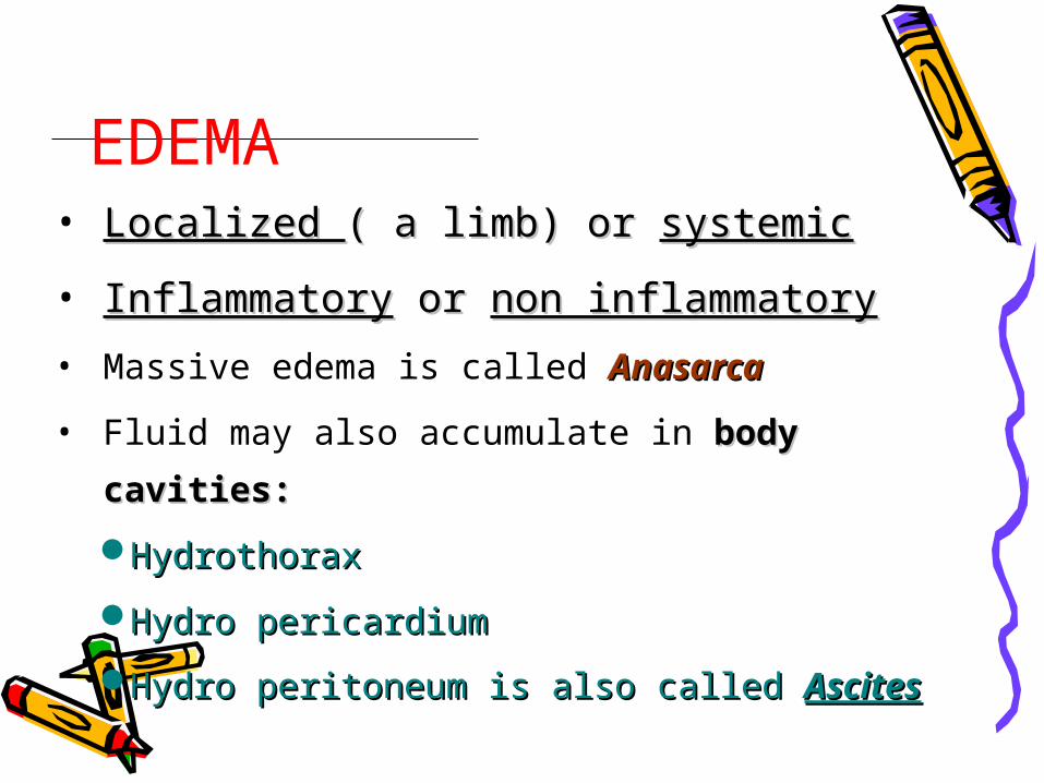
EDEMA• Localized Localized ( a limb) or ( a limb) or systemicsystemic
• InflammatoryInflammatory or or non inflammatorynon inflammatory
• Massive edema is called AnasarcaAnasarca
• Fluid may also accumulate in body body
cavities:cavities:
HydrothoraxHydrothorax
Hydro pericardiumHydro pericardium
Hydro peritoneum is also called Hydro peritoneum is also called AscitesAscites
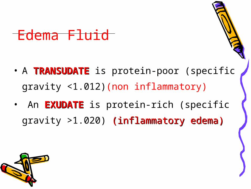
Edema Fluid
• A TRANSUDATETRANSUDATE is protein-poor (specific
gravity <1.012)(non inflammatory)
• An EXUDATEEXUDATE is protein-rich (specific
gravity >1.020) (inflammatory edema)(inflammatory edema)
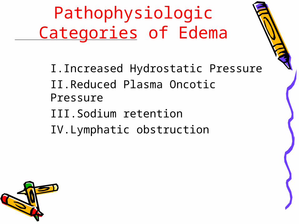
Pathophysiologic Categories of Edema
I.Increased Hydrostatic PressureII.Reduced Plasma Oncotic PressureIII.Sodium retentionIV.Lymphatic obstruction
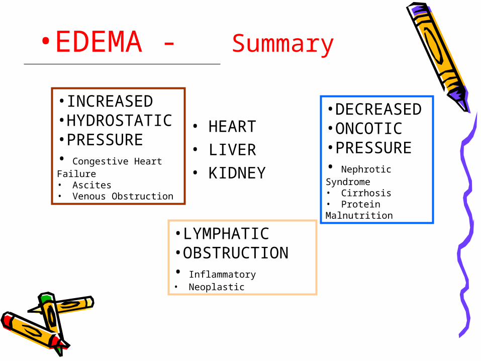
•EDEMA - Summary
•INCREASED•HYDROSTATIC•PRESSURE• Congestive Heart Failure• Ascites• Venous Obstruction
•DECREASED •ONCOTIC•PRESSURE• Nephrotic Syndrome• Cirrhosis• Protein Malnutrition
•LYMPHATIC•OBSTRUCTION• Inflammatory• Neoplastic
• HEART• LIVER• KIDNEY
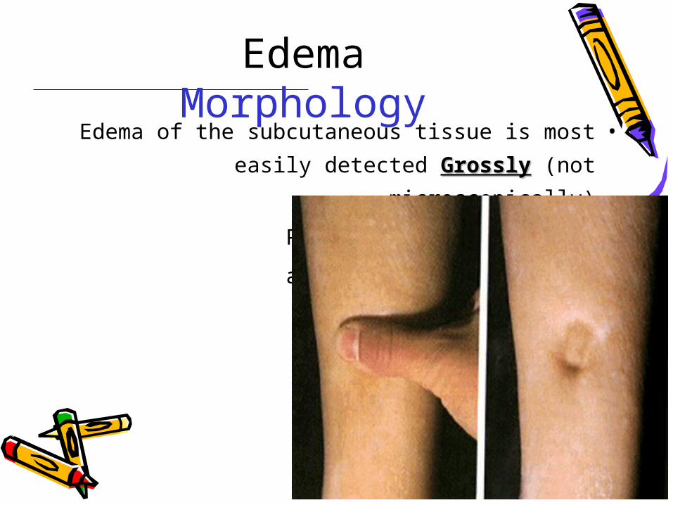
EdemaMorphology
•Edema of the subcutaneous tissue is most
easily detected GrosslyGrossly (not microscopically)
•Push your finger into it
•and a depression remains
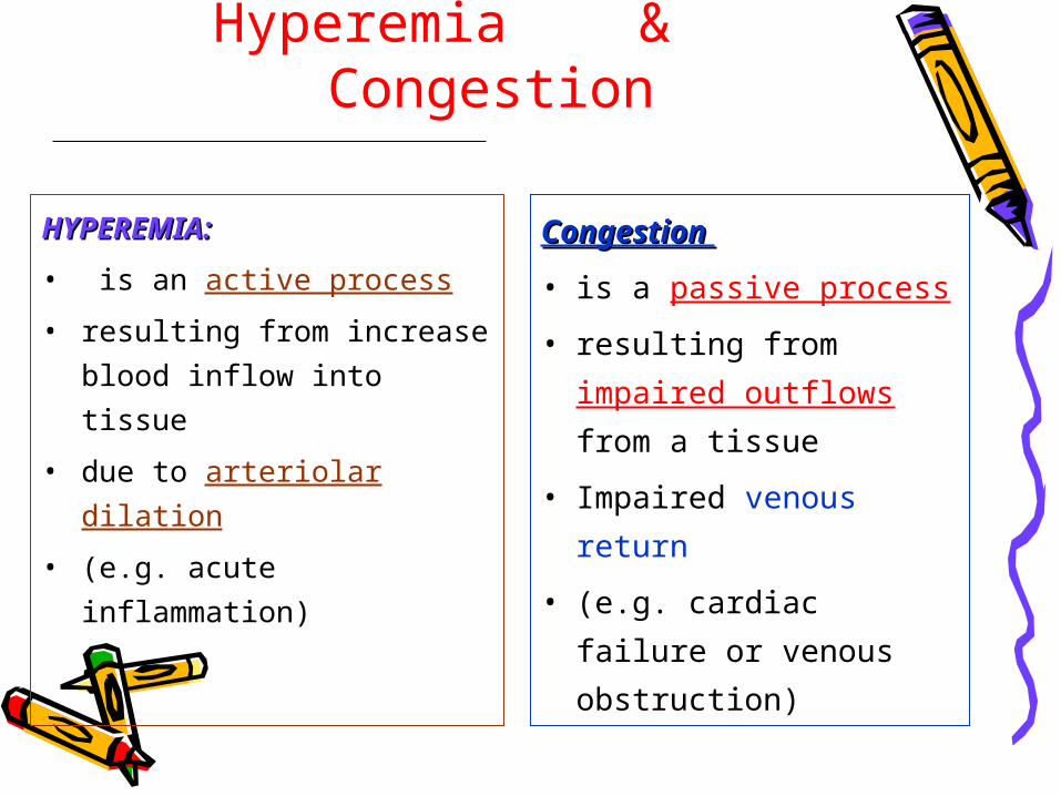
Compare between:Hyperemia & Congestion
HYPEREMIA:HYPEREMIA:
• is an active process
• resulting from increase
blood inflow into tissue
• due to arteriolar dilation
• (e.g. acute inflammation)
Congestion Congestion
• is a passive process
• resulting from
impaired outflows
from a tissue
• Impaired venous
return
• (e.g. cardiac failure or
venous obstruction)
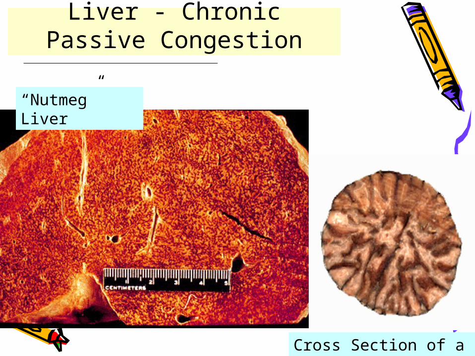
Cross Section of a Nutmeg
“Nutmeg” Liver
Liver - Chronic Passive Congestion
HemorrhageHemorrhageHemorrhageHemorrhage
Extravasation of bloodExtravasation of blood
due to rupture of blood vesselsdue to rupture of blood vessels
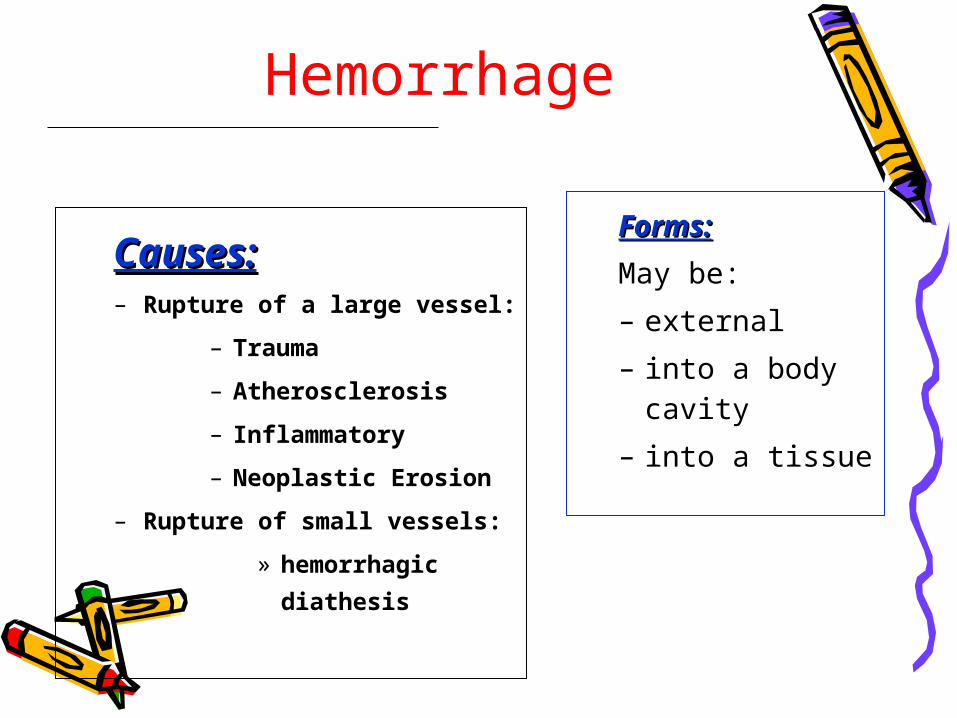
Hemorrhage
Causes:Causes:– Rupture of a large vessel:
– Trauma
– Atherosclerosis
– Inflammatory
– Neoplastic Erosion
– Rupture of small vessels:
» hemorrhagic
diathesis
Forms:Forms:
May be:
– external
– into a body cavity
– into a tissue
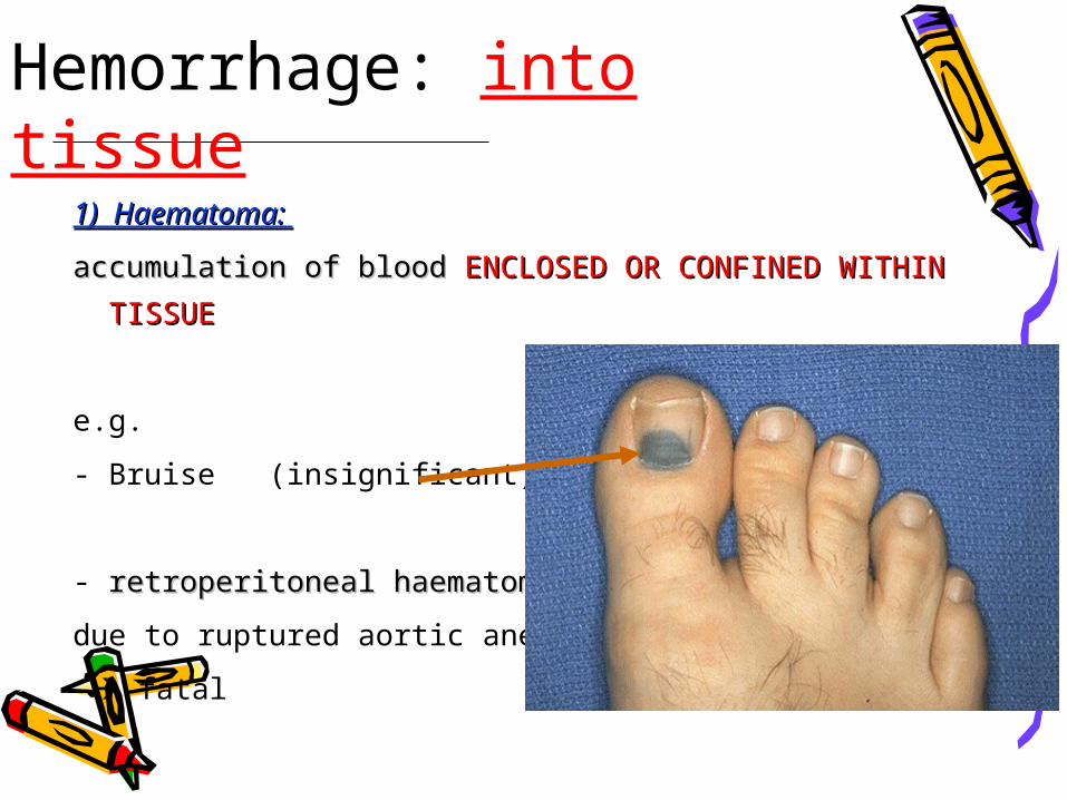
Hemorrhage: into tissue
1) Haematoma: 1) Haematoma:
accumulation of blood accumulation of blood ENCLOSED OR CONFINED WITHIN ENCLOSED OR CONFINED WITHIN
TISSUETISSUE
e.g.
- Bruise (insignificant)
- retroperitoneal haematomaretroperitoneal haematoma
due to ruptured aortic aneurysm
fatal
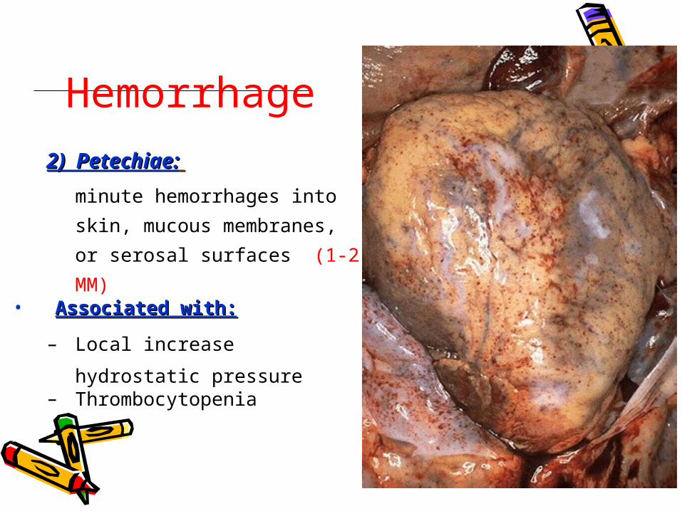
Hemorrhage
2) Petechiae:2) Petechiae:
minute hemorrhages into
skin, mucous membranes, or
serosal surfaces (1-2 MM) • Associated with:Associated with:
– Local increase hydrostatic
pressure– Thrombocytopenia
Hemorrhage
3) Purpura:3) Purpura:
– Slightly larger hemorrhages than Petechiae (3-5
MM)– Causes:Causes:
• Causes as Petechiae• Trauma• Vasculitis• Increased vascular fragility
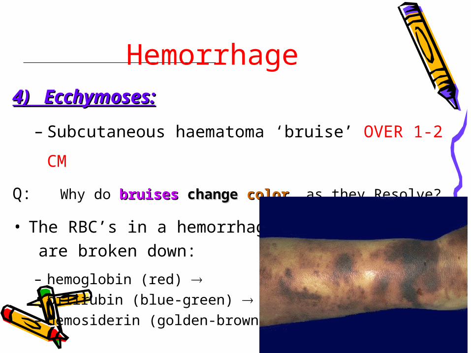
Hemorrhage4) Ecchymoses:4) Ecchymoses:
– Subcutaneous haematoma ‘bruise’ OVER 1-2
CM
Q: Why do bruisesbruises changechange colorcolor as they Resolve?
• The RBC’s in a hemorrhage are broken down:
– hemoglobin (red) – bilirubin (blue-green) – hemosiderin (golden-brown)
Hemorrhage: Accumulation of blood Accumulation of blood in a body cavityin a body cavity::
– Hemothorax
– Hemopericardium
– Hemoperitoneum
– Hemarthrosis
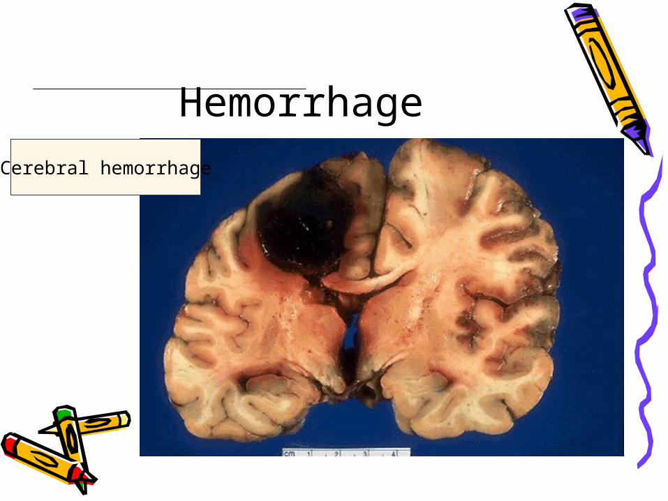
HemorrhageCerebral hemorrhage