Embed Size (px)
Citation preview
Edinburgh Research Explorer
High-pressure structural study of L-alpha-glutamine and the useof Hirshfeld surfaces and graph-set notation to investigate thehydrogen bonding present in the structure up to 4.9 GPa
Citation for published version:Lozano-Casal, P, Allan, DR & Parsons, S 2008, 'High-pressure structural study of L-alpha-glutamine andthe use of Hirshfeld surfaces and graph-set notation to investigate the hydrogen bonding present in thestructure up to 4.9 GPa' Acta Crystallographica Section B - Structural Science, vol 64, pp. 466-475. DOI:10.1107/S010876810801793X
Digital Object Identifier (DOI):10.1107/S010876810801793X
Link:Link to publication record in Edinburgh Research Explorer
Document Version:Publisher's PDF, also known as Version of record
Published In:Acta Crystallographica Section B - Structural Science
Publisher Rights Statement:Copyright © 2008 International Union of Crystallography; all rights reserved.
General rightsCopyright for the publications made accessible via the Edinburgh Research Explorer is retained by the author(s)and / or other copyright owners and it is a condition of accessing these publications that users recognise andabide by the legal requirements associated with these rights.
Take down policyThe University of Edinburgh has made every reasonable effort to ensure that Edinburgh Research Explorercontent complies with UK legislation. If you believe that the public display of this file breaches copyright pleasecontact [email protected] providing details, and we will remove access to the work immediately andinvestigate your claim.
Download date: 09. Jun. 2018
electronic reprint
Acta Crystallographica Section B
Structural Science,Crystal Engineeringand Materials
ISSN 2052-5192
High-pressure structural study of L-α-glutamine and the useof Hirshfeld surfaces and graph-set notation to investigate thehydrogen bonding present in the structure up to 4.9 GPa
P. Lozano-Casal, D. R. Allan and S. Parsons
Acta Cryst. (2008). B64, 466–475
Copyright c© International Union of Crystallography
Author(s) of this paper may load this reprint on their own web site or institutional repository provided thatthis cover page is retained. Republication of this article or its storage in electronic databases other than asspecified above is not permitted without prior permission in writing from the IUCr.
For further information see http://journals.iucr.org/services/authorrights.html
Acta Crystallographica Section B: Structural Science publishes papers in structural chem-istry and solid-state physics in which structure is the primary focus of the work reported.The central themes are the acquisition of structural knowledge from novel experimentalobservations or from existing data, the correlation of structural knowledge with physico-chemical and other properties, and the application of this knowledge to solve problemsin the structural domain. The journal covers metals and alloys, inorganics and minerals,metal-organics and purely organic compounds.
Crystallography Journals Online is available from journals.iucr.org
Acta Cryst. (2008). B64, 466–475 P. Lozano-Casal et al. · High-pressure structural study
research papers
466 doi:10.1107/S010876810801793X Acta Cryst. (2008). B64, 466–475
Acta Crystallographica Section B
StructuralScience
ISSN 0108-7681
High-pressure structural study of L-a-glutamine andthe use of Hirshfeld surfaces and graph-set notationto investigate the hydrogen bonding present in thestructure up to 4.9 GPa
P. Lozano-Casal,a* D. R. Allanb
and S. Parsonsc
aInstitute for Materials and Processes, School of
Science and Engineering, The University of
Edinburgh, King’s Buildings, Mayfield Road,
Edinburgh EH9 3JL, Scotland, bDiamond Light
Source, Harwell Science and Innovation
Campus, Chilton, Oxon OX11 ODU, England,
and cSchool of Chemistry and Centre for Science
at Extreme Conditions, The University of Edin-
burgh, King’s Buildings, West Mains Road,
Edinburgh EH9 3JJ, Scotland
Correspondence e-mail:
# 2008 International Union of Crystallography
Printed in Singapore – all rights reserved
The crystal structure of l-�-glutamine has been elucidated at
room temperature at pressures between 0 and 4.9 GPa by
using single-crystal high-pressure X-ray diffraction techniques.
The structure is primarily stabilized by five N—H� � �Ointermolecular interactions, which link molecules in a
herringbone-like layer arrangement, giving rise to voids
within the solid. The application of pressure on the structure
results in a reduction in the size of the voids, as a consequence
of the shortening of the N—H� � �O hydrogen bonds, which
compress to minimum N� � �O distances of around 2.6 A,
without driving the crystal structure to a phase transition. The
decrease in the hydrogen-bond distances is due to the
necessary stabilization of the structure, which arises from
molecules modifying their positions to optimize electrostatic
contacts and minimize the occupied space. Hirshfeld surfaces
and fingerprint plots have been used to rapidly assess the
structural changes that occur on application of pressure.
Received 13 February 2008
Accepted 12 June 2008
1. Introduction
l-�-Glutamine is an amino acid found in all forms of life. It is
classified as a semi-essential or conditionally essential amino
acid. l-�-Glutamine is very versatile, participating in many
reactions in the body and, for example, is important in the
regulation of acid–base balance. l-�-Glutamine participates in
the formation of purine and pyrimidine nucleotides, amino
sugars (such as glucosamine and l-glutamate) and other
amino acids (e.g. nicotinamide, adenine, dinucleotide and
glutathione). It also participates in protein synthesis, energy
production and the formation of d-glucose and glycogen.
Importantly, l-�-glutamine can serve as the primary respira-
tory substrate for the production of energy in enterocytes and
lymphocytes. It is considered to be an immunonutrient and
supplementary l-�-glutamine is used in medical foods for such
stress situations as trauma, cancer, infections and burns
(Skubitz & Anderson, 1996; Anderson et al., 1998).
The first structural studies performed on l-�-glutamine
were published by Cochran & Penfold (1952) and Koetzle et
al. (1973), later followed by the analysis of the charge density
and the topology of the crystal structure, reported by Wagner
& Luger (2001). The l-�-glutamine structure is formed from
five unique and significantly bent N—H� � �O hydrogen bonds,
one for each H atom attached to the nitrogen, which stabilize
the structure to form a three-dimensional arrangement of l-�-
glutamine molecules linked by a complicated hydrogen-bond
network. Owing to the limited description of the crystal
structure of l-�-glutamine available in the literature, a more
exhaustive study of the molecular packing was performed as
part of the work presented here, allowing us to provide a
better description of the l-�-glutamine structure.
electronic reprint
The work presented is one of a number of investigations we
are conducting on the effect of pressure on the crystal struc-
tures of different organic and biological compounds such as
acetone, cyclopropylamine, ethanol, methanol, l-serine and l-
cysteine, among others (Lozano-Casal et al., 2005; Allan et al.,
1998, 2001; Allen & Clark, 1999; Moggach, Allan et al., 2005;
Moggach, Clark & Parsons, 2005). The aim of these studies is
to understand how the various inter- and intramolecular
interactions respond to pressure, which can often result in the
formation of new high-pressure polymorphs. Indeed, the
degree to which bond compressibility can be explained and
the extent to which a structure can be compressed before a
phase transition takes place are profoundly important ques-
tions in the field of high-pressure structural chemistry.
Strong hydrogen bonds are rare in biological structures
since they are very rigid and not easily broken (D� � �A
distance less than 2.5 A and a D—H� � �A angle close to 180�,
where D and A are the donor and acceptor atoms, respec-
tively), and can hinder processes such as protein folding or
unfolding. On the other hand, the salt-bridge intermolecular
hydrogen bond, N+—H� � ��O C, which is present in l-�-
glutamine, is one of the two strongest intermolecular inter-
actions that exist in biological compounds, the other being P—
OH� � �O P. It exists in nucleic acids, owing to the strong
electrostatic component of the interaction, arising from the
charged N and O atoms in the zwitterionic molecules (Steiner,
2002).
This paper is organized as follows. First, we perform a
structural analysis of the l-�-glutamine structure by using
high-pressure X-ray diffraction techniques to investigate how
the crystal packing reacts to increasing pressure. Then the
most important structural changes with pressure are studied
research papers
Acta Cryst. (2008). B64, 466–475 P. Lozano-Casal et al. � High-pressure structural study 467
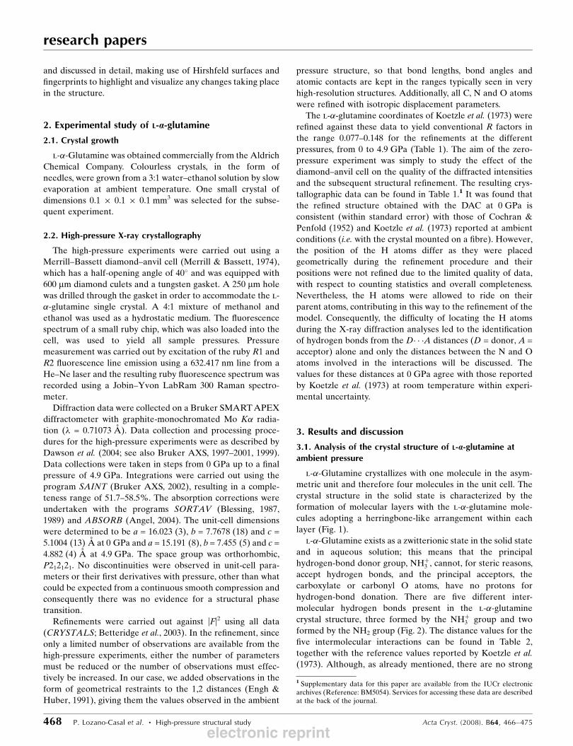
Table 1Crystallographic data for l-�-glutamine at increasing pressures.
0 GPa 0.1 GPa 1.4 GPa 4.9 GPa
Crystal dataChemical formula C5H10N2O3 C5H10N2O3 C5H10N2O3 C5H10N2O3
Mr 146.15 146.15 146.15 146.15Cell setting, space group Orthorhombic, P212121 Orthorhombic, P212121 Orthorhombic, P212121 Orthorhombic, P212121
Temperature (K) 293 293 293 293Z 4 4 4 4Dx (Mg m�3) 1.529 1.536 1.616 1.756Radiation type Mo K� Mo K� Mo K� Mo K�� (mm�1) 0.13 0.13 0.13 0.15Crystal form, colour Block, colourless Block, colourless Block, colourless Block, colourless
Data collectionDiffractometer Bruker SMART Bruker SMART Bruker SMART Bruker SMARTData collection method ’ and ! scans ’ and ! scans ’ and ! scans ’ and ! scansAbsorption correction Multi-scan† Multi-scan† Multi-scan† Multi-scan†Tmin 0.407 0.506 0.657 0.722Tmax 1.000 1.000 1.000 1.000
No. of measured, indepen-dent and observed reflec-tions
337, 326, 202 336, 324, 205 305, 295, 202 292, 283, 182
Criterion for observed reflec-tions
I > 2�(I) I > 2�(I) I > 2�(I) I > 2�(I)
Rint 0.184 0.224 0.155 0.214�max (�) 23.2 23.3 23.3 23.2
RefinementRefinement on F2 F2 F2 F2
R[F2 > 2�(F2)], wR(F2), S 0.105, 0.263, 1.05 0.148, 0.284, 1.16 0.107, 0.243, 1.04 0.114, 0.183, 1.10No. of reflections 297 267295 330 278No. of parameters 41 41 41 41H-atom treatment Fixed Not refined Mixture‡ Mixture‡Weighting scheme w = q/[�2(F*) + (P(1)p)2 + P(2)p
+ P(4) + P(5)sin �]; 0.106,4.74, 0.00, 0.00, 0.00, 0.333
P = P(6)*max(F2o ,0) + (1 �
P(6))Fc2; w = 1/[�2(F*) +
(P(1)p)2 + P(2)p + P(4) +P(5)sin �]; P(i) are: 0.00,11.6, 0.00, 0.00, 0.00, 0.333
w = q/[�2(F*) + (P(1)p)2 + P(2)p+ P(4) + P(5)sin �]0.897E�01, 4.49, 0.00, 0.00,0.00, 0.333
w = q/[�2(F*) + (P(1)p)2 + P(2)p+ P(4) + P(5)sin �], 0.00,3.26, 0.00, 0.00, 0.00, 0.333
(�/�)max < 0.0001 < 0.0001 < 0.0001 < 0.0001��max, ��min (e A�3) 0.57, �0.54 1.01, �0.76 0.76, �0.55 1.00, �1.05
Computer programs used: SMART (Bruker AXS, 1997–2001), SAINT (Bruker AXS, 2002), SIR92 (Altomare et al., 1993), CRYSTALS (Watkin et al., 1996), CRYSTALS (Betteridge etal. 2003), CAMERON (Watkin et al., 1996). † Based on symmetry-related measurements. ‡ Mixture of independent and constrained refinement.
electronic reprint
and discussed in detail, making use of Hirshfeld surfaces and
fingerprints to highlight and visualize any changes taking place
in the structure.
2. Experimental study of L-a-glutamine
2.1. Crystal growth
l-�-Glutamine was obtained commercially from the Aldrich
Chemical Company. Colourless crystals, in the form of
needles, were grown from a 3:1 water–ethanol solution by slow
evaporation at ambient temperature. One small crystal of
dimensions 0.1 � 0.1 � 0.1 mm3 was selected for the subse-
quent experiment.
2.2. High-pressure X-ray crystallography
The high-pressure experiments were carried out using a
Merrill–Bassett diamond–anvil cell (Merrill & Bassett, 1974),
which has a half-opening angle of 40� and was equipped with
600 mm diamond culets and a tungsten gasket. A 250 mm hole
was drilled through the gasket in order to accommodate the l-
�-glutamine single crystal. A 4:1 mixture of methanol and
ethanol was used as a hydrostatic medium. The fluorescence
spectrum of a small ruby chip, which was also loaded into the
cell, was used to yield all sample pressures. Pressure
measurement was carried out by excitation of the ruby R1 and
R2 fluorescence line emission using a 632.417 nm line from a
He–Ne laser and the resulting ruby fluorescence spectrum was
recorded using a Jobin–Yvon LabRam 300 Raman spectro-
meter.
Diffraction data were collected on a Bruker SMART APEX
diffractometer with graphite-monochromated Mo K� radia-
tion (� = 0.71073 A). Data collection and processing proce-
dures for the high-pressure experiments were as described by
Dawson et al. (2004; see also Bruker AXS, 1997–2001, 1999).
Data collections were taken in steps from 0 GPa up to a final
pressure of 4.9 GPa. Integrations were carried out using the
program SAINT (Bruker AXS, 2002), resulting in a comple-
teness range of 51.7–58.5%. The absorption corrections were
undertaken with the programs SORTAV (Blessing, 1987,
1989) and ABSORB (Angel, 2004). The unit-cell dimensions
were determined to be a = 16.023 (3), b = 7.7678 (18) and c =
5.1004 (13) A at 0 GPa and a = 15.191 (8), b= 7.455 (5) and c =
4.882 (4) A at 4.9 GPa. The space group was orthorhombic,
P212121. No discontinuities were observed in unit-cell para-
meters or their first derivatives with pressure, other than what
could be expected from a continuous smooth compression and
consequently there was no evidence for a structural phase
transition.
Refinements were carried out against |F|2 using all data
(CRYSTALS; Betteridge et al., 2003). In the refinement, since
only a limited number of observations are available from the
high-pressure experiments, either the number of parameters
must be reduced or the number of observations must effec-
tively be increased. In our case, we added observations in the
form of geometrical restraints to the 1,2 distances (Engh &
Huber, 1991), giving them the values observed in the ambient
pressure structure, so that bond lengths, bond angles and
atomic contacts are kept in the ranges typically seen in very
high-resolution structures. Additionally, all C, N and O atoms
were refined with isotropic displacement parameters.
The l-�-glutamine coordinates of Koetzle et al. (1973) were
refined against these data to yield conventional R factors in
the range 0.077–0.148 for the refinements at the different
pressures, from 0 to 4.9 GPa (Table 1). The aim of the zero-
pressure experiment was simply to study the effect of the
diamond–anvil cell on the quality of the diffracted intensities
and the subsequent structural refinement. The resulting crys-
tallographic data can be found in Table 1.1 It was found that
the refined structure obtained with the DAC at 0 GPa is
consistent (within standard error) with those of Cochran &
Penfold (1952) and Koetzle et al. (1973) reported at ambient
conditions (i.e. with the crystal mounted on a fibre). However,
the position of the H atoms differ as they were placed
geometrically during the refinement procedure and their
positions were not refined due to the limited quality of data,
with respect to counting statistics and overall completeness.
Nevertheless, the H atoms were allowed to ride on their
parent atoms, contributing in this way to the refinement of the
model. Consequently, the difficulty of locating the H atoms
during the X-ray diffraction analyses led to the identification
of hydrogen bonds from the D� � �A distances (D = donor, A =
acceptor) alone and only the distances between the N and O
atoms involved in the interactions will be discussed. The
values for these distances at 0 GPa agree with those reported
by Koetzle et al. (1973) at room temperature within experi-
mental uncertainty.
3. Results and discussion
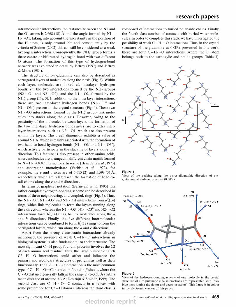
3.1. Analysis of the crystal structure of L-a-glutamine atambient pressure
l-�-Glutamine crystallizes with one molecule in the asym-
metric unit and therefore four molecules in the unit cell. The
crystal structure in the solid state is characterized by the
formation of molecular layers with the l-�-glutamine mole-
cules adopting a herringbone-like arrangement within each
layer (Fig. 1).
l-�-Glutamine exists as a zwitterionic state in the solid state
and in aqueous solution; this means that the principal
hydrogen-bond donor group, NHþ3 , cannot, for steric reasons,
accept hydrogen bonds, and the principal acceptors, the
carboxylate or carbonyl O atoms, have no protons for
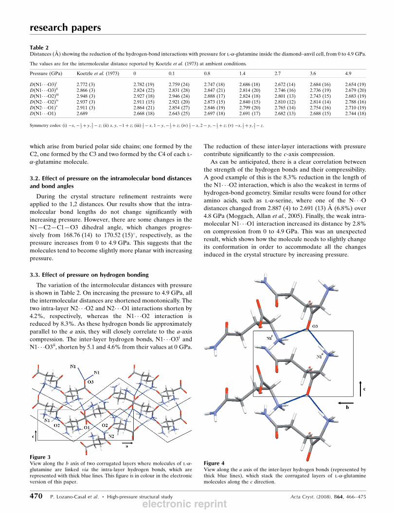
hydrogen-bond donation. There are five different inter-
molecular hydrogen bonds present in the l-�-glutamine
crystal structure, three formed by the NHþ3 group and two
formed by the NH2 group (Fig. 2). The distance values for the
five intermolecular interactions can be found in Table 2,
together with the reference values reported by Koetzle et al.
(1973). Although, as already mentioned, there are no strong
research papers
468 P. Lozano-Casal et al. � High-pressure structural study Acta Cryst. (2008). B64, 466–475
1 Supplementary data for this paper are available from the IUCr electronicarchives (Reference: BM5054). Services for accessing these data are describedat the back of the journal.
electronic reprint
intramolecular interactions, the distance between the N1 and
the O1 atoms is 2.668 (18) A and the angle formed by N1—
H� � �O1, taking into account the uncertainty in the position of
the H atom, is only around 90� and consequently by the
criteria of Steiner (2002) this can still be considered as a weak
hydrogen interaction. Consequently, the NHþ3 group forms a
three-centre or bifurcated hydrogen bond with two different
O atoms. The formation of this type of hydrogen-bond
network was explained in detail by Jeffrey (1997) and Jeffrey
& Mitra (1984).
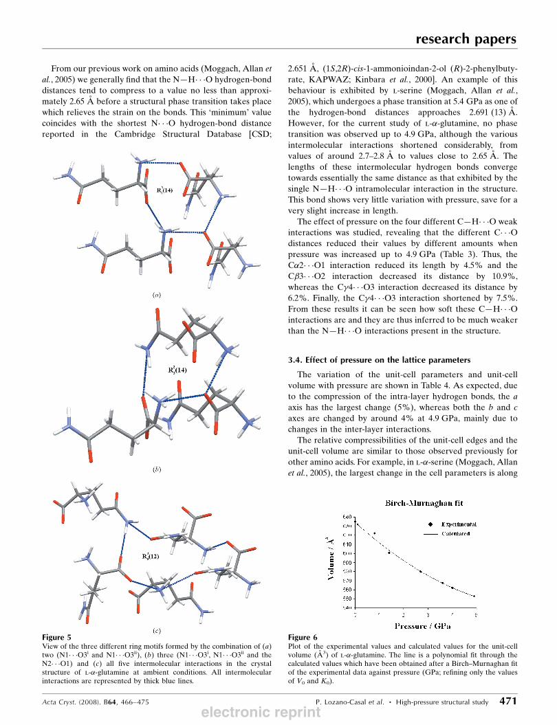
The structure of l-�-glutamine can also be described as
corrugated layers of molecules along the a axis (Fig. 3). Within
each layer, molecules are linked via intralayer hydrogen
bonds: via the two interactions formed by the NH2 groups
(N2� � �O1 and N2� � �O2), and the N1� � �O2, formed by the
NHþ3 group (Fig. 3). In addition to the intra-layer interactions,
there are two inter-layer hydrogen bonds (N1� � �O3i and
N1� � �O3ii) present in the crystal structure (Fig. 4). These two
N1� � �O3 interactions, formed by the NHþ3 group, link mole-
cules into stacks along the c axis. However, owing to the
proximity of the molecules between layers, the formation of
the two inter-layer hydrogen bonds gives rise to extra inter-
layer interactions, such as N2� � �O1, which are also present
within the layers. The c cell dimension exhibits a value of
around 5.1 A, which is mainly associated with the formation of
two head-to-head hydrogen bonds [N1� � �O3i and N1� � �O3ii],
which actively participate in the stacking of layers along this
direction. This feature is also present in other amino acids,
where molecules are arranged in different chain motifs formed
by N—H� � �OOC interactions. In serine (Benedetti et al., 1973)
and asparagine monohydrate (Verbist et al., 1972), for
example, the c and a axes are of 5.615 (2) and 5.593 (5) A,
respectively, which are related with the formation of head-to-
tail chains along the c and a directions.
In terms of graph-set notation (Bernstein et al., 1995) this
rather complex hydrogen-bonding scheme can be described in
terms of three neighbouring, and coupled, rings (Fig. 5). Thus,
the N1� � �O3i, N1� � �O3ii and N2� � �O1 interactions form R23ð14Þ
rings, which link molecules to form the layers running along
the c direction, whereas the N1� � �O3i, N1� � �O3ii and N2� � �O2
interactions form R33ð14Þ rings, to link molecules along the a
and b directions. Finally, the five different intermolecular
interactions can be combined to form R33ð12Þ rings to form the
corrugated layers, which run along the a and c directions.
Apart from the strong electrostatic interactions already
mentioned, the presence of weak C—H� � �O interactions in
biological systems is also fundamental to their structure. The
most significant C—H group found in proteins involves the C2
of each amino acid residue. Thus, the large number of such
C2—H� � �O interactions could affect and influence the
primary and secondary structures of proteins as well as their
functionality. The C2—H� � �O interaction is the most common
type of C—H� � �O C interaction found in � sheets, where the
C� � �O distance generally falls in the range 2.91–3.50 A (with a
mean distance of around 3.3 A; Desiraju & Steiner, 1999). The
second class are C—H� � �O C contacts in �-helices with
some preference for C3—H donors, whereas the third class is
composed of interactions to buried polar-side chains. Finally,
the fourth class consists of contacts with buried water mole-
cules. In order to complete this study, we have investigated the
possibility of weak C—H� � �O interactions. Thus, in the crystal
structure of l-�-glutamine at 0 GPa presented in this work,
there are four C—H� � �O interactions (where the O atom
belongs both to the carboxylic and amide groups; Table 3),
research papers
Acta Cryst. (2008). B64, 466–475 P. Lozano-Casal et al. � High-pressure structural study 469
Figure 2View of the hydrogen-bonding scheme of one molecule in the crystalstructure of l-�-glutamine (the interactions are represented with thickblue lines joining the donor and acceptor atoms). This figure is in colourin the electronic version of this paper.
Figure 1View of the packing along the c-crystallographic direction of l-�-glutamine at ambient pressure (0 GPa).
electronic reprint
which arise from buried polar side chains; one formed by the
C2, one formed by the C3 and two formed by the C4 of each l-
�-glutamine molecule.
3.2. Effect of pressure on the intramolecular bond distancesand bond angles
During the crystal structure refinement restraints were
applied to the 1,2 distances. Our results show that the intra-
molecular bond lengths do not change significantly with
increasing pressure. However, there are some changes in the
N1—C2—C1—O3 dihedral angle, which changes progres-
sively from 168.76 (14) to 170.52 (15)�, respectively, as the
pressure increases from 0 to 4.9 GPa. This suggests that the
molecules tend to become slightly more planar with increasing
pressure.
3.3. Effect of pressure on hydrogen bonding
The variation of the intermolecular distances with pressure
is shown in Table 2. On increasing the pressure to 4.9 GPa, all
the intermolecular distances are shortened monotonically. The
two intra-layer N2� � �O2 and N2� � �O1 interactions shorten by
4.2%, respectively, whereas the N1� � �O2 interaction is
reduced by 8.3%. As these hydrogen bonds lie approximately
parallel to the a axis, they will closely correlate to the a-axis
compression. The inter-layer hydrogen bonds, N1� � �O3i and
N1� � �O3ii, shorten by 5.1 and 4.6% from their values at 0 GPa.
The reduction of these inter-layer interactions with pressure
contribute significantly to the c-axis compression.
As can be anticipated, there is a clear correlation between
the strength of the hydrogen bonds and their compressibility.
A good example of this is the 8.3% reduction in the length of
the N1� � �O2 interaction, which is also the weakest in terms of
hydrogen-bond geometry. Similar results were found for other
amino acids, such as l-�-serine, where one of the N� � �Odistances changed from 2.887 (4) to 2.691 (13) A (6.8%) over
4.8 GPa (Moggach, Allan et al., 2005). Finally, the weak intra-
molecular N1� � �O1 interaction increased its distance by 2.8%
on compression from 0 to 4.9 GPa. This was an unexpected
result, which shows how the molecule needs to slightly change
its conformation in order to accommodate all the changes
induced in the crystal structure by increasing pressure.
research papers
470 P. Lozano-Casal et al. � High-pressure structural study Acta Cryst. (2008). B64, 466–475
Table 2Distances (A) showing the reduction of the hydrogen-bond interactions with pressure for l-�-glutamine inside the diamond–anvil cell, from 0 to 4.9 GPa.
The values are for the intermolecular distance reported by Koetzle et al. (1973) at ambient conditions.
Pressure (GPa) Koetzle et al. (1973) 0 0.1 0.8 1.4 2.7 3.6 4.9
D(N1� � �O3)i 2.772 (3) 2.782 (19) 2.759 (24) 2.747 (18) 2.686 (18) 2.672 (14) 2.684 (16) 2.654 (19)D(N1� � �O3)ii 2.866 (3) 2.824 (22) 2.831 (28) 2.847 (21) 2.814 (20) 2.746 (16) 2.736 (19) 2.679 (20)D(N1� � �O2)iii 2.948 (3) 2.927 (18) 2.946 (24) 2.888 (17) 2.824 (18) 2.801 (13) 2.743 (15) 2.683 (19)D(N2� � �O2)iv 2.937 (3) 2.911 (15) 2.921 (20) 2.873 (15) 2.840 (15) 2.810 (12) 2.814 (14) 2.788 (16)D(N2� � �O1)v 2.911 (3) 2.864 (21) 2.854 (27) 2.846 (19) 2.799 (20) 2.765 (14) 2.754 (16) 2.710 (19)D(N1� � �O1) 2.689 2.668 (18) 2.643 (25) 2.697 (18) 2.691 (17) 2.682 (13) 2.688 (15) 2.744 (18)
Symmetry codes: (i) �x;� 12 þ y; 5
2 � z; (ii) x; y;�1 þ z; (iii) 12 � x; 1 � y;� 1
2 þ z; (iv) 12 � x; 2 � y;� 1
2 þ z; (v) �x; 12 þ y; 3
2 � z.
Figure 4View along the a axis of the inter-layer hydrogen bonds (represented bythick blue lines), which stack the corrugated layers of l-�-glutaminemolecules along the c direction.
Figure 3View along the b axis of two corrugated layers where molecules of l-�-glutamine are linked via the intra-layer hydrogen bonds, which arerepresented with thick blue lines. This figure is in colour in the electronicversion of this paper.
electronic reprint
From our previous work on amino acids (Moggach, Allan et
al., 2005) we generally find that the N—H� � �O hydrogen-bond
distances tend to compress to a value no less than approxi-
mately 2.65 A before a structural phase transition takes place
which relieves the strain on the bonds. This ‘minimum’ value
coincides with the shortest N� � �O hydrogen-bond distance
reported in the Cambridge Structural Database [CSD;
2.651 A, (1S,2R)-cis-1-ammonioindan-2-ol (R)-2-phenylbuty-
rate, KAPWAZ; Kinbara et al., 2000]. An example of this
behaviour is exhibited by l-serine (Moggach, Allan et al.,
2005), which undergoes a phase transition at 5.4 GPa as one of
the hydrogen-bond distances approaches 2.691 (13) A.
However, for the current study of l-�-glutamine, no phase
transition was observed up to 4.9 GPa, although the various
intermolecular interactions shortened considerably, from
values of around 2.7–2.8 A to values close to 2.65 A. The
lengths of these intermolecular hydrogen bonds converge
towards essentially the same distance as that exhibited by the
single N—H� � �O intramolecular interaction in the structure.
This bond shows very little variation with pressure, save for a
very slight increase in length.
The effect of pressure on the four different C—H� � �O weak
interactions was studied, revealing that the different C� � �Odistances reduced their values by different amounts when
pressure was increased up to 4.9 GPa (Table 3). Thus, the
C�2� � �O1 interaction reduced its length by 4.5% and the
C�3� � �O2 interaction decreased its distance by 10.9%,
whereas the C4� � �O3 interaction decreased its distance by
6.2%. Finally, the C4� � �O3 interaction shortened by 7.5%.
From these results it can be seen how soft these C—H� � �Ointeractions are and they are thus inferred to be much weaker
than the N—H� � �O interactions present in the structure.
3.4. Effect of pressure on the lattice parameters
The variation of the unit-cell parameters and unit-cell
volume with pressure are shown in Table 4. As expected, due
to the compression of the intra-layer hydrogen bonds, the a
axis has the largest change (5%), whereas both the b and c
axes are changed by around 4% at 4.9 GPa, mainly due to
changes in the inter-layer interactions.
The relative compressibilities of the unit-cell edges and the
unit-cell volume are similar to those observed previously for
other amino acids. For example, in l-�-serine (Moggach, Allan
et al., 2005), the largest change in the cell parameters is along
research papers
Acta Cryst. (2008). B64, 466–475 P. Lozano-Casal et al. � High-pressure structural study 471
Figure 5View of the three different ring motifs formed by the combination of (a)two (N1� � �O3i and N1� � �O3ii), (b) three (N1� � �O3i, N1� � �O3ii and theN2� � �O1) and (c) all five intermolecular interactions in the crystalstructure of l-�-glutamine at ambient conditions. All intermolecularinteractions are represented by thick blue lines.
Figure 6Plot of the experimental values and calculated values for the unit-cellvolume (A3) of l-�-glutamine. The line is a polynomial fit through thecalculated values which have been obtained after a Birch–Murnaghan fitof the experimental data against pressure (GPa; refining only the valuesof V0 and K0).
electronic reprint
the b direction (6.2%), which suffers three times the change
along the a and c directions (2.6 and 2.1%). The corresponding
change in the unit-cell volume was of 11% over 4.8 GPa,
which is similar to the 13% reduction in unit-cell volume
found here for l-�-glutamine over approximately the same
pressure range.
Finally, the program EOSFIT5.2 (Angel, 2002) was used to
fit the pressure dependence of the unit-cell volume of l-�-
glutamine with the Birch–Murnaghan equation-of-state.
Given the relatively low number of observations, k 0 was fixed
to a value of 4.0 and k00 was set at �0.01498 in order that the
higher-order terms of the equation were eliminated. The
refined values for V0 and k0 are 635 (2) A3 and 260 (11) GPa,
respectively. The value of V0 obtained from this least-squares
refinement is in good agreement with the measured ambient
pressure value [634.8 (2) A3] and the value of k0 is very similar
to that observed in other amino acid systems [e.g. l-�-aspar-
agine monohydrate, 176 (9) GPa]. Finally, a plot of the unit-
cell volume versus pressure, including the Birch–Murnaghan
fit, is shown in Fig. 6.
3.5. Comparison of the ambientpressure and high-pressure crystalstructures of L-a-glutamine:Hirshfeld surfaces
The program CrystalExplorer
(Wolff et al., 2005) is a recently
developed tool that allows the use
of Hirshfeld surfaces to partition
crystal space in order to explore
packing modes and intermolecular
interactions in molecular crystals
(McKinnon et al., 2004). We have
used this program to visualize the
structure of l-�-glutamine at
ambient pressure (i.e. 0 GPa) and
at high pressure (i.e. 4.9 GPa) in
order to make a more detailed
comparison between them.
Hirshfeld surfaces (McKinnon et
al., 2004) are shown for the struc-
ture of l-�-glutamine at ambient
pressure and high pressure in Fig. 7.
It can be seen how the three H
atoms of the NHþ3 group and the
two H atoms of the NH2 group
actively participate in the forma-
tion of hydrogen bonding. This is
shown by the orange–red region on
the surface adjacent to the O
atoms, which act as acceptors.
From Fig. 7 it is possible to see
research papers
472 P. Lozano-Casal et al. � High-pressure structural study Acta Cryst. (2008). B64, 466–475
Table 3Donor-to-acceptor atom distances (A) for the four most significant C� � �Ointermolecular interactions present in the crystal structure of l-�-glutamine.
These values arose from the data collected on a crystal inside the diamond–anvil cell, at 0 GPa.
Pressure (GPa) 0 4.9(C� � �O) interaction D(C� � �O) (A)
C�(C2)� � �O1 3.54 (2) 3.38 (2)C�(C3)� � �O2 3.44 (3) 3.07 (3)C(C4)� � �O3i 3.43 (3) 3.22 (2)C(C4)� � �O3ii 3.518 (18) 3.254 (18)
Symmetry codes: (i) �x;� 12 þ y; 5
2 � z; (ii) x; y;�1 þ z.
Table 4Lattice parameters obtained in the l-�-glutamine X-ray diffractionexperiments between ambient pressure and 4.9 GPa.
The values obtained by Koetzle et al. (1973) are also given for comparison.
Pressure (GPa) a (A) b (A) c (A) V (A3)
Koetzle et al. (1973) 16.020 (10) 7.762 (6) 5.119 (4) 636.50 16.023 (3) 7.7678 (18) 5.1004 (3) 634.8 (2)0.1 15.992 (2) 7.7558 (12) 5.0941 (9) 631.83 (17)0.8 15.879 (6) 7.705 (3) 5.084 (2) 622.0 (4)1.4 15.679 (11) 7.628 (6) 5.023 (5) 600.8 (9)2.7 15.450 (8) 7.55 (6) 4.972 (5) 580.0 (7)3.6 15.328 (7) 7.497 (5) 4.941 (4) 567.8 (6)4.9 15.191 (8) 7.455 (5) 4.882 (14) 552.8 (6)
Figure 7Hirshfeld surfaces for the ambient pressure (top) and high-pressure (bottom) structures of l-�-glutamine.Each molecule is shown with the Hirshfeld surface mapped with de [left; for this series mapped between1.0 (red) and 2.0 A (blue)] and dnorm [(mapped between �0.66 (red) and �0.89 (blue)], where de is thedistance to the nearest atom centre exterior to the surface and dnorm is the normalized contact distance,which takes into account the van der Waals radio of the atoms. (The different interactions are labelled 1–5, as shown in the text: (1) N1� � �O3i, (2) N1� � �O3ii, (3) N1� � �O2, (4) N2� � �O2, (5) N2� � �O1.
electronic reprint
significant differences in the
hydrogen bonding between the
ambient and high-pressure structures.
One of the main differences is the
shortening of the hydrogen bonds at
high pressure, which is illustrated by
an increase in the redness of the
contact areas (yellow–orange) in the
de surface as well as the formation of
other intermolecular interactions
(extra red regions in high-pressure
surfaces), which only become signifi-
cant when they shorten. In addition
to this, the voids ‘close up’ on pres-
sure increase. This can be seen by
comparing the blue areas in the de
surface, which are much larger in the
ambient pressure structure than those
at high pressure. Additionally, Fig. 7
illustrates a second type of surface,
known as the dnorm surface, which
includes the van der Waals (vdW)
radius of the internal and external
atoms involved in the contact. In
these surfaces, red areas highlight
shorter contacts, white represent
contacts around the vdW separation
and blue is for longer contacts. In
contrast to de surfaces, the dnorm
surface highlights both donor and
acceptor equally. These differences
are also shown by the total and partial
fingerprint plots (McKinnon et al.,
2007) in Fig. 8.
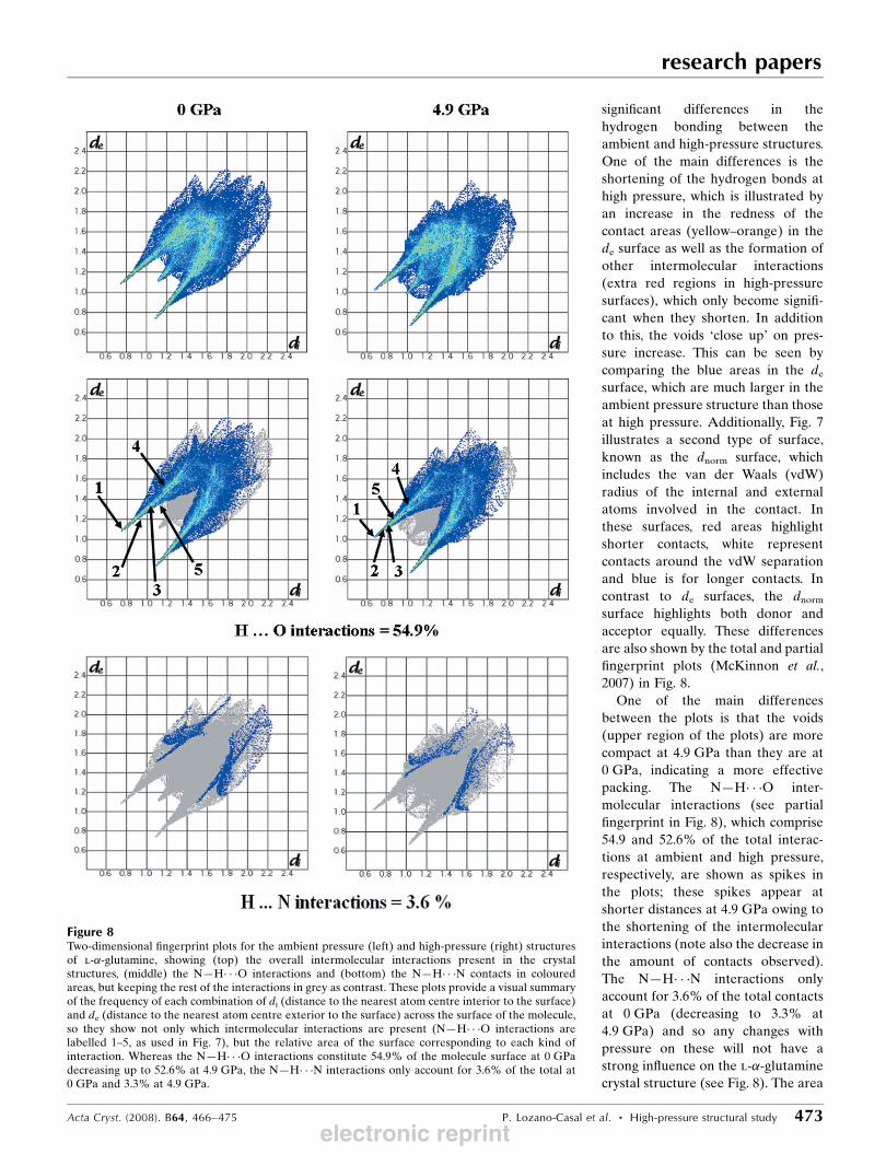
One of the main differences
between the plots is that the voids
(upper region of the plots) are more
compact at 4.9 GPa than they are at
0 GPa, indicating a more effective
packing. The N—H� � �O inter-
molecular interactions (see partial
fingerprint in Fig. 8), which comprise
54.9 and 52.6% of the total interac-
tions at ambient and high pressure,
respectively, are shown as spikes in
the plots; these spikes appear at
shorter distances at 4.9 GPa owing to
the shortening of the intermolecular
interactions (note also the decrease in
the amount of contacts observed).
The N—H� � �N interactions only
account for 3.6% of the total contacts
at 0 GPa (decreasing to 3.3% at
4.9 GPa) and so any changes with
pressure on these will not have a
strong influence on the l-�-glutamine
crystal structure (see Fig. 8). The area
research papers
Acta Cryst. (2008). B64, 466–475 P. Lozano-Casal et al. � High-pressure structural study 473
Figure 8Two-dimensional fingerprint plots for the ambient pressure (left) and high-pressure (right) structuresof l-�-glutamine, showing (top) the overall intermolecular interactions present in the crystalstructures, (middle) the N—H� � �O interactions and (bottom) the N—H� � �N contacts in colouredareas, but keeping the rest of the interactions in grey as contrast. These plots provide a visual summaryof the frequency of each combination of di (distance to the nearest atom centre interior to the surface)and de (distance to the nearest atom centre exterior to the surface) across the surface of the molecule,so they show not only which intermolecular interactions are present (N—H� � �O interactions arelabelled 1–5, as used in Fig. 7), but the relative area of the surface corresponding to each kind ofinteraction. Whereas the N—H� � �O interactions constitute 54.9% of the molecule surface at 0 GPadecreasing up to 52.6% at 4.9 GPa, the N—H� � �N interactions only account for 3.6% of the total at0 GPa and 3.3% at 4.9 GPa.
electronic reprint
between the spikes corresponds to C—H� � �O interactions and
the spike within the middle area is representative of short
H� � �H contacts. From the plots it is possible to see how the
weak C—H� � �O interactions shorten when pressure is applied
to the l-�-glutamine crystal structure. This can be seen from
the increment of the contacts in the area characteristic for C—
H� � �O contacts (i.e. area between the spikes). In addition to
this, the spikes characteristic of the N—H� � �O interactions
shortened slightly owing to the decrease in the hydrogen-bond
distances.
4. Conclusions
High-pressure X-ray diffraction techniques were used to study
the compressibility of the l-�-glutamine crystal structure, from
0 to 4.9 GPa. Pressure induces the different intra- and inter-
layer hydrogen-bond distances to shorten by varying amounts
and changes ranging between 8.3 and 4.2% were observed.
The hydrogen bond which is reduced the most (8.3%) is
N1� � �O2, followed by changes in N1� � �O3i (5.1%), N1� � �O3ii
(4.6%), N2� � �O2 and N2� � �O1 (4.2%). Consequently, the
molecules within layers are pushed together and the layers are
compressed along the c axis. The reduction in the hydrogen-
bond distances can also be described using the Hirshfeld
surfaces and fingerprint plots presented in this work. Addi-
tionally, significant changes occurred on the weak C—H� � �Ointeractions, with C� � �O distances shortened by diverse
amounts, from 4.5 to 10.9% of their values at 0 GPa, therefore
giving an insight of their soft nature.
The unit-cell volume data of l-�-glutamine, from 0 to
4.9 GPa, were fitted to the Birch–Murnaghan equation-of-
state. Thus, values for the V0 and k0 were found to be
635 (2) A3 and 260 (11), respectively. The value of k0 is similar
to the value obtained from a high-pressure study of l-�-
asparagine monohydrate and falls broadly within the range of
other hydrogen-bonded systems.
l-�-Glutamine does not undergo a phase transition up to a
pressure of 4.9 GPa, as was also found to be the case for l-�-
aspartic acid up to a similar pressure (Lozano-Casal, 2006).
However, the much simpler amino acid, l-serine, does
undergo a phase transition at about 4.8 GPa, to a previously
unobserved polymorph (Moggach, Allan et al., 2005). A
reason for this can be found in the shape and size of the
molecules. For example, the smallest amino acid, glycine,
which forms simple hydrogen-bond networks, presents several
phase transitions (Boldyreva, 2003, 2004; Boldyreva et al.,
2004; Dawson et al., 2005). However, for larger amino acids
such as l-�-glutamine and l-�-aspartic acid, this is not found
to be the case. Owing to the flexibility of the molecules, the
presence of several hydrogen donor and acceptor atoms, and
the necessity to form multiple hydrogen bonds to hold the
crystal structure together, these larger molecules have diffi-
culty achieving a minimum in their packing energy in order to
go through a phase transition, probably due to kinetic impe-
diments. Nevertheless, in the compression of the structure of
l-�-glutamine, the ‘limit’ distance was reached but no phase
transition took place. This question of how much pressure is
required to drive the structure to a new polymorph is thus still
open.
References
Allan, D. R. & Clark, S. J. (1999). Phys. Rev. B, 60, 6328—6334.Allan, D. R., Clark, S. J., Brugmans, M. J. P., Ackland, G. J. & Vos,
W. L. (1998). Phys. Rev. B, 58, R11809–R11812.Allan, D. R., Parsons, S. & Teat, S. J. (2001). J. Synchrotron Rad. 8, 10–
17.Altomare, A., Cascarano, G., Giacovazzo, C. & Guagliardi, A. (1993).J. Appl. Cryst. 26, 343–350.
Anderson, P. M., Ramsay, N. K., Shu, X. O., Rydholm, N.,Rogosheske, J., Nicklow, R., Weisdorf, D. J. & Skubitz, K. M.(1998). Bone Marrow Transplant. 22, 339–344.
Angel, R. J. (2002). EOSFIT5.2. Virginia Tech, Blacksburgh, USA.Angel, R. J. (2004). J. Appl. Cryst. 37, 486–492.Benedetti, E., Pedone, C. & Sirigu, A. (1973). Gazz. Chim. Ital. 103,
555–561.Bernstein, J., Davis, R. E., Shimoni, L. & Chang, N. L. (1995). Angew.Chem. Int. Ed. Engl. 34, 1555–1573.
Betteridge, P. W., Carruthers, J. R., Cooper, R. I., Prout, K. & Watkin,D. J. (2003). J. Appl. Cryst. 36, 1487.
Blessing, R. H. (1987). Cryst. Rev. 1, 3–58.Blessing, R. H. (1989). J. Appl. Cryst. 22, 396–397.Boldyreva, E. V. (2003). J. Mol. Struct. 647, 159–179.Boldyreva, E. V. (2004). Cryst. Eng. 6, 235–254.Boldyreva, E. V., Ivashevskaya, S. N., Sowa, H., Ashbahs, H. &
Weber, H. P. (2004). Dokl. Phys. Chem. 396, 111–114.Bruker AXS (1997–2001). SMART, Version 5.049–5.059. Bruker-
AXS, Madison, Wisconsin, USA.Bruker AXS (1999). GEMINI, Version 1.01. Bruker-AXS, Madison,
Wisconsin, USA.Bruker AXS (2002). SAINT, Version 6. Bruker-AXS, Madison,
Wisconsin, USA.Cochran, W. & Penfold, B. R. (1952). Acta Cryst. 5, 644–653.Dawson, A., Allan, D. R., Belmonte, S. A., Clark, S. J., David, W. I. F.,
McGregor, P. A., Parsons, S., Pulham, C. R. & Sawyer, L. (2005).Cryst. Growth Des. 5, 1415–1427.
Dawson, A., Allan, D. R., Parsons, S. & Ruf, M. (2004). J. Appl. Cryst.37, 410–416.
Desiraju, G. R. & Steiner, T. (1999). The Weak Hydrogen Bond inStructural Chemistry and Biology, pp. 350–363. New York: OxfordUniversity Press.
Engh, R. A. & Huber, R. (1991). Acta Cryst. A47, 392–400.Jeffrey, G. A. (1997). An Introduction to Hydrogen Bonding. New
York: Oxford University Press.Jeffrey, G. A. & Mitra, J. (1984). J. Am. Chem. Soc. 106, 5546–
5553.Kinbara, K., Kobayashi, Y. & Saigo, K. (2000). J. Chem. Soc. PerkinTrans. 2, 111–119.
Koetzle, T. F., Frey, M. N., Lehmann, M. S. & Hamilton, W. C. (1973).Acta Cryst. B29, 2571–2575.
Lozano-Casal, P. (2006). PhD thesis. The University of Edinburgh.Lozano-Casal, P., Allan, D. R. & Parsons, S. (2005). Acta Cryst. B61,
717–723.McKinnon, J. J., Jayatilaka, D. & Spackman, M. A. (2007). Chem.Commun. pp. 3814–3816.
McKinnon, J. J., Spackman, M. A. & Mitchell, A. S. (2004). ActaCryst. B60, 627–668.
Merrill, L. & Bassett, W. A. (1974). Rev. Sci. Instrum. 45, 290–294.
Moggach, S. A., Allan, D. R., Morrison, C. A., Parsons, S. & Sawyer,L. (2005). Acta Cryst. B61, 58–68.
Moggach, S. A., Clark, S. J. & Parsons, S. (2005). Acta Cryst. E61,o2739–o2742.
research papers
474 P. Lozano-Casal et al. � High-pressure structural study Acta Cryst. (2008). B64, 466–475
electronic reprint
Skubitz, K. M. & Anderson, P. M. (1996). J. Lab. Clin. Med. 127, 223–228.
Steiner, T. (2002). Angew. Chem. Int. Ed. 41, 48–76.Verbist, J. J., Lehmann, M. S., Koetzle, T. F. & Hamilton, W. C. (1972).Acta Cryst. B28, 3006–3013.
Wagner, A. & Luger, P. (2001). J. Mol. Struct. 595, 39–49.
Watkin, D. J., Prout, C. K., Carruthers, J. R. & Betteridge, P. W.(1996). CRYSTALS, Issue 10. Chemical Crystallography Labora-tory, Oxford, England.
Wolff, S. K., Grimwood, D., McKinnon, J., Jayatilaka, D. & Spackman,M. (2005). CrystalExplorer, Version 1.5. University of WesternAustralia, Australia.
research papers
Acta Cryst. (2008). B64, 466–475 P. Lozano-Casal et al. � High-pressure structural study 475electronic reprint