Embed Size (px)
Citation preview
Edinburgh Research Explorer
The chemical basis of serine palmitoyltransferase inhibition bymyriocin
Citation for published version:Wadsworth, JM, Clarke, DJ, McMahon, SA, Lowther, JP, Beattie, AE, Langridge-Smith, PRR, Broughton,HB, Dunn, TM, Naismith, JH & Campopiano, DJ 2013, 'The chemical basis of serine palmitoyltransferaseinhibition by myriocin' Journal of the American Chemical Society, vol 135, no. 38, pp. 14276-85. DOI:10.1021/ja4059876
Digital Object Identifier (DOI):10.1021/ja4059876
Link:Link to publication record in Edinburgh Research Explorer
Document Version:Peer reviewed version
Published In:Journal of the American Chemical Society
Publisher Rights Statement:Copyright © 2013 by the American Chemical Society. All rights reserved.
General rightsCopyright for the publications made accessible via the Edinburgh Research Explorer is retained by the author(s)and / or other copyright owners and it is a condition of accessing these publications that users recognise andabide by the legal requirements associated with these rights.
Take down policyThe University of Edinburgh has made every reasonable effort to ensure that Edinburgh Research Explorercontent complies with UK legislation. If you believe that the public display of this file breaches copyright pleasecontact [email protected] providing details, and we will remove access to the work immediately andinvestigate your claim.
Download date: 08. Jun. 2018
The chemical basis of serine palmitoyltransferase inhibition
by myriocin**
John M. Wadsworth,1,#
David J. Clarke,1,#
Stephen A. McMahon,2 Jonathan P. Lowther,
1 Ashley E. Beattie,
2
Pat Langridge-Smith,2 Howard Broughton,
3 Teresa M. Dunn,
4 James H. Naismith
2
and Dominic J. Campopiano1,*
[1]EaStCHEM,
School of Chemistry, Joseph Black Building, University of Edinburgh, West Mains Road,
Edinburgh, EH9 3JJ, UK.
[2]Biomedical Sciences Research Complex, The University, St Andrews, KY16 9ST, UK.
[3]Molecular Modelling Laboratory, Centro de Investigación Lilly, S.A., Avda. de la Industria, 30. 28108-
Alcobendas. Madrid. Spain.
[4]Department of Biochemistry and Molecular Biology, Uniformed Services University of the Health Sciences,
Bethesda, Maryland, USA.
[*
]Corresponding author; [email protected]
[**
]We wish to thank the BBSRC for awarding grants to D.J.C. (BB/I013687/1) and J.H.N. (BB/F009739/1)
that support D.J.Cla/J.P.L. and S.A.McM. respectively. J.M.W. is supported by a joint PhD studentship
between the University of Edinburgh and Lily pharmaceuticals. A.E.B. is supported by a joint PhD
studentship between the University of Edinburgh and The Derek Stewart Charitable Trust. The collaboration
between D.J.C. and T.M.D. is supported by BBSRC grant (BB/G53045X/1).
Supporting information: Supporting material is available free of charge via the Internet at http://pubs.acs.org
Author contributions: [#
] These authors contributed equally.
This document is the Accepted Manuscript version of a Published Work that appeared in final form
in Journal of the American Chemical Society, copyright © American Chemical Society after peer
review and technical editing by the publisher. To access the final edited and published work see
http://dx.doi.org/10.1021/ja4059876
Cite as:
Wadsworth, J. M., Clarke, D. J., McMahon, S. A., Lowther, J. P., Beattie, A. E., Langridge-Smith,
P. R. R., Broughton, H. B., Dunn, T. M., Naismith, J. H., & Campopiano, D. J. (2013). The chemical
basis of serine palmitoyltransferase inhibition by myriocin. Journal of the American Chemical
Society, 135(38), 14276-85.
Manuscript received: 14/06/2013; Accepted: 19/08/2013; Article published: 11/09/2013
Page 1 of 23
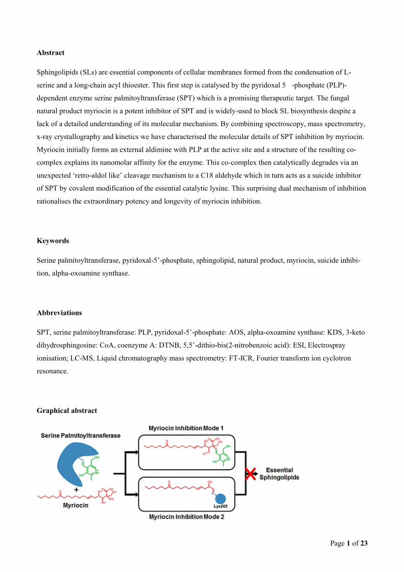
Abstract
Sphingolipids (SLs) are essential components of cellular membranes formed from the condensation of L-
serine and a long-chain acyl thioester. This first step is catalysed by the pyridoxal 5 -phosphate (PLP)-
dependent enzyme serine palmitoyltransferase (SPT) which is a promising therapeutic target. The fungal
natural product myriocin is a potent inhibitor of SPT and is widely-used to block SL biosynthesis despite a
lack of a detailed understanding of its molecular mechanism. By combining spectroscopy, mass spectrometry,
x-ray crystallography and kinetics we have characterised the molecular details of SPT inhibition by myriocin.
Myriocin initially forms an external aldimine with PLP at the active site and a structure of the resulting co-
complex explains its nanomolar affinity for the enzyme. This co-complex then catalytically degrades via an
unexpected ‘retro-aldol like’ cleavage mechanism to a C18 aldehyde which in turn acts as a suicide inhibitor
of SPT by covalent modification of the essential catalytic lysine. This surprising dual mechanism of inhibition
rationalises the extraordinary potency and longevity of myriocin inhibition.
Keywords
Serine palmitoyltransferase, pyridoxal-5’-phosphate, sphingolipid, natural product, myriocin, suicide inhibi-
tion, alpha-oxoamine synthase.
Abbreviations
SPT, serine palmitoyltransferase: PLP, pyridoxal-5’-phosphate: AOS, alpha-oxoamine synthase: KDS, 3-keto
dihydrosphingosine: CoA, coenzyme A: DTNB, 5,5’-dithio-bis(2-nitrobenzoic acid): ESI, Electrospray
ionisation; LC-MS, Liquid chromatography mass spectrometry: FT-ICR, Fourier transform ion cyclotron
resonance.
Graphical abstract
Page 2 of 23
Introduction
Sphingolipids (SLs) are a large sub class of lipids which are defined by the presence of an amino alcohol
functionality of sphingosine (or similar).1 SLs have been implicated in a wide range of cellular functions and
linked to diseases such as diabetes, Alzheimer’s and asthma.2 Controlling their production is under intense
investigation as a new strategy in pharmaceutical therapy. For example, the drug fingolimod, which mimics
sphingosine, is used for the treatment of multiple sclerosis and is the first SL-derived therapeutic agent used in
the clinic.3 Serine palmitoyltransferase (SPT, EC 2.3.1.50) is a member of the α-oxoamine synthase (AOS)
family of PLP-dependent enzymes along with 8-amino-7-oxononanoate synthase (AONS)4, 2-amino-3-
ketobutyrate ligase (KBL)5, 5-aminolevulinate synthase (ALAS)
6 and cholera quorum-sensing autoinducer
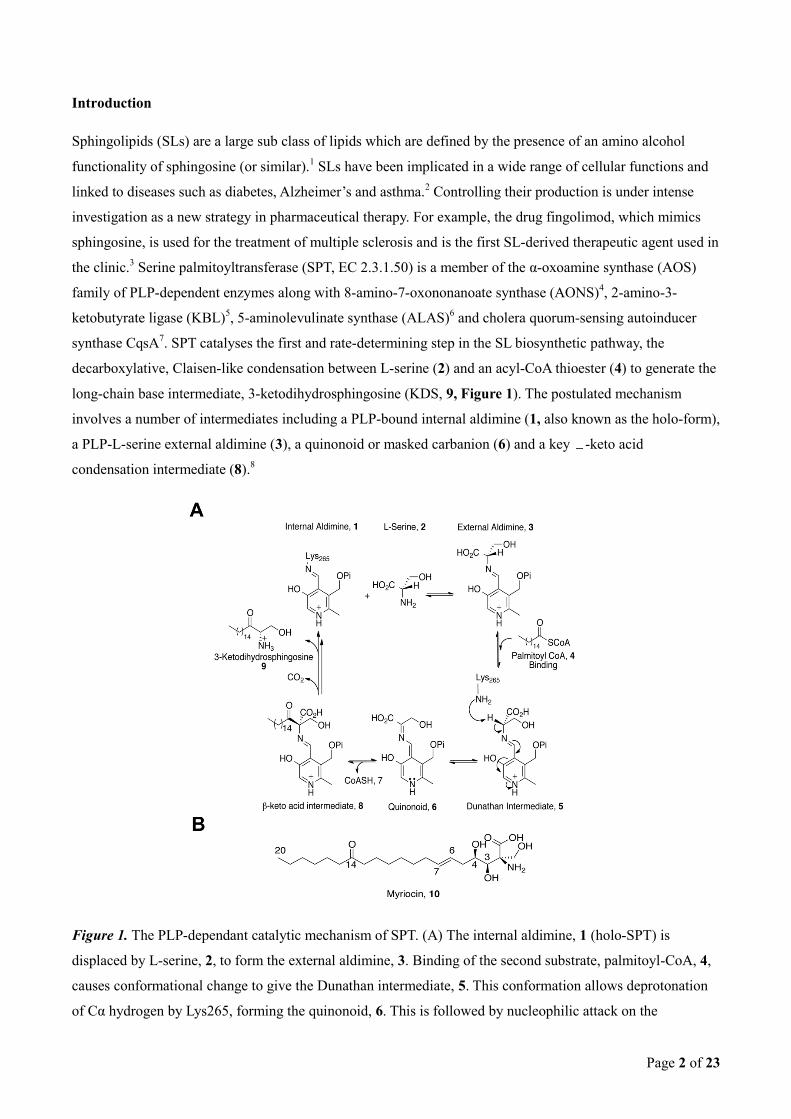
synthase CqsA7. SPT catalyses the first and rate-determining step in the SL biosynthetic pathway, the
decarboxylative, Claisen-like condensation between L-serine (2) and an acyl-CoA thioester (4) to generate the
long-chain base intermediate, 3-ketodihydrosphingosine (KDS, 9, Figure 1). The postulated mechanism
involves a number of intermediates including a PLP-bound internal aldimine (1, also known as the holo-form),
a PLP-L-serine external aldimine (3), a quinonoid or masked carbanion (6) and a key -keto acid
condensation intermediate (8).8
Figure 1. The PLP-dependant catalytic mechanism of SPT. (A) The internal aldimine, 1 (holo-SPT) is
displaced by L-serine, 2, to form the external aldimine, 3. Binding of the second substrate, palmitoyl-CoA, 4,
causes conformational change to give the Dunathan intermediate, 5. This conformation allows deprotonation
of Cα hydrogen by Lys265, forming the quinonoid, 6. This is followed by nucleophilic attack on the
Page 3 of 23
palmitoyl-CoA thioester from Cα, releasing CoASH, 7, forming the β-keto acid intermediate, 8. This
intermediate undergoes decarboxylation, before release of the product 3-ketodihydrosphingosine (KDS), 9,
and regeneration of the internal aldimine, 1. (B) The natural product SPT inhibitor myriocin, 10.
Over 160 PLP-dependent enzymes have been characterised9 and mechanistic studies of non-specific PLP
inhibitors such as cycloserine, penicillamine, and β-chloro-alanine10
have greatly enhanced the understanding
of PLP chemistry as well as highlighting the diversity of reactions that use this organic cofactor as a catalyst.
Consequently, PLP-dependent enzymes are now recognised as attractive drug targets.11
Despite the importance
of both PLP enzymes in general and SPT in particular, only a small number of SPT-specific inhibitors have
been discovered, all of which are natural products - sphingofungins, viridiofungin A, lipoxamycin and
myriocin (10, Figure 1B).12
Myriocin [(2S,3R,4R,6E)-2-Amino-3,4-dihydroxy-2-(hydroxymethyl)-14-oxo-6-
eicosenoic acid], also known as thermozymocidin and ISP-1, was first discovered in 1972 by two independent
groups13
from the thermophilic moulds Myriococcum albomyces and Mycelia sterilia and both found the
natural product to be a potent antifungal agent. It was re-isolated from the fungus Isaria sinclairii in 1994 and
shown to be a potent immunosuppressant.14
Kawasaki and colleagues12d
determined an IC50 value of 15 nM,
using the mouse cytotoxic T-cell line, CTLL-2 as a model for myriocin-dependent inhibition of cell growth
and elimination of ceramide production. Since growth was restored by the addition of sphingosine, it
suggested that SPT was the primary target of myriocin. In 1999 Schreiber and colleagues used a myriocin-like
affinity resin to identify the two subunits of murine SPT (SPT1 and SPT2 encoded by the genes, lcb1 and lcb2
respectively) as the primary targets of the natural product myriocin.15
Myriocin remains the most valuable and widely used chemical probe in SL research; for example recent
studies using the natural product in whole eukaryotic cells and cell extracts have revealed unexpected
structural complexity in the membrane-bound SPTs from higher organisms.2c, 16
Despite its use, the molecular
basis of myriocin inhibition of SPT is largely unknown. To shed light on the chemistry of this important
enzyme:inhibitor complex we have used a soluble, recombinant form of the enzyme from the SL-producing
bacterium Sphingomonas paucimobilis which has proven an informative model system. Here we present a
detailed chemical analysis of the SPT:myriocin inhibition mechanism including a structural description of the
covalent PLP:external aldimine of myriocin. We also reveal an unexpected, enzyme-catalysed myriocin
degradation mechanism that generates a second species which acts as a suicide inhibitor of its enzyme target;
thus we propose that myriocin acts as a novel dual-mode inhibitor of SPT.
Experimental section
Chemicals and Molecular Biology Tools. Plasmids and competent cells were purchased from Novagen. All
buffers and reagents were from Sigma. Palmitoyl-CoA was purchased from Avanti Lipids, and myriocin was
purchased from Sigma.
Page 4 of 23
Gene cloning and mutagenesis. The S. paucimobilis SPT used in this study is in vector pET28a and contains
a six histidine tag at the N-terminus. This generates an SPT of 441 amino acids with a short extension at the
N-terminus containing the His tag and a 12 amino acid linker. The K265A mutant was made using the Liu
mutagenesis protocol17
with the following primers:
5’-gtcggcactttctctgcgtctgttggaactgttggc-3’ (Forward),
5’-acccgccaacagttccaacagacgcagagaaagtgccgac-3’ (Reverse).
The bases mutated are shown in bold and the isolated SPT K265A clone was verified by DNA sequencing.
Mass spectrometry analysis of both wild type and K265A mutant SPTs showed the proteins to be intact with
only the N-terminal methionine removed. To avoid confusion and allow comparison with previous data we
number the N-terminally-tagged SPT using the same sequence as before.8 Key active site residues are His159,
Asp231, His234, and Lys265.
Expression and purification of SPT WT and SPT K265A mutant. Expression and purification of the WT
protein has been reported previously.8 A single colony of pET-28a SPT BL21 (DE3) (or SPT K265A mutant)
was used to inoculate 250 ml of LB media with kanamycin (30 μg mL-1)
and grown overnight to saturation.
This culture was diluted 1:100 in fresh LB / kanamycin and grown to an OD600 of 0.6 before expression was
induced with 0.1 mM Isopropyl β-D-1-thiogalactopyranoside (IPTG). Protein expression continued for 5
hours at 30 °C, 200 rpm. Harvested cells where re-suspended in lysis buffer 20 mM KPhos, pH 7.5, 150 mM
NaCl, 10 mM imidazole, 25 μM PLP and complete protease inhibitor cocktail (Roche) and lysed by sonication
(Soniprep 150, 15 cycles of 30 sec on followed by 30 sec off) on ice. The resulting lysate was centrifuged for
30 mins at 47000 g. The supernatant was incubated with Ni resin (Ni-NTA Superflow, Qiagen) for 1 hour at 4
°C. The resin was removed, washed and the protein eluted in lysis buffer supplemented with 300 mM
imidazole. Imidazole was removed by dialysis of the protein into 20 mM KPhos, pH 7.5,150 mM NaCl, 25
μM PLP before a final gel filtration purification step using a Superdex S200 column (GE Healthcare)
equilibrated and eluted with 20 mM Tris, pH 7.5,150 mM NaCl, 25 μM PLP buffer. The purity of the eluted
protein was checked by SDS PAGE and the protein identity confirmed by mass spectrometry.
UV-vis Spectroscopic Measurements. All UV-visible spectra were recorded on a Cary 50 UV-visible
spectrophotometer (Varian) and analyzed using Cary WinUV software (Varian). Immediately prior to
enzymatic assays, SPT was converted to the holo-form by dialysis against freshly-prepared 20 mM KPhos
(pH 7.5) containing 150 mM NaCl and 25 μM PLP for 1 h at 4 °C. Excess PLP was removed by passing the
protein through a PD-10 (Sephadex G-25M) desalting column (GE Healthcare) before concentration to 40 μM
using a VivaSpin 30 kDa cut-off concentration filter. For UV-visible analysis, the concentration of
recombinant SPT was 40 μM, and the spectrophotometer was blanked with 20 mM potassium phosphate (pH
7.5) containing 150 mM NaCl. The spectrophotometer was maintained at a constant temperature throughout
all time-dependant assays using a Cary PCB-150 single cell Peltier.
Page 5 of 23
SPT Activity Assay. SPT activity was measured using a previously published method that uses a continuous
spectrophotometric assay to monitor the release of CoASH from acyl-CoA substrates and reaction with 5,5′-
dithiobis-2-nitrobenzoic acid (DTNB).8b
Assays were performed on a 250 μL scale on a 96-well format in a
Biotek Synergy HT plate reader. The enzyme was incubated with L-serine and myriocin in a buffered solution
containing DTNB, and the assay was started by the addition of the second substrate, palmitoyl-CoA. The
CoASH thiol product was monitored by observation of the TNB− anion at 412 nm (εmax = 14,150 M
−1 cm
−1)
for 45 minutes. A typical experiment contained 0.2 μM SPT, 20 mM L-serine, 250 μM palmitoyl-CoA, and 0.2
mM DTNB in 100 mM HEPES, pH 8.0. Kinetic constants were calculated using GraphPad Prism 6 software.
Km and competition experiments where performed and calculated using Michaelis-Menten kinetics.
Inhibition Studies. Due to the tight binding nature of myriocin, it was necessary to calculate inhibition
kinetics using the regimen described by Williams and Morrison.18
The Michaelis-Menten equation can not be
used as the assumption that the free inhibitor concentration is equal to the total inhibitor concentration is not
valid; instead the Ki is calculated using the quadratic Morrison equation. The Km for L-serine was calculated
from rates measured with increasing concentrations of L-serine (0.1-40 mM) while maintaining the palmitoyl-
CoA concentration at 250 μM in excess of its Km (35 μM). Similarly, the Km for palmitoyl-CoA was calculated
from rates measured with increasing concentrations of palmitoyl-CoA (2.5-1500 μM) and excess L-serine (20
mM).
In order to determine the type of inhibition between SPT and myriocin, IC50 values for the inhibitor were
determined in the presence of a number of different substrate concentrations (both L-serine and palmitoyl-
CoA). During these experiments the enzyme concentration remained fixed at 200 nM. In order to ensure
solubility of myriocin in the assay mixture, myriocin was dissolved in DMSO then diluted to its final desired
concentration with the DMSO concentration at less than or equal to 1%. For the experiments varying
palmitoyl-CoA concentration it was necessary to add L-ser and palmitoyl-CoA prior to addition of myriocin.
The resulting IC50 values were plotted against substrate concentrations in a manner described by Cha,
Williams and Morrison 19
. All kinetic experiments were performed in triplicate.
To determine the reversibility of inhibition, 40 μM SPT was treated with 200 μM myriocin and incubated for
either 10 minutes or 16 hours at 25 °C. Excess inhibitor was then removed by extensive dialysis against 20
mM KPhos, 150 mM NaCl, pH 7.5 with 25 μM fresh PLP. Aliquots were then removed at specific timepoints
and assayed for SPT activity as above.
Mass Spectrometry. All mass spectra were acquired using a 12 Telsa SolariX FT-ICR mass spectrometer
equipped with an electrospray ion source (Bruker Daltonics). LC-MS experiments were performed using an
Ultimate 3000 HPLC system (Dionex).
For the detection of the PLP-myriocin aldimine (11) and the PLP-decarboxymyriocin aldimine (15), samples
were prepared by incubating myriocin (200 μM) with either wildtype or K265A SPT (40 μM) at 25 °C. At
Page 6 of 23
specific time points, aliquots were removed and analysed by LC-MS using a Phenomenex C18 Aeris
Widepore 50 x 2.1mm column, operating at a flow rate of 150 μl/min. A gradient of 2 to 98% acetonitrile was
performed over 20 minutes. In order to preserve the acid labile imine bond, no acidic modifiers were added to
either mobile phase. For single ion monitoring, the mass resolving quadrupole was set to a specific m/z, with a
10 m/z window, and ions were typically accumulated for 2000 milliseconds.
For intact protein mass spectrometry, LC-MS was performed using a monolithic PS-DVB (500 μm x 50 mm)
reverse-phase analytical column (Dionex). Protein (100 pmoles) was loaded onto the column (maintained at
60 °C) followed by a 10 min linear gradient from 2 to 95% acetonitrile (flow rate 20 μl/min). Mass spectra
were collected every 200 ms between m/z 600-2500. Data analysis was performed using DataAnalysis
software (Bruker Daltonics). Neutral spectra were created using Maximum Entropy deconvolution.
Peptide Mass Mapping. For peptide mass mapping, SPT (40 μM) was treated with 200 μM myriocin.
Derivitization was allowed to proceed for 18 hours at 25 °C, whilst being monitored by UV-vis spectroscopy.
After loss of the peak at 430 nm in the UV-vis spectrum, the sample was chemically reduced by treatment
with 10 mM NaBH4. Finally, this sample was treated with the protease trypsin (Promega, sequence grade) for
18 h at 37 °C at an enzyme:protein ratio of approximately 1:20 by weight. The resulting peptide mixture was
analysed by LC-MS using a monolithic PS-DVB (500 μm x 50 mm) reverse-phase analytical column
(Dionex). The column was maintained at 60 °C, and a linear gradient of 5-70% acetonitrile was performed
over 30 minutes.
The resulting data was analysed by DataAnalysis software (Bruker Daltonics). A mass list was created using
the SNAP 2.0 algorithm (Bruker Daltonics) and searched against the known primary sequence of SPT using
the MS-Fit software (University of California). For data searching, error tolerances were set to 10 ppm.
Structural Biology. Protein for crystallization was concentrated to 20 mg mL-1
and incubated with 5
equivalents of myriocin immediately prior to crystallization trials. Crystals of K265A SPT were grown by
vapour drop diffusion at 20 °C over the course of two weeks and are readily reproducible. The optimum
growth conditions are 32% PEG MME 2000, 0.1 M HEPES pH 7.5 and a protein:precipitant ratio of 1:1. Prior
to data collection the crystals were cryo-cooled in mother liquor doped with 20% glycerol. Data were
collected at Beamline I04-1 at the Diamond synchrotron light source, Oxfordshire, England and processed in
an automated manner using Xia2.20
The structure was solved by molecular replacement using PHASER21
and
PDB code 2JG2 as a model. The myriocin dictionary was created using PRODRG.22
The model was refined
using REFMAC523
with TLS. COOT24
was used for manual manipulation of the structure and model quality
was assessed throughout with MOLPROBITY25
. Data collection and refinement statistics can be found in
supplementary Table 2.
Accession codes. Protein Data Bank (PDB): The crystallographic data for the SPT -PLP:decarboxymyriocin
structure is deposited under accession code 4BMK.
Page 7 of 23
Results
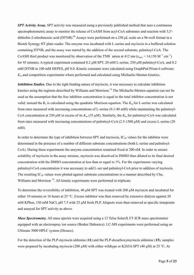
Formation of a PLP-myriocin aldimine in wild-type SPT. Holo-SPT (1) displays a characteristic UV-vis
spectrum for a PLP-containing protein with absorption maxima at 333 nm and 420 nm (Figure 2A),
corresponding to the equilibrium between the ketoenamine and enolimine forms of PLP.8b
Addition of fivefold
molar excess of myriocin to holo-SPT led to an immediate increased absorbance with a maximum at 430 nm
and loss of the 333 nm peak as previously observed by Ikushiro et al.12b
Hanada et al. proposed that this
species corresponds to a PLP-myriocin aldimine (11) formed as a result of transimination (Figure 2B) that
acts as a stable mimic of the β-keto acid intermediate (8) in the SPT mechanism (Figure 1).26
LC ESI-MS
analysis detects a species of monoisotopic mass 631.30153 Da consistent with the PLP-myriocin aldimine (11)
(Predicted monoisotopic mass 631.29902 Da; [(C29H47N2O11P)+H]+) (Figure 2C). Attempts to trap the
reduced form of (11) using NaBH4 were unsuccessful.
Figure 2. SPT inhibition occurs via formation of a PLP-myriocin aldimine. (A) UV-Vis spectrum of 40 μM
SPT before (solid line) and after 200 μM myriocin addition (dotted line). (B) The proposed structure of the
inhibitory complex - a PLP-myriocin aldimine, 11. (C) Detection of the PLP-myriocin aldimine by LC-MS.
Top, Extracted Ion Chromatogram at m/z 631. Bottom, high resolution mass spectrum of the PLP-myriocin
Page 8 of 23
aldimine, obtained by summing the spectra between t = 8-12 minutes. ([M+H]+ C29H48N2O11P; predicted m/z
631.29902; observed error 4.0ppm). * denotes a contaminant. (D) Inhibition of SPT by myriocin, data fitted
using the Morrison equation. The Ki obtained for the PLP-myriocin aldimine is 967 ± 98 nM.
Using the method by Cha,19a
IC50 values for the inhibitor were determined at fixed enzyme concentrations
whilst systematically varying each substrate concentration. IC50 values increase linearly with varying L-serine
and palmitoyl-CoA concentration, establishing myriocin as a competitive inhibitor for both L-serine and
palmitoyl-CoA substrates (Supplementary Figure 1). The Morrison equation19b
, yields a Ki for myriocin of
967 ± 98 nM (Figure 2D).
The PLP-myriocin inhibitory complex undergoes slow catalytic degradation. The PLP-myriocin aldimine
(11) at 25 oC was stable for 90 minutes before a slow spectral transition took place over 16 hours (decrease at
430 nm and a concomitant increase at 331 and 400 nm) (Figure 3A); indicating conversion of 11 to a
previously unseen PLP aldimine. To our surprise the new species remained catalytically compromised,
indicating the new PLP aldimine is also inhibitory. Immediately after addition of myriocin only 7.7% of
enzyme activity remains (as expected); however after complete conversion (judged by no further change in the
UV-vis spectrum) activity remained low at 10.1% relative to the starting value (Figure 3B). Lowering the
temperature to 4 °C arrested the decomposition of the PLP-myriocin aldimine (Supplementary Figure 2).
Interestingly, a catalytically-inactive mutant, SPT K265A, displayed two peaks with λmax values of 326 nm
and 402 nm, consistent with PLP bound non-covalently as the aldehyde (Figure 3C). Upon incubation with
five-fold molar excess of myriocin we observed a large shift to a single absorbance maximum at 425 nm
similar to that observed in the wild type SPT. In contrast to the wild-type incubation this spectrum remained
unchanged over 16 hrs at 25 °C indicating a stable PLP-myriocin external aldimine. Taken together, these
intriguing observations suggest that the initial SPT:PLP-myriocin inhibitor complex breaks down to form a
second species that also inhibits SPT.
To test the reversibility of inhibition we first generated the SPT:PLP-myriocin complex by incubation of SPT
with the inhibitor for 10 minutes and 16 hrs at 25 °C. After this incubation period the temperature was
was performed for 24 hours. Aliquots were removed from the dialysate at specific time points (0, 3 and 24
hours) and assayed for SPT activity (Figure 3D). For the enzyme incubated with myriocin for the shorter time
(10 mins), then dialysed for 3 hours, no significant activity was recovered; however, dialysis for 24 hours
recovered 60% enzymatic activity (Figure 3D, white bars). This regain in enzymatic activity was also
accompanied by a change in the UV-vis spectrum back to the internal aldimine form – i.e. λmax 333 and 420
nm (data not shown). Taken together, this data from short, 10 minute incubations of SPT with myriocin is
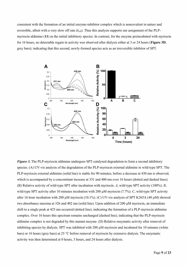
Page 9 of 23
consistent with the formation of an initial enzyme-inhibitor complex which is noncovalent in nature and
reversible, albeit with a very slow off rate (koff). Thus this analysis supports our assignment of the PLP-
myriocin aldimine (11) as the initial inhibitory species. In contrast, for the enzyme preincubated with myriocin
for 16 hours, no detectable regain in activity was observed after dialysis either at 3 or 24 hours (Figure 3D,
grey bars); indicating that this second, newly-formed species acts as an irreversible inhibitor of SPT.
Figure 3. The PLP-myriocin aldimine undergoes SPT-catalysed degradation to form a second inhibitory
species. (A) UV-vis analysis of the degradation of the PLP-myriocin external aldimine in wild-type SPT. The
PLP-myriocin external aldimine (solid line) is stable for 90 minutes, before a decrease at 430 nm is observed,
which is accompanied by a concomitant increase at 331 and 400 nm over 16 hours (dotted and dashed lines).
(B) Relative activity of wild-type SPT after incubation with myriocin. A, wild type SPT activity (100%). B,
wild-type SPT activity after 10 minutes incubation with 200 μM myriocin (7.7%). C, wild-type SPT activity
after 16 hour incubation with 200 μM myriocin (10.1%). (C) UV-vis analysis of SPT K265A (40 μM) showed
two absorbance maxima at 326 and 402 nm (solid line). Upon addition of 200 μM myriocin, an immediate
shift to a single peak at 425 nm occurred (dotted line), indicating the formation of a PLP-myriocin aldimine
complex. Over 16 hours this spectrum remains unchanged (dashed line), indicating that the PLP-myriocin
aldimine complex is not degraded by this mutant enzyme. (D) Relative enzymatic activity after removal of
inhibiting species by dialysis. SPT was inhibited with 200 μM myriocin and incubated for 10 minutes (white
bars) or 16 hours (grey bars) at 25 °C before removal of myriocin by extensive dialysis. The enzymatic
activity was then determined at 0 hours, 3 hours, and 24 hours after dialysis.
Page 10 of 23
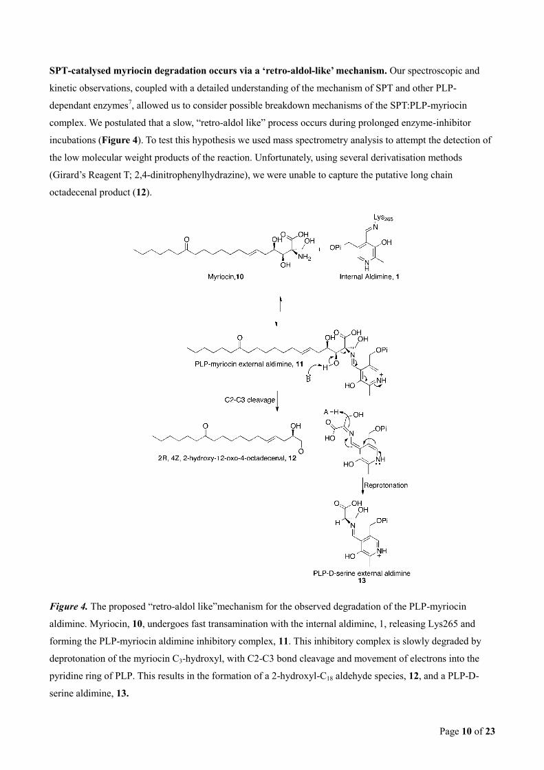
SPT-catalysed myriocin degradation occurs via a ‘retro-aldol-like’ mechanism. Our spectroscopic and
kinetic observations, coupled with a detailed understanding of the mechanism of SPT and other PLP-
dependant enzymes7, allowed us to consider possible breakdown mechanisms of the SPT:PLP-myriocin
complex. We postulated that a slow, “retro-aldol like” process occurs during prolonged enzyme-inhibitor
incubations (Figure 4). To test this hypothesis we used mass spectrometry analysis to attempt the detection of
the low molecular weight products of the reaction. Unfortunately, using several derivatisation methods
(Girard’s Reagent T; 2,4-dinitrophenylhydrazine), we were unable to capture the putative long chain
octadecenal product (12).
Figure 4. The proposed “retro-aldol like”mechanism for the observed degradation of the PLP-myriocin
aldimine. Myriocin, 10, undergoes fast transamination with the internal aldimine, 1, releasing Lys265 and
forming the PLP-myriocin aldimine inhibitory complex, 11. This inhibitory complex is slowly degraded by
deprotonation of the myriocin C3-hydroxyl, with C2-C3 bond cleavage and movement of electrons into the
pyridine ring of PLP. This results in the formation of a 2-hydroxyl-C18 aldehyde species, 12, and a PLP-D-
serine aldimine, 13.
Page 11 of 23
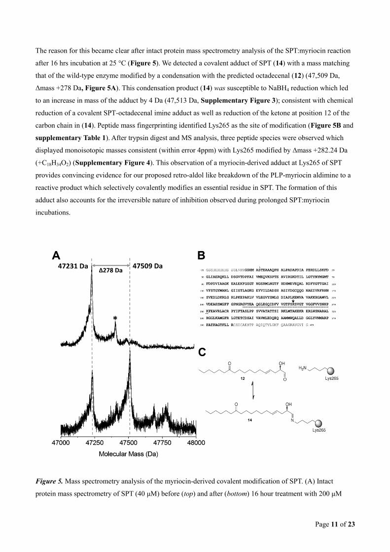
The reason for this became clear after intact protein mass spectrometry analysis of the SPT:myriocin reaction
after 16 hrs incubation at 25 °C (Figure 5). We detected a covalent adduct of SPT (14) with a mass matching
that of the wild-type enzyme modified by a condensation with the predicted octadecenal (12) (47,509 Da,
Δmass +278 Da, Figure 5A). This condensation product (14) was susceptible to NaBH4 reduction which led
to an increase in mass of the adduct by 4 Da (47,513 Da, Supplementary Figure 3); consistent with chemical
reduction of a covalent SPT-octadecenal imine adduct as well as reduction of the ketone at position 12 of the
carbon chain in (14). Peptide mass fingerprinting identified Lys265 as the site of modification (Figure 5B and
supplementary Table 1). After trypsin digest and MS analysis, three peptide species were observed which
displayed monoisotopic masses consistent (within error 4ppm) with Lys265 modified by Δmass +282.24 Da
(+C18H34O2) (Supplementary Figure 4). This observation of a myriocin-derived adduct at Lys265 of SPT
provides convincing evidence for our proposed retro-aldol like breakdown of the PLP-myriocin aldimine to a
reactive product which selectively covalently modifies an essential residue in SPT. The formation of this
adduct also accounts for the irreversible nature of inhibition observed during prolonged SPT:myriocin
incubations.
Figure 5. Mass spectrometry analysis of the myriocin-derived covalent modification of SPT. (A) Intact
protein mass spectrometry of SPT (40 μM) before (top) and after (bottom) 16 hour treatment with 200 μM
Page 12 of 23
myriocin at 25 °C. SPT displays an average neutral mass of 47231 Da. After myriocin treatment a prominent
new species of average mass 47509 is observed (Δmass +278 Da). Minor peaks highlighted by * arise from α-
N-gluconoylation of the His-tag. (B) Sequence coverage achieved when analysing myriocin-modified SPT
enzyme. Amino acids highlighted in bold were observed in the peptide mass fingerprint (see also
supplementary Table 1). The myriocin derived modification was isolated to a single site between Glu245 and
Lys280 (underlined); this sequence contains a single internal lysine residue, Lys265 (highlighted by the
arrow). (C) The myriocin-derived modification displayed a Δmass + 282.24 Da (after hydride reduction; see
also Supplementary Figure 4), this is consistent with condensation of the C18-aldehyde (12) onto the amine
of Lys265 forming the imine (14), with subsequent chemical reduction of the imine and ketone functional
groups (+C18H34O2).
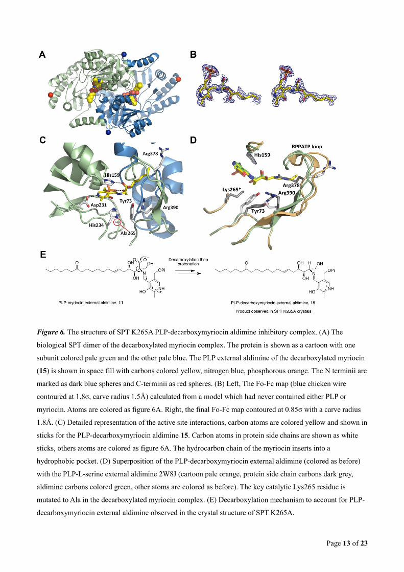
Structure of a SPT K265A:PLP-myriocin external aldimine complex. We were unable to prepare crystals
of the wild type SPT:PLP-myriocin aldimine complex, most likely due to aldimine degradation. However
crystals of SPT K265A were obtained that diffracted to 1.6 Å resolution and contained a canonical dimer in
the asymmetric unit (Figure 6A). The mobility/flexibility of the acyl chain prevented us from assigning a
structure beyond carbon 9 of myriocin. Nevertheless difference electron density revealed a PLP-myriocin
aldimine complex had formed (Figure 6B). The myriocin stereochemistry is retained - the Z configuration of
the double bond at position C6 and cis diol geometry at positions C3 and C4. We were surprised to discover
that the density indicates that myriocin had undergone loss of the carboxylate from the C2 position to form a
PLP-decarboxymyriocin aldimine (15). We built the model reasoning the replacement of the carboxylate with
a proton (sp3 hybridised carbon) but the resolution on its own is not sufficient to distinguish an sp
3 from an sp
2
carbon (Figure 6B). As had been noted previously8b
residues from both SPT subunits are involved in PLP
binding and the active site is at the dimer interface (Figure 6C).
Page 13 of 23
Figure 6. The structure of SPT K265A PLP-decarboxymyriocin aldimine inhibitory complex. (A) The
biological SPT dimer of the decarboxylated myriocin complex. The protein is shown as a cartoon with one
subunit colored pale green and the other pale blue. The PLP external aldimine of the decarboxylated myriocin
(15) is shown in space fill with carbons colored yellow, nitrogen blue, phosphorous orange. The N terminii are
marked as dark blue spheres and C-terminii as red spheres. (B) Left, The Fo-Fc map (blue chicken wire
contoured at 1.8σ, carve radius 1.5Å) calculated from a model which had never contained either PLP or
myriocin. Atoms are colored as figure 6A. Right, the final Fo-Fc map contoured at 0.85σ with a carve radius
1.8Å. (C) Detailed representation of the active site interactions, carbon atoms are colored yellow and shown in
sticks for the PLP-decarboxymyriocin aldimine 15. Carbon atoms in protein side chains are shown as white
sticks, others atoms are colored as figure 6A. The hydrocarbon chain of the myriocin inserts into a
hydrophobic pocket. (D) Superposition of the PLP-decarboxymyriocin external aldimine (colored as before)
with the PLP-L-serine external aldimine 2W8J (cartoon pale orange, protein side chain carbons dark grey,
aldimine carbons colored green, other atoms are colored as before). The key catalytic Lys265 residue is
mutated to Ala in the decarboxylated myriocin complex. (E) Decarboxylation mechanism to account for PLP-
decarboxymyriocin external aldimine observed in the crystal structure of SPT K265A.
Page 14 of 23
Comparison of this complex with the wild type SPT:PLP-L-serine external aldimine complex (PDB code
2W8J)8b
reveals essentially the same positioning of the PLP rings in both structures (Figure 6D). Moreover,
many key active site residues (His159, Asp231, His234) adopt similar positions. However Arg378, which is
involved in a key electrostatic interaction with the carboxylate of L-serine in the SPT:PLP-L-serine external
aldimine structure, is swung out of the active site with concomitant change in the important mobile loop 378
RPPATP 383; both the loop and Arg378 are partially disordered in the new complex. These changes are the
result of accommodating the extended hydrophobic tail of 15. We also note that Arg390, a conserved residue
which is required for product formation in the SPT catalytic cycle27
, adjusts its side chain conformation and
makes interactions with both Tyr73 and the C4 hydroxyl of myriocin. In the experimental maps the aromatic
portion of the side chain of Tyr73 is not visible in the experimental density despite other plausible
conformations being precluded by packing. We confirmed by DNA sequencing and mass spectrometry the
residue was tyrosine.
Discussion
Natural products and their derivatives are responsible for over half of all Food and Drug Administration
(FDA)-approved drugs28
and continue to provide excellent lead molecules.29
Natural products also serve as
excellent chemical probes that can be used to tease out the fine details of biochemical pathways and
networks.30
Myriocin, as well as possessing potent antifungal, antibacterial and immune suppression activity,
is also a valuable chemical tool. Recently Breslow et al. used myriocin to probe the multi-protein, membrane-
bound SPT complex (denoted SPOTs) from the endoplasmic reticulum (ER) of yeast that contains SPT1,
SPT2, TSC3P, ORM proteins and Sac1 phosphatase.2c
The SPOTs complex is now known to be central to SL
biosynthesis in higher organisms acting as a metabolic rheostat.31
SLs are now recognised as important in the
aetiology of many significant human diseases. The pro-drug fingolimod, 2-amino-2-[2-(4-
octylphenyl)ethyl]propane-1,3-diol, becomes phosphorylated and mimics sphingosine 1-phosphate (S1P) in
the body. The drug suppresses the immune system and is used in the treatment of relapsing remitting multiple
sclerosis where it is thought to work by binding to S1P receptors. There is now widespread interest in
regulating the production of SLs in the body and the most obvious target is the SPOTs complex which
contains SPT. There are two related obstacles to progress, firstly the human SPOTs complex is not currently
tractable to biochemical study and there is very limited understanding of how the exemplar inhibitor myriocin
works at a molecular level. To overcome these obstacles we have employed the bacterial SPT homolog to
reveal the molecular details of myriocin inhibition for the first time.
We have experimentally confirmed the existence of the previously assumed SPT:PLP-myriocin external
aldimine inhibitor complex (11) by mass spectrometry and spectroscopy (Figure 2B and 2C). We observed
that myriocin (Ki 967 nM) was a potent competitive inhibitor for both L-serine and palmitoyl-CoA, an
observation consistent with the ordered, bi-bi mechanism of AOS enzymes and the inhibitor acting as a
intermediate mimic. Despite this nanomolar potency we were unable to completely abolish the activity of the
Page 15 of 23
enzyme, our data suggest that this is because at the con-centration of the enzyme required for reliable assay,
we cannot dissolve sufficient myriocin to saturate the binding site. In vivo, with endogenous SPT levels (found
within an ER-bound, SPOTs complex) where the enzyme is in a hydrophobic lipid environment, this
limitation is not expected to occur. The Schiff’s base external aldimine formed between PLP and myriocin
(11) is analogous to that formed between PLP and D-serine, a known, but weak, SPT inhibitor.32
This
inhibition should be reversible by incubation with fresh PLP which would displace the inhibitor and restore
the holo, internal aldimine form. Indeed, this was the case but only if the PLP was added to the
enzyme:inhibitor complex relatively soon after it was formed. However, addition of PLP could not rescue the
enzyme if the SPT:PLP-myriocin complex was subjected to prolonged incubation.
Coupled to this surprising result regarding the kinetics we also observed interesting, time-dependent
spectroscopic changes of the SPT:PLP-myriocin external aldimine complex (11). This suggested to us that this
initial complex was breaking down to an unanticipated second inhibitory species. We generated an inactive
SPT (K265A) by removal of the key, conserved active site lysine which not only binds the PLP but also plays
an essential role in acid/base catalysis during the mechanism. The SPT K265A mutant binds PLP (albeit non-
covalently) and forms the PLP-myriocin external aldimine 11 but it does not undergo the apparent breakdown
observed with the wild-type enzyme suggesting Lys265 is crucial for this enzyme-catalysed reaction. Mass
spectrometry and chemical reduction establishes that in the wild type SPT the PLP-myriocin external aldimine
converts to a C18 imine adduct of Lys265, the key catalytic residue. This covalent modification results in
irreversible inhibition of SPT that cannot be rescued by incubation with PLP. This result was entirely
unexpected and suggested both a previously unsuspected catalytic activity of SPT, as well as a novel duel
mechanism of action of SPT inhibition by myriocin.
We were unable to obtain crystals of the wild-type enzyme with myriocin in the active site presumably
because the PLP-myriocin aldimine degrades during the two week timescale of crystallisation. However, the
catalytically inactive K265A mutant did allow us to capture an external aldimine complex; but even then the
refined structure revealed that it had undergone a surprising decarboxylation leaving a PLP-
decarboxymyriocin aldmine 15 in the active site. To investigate the time-scale of this decarboxylation process
we used mass spectrometry analysis of the SPT K265A:myriocin incubation and found that the PLP-myriocin
aldimine (11) is stable at room temperature for at least seven days with decarboxylation only observed to
occur after this time (Supplementary Figure 5).
PLP is a versatile cofactor that can use the electron sink properties of the protonated pyridine ring to catalyse a
wide range of reactions of amino acid substrates including racemisation and decarboxylation.33
These different
chemical reactions proceed from the same key, PLP-amino acid external aldimine and are controlled by the
architecture of the particular enzyme active site. Dunathan put forward a hypothesis that outlined the stereo-
electronic constraints that govern PLP-dependent enzymes.34
Modelling the SPT K265A PLP-myriocin
external aldimine complex (11) places the carboxylate perpendicular to the PLP (“Dunathan conformation”)
Page 16 of 23
that would allow decarboxylation by the mechanism proposed in Figure 6E. However, its extreme slowness
relative to bone fide PLP-dependent decarboxylases, cautions that the PLP-myriocin aldimine in K265A is not
optimally aligned for this reaction. It is worth noting here that the precise orientation of the PLP-myriocin
aldimine required for decarboxylation may not be achievable to the wild type enzyme. In the wild type SPT
the side chain of K265 would be very close to/clash with the carboxylate, perhaps distorting it out of the
Dunathan conformation or stabilising the carboxylate by an ionic interaction. In our previous study of the
SPT:PLP-L-serine external aldimine complex of S. paucimoblis SPT we revealed that the carboxylate group is
held in a specific orientation by salt bridges with Arg378 and His159.8b, 27
In this arrangement decarboxylation
is not favoured, rather deprotonation of the L-serine at C α by K265 is achieved only upon a conformational
change caused by palmitoyl-CoA binding (Figure 1). 8b, 35
An overlay of the wild-type PLP-L-serine and the K265A PLP-decarboxymyriocin structures reveals that the
configuration of the SPT active site remains relatively well conserved (Figure 6D) as well as providing a
molecular insight into why myriocin is a nanomolar inhibitor (Supplementary Figure S6a). Conserved
residues His159, Asp231 and His234 are all in the same relative positions within the active site. Moreover, the
CH2OH head group of myriocin interacts with the 5′-phosphate of PLP in the same manner as the hydroxyl
group from L-serine (Supplementary Figure S6b). Of key note, the 3,4 cis diol of decarboxymyriocin makes
hydrogen bonds to the protein, notably the 3-hydroxy of myriocin with the important catalytic residue His159;
this interaction would be expected to be preserved in the wild type SPT:myriocin complex. The hydrogen
bond network that surrounds and includes the 4-hydroxy of decarboxymyriocin may be changed by the
presence of Lys265 but at least some of the same network seems certain to persist and this too involves the
same residues that interact with the carboxylate of the PLP-L-serine external aldimine. These interactions
rationalise the competitive inhibition with L-serine. Accompanying these interactions are movements of the
side-chains of Tyr73, Arg378 and Arg390 as well as a displacement of a key conserved stretch of amino acids
(RPPATP) that constitute a mobile loop that undergoes conformational changes during the catalytic cycle. The
6,7 trans double bond geometry of myriocin is clearly defined and we can see electron density for the carbon
chain up to C9 which sits in the hydrophobic cleft adjacent to PLP. The carbon tail of myriocin binds in a
similar orientation to the decanoyl-tail of the PLP-product external aldimine observed bound in the crystal
structure of the related AOS enzyme CqsA from Vibrio cholera (Supplementary Figure S7)7 consistent with
our hypothesis that myriocin mimics the condensation intermediate.
The structure also rationalises the unexpected retro-aldol degradation of the PLP-myriocin external aldimine
11 into the C18 aldehyde 12. This mechanism requires a base to abstract the proton from the 3-hydroxy of
myriocin. Based on its role in the SPT reaction and the fact that a K265A mutant was unable to catalyse the
retro-aldol myriocin degradation, Lys265 was a prime candidate for this role. However, structural overlay
suggests that Lys265 would be on the wrong face to perform this role (Figure 6D). However, the absolutely
conserved His159 is positioned 2.6 Å away from the 3-hydroxy of myriocin and we propose that it initiates
the breakage of the C2-C3 bond with the electrons sinking into the PLP ring (Figure 4 and Supplementary
Page 17 of 23
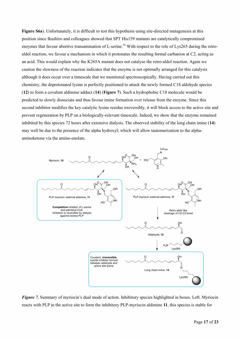
Figure S6a). Unfortunately, it is difficult to test this hypothesis using site-directed mutagenesis at this
position since Ikushiro and colleagues showed that SPT His159 mutants are catalytically compromised
enzymes that favour abortive transamination of L-serine.36
With respect to the role of Lys265 during the retro-
aldol reaction, we favour a mechanism in which it protonates the resulting formal carbanion at C2, acting as
an acid. This would explain why the K265A mutant does not catalyse the retro-aldol reaction. Again we
caution the slowness of the reaction indicates that the enzyme is not optimally arranged for this catalysis
although it does occur over a timescale that we monitored spectroscopically. Having carried out this
chemistry, the deprotonated lysine is perfectly positioned to attack the newly formed C18 aldehyde species
(12) to form a covalent aldimine adduct (14) (Figure 7). Such a hydrophobic C18 molecule would be
predicted to slowly dissociate and thus favour imine formation over release from the enzyme. Since this
second inhibitor modifies the key catalytic lysine residue irreversibly, it will block access to the active site and
prevent regeneration by PLP on a biologically-relevant timescale. Indeed, we show that the enzyme remained
inhibited by this species 72 hours after extensive dialysis. The observed stability of the long chain imine (14)
may well be due to the presence of the alpha hydroxyl; which will allow tautomerization to the alpha-
aminoketone via the amino-enolate.
Figure 7. Summary of myriocin’s dual mode of action. Inhibitory species highlighted in boxes. Left. Myriocin
reacts with PLP in the active site to form the inhibitory PLP-myriocin aldimine 11, this species is stable for
Page 18 of 23
greater than an hour at physiological temperature with inhibition being reversible upon addition of excess
PLP. Right. PLP-myriocin aldimine 11 decomposes over 16 hours, at physiological temperature, to produce a
long chain aldehyde (12) that react with the active site lysine to form an imine, thus rendering the enzyme
inactive. This covalent modification can be classed as suicide inhibition.
Conclusion
We set out to delineate the structural and mechanistic details of how myriocin inhibits SPT and in doing so we
have revealed unexpected and hitherto unprecedented chemistry (summarised in Figure 7). Our results
demonstrate that, as predicted, myriocin acts as a classical intermediate mimic inhibitor with nanomolar
affinity for its target. However, once bound in the active site the inhibitory species is broken down by the
enzyme to generate a reactive product which acts a suicide inhibitor by covalent modification of a key active
site residue conserved in all SPTs.
The PLP-catalysed retro-aldol reaction that breaks down the myriocin calls to mind other important PLP-
dependent enzymes that use the cofactor to catalyse C-C bond cleavage. Serine hydroxymethyltransferase37
,
which is involved in one carbon metabolism; and CqsA7, which produces quorum sensing molecules, both use
a proposed retro-aldol mechanism to generate glycine from L-serine and L-threonine respectively.
Furthermore, the proposed mechanism for myriocin degradation is reminiscent of the mechanism of the PLP-
dependent sphingosine-1-phosphate (S1P) lyase (S1PL), the terminal enzyme of SL biosynthesis that catalyses
the breakdown of S1P.38
During the catalytic cycle of S1PL, the C3 hydroxyl group of the key PLP-S1P
external aldimine is deprotonated, which leads to C2-C3 bond cleavage and production of hexadecenal and
phosphoethanolamine. The similarity of the long chain aldehyde product of S1PL to the suicide-inhibitor
produced by SPT-catalysed breakdown of myriocin, 12, is striking. This suggested to us a possible feedback
mechanism whereby the end product of the SL pathway, hexadecenal, may regulate SL biosynthesis by
inhibiting SPT, the enzyme that catalyses the first step in de novo SL synthesis. Indeed, in preliminary in vitro
studies, we have found that hexadecanal inhibits SPT with an IC50 of 144 μM (Supplementary Figure S8).
The high sequence homology between the SPTs from bacteria and other higher order species suggest that the
mechanism of myriocin inhibition is conserved, and studies on the human enzyme are underway. The
unprecedented combination of tight binding and mechanism-based inactivation within a single molecule
described herein has the potential to inform the design of a new class of inhibitors for SPT and other PLP-
dependent enzymes.
Page 19 of 23
Notes and references
[1] (a) Pruett, S. T.; Bushnev, A.; Hagedorn, K.; Adiga, M.; Haynes, C. A.; Sullards, M. C.; Liotta, D. C.;
Merrill, A. H., Thematic Review Series: Sphingolipids. Biodiversity of sphingoid bases "sphingosines"
and related amino alcohols. J. Lipid Res. 2008, 49 (8), 1621-1639; (b) Merrill, A. H., Sphingolipid and
Glycosphingolipid Metabolic Pathways in the era of Sphingolipidomics. Chem. Rev. 2011, 111 (10), 6387-
6422.
[2] (a) Summers, S. A.; Nelson, D. H., A Role for Sphingolipids in Producing the Common Features of Type
2 Diabetes, Metabolic Syndrome X, and Cushing's Syndrome. Diabetes 2005, 54 (3), 591-602; (b) van
Echten-Deckert, G.; Walter, J., Sphingolipids: Critical players in Alzheimer’s disease. Prog. Lipid Res.
2012, 51 (4), 378-393; (c) Breslow, D. K.; Collins, S. R.; Bodenmiller, B.; Aebersold, R.; Simons, K.;
Shevchenko, A.; Ejsing, C. S.; Weissman, J. S., Orm family proteins mediate sphingolipid homeostasis.
Nature 2010, 463 (7284), 1048-1053.
[3] Yeung, B. K. S., Natural product drug discovery: the successful optimization of ISP-1 and halichondrin B.
Curr. Opin. Chem. Biol. 2011, 15 (4), 523-528.
[4] Webster, S. P.; Alexeev, D.; Campopiano, D. J.; Watt, R. M.; Alexeeva, M.; Sawyer, L.; Baxter, R. L.,
Mechanism of 8-Amino-7-oxononanoate Synthase: Spectroscopic, Kinetic, and Crystallographic
Studies†,‡. Biochemistry 2000, 39 (3), 516-528.
[5] Schmidt, A.; Sivaraman, J.; Li, Y.; Larocque, R.; Barbosa, J. A. R. G.; Smith, C.; Matte, A.; Schrag, J. D.;
Cygler, M., Three-Dimensional Structure of 2-Amino-3-ketobutyrate CoA Ligase from Escherichia coli
Complexed with a PLP−Substrate Intermediate: Inferred Reaction Mechanism. Biochemistry 2001, 40
(17), 5151-5160.
[6] Astner, I.; Schulze, J. O.; van den Heuvel, J.; Jahn, D.; Schubert, W.-D.; Heinz, D. W., Crystal structure of
5-aminolevulinate synthase, the first enzyme of heme biosynthesis, and its link to XLSA in humans.
EMBO J. 2005, 24 (18), 3166-3177.
[7] Jahan, N.; Potter, J. A.; Sheikh, M. A.; Botting, C. H.; Shirran, S. L.; Westwood, N. J.; Taylor, G. L.,
Insights into the Biosynthesis of the Vibrio cholerae Major Autoinducer CAI-1 from the Crystal Structure
of the PLP-Dependent Enzyme CqsA. J. Mol. Biol. 2009, 392 (3), 763-773.
[8] (a) Yard, B. A.; Carter, L. G.; Johnson, K. A.; Overton, I. M.; Dorward, M.; Liu, H.; McMahon, S. A.;
Oke, M.; Puech, D.; Barton, G. J.; Naismith, J. H.; Campopiano, D. J., The Structure of Serine
Palmitoyltransferase; Gateway to Sphingolipid Biosynthesis. J. Mol. Biol. 2007, 370 (5), 870-886; (b)
Raman, M. C. C.; Johnson, K. A.; Yard, B. A.; Lowther, J.; Carter, L. G.; Naismith, J. H.; Campopiano, D.
Page 20 of 23
J., The External Aldimine Form of Serine Palmitoyltransferase. J. Biol. Chem. 2009, 284 (25), 17328-
17339.
[9] Raboni, S.; Spyrakis, F.; Campanini, B.; Amadasi, A.; Bettati, S.; Peracchi, A.; Mozzarelli, A.;
Contestabile, R.; Lew, M.; Hung-Wen, L., Pyridoxal 5'-Phosphate-Dependent Enzymes: Catalysis,
Conformation, and Genomics. In Comprehensive Natural Products II, Elsevier: Oxford, 2010; pp 273-
350.
[10] (a) Lowther, J.; Yard, B. A.; Johnson, K. A.; Carter, L. G.; Bhat, V. T.; Raman, M. C. C.; Clarke, D. J.;
Ramakers, B.; McMahon, S. A.; Naismith, J. H.; Campopiano, D. J., Inhibition of the PLP-dependent
enzyme serine palmitoyltransferase by cycloserine: evidence for a novel decarboxylative mechanism of
inactivation. Molecular BioSystems 2010, 6 (9), 1682-1693; (b) Lowther, J.; Beattie, A. E.; Langridge-
Smith, P. R. R.; Clarke, D. J.; Campopiano, D. J., L-Penicillamine is a mechanism-based inhibitor of
serine palmitoyltransferase by forming a pyridoxal-5'-phosphate-thiazolidine adduct. MedChemComm
2012, 3 (8), 1003-1008; (c) Badet, B.; Roise, D.; Walsh, C. T., Inactivation of the dadB Salmonella
typhimurium alanine racemase by D and L isomers of beta-substituted alanines: kinetics, stoichiometry,
active site peptide sequencing, and reaction mechanism. Biochemistry 1984, 23 (22), 5188-94.
[11] (a) Conti, P.; Tamborini, L.; Pinto, A.; Blondel, A.; Minoprio, P.; Mozzarelli, A.; De Micheli, C., Drug
Discovery Targeting Amino Acid Racemases. Chem. Rev. 2011, 111 (11), 6919-6946; (b) Amadasi, A.;
Bertoldi, M.; Contestabile, R.; Bettati, S.; Cellini, B.; di Salvo, M. L.; Borri-Voltattorni, C.; Bossa, F.;
Mozzarelli, A., Pyridoxal 5'-phosphate enzymes as targets for therapeutic agents. Curr Med Chem 2007,
14 (12), 1291-324.
[12] (a) Fujita, T.; Hirose, R.; Yoneta, M.; Sasaki, S.; Inoue, K.; Kiuchi, M.; Hirase, S.; Chiba, K.; Sakamoto,
H.; Arita, M., Potent Immunosuppressants, 2-Alkyl-2-aminopropane-1,3-diols1. J. Med. Chem. 1996, 39
(22), 4451-4459; (b) Ikushiro, H.; Hayashi, H.; Kagamiyama, H., Reactions of Serine Palmitoyltransferase
with Serine and Molecular Mechanisms of the Actions of Serine Derivatives as Inhibitors Biochemistry
2004, 43 (4), 1082-1092; (c) Hanada, K.; Nishijima, M.; Fujita, T.; Kobayashi, S., Specificity of inhibitors
of serine palmitoyltransferase (SPT), a key enzyme in sphingolipid biosynthesis, in intact cells: A novel
evaluation system using an SPT-defective mammalian cell mutant. Biochem. Pharmacol. 2000, 59 (10),
1211-1216; (d) Miyake, Y.; Kozutsumi, Y.; Nakamura, S.; Fujita, T.; Kawasaki, T., Serine
Palmitoyltransferase Is the Primary Target of a Sphingosine-like Immunosuppressant, ISP-1/Myriocin.
Biochem. Biophys. Res. Commun. 1995, 211 (2), 396-403.
[13] (a) Kluepfel, D.; Bagli, J.; Baker, H.; Charest, M. P.; Kudelski, A., Myriocin, a new antifungal antibiotic
from Myriococcum albomyces. J Antibiot (Tokyo) 1972, 25 (2), 109-15; (b) Aragozzini, F.; Manachini, P.
L.; Craveri, R.; Rindone, B.; Scolastico, C., Isolation and structure determination of a new antifungal α-
hydroxymethyl-α-amino acid. Tetrahedron 1972, 28 (21), 5493-5498.
Page 21 of 23
[14] Fujita, T.; Inoue, K.; Yamamoto, S.; Ikumoto, T.; Sasaki, S.; Toyama, R.; Chiba, K.; Hoshino, Y.;
Okumoto, T., Fungal metabolites. Part 11. A potent immunosuppressive activity found in Isaria sinclairii
metabolite. J Antibiot (Tokyo) 1994, 47 (2), 208-15.
[15] Chen, J. K.; Lane, W. S.; Schreiber, S. L., The identification of myriocin-binding proteins. Chemistry &
Biology 1999, 6 (4), 221-235.
[16] Han, S.; Lone, M. A.; Schneiter, R.; Chang, A., Orm1 and Orm2 are conserved endoplasmic reticulum
membrane proteins regulating lipid homeostasis and protein quality control. Proceedings of the National
Academy of Sciences 2010, 107 (13), 5851-5856.
[17] Liu, H.; Naismith, J., An efficient one-step site-directed deletion, insertion, single and multiple-site
plasmid mutagenesis protocol. BMC Biotechnology 2008, 8 (1), 1-10.
[18] Williams, J. W.; Morrison, J. F., [17] The kinetics of reversible tight-binding inhibition. In Methods
Enzymol., Daniel, L. P., Ed. Academic Press: 1979; Vol. Volume 63, pp 437-467.
[19] (a) Cha, S., Tight-binding inhibitors: Kinetic behavior. Biochem. Pharmacol. 1975, 24 (23), 2177-2185;
(b) Williams, J.; Morrison, J., The kinetics of reversible tight-binding inhibition. Methods Enzymol 1979,
63, 437-467.
[20] Winter, G., xia2: an expert system for macromolecular crystallography data reduction. J. Appl.
Crystallogr. 2010, 43 (1), 186-190.
[21] McCoy, A. J.; Grosse-Kunstleve, R. W.; Adams, P. D.; Winn, M. D.; Storoni, L. C.; Read, R. J., Phaser
crystallographic software. J. Appl. Crystallogr. 2007, 40 (Pt 4), 658-674.
[22] Schuttelkopf, A. W.; van Aalten, D. M. F., PRODRG: a tool for high-throughput crystallography of
protein-ligand complexes. Acta Crystallographica Section D 2004, 60 (8), 1355-1363.
[23] Winn, M. D.; Isupov, M. N.; Murshudov, G. N., Use of TLS parameters to model anisotropic
displacements in macromolecular refinement. Acta Crystall D Biol Crystallogr 2001, 57 (pt 1), 122-33.
[24] Emsley, P.; Cowtan, K., Coot: model-building tools for molecular graphics. Acta Crystallogr D Biol
Crystallogr 2004, 60 (Pt 12 Pt 1), 2126-32.
[25] Davis, I. W.; Leaver-Fay, A.; Chen, V. B.; Block, J. N.; Kapral, G. J.; Wang, X.; Murray, L. W.; Arendall,
W. B., 3rd; Snoeyink, J.; Richardson, J. S.; Richardson, D. C., MolProbity: all-atom contacts and structure
validation for proteins and nucleic acids. Nucleic Acids Res. 2007, 35 (Web Server issue), W375-83.
[26] Hanada, K., Serine palmitoyltransferase, a key enzyme of sphingolipid metabolism. Biochimica et
Biophysica Acta (BBA) - Molecular and Cell Biology of Lipids 2003, 1632 (1-3), 16-30.
Page 22 of 23
[27] Lowther, J.; Charmier, G.; Raman, M. C.; Ikushiro, H.; Hayashi, H.; Campopiano, D. J., Role of a
conserved arginine residue during catalysis in serine palmitoyltransferase. FEBS Lett. 2011, 585 (12),
1729-1734.
[28] (a) Newman, D. J.; Cragg, G. M., Natural products as sources of new drugs over the 30 years from 1981
to 2010. J Nat Prod 2012, 75 (3), 311-35; (b) Swinney, D.; Anthony, J., How were new medicines
discovered? Nature Reviews Drug Discovery 2011, 10, 507-519; (c) Schulze, C. J.; Bray, W. M.;
Woerhmann, M. H.; Stuart, J.; Lokey, R. S.; Linington, R. G., "Function-first" lead discovery: mode of
action profiling of natural product libraries using image-based screening. Chem Biol 2013, 20 (2), 285-95.
[29] (a) Jesse, W.-H. L.; Vederas, J. C., Drug Discovery and Natural Products: End of an Era or an Endless
Frontier? Science 2009, 325 (5937), 161-165; (b) Fischbach, M. A.; Walsh, C. T., Antibiotics for
Emerging Pathogens. Science 2009, 325 (5944), 1089-1093.
[30] Carlson, E. E., Natural Products as Chemical Probes. ACS Chemical Biology 2010, 5 (7), 639-653.
[31] Tafesse, F. G.; Holthuis, J. C. M., Cell biology: A brake on lipid synthesis. Nature 2010, 463 (7284),
1028-1029.
[32] Hanada, K.; Hara, T.; Nishijima, M., D-Serine inhibits serine palmitoyltransferase, the enzyme catalyzing
the initial step of sphingolipid biosynthesis. FEBS Lett. 2000, 474 (1), 63-65.
[33] Eliot, A. C.; Kirsch, J. F., PYRIDOXAL PHOSPHATE ENZYMES: Mechanistic, Structural, and
Evolutionary Considerations. Annu. Rev. Biochem 2004, 73 (1), 383-415.
[34] Dunathan, H. C., Conformation and reaction specificity in pyridoxal phosphate enzymes. Proceedings of
the National Academy of Sciences of the United States of America 1966, 55 (4), 712-716.
[35] Ikushiro, H.; Fujii, S.; Shiraiwa, Y.; Hayashi, H., Acceleration of the Substrate Cα Deprotonation by an
Analogue of the Second Substrate Palmitoyl-CoA in Serine Palmitoyltransferase. J. Biol. Chem. 2008, 283
(12), 7542-7553.
[36] Shiraiwa, Y.; Ikushiro, H.; Hayashi, H., Multifunctional Role of His159in the Catalytic Reaction of
Serine Palmitoyltransferase. J. Biol. Chem. 2009, 284 (23), 15487-15495.
[37] Florio, R.; di Salvo, M. L.; Vivoli, M.; Contestabile, R., Serine hydroxymethyltransferase: A model
enzyme for mechanistic, structural, and evolutionary studies. Biochimica et Biophysica Acta (BBA) -
Proteins and Proteomics 2011, 1814 (11), 1489-1496.
[38] 38.Bourquin, F.; Capitani, G.; Grutter, M. G., PLP-dependent enzymes as entry and exit gates of
sphingolipid metabolism. Protein Sci 2011, 20 (9), 1492-508.